Embed Size (px)
Citation preview
Infections of the External EarInfections of the External Ear
Bastaninejad Shahin, MD, Otolaryngologist
Prof. Cummings
Otitis Externa
• Bacterial infection of external auditory canal
• Categorized by time course– Acute– Chronic
Acute Otitis Externa (AOE)
• Swimmer’s ear
• Preinflammatory stage: first stage starts with:– Symptoms: pruritus and sense of fullness– Signs: mild edema
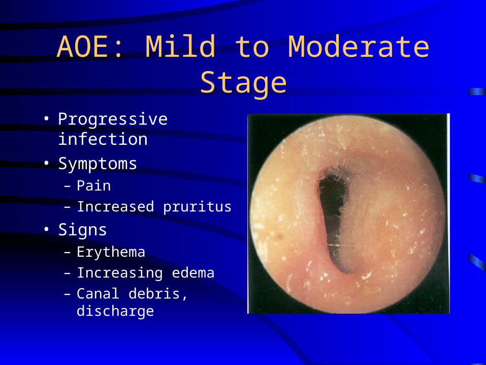
AOE: Mild to Moderate Stage
• Progressive infection• Symptoms
– Pain
– Increased pruritus
• Signs– Erythema
– Increasing edema
– Canal debris, discharge
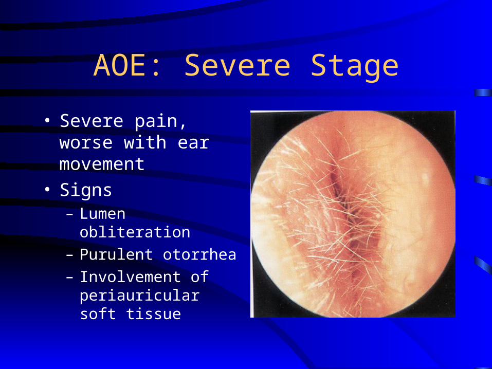
AOE: Severe Stage
• Severe pain, worse with ear movement
• Signs– Lumen obliteration
– Purulent otorrhea
– Involvement of periauricular soft tissue
AOE: Treatment
• Most common pathogens: P. aeruginosa and S. aureus
• Four principles– Frequent canal cleaning– Topical antibiotics– Pain control– Instructions for prevention
Chronic Otitis Externa (COE)
• It’s a chronic inflammatory process
• Persistent symptoms (> 2 months)
• Bacterial, fungal, dermatological etiologies
COE: Symptoms
• Unrelenting pruritus
• Mild discomfort
• Dryness of canal skin
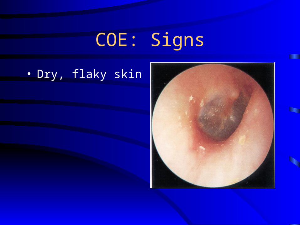
COE: Signs
• Dry, flaky skin

COE: Treatment
• Similar to that of AOE
• Topical antibiotics, frequent cleanings
• Topical Steroids
• Surgical intervention
Furunculosis
• Acute localized infection
• Lateral 1/3 canal
• Obstructed apopilosebaceous unit
• Pathogen: S. aureusS. aureus
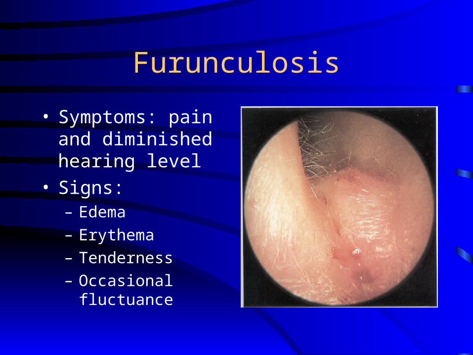
Furunculosis
• Symptoms: pain and diminished hearing level
• Signs:– Edema
– Erythema
– Tenderness
– Occasional fluctuance
Furunculosis: Treatment
• Local heat
• Analgesics
• Oral anti-staphylococcal antibiotics
• Incision and drainage reserved for localized abscess
• IV antibiotics for soft tissue extension
Otomycosis
• Fungal infection of EAC skin
• Primary or secondary
• Most common organisms: Aspergillus and Candida
Otomycosis: Symptoms
• Often indistinguishable from bacterial OE
• Pruritus deep within the ear
• Dull pain
• Hearing loss (obstructive)
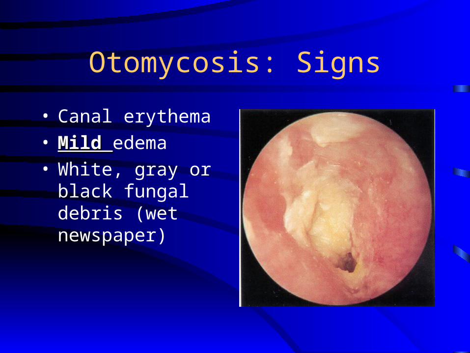
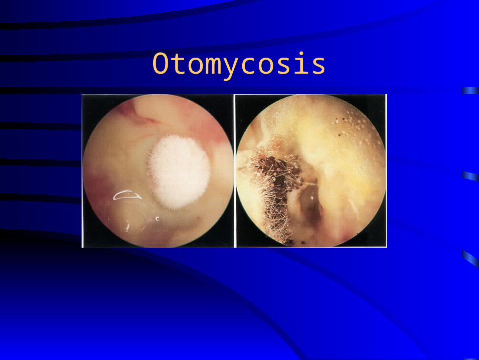
Otomycosis: Signs
• Canal erythema• Mild Mild edema• White, gray or black
fungal debris (wet newspaper)
Otomycosis
Otomycosis: Treatment
• Thorough cleaning and drying of canal
• Topical antifungals
• Canal Acidification
• Treat coexisted bacterial superinfection
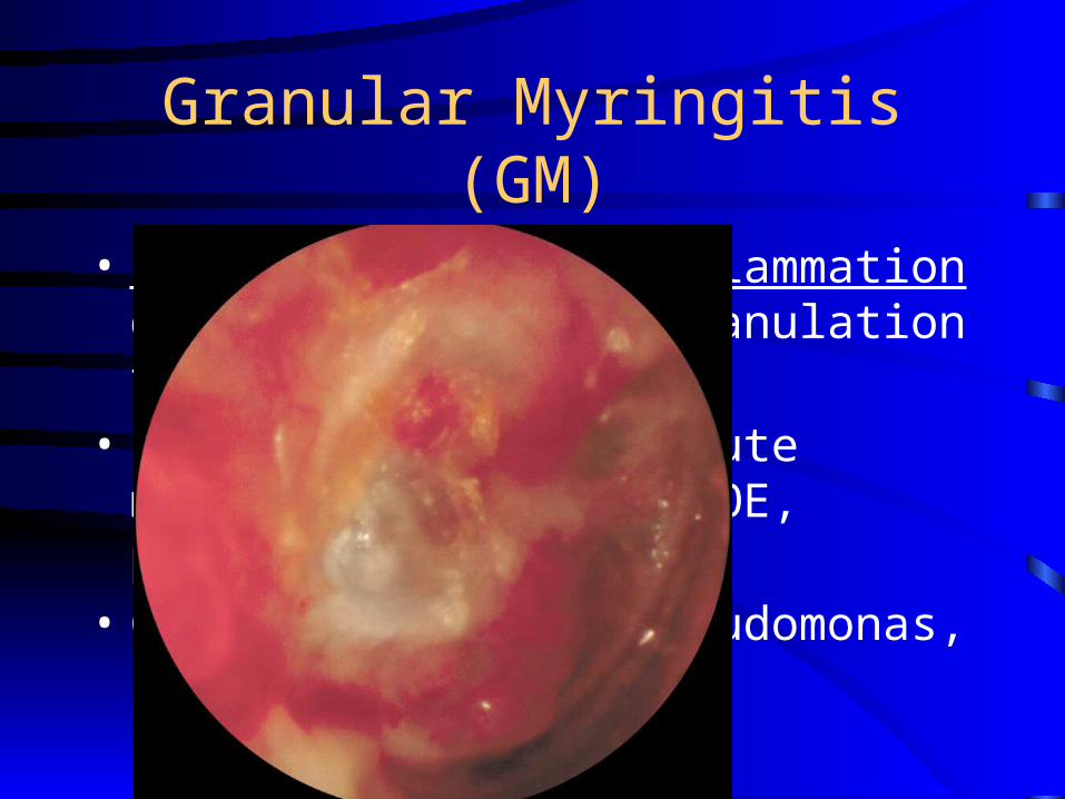
Granular Myringitis (GM)
• Localized chronic inflammation of pars tensa with granulation tissue
• Sequela of primary acute myringitis, previous OE, perforation of TM
• Common organisms: Pseudomonas, Proteus
GM: Symptoms
• Foul smelling discharge from one ear
• Often asymptomatic
• Slight irritation or fullness
• No hearing loss or significant pain
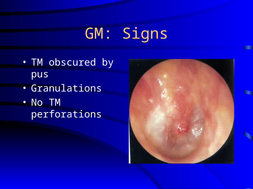
GM: Signs
• TM obscured by pus • Granulations• No TM perforations
GM: Treatment
• Careful and frequent debridement
• Topical anti-pseudomonal antibiotics
• Occasionally combined with steroids
• At least 2 weeks of therapy
• May warrant careful destruction of granulation tissue if no response
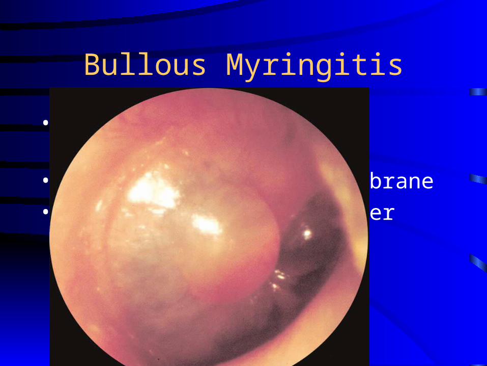
Bullous Myringitis
• Due to the Virus or Mycoplasma
• Confined to tympanic membrane
• Primarily involves younger children
Bullous Myringitis: Symptoms
• Sudden onset of severe pain
• No fever
• No hearing impairment
• Bloody otorrhea (significant) if rupture
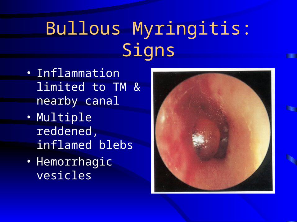
Bullous Myringitis: Signs
• Inflammation limited to TM & nearby canal
• Multiple reddened, inflamed blebs
• Hemorrhagic vesicles
Bullous Myringitis: Treatment
• Self-limiting
• Analgesics
• Topical antibiotics to prevent secondary infection
• Incision of blebs is unnecessary
• Azithromycin or AOM antimicrobial therapy
Necrotizing External Otitis(NEO)
• Potentially lethal infection of EAC and surrounding structures
• Typically seen in diabetics and immunocompromised patients
• Pseudomonas aeruginosa is the usual culprit
NEO: Symptoms
• Poorly controlled diabetic with OE
• Deep-seated aural pain
• Chronic otorrhea
• Aural fullness
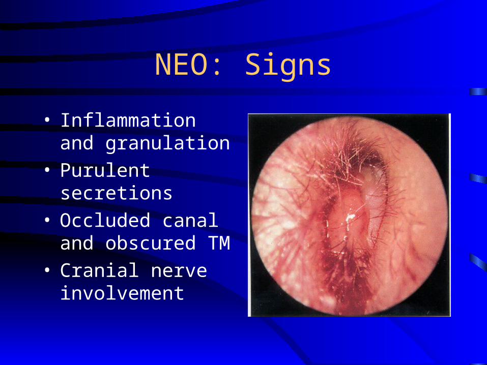
NEO: Signs
• Inflammation and granulation
• Purulent secretions• Occluded canal and
obscured TM• Cranial nerve
involvement
NEO: Imaging
• Plain films
• Computerized tomography – most used
• Technetium-99 – reveals osteomyelitis
• Gallium scan – useful for evaluating RxGallium scan – useful for evaluating Rx
• Magnetic Resonance Imaging
NEO: Diagnosis
• Clinical findings
• Laboratory evidence
• Imaging
• Physician’s suspicion
NEO: Treatment
• Intravenous antibiotics for at least 4 weeks (Ceftazidim) – with serial gallium scans monthly
• Local canal debridement until healed
• Pain control
• Use of topical agents
• Surgical debridement for refractory cases

NEO: Mortality
• Death rate essentially unchanged despite newer antibiotics (37% to 23%)
• Higher with multiple cranial neuropathies (60%)
Herpes Zoster Oticus
• Viral infection caused by varicella zoster
• Infection along one or more cranial nerve dermatomes (shingles)
• Ramsey Hunt syndrome: herpes zoster of the pinna with otalgia and facial paralysis
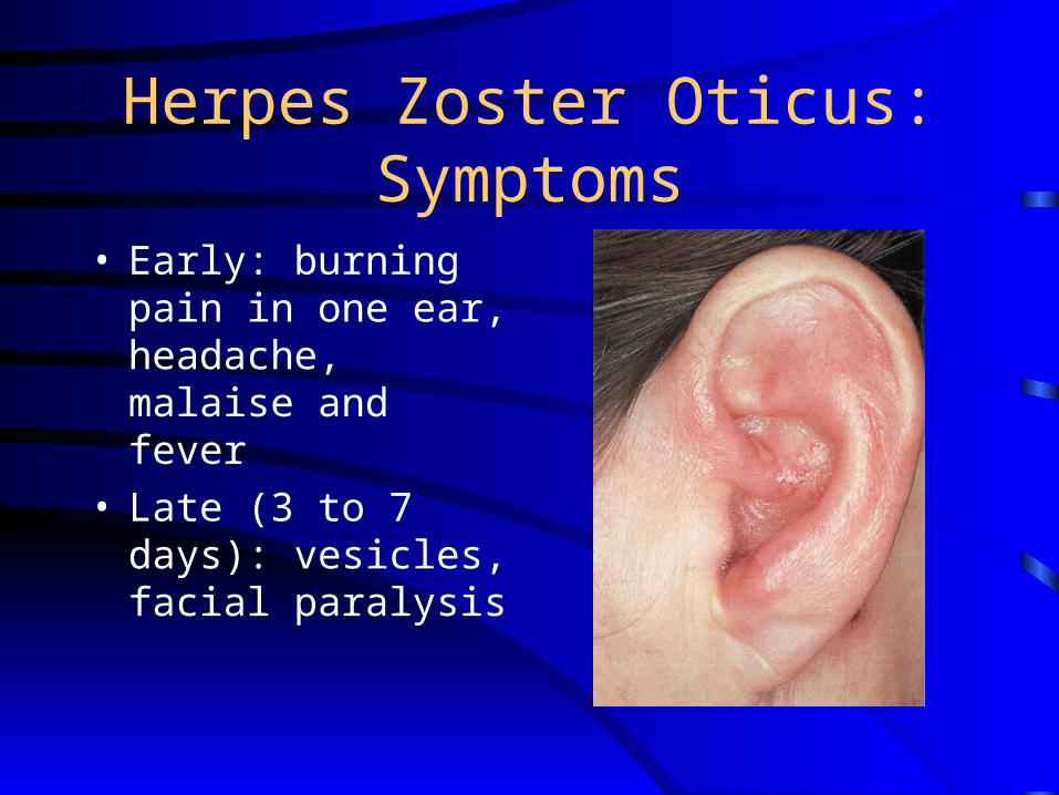
Herpes Zoster Oticus: Symptoms
• Early: burning pain in one ear, headache, malaise and fever
• Late (3 to 7 days): vesicles, facial paralysis
Herpes Zoster Oticus: Treatment
• Oral steroid taper (10 to 14 days)
• Antivirals