Embed Size (px)
Citation preview

INFINITY™ TOTAL ANKLE SYSTEM149336-0
The following languages are included in this packet:
English (en)
For additional languages, visit our website www.wmt.com. Then click on the Prescribing Information option.
For additional information and translations please contact the manufacturer or local distributor.
M C 0086* PWright Medical Technology, Inc Wright Medical EMEA5677 Airline Road Hoogoorddreef 5Arlington, TN 38002 1101 BA AmsterdamU.S.A The Netherlands* The CE-Marking of Conformity is applied per catalog number and appears on the outer label, if applicable.
December 2012
EN

Attention Operating Surgeon
IMPORTANT MEDICAL INFORMATION INFINITY™ TOTAL ANKLE SYSTEM
(149336-0)
OUTLINE:DEFINITIONSGENERAL PRODUCT INFORMATION
A. INTENDED USEB. INDICATIONSC. CONTRAINDICATIONSD. POTENTIAL COMPLICATIONSE. PRECAUTIONSF. ADVERSE EFFECTSG. HANDLING & STERILIZATIONH. STORAGE CONDITIONS

DEFINITIONSSymbols and abbreviations may be used on the package label. The following table provides the definition of these symbols and abbreviations.
Table 1. Definitions of Symbols and Abbreviations
Symbol Definition
g Batch code
h Catalog number
D Do not re-use
Y Caution, consult accompanying documents
i Consult operating instructions
H Use by
l Temperature limitation
p Keep dry

Keep away from sunlight
N Date of manufacture
M Manufacturer
P Authorized EC Representative in the European Community
I Sterilized using ethylene oxide
K Sterilized using radiation
Sterilized using gas plasma
J Sterilized using aseptic processing techniques
Non-sterile
B Do not resterilize
Caution: U.S. federal law restricts this device to sale by or on the order of a physician.
L Do not use if packaging is ripped or damaged
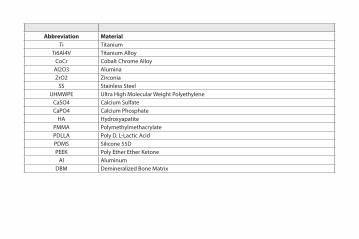
Abbreviation MaterialTi Titanium
Ti6Al4V Titanium AlloyCoCr Cobalt Chrome Alloy
Al2O3 AluminaZrO2 Zirconia
SS Stainless SteelUHMWPE Ultra High Molecular Weight Polyethylene
CaSO4 Calcium SulfateCaPO4 Calcium Phosphate
HA HydroxyapatitePMMA PolymethylmethacrylatePDLLA Poly D, L-Lactic AcidPDMS Silicone 55DPEEK Poly Ether Ether Ketone
Al AluminumDBM Demineralized Bone Matrix

GENERAL PRODUCT INFORMATIONThrough the advancement of partial and total joint replacement, the surgeon has been provided with a means of restoring mobility, correcting deformity, and reducing pain for many patients. While the prostheses used are largely successful in attaining these goals, it must be recognized that they are manufactured from a variety of materials and that any joint replacement system, therefore, cannot be expected to withstand activity levels and loads as would normal healthy bone. In addition, the system, including the implant/bone interface, will not be as strong, reliable, or durable as a natural human joint.
Ankle joint replacement components consist of a talar dome, a tibial platform, and a UHMWPE component. Components are available in a variety of sizes and design configurations intended for both primary and revision applications.
In using joint prostheses, the surgeon should be aware of the following:
• The correct selection of the prosthesis is extremely important. The potential for success in joint replacement is increased by selection of the proper size, shape, and design of the prosthesis. Joint prostheses require careful seating and adequate bone support. Surgeons are encouraged to use their best medical judgment when choosing the proper implant size regardless of the endosteal area of the bone. Surgeons must be familiar with the applicable operative techniques and instructions for use for each implant system.
• In selecting patients for total joint replacements, the following factors can be critical to the eventual success of the procedure.1. Patient's weight. An overweight or obese patient can produce high loads on the prosthesis, which can lead to
failure of the prosthesis. This becomes a major consideration when the patient is small boned and a small size prosthesis must be used.
2. Patient's occupation or activity. If the patient is involved in an occupation or activity, which includes substantial walking, running, lifting, or muscle strain, the resultant forces can cause failure of the fixation or the device, or both. The prosthesis will not restore function to the level expected with normal healthy bone, and the patient should not have unrealistic functional expectations.
3. Condition of senility, mental illness, or alcoholism. These conditions, among others, may cause the patient to ignore certain necessary limitations and precautions in the use of the prosthesis, leading to failure or other complications.
4. Foreign body sensitivity. Where material sensitivity is suspected, appropriate tests should be made prior to material selection or implantation.
A. INTENDED USEThe INFINITY™ Total Ankle System is intended to give a patient limited mobility by reducing pain, restoring alignment and replacing the flexion and extension movement in the ankle joint.
B. INDICATIONSThe INFINITY™ Total Ankle is indicated for patients with ankle joints damaged by severe rheumatoid, post-traumatic, or degenerative arthritis.
The INFINITY ™ Total Ankle is additionally indicated for patients with a failed previous ankle surgery.
CAUTION: The ankle prosthesis is intended for cement use only.
C. CONTRAINDICATIONSAbsolute contraindications include:1. Osteomyelitis;
2. Excessive bone loss at the ankle joint site;
3. Steroid use;
4. Infection at the ankle site or infections at distant sites that could migrate to the ankle;
5. Sepsis;
6. Muscular atrophy;
7. Dementia;

8. Vascular deficiency in the ankle joint;
9. Skeletally immature patients (patient is less than 21 years of age at the time of surgery);
10. Cases where there is inadequate neuromuscular status (e.g., prior paralysis, fusion and/or inadequate abductor strength), poor bone stock, poor skin coverage around the joint which would make the procedure unjustifiable;
11. Neuropathic joints;
12. Hepatitis or HIV infection;
13. Excessive loads as caused by activity or patient weight;
14. Female of childbearing age, for whom a negative pregnancy test is not obtained; and,
15. Neurological or musculoskeletal disease that may adversely affect gait or weight-bearing.
Conditions presenting increased risk of failure include:1. Uncooperative patient or patient with neurologic disorders, incapable of following instructions;
2. Marked bone loss, severe osteoporosis, or revision procedures for which an adequate fit of the prosthesis cannot be achieved;
3. Metabolic disorders that may impair bone formation;
4. Osteomalacia; and,
5. Poor prognosis for good wound healing (e.g., decubitus ulcer, end-stage diabetes, severe protein deficiency and/or malnutrition).
WARNING: This device is not intended for subtalar joint fusion or subtalar joint impingement. Please carefully evaluate the anatomy of each patient before implantation.

D. POTENTIAL COMPLICATIONSImproper selection, placement, positioning, and fixation of the prosthetic components may result in unusual stress conditions and a subsequent reduction in service life of the prosthetic component. The surgeon must be thoroughly familiar with the implant, instruments, and surgical procedure prior to performing surgery. Periodic, long-term follow-up is recommended to monitor the position and state of the prosthetic components, as well as the condition of the adjoining bone.
Proper surgical procedures and techniques are the responsibility of the medical professional. Each surgeon must evaluate the appropriateness of the procedure based on personal medical training and experience. Although Wright Medical Technology, Inc. (Wright) cannot recommend a particular surgical technique suitable for all patients, a detailed surgical technique is available for surgeon reference. Medical procedures for optimal utilization of the prosthesis should be determined by the physician. However, the physician is advised that there is recent evidence that the potential for deep sepsis following total joint arthoplasty may be reduced by:
1. Consistent use of prophylactic antibiotics.
2. Utilizing a laminar flow clean air system.
3. Having all operating room personnel, including observers, properly attired.
4. Protecting instruments from airborne contamination.
5. Impermeable draping.
Materials. The prosthetic components are manufactured from a variety of materials which include cobalt-chromium-molybdenum alloy, titanium alloy, ultra high molecular weight polyethylene (UHMWPE), and commercially pure titanium all of which conform to ASTM or ISO standards, or internal standards.

E. PRECAUTIONS1. The patient must be advised of the limitations of the reconstruction and the need for protection of the prosthesis
from full weight bearing until adequate fixation and healing have occurred. Excessive activity and trauma affecting the joint replacement have been implicated with failure of the reconstruction by loosening, fracture and/or wear of the prosthetic components. Loosening of the components can result in increased production of wear particles, as well as damage to the bone, making successful revision surgery more difficult.
2. The patient should be cautioned to limit activities and protect the replaced joint from unreasonable stresses, and follow the instructions of the physician with respect to follow-up care and treatment. The patient should be closely monitored if a change at the operative site has been detected. The possibility of deterioration of the joint should be evaluated and possible revision surgery considered.
3. The patient should be warned of surgical risks, and made aware of possible adverse effects. The patient should be warned that the prosthesis does not replace normal healthy bone, that the prosthesis can break or become damaged as a result of certain activity or trauma, has a finite expected service life, and may need to be replaced at some time in the future. The patient should also be advised of other risks that the surgeon believes should be disclosed. The patient should be advised that any noise or unusual sensation should be reported to the surgeon as it may indicate implant malfunction.
4. Specialized instruments are available and must be used to assure the accurate implantation of prosthetic components. Careful attention must be given to accurately installing the prosthesis. Do not mix instruments from different manufacturers. While rare, breakage of instruments may occur especially with extensive use or excessive force. For this reason, instruments should be examined for wear or damage prior to surgery.
5. Preoperative templates should be used to assure proper sizing of prostheses. Use only with mating Wright Medical prosthetic components of appropriate size. Mismatching of components could impede component articulation, leading to wear and possible failure of the component and also contribute to joint laxity.
6. Periodic post-operative x-rays are recommended for close comparison with early post-op conditions to detect long term evidence of changes in position, loosening, bending, or cracking of components.

7. As with any surgical procedure, care should be exercised in treating individuals with preexisting conditions that may affect the success of the surgical procedure. This includes individuals with bleeding disorders of any etiology, long-term steroidal therapy, immunosuppressive therapy, or high dosage radiation therapy.
Recommendations Regarding Device Fragments1. Use medical devices in accordance with their labeled indications and the manufacturer’s instructions for use,
especially during insertion and removal.
2. Inspect devices prior to use for damage during shipment or storage or any out-of-box defects that might increase the likelihood of fragmentation during a procedure.
3. Inspect devices immediately upon removal from the patient for any signs of breakage or fragmentation.
4. If the device is damaged, retain it to assist with the manufacturer’s analysis of the event.
5. Carefully consider and discuss with the patient (if possible) the risks and benefits of retrieving vs. leaving the fragment in the patient.
6. Advise the patient of the nature and safety of unretrieved device fragments including the following information:
a. The material composition of the fragment (if known);
b. The size of the fragment (if known);
c. The location of the fragment;
d. The potential mechanisms for injury, e.g., migration, infection;
e. Procedures or treatments that should be avoided such as MRI exams in the case of metallic fragments. This may help to reduce the possibility of a serious injury from the fragment.
Concerning Magnetic Resonance EnvironmentsThe devices described in this package insert have not been evaluated for safety and compatibility in the MR environment. The devices described in this package insert have not been tested for heating or migration in the MR environment.

F. ADVERSE EFFECTS1. With all joint replacements, asymptomatic, localized, progressive bone resorption (osteolysis) may occur around
the prosthetic components as a consequence of foreign-body reaction to particulate matter. Particulate is generated by interaction between components, as well as between the components and bone, primarily through wear mechanisms of adhesion, abrasion, and fatigue including third-body wear. Osteolysis can lead to future complications necessitating the removal and replacement of prosthetic components. See Important Physician Information section for more information.
2. Although rare, metal sensitivity reactions in patients following joint replacement have been reported. Implantation of foreign material in tissues can result in histological reactions involving production of macrophages and fibroblasts.
3. Peripheral neuropathies have been reported following total joint surgery. Subclinical nerve damage has been reported, and may occur as the result of surgical trauma.
4. Dislocation and subluxation of prosthetic components can result from improper positioning and/or migration of the components. Muscle and fibrous tissue laxity can also contribute to these conditions.
5. Prosthetic components can loosen or migrate due to trauma or loss of fixation.
6. Infection can lead to failure of the joint replacement.
7. While rare, fatigue fracture of the prosthetic component can occur as a result of trauma, strenuous activity, improper alignment, incomplete implant seating, or duration of service.
8. Bone damage or fracture may occur during installation due to compromised bone quality, osteoporosis, or previous bone injury or surgery.
9. Allergic reactions to the prosthetic component materials can occur.

Intraoperative and early postoperative complications can include:1. pain;
2. a sudden drop in blood pressure intra-operatively due to the use of bone cement;
3. damage to blood vessels;
4. temporary or permanent nerve damage resulting in pain or numbness of the affected limb;
5. cardiovascular disorders including venous thrombosis, pulmonary embolism, or myocardial infarction.
6. hematoma;
7. delayed wound healing; and
8. deep wound infection (early or late) which may necessitate removal of the prosthesis. On rare occasions, arthrodesis of the involved joint or amputation of the limb may be required.
Late postoperative complications can include:1. pain;
2. bone fracture by trauma or excessive loading, particularly in the presence of poor bone stock;
3. periarticular calcification or ossification, with or without impediment to joint mobility; and
4. inadequate range of motion due to improper selection or positioning of components or periarticular calcification.

Important Physician InformationBone resorption is a natural consequence of total joint arthroplasty due to changes in bone remodeling patterns. Bone remodeling is mediated by the changes in stress distribution caused by implantation. Extensive resorption around the prosthesis may lead to implant loosening and failure. It is generally agreed that osteolysis is the result of localized foreign-body reaction to particulate debris generated by cement, metal, ultra-high molecular-weight polyethylene (UHMWPE), and ceramic. Regarding the etiology, it has been hypothesized that particulate debris generated by the components of a prosthesis migrate into the synovial cavity and the bone-implant interface, where they recruit macrophages and stimulate phagocytic action. The degree of recruitment is determined by the size, distribution, and amount of particulate debris (rate of debris generation). The phagocytic action results in the release of cytokines and intercellular mediators (IL-1, 2, PE2) which encourage osteoclastic bone resorption. Clinical and basic research is continuing in order to provide scientific basis for the causes of this phenomenon and potential ways to reduce its occurrence. Osteolysis can be asymptomatic and therefore routine periodic radiographic examination is vital to prevent any serious future complication. Presence of focal lesions that are progressive may necessitate replacement of the prosthetic component(s).
G. HANDLING AND STERILIZATIONImplantsThis product has been sterilized and should be considered sterile unless the inner package has been opened or damaged. If the inner package integrity has been compromised, contact the manufacturer for instructions. Remove from package, using aseptic OR technique, only after the correct size has been determined and the operative site has been prepared for final implantation. Always handle the product with powder-free gloves, and avoid contact with hard objects that may damage the product.
This product is for single use only. An implant should never be re-sterilized after contact with body tissues or fluids.
Devices labeled for single-use only should never be reused. Reuse of these devices may potentially result in serious patient harm. Examples of hazards related to the reuse of these devices include, but are not limited to: significant degradation in device performance, cross-infection, and contamination.

WARNING: All packaging materials MUST be removed from the implant prior to implantation.
WARNING: You must NEVER steam sterilize/resterilize the components of the INFINITY™ Total Ankle System.
InstrumentsCleaning1. Disassemble all components as per manufacturer instructions (if appropriate).
2. Rinse with cold tap water to remove any gross contamination.
3. Bathe in an enzymatic detergent solution prepared per manufacturer directions for 5 minutes.
4. Scrub thoroughly with a soft brush and/or pipe cleaner; repeatedly flush any very narrow lumens with enzymatic detergent solution using a syringe.
5. Rinse with cold tap water for a minimum of one minute; use a syringe to repeatedly flush any very narrow lumens.
6. Bathe in a detergent solution prepared per manufacturer directions for 5 minutes.
7. Scrub thoroughly with a soft brush and/or pipe cleaner; repeatedly flush any very narrow lumens with detergent solution using a syringe.
8. Rinse thoroughly/flush with reverse osmosis/deionized (RO/DI) water.
9. Sonicate for a minimum of 10 minutes in an enzymatic detergent solution prepared per manufacturer directions.
10. Rinse thoroughly/flush with RO/DI water.
11. Dry with a clean, soft, absorbent, disposable cloth.
12. Visually inspect for cleanliness. All visible surfaces, internal and external, should be visually inspected. If necessary re-clean until it is visibly clean.
Note: Brushes (i.e. pipe cleaners) could be used for cleaning most lumens; however, the use of a syringe to flush narrow lumens with diameters less than or equal to 0.041 inches is recommended.

Steam SterilizationThe minimum recommended steam sterilization conditions for Wright reusable instruments are as follows:
1. Double wrap the component in an FDA-cleared CSR wrap or similar type non-woven medical grade wrapping material.
2. Autoclave according to the following parameters:Steam Sterilization
Cycle Type Parameter Minimum Set PointPrevacuum
270˚F (132˚C)Exposure Temperature 270˚F (132˚C)
Exposure Time 4 minutesDry Time 20 minutes
3. After sterilization, remove the component from its wrapping using accepted sterile technique with powder-free gloves. Ensure that implants are at room temperature prior to implantation. Avoid contact with hard objects that may cause damage. These recommendations are consistent with ANSI/AAMI ST79: 2006 Table 5 guidelines1 and have been developed and validated using specific equipment. Due to variations in environment and equipment, it must be demonstrated that these recommendations produce sterility in your environment. If processing conditions, wrapping materials, or equipment changes occur, the effectiveness of the sterilization process must be demonstrated.
H. STORAGE CONDITIONSAll implants must be stored in a clean, dry environment and be protected from sunlight and extremes in temperature.
Trademarks™ and Registered Trademarks® are owned or licensed by Wright Medical Technology, Inc.
1 Comprehensive guide to steam sterilization and sterility assurance in health care facilities (ANSI/AAMI ST79:2006).