Embed Size (px)
Citation preview
Accepted Manuscript
Title: Inflammation as a link between obesity, metabolicsyndrome and type 2 diabetes
Author: Nathalie Esser Sylvie Legrand-Poels Jacques PietteAndre J. Scheen Nicolas Paquot
PII: S0168-8227(14)00187-9DOI: http://dx.doi.org/doi:10.1016/j.diabres.2014.04.006Reference: DIAB 6044
To appear in: Diabetes Research and Clinical Practice
Received date: 28-3-2014Accepted date: 7-4-2014
Please cite this article as: N. Esser, S. Legrand-Poels, J. Piette, A.J. Scheen,N. Paquot, Inflammation as a link between obesity, metabolic syndromeand type 2 diabetes, Diabetes Research and Clinical Practice (2014),http://dx.doi.org/10.1016/j.diabres.2014.04.006
This is a PDF file of an unedited manuscript that has been accepted for publication.As a service to our customers we are providing this early version of the manuscript.The manuscript will undergo copyediting, typesetting, and review of the resulting proofbefore it is published in its final form. Please note that during the production processerrors may be discovered which could affect the content, and all legal disclaimers thatapply to the journal pertain.
Page 1 of 24
Accep
ted
Man
uscr
ipt
1
Inflammation as a link between obesity, metabolic syndrome and
type 2 diabetes
Nathalie Esser1,2
, Sylvie Legrand-Poels1, Jacques Piette
1, André J. Scheen
2,
Nicolas Paquot1,2
1. Virology and Immunology Unit, GIGA-Research, University of Liege, Liege,
Belgium
2. Division of Diabetes, Nutrition and Metabolic Disorders, Department of Medicine,
University Hospital of Liege, Liege, Belgium
Corresponding author:
Dr. Nathalie Esser
Division of Diabetes, Nutrition and Metabolic Disorders,
Department of Medicine, CHU Sart Tilman, University of Liege,
B-4000 Liege, Belgium
Tel: +3243667238; Fax: +3243667068;
Email: [email protected]
Word count: 4721
*Manuscript
Page 2 of 24
Accep
ted
Man
uscr
ipt
2
ABSTRACT : It is recognized that a chronic low-grade inflammation and an activation of the immune
system are involved in the pathogenesis of obesity-related insulin resistance and type 2
diabetes. Systemic inflammatory markers are risk factors for the development of type 2
diabetes and its macrovascular complications. Adipose tissue, liver, muscle and pancreas are
themselves sites of inflammation in presence of obesity. An infiltration of macrophages and
other immune cells is observed in these tissues associated with a cell population shift from an
anti-inflammatory to a pro-inflammatory profile. These cells are crucial for the production of
pro-inflammatory cytokines, which act in an autocrine and paracrine manner to interfere with
insulin signaling in peripheral tissues or induce β-cell dysfunction and subsequent insulin
deficiency. Particularly, the pro-inflammatory interleukin-1β is implicated in the pathogenesis
of type 2 diabetes through the activation of the NLRP3 inflammasome. The objectives of this
review are to expose recent data supporting the role of the immune system in the pathogenesis
of insulin resistance and type 2 diabetes and to examine various mechanisms underlying this
relationship. If type 2 diabetes is an inflammatory disease, anti-inflammarory therapies could
have a place in prevention and treatment of type 2 diabetes.
Keywords:
Type 2 diabetes – obesity – insulin resistance – macrophages – NLRP3 inflammasome –
inflammation – metabolic syndrome
Page 3 of 24
Accep
ted
Man
uscr
ipt
3
INTRODUCTION Obesity, in particular excess visceral adiposity, is associated with insulin resistance,
hyperglycaemia, dyslipidaemia and hypertension, which together are termed “metabolic
syndrome” [1]. These metabolic disorders increase the risk of development of type 2 diabetes
mellitus (T2DM) and cardiovascular diseases and contribute to high rates of mortality and
morbidity [1]. T2DM is the most prevalent metabolic disease in the world and is characterized
by defects in insulin secretion and a peripheral insulin resistance in the skeletal muscle, the
adipose tissue and the liver. The progression from obesity-related insulin resistance to T2DM
remains poorly understood but implicates a failure of pancreatic β-cells to compensate for
insulin resistance leading to chronic hyperglycaemia. A chronic low-grade inflammation and
an activation of the immune system are observed in abdominal obesity and may have a role in
the pathogenesis of obesity-related metabolic disorders [2-5]. This review summarizes data
implicating the immune system in the pathophysiogy of insulin resistance and T2DM. We
will also examine the biological, tissular and cellular inflammatory markers associated with
obesity-related metabolic disorders that may predict the development of T2DM. Molecular
mechanisms underlying this inflammatory activation state will be reviewed and preliminary
results obtained with anti-inflammatory therapies in the prevention and treatment of T2DM
will be described.
1. SYSTEMIC MARKERS OF INFLAMMATION
1.1. Inflammatory markers in obesity, metabolic syndrome and T2DM
White blood cell counts and plasma levels of coagulation factors (fibrinogen and plasminogen
activator inhibitor 1 (PAI-1)), acute-phase proteins such as C-reactive protein (CRP) and
serum amyloid A (SAA), pro-inflammatory cytokines (tumor necrosis factor (TNF)-α,
interleukin (IL)-1β and IL-6), and chemokines are elevated in obese and T2DM patients and
shown to be reduced when these patients are engaged in a more intensive lifestyle causing
weight loss [6-11]. These pro-inflammatory markers also positively correlated with insulin
resistance and the features of the metabolic syndrome, in most cases, independently of the
degree of obesity [6, 7, 12-14].
Page 4 of 24
Accep
ted
Man
uscr
ipt
4
1.2. Inflammatory markers for development of T2DM in obese patients
Subclinical chronic inflammation seems to be an independent risk factor for the development
of T2DM. Indeed, high levels of many inflammatory factors at baseline in diverse human
populations are correlated with incident T2DM, regardless of the initial degree of insulin
resistance and obesity. Prospective studies have identified white blood cell count [12, 15],
pro-inflammatory cytokines [16], chemokines [17], and other several indirect markers of
inflammation such as fibrinogen, sialic acid and PAI-1 [15, 18] as predictors of T2DM. In
contrast to all these inflammatory biomarkers, CRP measurement is less expensive,
standardized and widely available. Particularly a highly sensitive measurement of CRP has
been developed to detect this protein with greater accuracy at lower levels. A number of
prospective studies have shown that high sensitivity-CRP (hs-CRP) levels predict
development of T2DM in different non-diabetic populations regardless the degree of
adiposity, fat distribution and insulin resistance. All these studies were included in a recent
review and meta-analysis which provided further evidence that elevated CRP levels are
significantly associated with increased risk of T2DM (relative risk [RR] 1.26 [95%
confidence interval or CI 1.16-1.37]) [19]. This meta-analysis also detected a significant dose-
response association between IL-6 (its inducer) and T2DM risk (RR 1.31 [95% CI 1.17-1.46])
[19].
1.3. Inflammatory markers for cardiovascular disease in T2DM patients
Since hs-CRP plasma levels have been associated with cardiovascular diseases and death in
the general population [20] and in patients with metabolic syndrome [21], an important
question is the possible association of inflammatory markers with these risks in T2DM
patients. The Hoorn population-based study was the first to show that CRP is a predictor of
mortality in T2DM individuals over a 5- to 7-year period [22]. Other observational studies in
T2DM populations then showed that markers of inflammation and coagulation, and in
particular CRP, are independent predictors of coronary heart disease [23, 24] and mortality
[25]. However, the association between CRP and cardiovascular risk seems to be much
weaker in T2DM patients than in the general population [24, 26] and needs to be more
extensively studied. Interest has also been focused on cardiovascular risk prediction by pro-
inflammatory cytokines plasma levels. For instance, in the ADVANCE trial, IL-6 plasma
levels significantly improved the prediction of macrovascular events and death in T2DM
patients [27].
Page 5 of 24
Accep
ted
Man
uscr
ipt
5
2. TISSUE INFLAMMATION IN OBESITY AND T2DM
A number of experimental and clinical data have clearly established that adipose tissue, liver,
muscle and pancreas are sites of inflammation in presence of obesity and T2DM. An
infiltration of macrophages into these tissues is seen in animal models of obesity and diabetes
as well as in obese human individuals with metabolic syndrome or T2DM. These cells are
crucial for the production of pro-inflammatory cytokines [4], including TNFα, IL-6 and IL-
1β. They act in an autocrine and paracrine manner to promote insulin resistance by interfering
with insulin signaling in peripheral tissues through activation of the c-JUN N-terminal kinase
(JNK) and nuclear factor-kappa B (NF-κB) pathways [2]. These pathways are activated in
multiple tissues in obesity and T2DM and have a central role in promoting tissue
inflammation.
2.1. Inflammation in insulin-sensitive tissues
2.1.1 Adipose tissue
Hotamisligil and colleagues were the first to show an increased expression and production of
TNFα in adipose tissue of obese individuals and its direct role in obesity-induced insulin
resistance [28]. Accumulating data then confirmed a specific up-regulation of genes encoding
inflammatory factors and an over-production of many cytokines and chemokines in enlarged
adipose tissue [5]. Furthermore, improvement in insulin sensitivity induced by weight loss
was accompanied by a decrease in the expression of pro-inflammatory genes [28, 29]. So
adipose tissue inflammation was considered as a crucial event leading to metabolic syndrome,
T2DM and atherosclerotic cardiovascular diseases.
More recently, adipose tissue has been associated with a marked accumulation of immune
cells in its stromovascular fraction during obesity [4] (Figure 1). Particularly, obesity induces
an infiltration of macrophages in adipose tissue of both mice and humans [30, 31]. Although it
was shown that enlarged adipocytes themselves produce pro-inflammatory cytokines and
chemokines [32], adipose tissue macrophages seem to be crucial for the production of adipose
tissue-derived pro-inflammatory cytokines. Indeed, their recruitment correlates with the
degree of obesity and is linked to systemic inflammation, insulin resistance [30] and
metabolic syndrome [33]. Moreover, weight loss induced by surgery [29] or diet and exercise
[10] results in a reduction in the number of adipose tissue macrophages in parallel to the
decreased expression of pro-inflammatory markers in both adipose tissue and plasma of obese
individuals.
Page 6 of 24
Accep
ted
Man
uscr
ipt
6
Macrophages can be classified into two distinct subtypes: the “classically activated
macrophages” phenotype, termed M1, which secrete pro-inflammatory cytokines such as IL-
1β, IL-6, TNF-α, and the “alternatively activated macrophages” phenotype, termed M2 which
produce anti-inflammatory cytokines such as IL-10 [4]. While well-established in mice [34,
35], the existence of distinct M1 and M2 subsets of adipose tissue macrophages has not been
confirmed in human, where macrophages have rather been described as being a mix between
M1 and M2 phenotypes [36]. In addition to adipose tissue macrophages infiltration, obesity
causes a phenotypic switch from the M2 to M1 phenotype, correlating with insulin resistance
both in mice and humans [34-36]. Direct and paracrine signals issued from M1 macrophages
can impair insulin signaling and adipogenesis in adipocytes whereas M2 macrophages seem
to protect against obesity-induced insulin resistance [4].
Although macrophages are the most abundant leukocyte population in expanding adipose
tissue, the adaptive immune system may also contribute to obesity-induced inflammation.
Infiltrating lymphocytes precede macrophage populations in obese adipose tissue concomitant
with early insulin resistance and may play a role in adipose tissue inflammation by modifying
the number and the activation state of adipose tissue macrophages [37-40]. In mouse models
of obesity, an increased numbers of cytotoxic CD8+ effector cells is suspected to initiate the
recruitment and activation of adipose tissue macrophages and promote pro-inflammatory
cascades associated with insulin resistance [38, 40]. Obesity also induces modification in the
balance between pro-inflammatory (T helper 1 and T helper 17 lymphocytes) and anti-
inflammatory (T helper 2 and regulatory T lymphocytes) CD4+ cells subsets, leading to
secretion of cytokines from newly recruited adipose tissue macrophages [39-41]. Of particular
interest, the number of anti-inflammatory regulatory T lymphocytes decreases with obesity in
adipose tissue of both mice and humans [37, 39, 40] and even more in obese patients with
metabolic syndrome [33]. The regulatory T lymphocytes express high amount of the anti-
inflammatory cytokine IL-10 which inhibit macrophage migration and induce the
differentiation of M2 macrophages [35, 37]. A boost in the number of these cells in obese
mice can improve insulin sensitivity and reduce macrophage infiltration in adipose tissue [37].
These data suggest that regulatory T cells may repress adipose tissue inflammation and play a
role in providing protection against insulin resistance-induced inflammation linked to obesity
[33, 37]. The number of many other immune cells is also modified in adipose tissue during
obesity and could regulate inflammation and insulin resistance[4] (Figure 1).
Overall, these data reveal a complex interplay between cells of innate and adaptive immunity
and the balance among these immune cells appears to be important for the homeostasis and
Page 7 of 24
Accep
ted
Man
uscr
ipt
7
control of adipose tissue inflammation in obesity and T2DM. However, the molecular events
that initiate immune cell recruitment and activation are not fully understood.
Subcutaneous versus visceral adipose tissues
Abdominal obesity is the key component of the metabolic syndrome, with a predominance of
intra-abdominal visceral fat accumulation, indirectly measured by waist circumference in
clinical practice. So, besides total adiposity, the pathogenic role of adipose tissue seems to be
determined by its specific anatomic location. Indeed, although both subcutaneous and visceral
adipose tissues are associated with metabolic risk profile, high visceral adipose tissue is more
strongly correlated with metabolic syndrome than its subcutaneous counterpart[42].
Furthermore, it is associated with ectopic lipid accumulation in the liver and skeletal muscle,
which participates to local insulin resistance and contributes to associated metabolic
complications [43]. Subcutaneous and visceral adipose tissues differ by phenotypic,
physiological and functional characteristics [43]. Specific differences in inflammatory profile
have also been reported, with more macrophages [29, 31, 33], T lymphocytes [31, 33], and
inflammatory molecules in the visceral vs the subcutaneous tissues of obese individuals [29,
33]. Moreover, a lower number of anti-inflammatory regulatory T lymphocytes was recently
found in the visceral adipose tissue of obese individuals with metabolic syndrome [33]. This
less favourable inflammatory profile of visceral adipose tissue is in line with the belief this
tissue has a more important role in the development of obesity-related insulin resistance.
2.1.2 Liver
Nonalcoholic fatty liver disease (NAFLD) often accompanies abdominal obesity, and its
prevalence increases in parallel to that of T2DM. NAFLD includes a large spectrum of
lesions, from simple benign steatosis to steatohepatitis (nonalcoholic steatohepatitis or
NASH), which can lead to cirrhosis and hepatocarcinoma [44]. Inflammation clearly plays a
pivotal role in the progression of this disease process. NAFLD and subsequent hepatic insulin
resistance in obesity are associated with increased expression and overproduction of
inflammatory mediators, including TNFα, IL-6 and IL-1β [45]. Unlike adipose tissue, the
liver is densely populated with resident macrophages, the Kupffer cells, which account for
over 10% of total liver cells. The number of Kupffer cells does not increase with obesity, but
their activation state changes [45, 46]. Although Kupffer cells have been less studied than
adipose tissue macrophages in the context of insulin resistance, they clearly contribute to the
Page 8 of 24
Accep
ted
Man
uscr
ipt
8
production of inflammatory mediators that promote insulin resistance in liver [46].
Interestingly, as earlier described for adipose tissue macrophages, alternative M2 activation
phenotype of Kupffer cells appears to ameliorate insulin resistance and to delay the
progression to NASH in mice [46]. These data suggest that steatosis might induce a sub-acute
inflammatory response in liver, similar to that observed in the adipose tissue inflammation
following adipocyte lipid accumulation. Alternatively, pro-inflammatory mediators in the
portal circulation, potentially produced by abdominal fat, might also initiate hepatic
inflammation.
2.1.3 Muscle
The muscle is another major site of insulin resistance in obesity and T2DM. Recent reports
have shown an infiltration of macrophages within skeletal muscles of obese mice, particularly
in the inter-muscular adipose depots [30]. Just as adipose tissue, these skeletal muscle
macrophages exhibit a pro-inflammatory M1 phenotype [47] accompanied with an increased
expression of inflammatory factors in muscle cells [30, 47] contributing locally to insulin
resistance. Moreover, the gene expression of phenotype markers of pro- and anti-
inflammatory macrophages in human skeletal muscle seems to correlate with insulin
sensitivity [48]. However, the content of macrophages in skeletal muscle in obesity is by far
lower than in adipose tissue or liver, and further research is needed to determine if skeletal
muscle is mainly a target of inflammation-induced insulin resistance or if local inflammatory
cascades may also play a role in insulin resistance.
2.2. INFLAMMATION IN PANCREAS AND INSULIN DEFICIENCY
The progression from obesity-related insulin resistance to T2DM implicates a failure of
pancreatic β cells to compensate for insulin resistance, leading to chronic hyperglycaemia. An
inflammation was also demonstrated in pancreatic islets of T2DM patients as shown by the
presence of amyloid deposits, fibrosis, increased β cell death and infiltration of macrophages
along with increased levels of pro-inflammatory cytokines and chemokines [3]. Moreover this
increase of immune cells in pancreatic islets is described before the onset of T2DM.
Particularly the expression and local release of the pro-inflammatory cytokine IL-1β are
increased in pancreatic islet of T2DM individuals [49]. This cytokine seems to be a master
regulator of islet inflammation in T2DM by increasing local expression of pro-inflammatory
cytokines and chemokines [50] leading to immune cell recruitment in islets. This local
Page 9 of 24
Accep
ted
Man
uscr
ipt
9
inflammation can reduce insulin secretion and trigger β-cell apoptosis leading to decrease islet
mass, all critical events in the progression of T2DM.
3. SENSORS AND MEDIATORS OF INFLAMMATION IN OBESITY
AND T2DM
Although subclinical inflammation is important in the pathogenesis of T2DM, the events
initiating this inflammatory process remain unclear and could involve different but synergic
mechanisms leading to the activation of NF-κB and JNK pathways, cytokines and
chemokines release and recruitment of immune cells.
3.1. IL-1β and NLRP3 inflammasome
IL-1β, one of the major pro-inflammatory cytokine produced by macrophages, has been
shown to be a key contributor to the pathogenesis of T2DM. IL-1β signaling occurs through
the IL-1 receptor-I and leads to the activation of NF-κB pathways and the generation of other
inflammatory mediators, such as TNFα and IL-1β itself, thus initiating a self-amplifying
cytokine network [50]. The control of IL-1β production is tightly regulated and depends on
two signals. First, a pro-inflammatory signal stimulates the transcription of IL1B with
subsequent storage of inactive pro-IL-1β into the cell. A second signal then induces the
production of active, mature IL-1β by cleavage of its inactive precursor by caspase-1, which
is activated in a large cytoplasmic multiprotein complex called the inflammasome [51].
Inflammasomes are central components of the innate immune response and recognize
microbial products (pathogen-associated molecular patterns (PAMPs)) or endogenous
molecules (danger-associated molecular pattern (DAMPs)) by innate pattern recognition
receptors (PRRs). Structurally, inflammasomes are usually formed through the interaction
between a PRR, mainly a member of the nucleotide-binding oligomerization domain like
receptor (NLR) family, the adaptor protein apoptosis-associated speck-like protein containing
a caspase-recruitment domain (ASC) and the pro-caspase-1 [51]. Among several NLRs that
form inflammasome platforms, there are compelling evidences for a major role of the NLR
family pyrin domain-containing 3 (NLRP3) inflammasome in the progression from obesity to
T2DM (Figure 2). NLRP3 inflammasome activation and subsequent IL-1β production have
been first described in pancreatic β-cells and islet-infiltrating macrophages. They contribute to
islet inflammation induced by chronic exposure to high levels of free fatty acids and glucose,
leading to increased apoptosis and impaired insulin secretion of β-cells [49, 52, 53].
Page 10 of 24
Accep
ted
Man
uscr
ipt
10
Moreover, islet amyloid polypeptide (IAPP), a protein that forms amyloid deposits in the
pancreas, seems to contribute to IL-1β production in islets through activation of the NLRP3
inflammasome [54].
Accumulating evidences also give to NLRP3 inflammasome a central role in obesity-induced
insulin resistance [33, 55, 56]. The expression of NLRP3 inflammasome components, the
activity of caspase-1 and the levels of IL-1β are increased in adipose tissue, mainly in
macrophages, of obese mice and humans and directly correlate with insulin resistance,
features of the metabolic syndrome and severity of T2DM [33, 56]. The deleterious effects of
visceral adipose tissue might be also related to an up-regulated expression and activation of
NLRP3 inflammasome compared to subcutaneous adipose tissue in overweight and obese
patients [33, 57]. The NLRP3 inflammasome seems to act as a sensor of metabolic danger
signals that accumulate during obesity, including high levels of glucose [52], saturated free
fatty acids [58, 59], lipid intermediates such as ceramides [56] and uric acid [51], and its
activation results in IL-1β production and induction of numerous cytokines and chemokines.
Moreover its inhibition has shown to have pleiotropic effects combining improved insulin
signaling in adipose tissue, liver and skeletal muscle, and increased insulin secretion in the
pancreas [55, 56].
We have recently strengthened the involvement of NLRP3 inflammasome in obesity-related
metabolic disorders by studying several features related to NLRP3 inflammasome activation
in a unique subgroup of obese individuals who not display the typical metabolic disorders
associated with obesity despite their excessive fatness, and are at lower risk of developing
T2DM and cardiovascular diseases. This phenotype is referred in the literature as
“metabolically healthy obesity” (MHO) and may account for around 30% of the obese
population [60]. Interestingly, the MHO phenotype is characterized by a more favourable
body fat distribution with lower visceral fat and greater subcutaneous fat [42], by a lower
ectopic fat depot in the liver [61], and by a less inflammatory profile with lower levels of
circulating inflammatory markers [14, 42] compared to unhealthy obese phenotype. We have
recently reported that the visceral adipose tissue of MHO phenotype is associated with lower
activation of the NLRP3 inflammasome in infiltrating macrophages, and with a more
favourable inflammatory and immunological profile compared to that of unhealthy obese
phenotype [33] (Figure 3). Identification of the triggers that determine differential activation
of the inflammasome between obesity phenotypes would likely help to better understand the
physiopathology of obesity-related metabolic disorders. Fatty acids may be good candidates
as saturated fatty acid can induce inflammatory cascades in macrophages and adipocytes
Page 11 of 24
Accep
ted
Man
uscr
ipt
11
through the activation of NLRP3 inflammasome [58] whereas unsaturated fatty acids exert
strong anti-inflammatory effects resulting in improved insulin sensitivity in obese and T2DM
individuals [62]. Moreover, unsaturated and omega-3 fatty acids do not activate NLRP3
inflammasome [58, 59, 63] and can prevent its activation by other inducers [59]. The
importance of unsaturated fatty acids in mediating inflammation is enhanced by a recent study
showing a distinct fatty acid profile between MHO and unhealthy obese individuals,
specifically for saturated fatty acids [64]. Further comparisons of these two groups are
required to better identify potential mechanisms of protection against obesity-related insulin
resistance and inflammation and to develop preventive and therapeutic strategies.
3.2. Adipocyte hypertrophy, hypoxia and cell death
Other hypothetical events have been proposed to initiate inflammatory process in adipose
tissue. Obesity leads to an increased adipocyte size and hypertrophic adipocytes may produce
themselves cytokines and chemokines [32], leading to macrophages recruitment. Adipocyte
hypertrophy may also be responsible for hypoxia or even adipocyte death. Areas of hypoxia
have been observed in expanding adipose tissue of obese mice and humans, as a result of
insufficient vasculature and oxygen supply. Hypoxia can induce recruitment of macrophages
and expression of pro-inflammatory cytokines in both adipocytes and immune cells,
contributing to adipose tissue inflammation and dysfunction [31]. In the absence of additional
nutrients supply, hypertrophy can also provoke adipocyte necrosis and the release of their
cellular contents into the extracellular space triggering an inflammatory response.
Particularly, some of the moribund or dead adipocytes become surrounded by macrophages to
form the “crown-like structures” observed in expanding adipose tissue [29-31].
3.3. Endoplasmic reticulum stress
One other potential mechanism of NLRP3 activation in macrophages involves the
endoplasmic reticulum, a major site for protein folding, maturation and trafficking. In obesity,
the chronic excess nutrient intake generates endoplasmic reticulum stress due to an increase in
synthetic demand, leading to activation of pro-inflammatory signaling pathways. Endoplasmic
reticulum stress has been shown in adipose tissue of obese insulin resistant individuals and
may be a cause for the development of insulin resistance and inflammation [65].
Page 12 of 24
Accep
ted
Man
uscr
ipt
12
4. ANTI-INFLAMMATORY THERAPEUTIC PERSPECTIVES
Given the obvious link between inflammation and pathogenesis of T2DM, anti-inflammatory
strategies have been proposed for its prevention and treatment. They are extensively reviewed
elsewhere [66].
4.1. Anti-IL-1β
Considering the central role of NLRP3 inflammasome and IL-1β in the pathogenesis of
T2DM, it is not surprising that the blockade of IL-1β activity has shown improvement in
glucose control in prediabetic or T2DM populations. Studies conducted with IL-1 receptor
antagonist (anakinra) or IL-1β antagonism (gevokizumab, canakizumab and LY2189102)
have shown beneficial effects on glycated haemoglobin and β cell function in parallel to a
decrease in systemic markers of inflammation [67-70]. Moreover, two studies have
demonstrated persistent effects up to several weeks after treatment cessation with anakinra
[71] or anti- IL-1β antibody [70]. Although the short duration of these studies does not
provide definitive conclusions, data suggests that IL-1β blocking activity enhances glucose
control in diabetic patients by improving the β-cell function and may even allow a partial
generation these cells.
4.2. Salicylate and salsalate
Sodium salicylate and aspirin have demonstrated beneficial effects in the treatment of T2DM
by improving glycaemic control through inhibition of NF-κB activity [72, 73]. Salsalate,
prodrug of salicylate, which unlike aspirin and sodium salicylate does not lead to bleeding
risk, can improve insulin sensitivity and glucose control in prediabetic and T2DM patients
with a good safety profile [74, 75]. More particularly, a large randomized trial, the Targeting
Inflammation with Salsalate in Type 2 Diabetes (TINSAL-T2D) trial, concluded that salsalate
improves glucose control in T2DM patients with decrease in fasting glucose and glycated
haemoglobin levels and enhances lipid profile [75]. These data suggest that NF-κB pathways
may represent a novel therapeutic target for prevention and treatment of T2DM. More
extensive studies are needed to confirm whether effects are sustainable with continued
administration of these drugs.
Page 13 of 24
Accep
ted
Man
uscr
ipt
13
4.3. Anti-TNFα
TNFα was the first pro-inflammatory cytokine implicated in pathogenesis of obesity-related
insulin resistance and T2DM [28]. However, TNF-α antagonism has not demonstrated
significant improvement on insulin sensitivity in patients with metabolic syndrome [76] or
T2DM [77, 78], but these pilot studies were probably underpowered as they were conducted
on a short-time period in a limited number of patients. Further longer and bigger studies are
warranted, especially because TNF-α antagonism treatment in non-diabetic patients with
rheumatoid polyarthritis improves their insulin sensitivity [79].
CONCLUSIONS The concept of T2DM as an inflammatory disease has recently emerged and seems to be
confirmed by accumulating evidences. A number of studies have shown that abdominal
obesity is associated with systemic low grade inflammation leading to insulin resistance and
metabolic disorders. Moreover, systemic inflammatory markers can predict development of
T2DM and cardiovascular diseases in the general population, and should be therefore used
more widely in clinical practice to detect individuals at risk. Adipose tissue appears to play a
central role in the induction of inflammation as over-nutrition leads to changes in its cellular
composition and production of pro-inflammatory cytokines and chemokines. A local
inflammation is also observed in the liver and skeletal muscle but its role in obesity-related
metabolic disorders still needs to be determined. Recent evidence implicates the
inflammasome NLRP3 in the pathogenesis of metabolic syndrome and T2DM. In pancreas
activation of NLRP3 inflammasome by high levels of glucose and fatty acids and subsequent
release of IL-1β lead to β cells dysfunction and apoptosis, insulin deficiency and progression
to T2DM. NLRP3 inflammasome is also activated in macrophages infiltrating visceral
adipose tissue from obese patients with metabolic disorders, and contributes to local
inflammation, defect in adipogenesis and insulin resistance. The exact events and triggers that
promote inflammatory cascade activation still need to be determined. Because an imbalance
in inflammatory profile and inflammasome activation has been detected in visceral adipose
tissue of metabolically healthy and unhealthy obese persons, study of these phenotypes may
help to better understand the molecular mechanisms, identify signal dangers in obesity and
develop preventive and therapeutic strategies. Targeting inflammation, especially NLRP3
inflammasome, may offer potential novel therapeutic perspectives in T2DM prevention and
treatment.

Page 14 of 24
Accep
ted
Man
uscr
ipt
14
Conflict of Interest Statement
None
Page 15 of 24
Accep
ted
Man
uscr
ipt
15
REFERENCES [1] Alberti KG, Eckel RH, Grundy SM, Zimmet PZ, Cleeman JI, Donato KA, et al.
Harmonizing the metabolic syndrome: a joint interim statement of the International Diabetes
Federation Task Force on Epidemiology and Prevention; National Heart, Lung, and Blood
Institute; American Heart Association; World Heart Federation; International Atherosclerosis
Society; and International Association for the Study of Obesity. Circulation. 2009;120:1640-
5.
[2] Shoelson SE, Lee J, Goldfine AB. Inflammation and insulin resistance. J Clin Invest.
2006;116:1793-801.
[3] Donath MY, Shoelson SE. Type 2 diabetes as an inflammatory disease. Nat Rev Immunol.
2011;11:98-107.
[4] Chawla A, Nguyen KD, Goh YP. Macrophage-mediated inflammation in metabolic
disease. Nat Rev Immunol. 2011;11:738-49.
[5] Ouchi N, Parker JL, Lugus JJ, Walsh K. Adipokines in inflammation and metabolic
disease. Nat Rev Immunol. 2011;11:85-97.
[6] Pickup JC, Mattock MB, Chusney GD, Burt D. NIDDM as a disease of the innate immune
system: association of acute-phase reactants and interleukin-6 with metabolic syndrome X.
Diabetologia. 1997;40:1286-92.
[7] Yudkin JS, Stehouwer CD, Emeis JJ, Coppack SW. C-reactive protein in healthy subjects:
associations with obesity, insulin resistance, and endothelial dysfunction: a potential role for
cytokines originating from adipose tissue? Arterioscler Thromb Vasc Biol. 1999;19:972-8.
[8] Bastard JP, Jardel C, Bruckert E, Blondy P, Capeau J, Laville M, et al. Elevated levels of
interleukin 6 are reduced in serum and subcutaneous adipose tissue of obese women after
weight loss. J Clin Endocrinol Metab. 2000;85:3338-42.
[9] Haffner S, Temprosa M, Crandall J, Fowler S, Goldberg R, Horton E, et al. Intensive
lifestyle intervention or metformin on inflammation and coagulation in participants with
impaired glucose tolerance. Diabetes. 2005;54:1566-72.
[10] Bruun JM, Helge JW, Richelsen B, Stallknecht B. Diet and exercise reduce low-grade
inflammation and macrophage infiltration in adipose tissue but not in skeletal muscle in
severely obese subjects. Am J Physiol Endocrinol Metab. 2006;290:E961-7.
[11] Belalcazar LM, Haffner SM, Lang W, Hoogeveen RC, Rushing J, Schwenke DC, et al.
Lifestyle intervention and/or statins for the reduction of C-reactive protein in type 2 diabetes:
from the look AHEAD study. Obesity. 2013;21:944-50.
[12] Vozarova B, Weyer C, Lindsay RS, Pratley RE, Bogardus C, Tataranni PA. High white
blood cell count is associated with a worsening of insulin sensitivity and predicts the
development of type 2 diabetes. Diabetes. 2002;51:455-61.
[13] Natali A, Toschi E, Baldeweg S, Ciociaro D, Favilla S, Sacca L, et al. Clustering of
insulin resistance with vascular dysfunction and low-grade inflammation in type 2 diabetes.
Diabetes. 2006;55:1133-40.
[14] Phillips CM, Perry IJ. Does inflammation determine metabolic health status in obese and
nonobese adults? J Clin Endocrinol Metab. 2013;98:E1610-9.
[15] Duncan BB, Schmidt MI, Pankow JS, Ballantyne CM, Couper D, Vigo A, et al. Low-
grade systemic inflammation and the development of type 2 diabetes: the atherosclerosis risk
in communities study. Diabetes. 2003;52:1799-805.
[16] Spranger J, Kroke A, Mohlig M, Hoffmann K, Bergmann MM, Ristow M, et al.
Inflammatory cytokines and the risk to develop type 2 diabetes: results of the prospective
population-based European Prospective Investigation into Cancer and Nutrition (EPIC)-
Potsdam Study. Diabetes. 2003;52:812-7.
Page 16 of 24
Accep
ted
Man
uscr
ipt
16
[17] Herder C, Baumert J, Thorand B, Koenig W, de Jager W, Meisinger C, et al. Chemokines
as risk factors for type 2 diabetes: results from the MONICA/KORA Augsburg study, 1984-
2002. Diabetologia. 2006;49:921-9.
[18] Festa A, D'Agostino R, Jr., Tracy RP, Haffner SM, Insulin Resistance Atherosclerosis S.
Elevated levels of acute-phase proteins and plasminogen activator inhibitor-1 predict the
development of type 2 diabetes: the insulin resistance atherosclerosis study. Diabetes.
2002;51:1131-7.
[19] Wang X, Bao W, Liu J, Ouyang YY, Wang D, Rong S, et al. Inflammatory markers and
risk of type 2 diabetes: a systematic review and meta-analysis. Diabetes Care. 2013;36:166-
75.
[20] Blake GJ, Ridker PM. Inflammatory bio-markers and cardiovascular risk prediction. J
Intern Med. 2002;252:283-94.
[21] Ridker PM, Buring JE, Cook NR, Rifai N. C-reactive protein, the metabolic syndrome,
and risk of incident cardiovascular events: an 8-year follow-up of 14 719 initially healthy
American women. Circulation. 2003;107:391-7.
[22] Jager A, van Hinsbergh VW, Kostense PJ, Emeis JJ, Yudkin JS, Nijpels G, et al. von
Willebrand factor, C-reactive protein, and 5-year mortality in diabetic and nondiabetic
subjects: the Hoorn Study. Arterioscler Thromb Vasc Biol. 1999;19:3071-8.
[23] Saito I, Folsom AR, Brancati FL, Duncan BB, Chambless LE, McGovern PG.
Nontraditional risk factors for coronary heart disease incidence among persons with diabetes:
the Atherosclerosis Risk in Communities (ARIC) Study. Ann Intern Med. 2000;133:81-91.
[24] Best LG, Zhang Y, Lee ET, Yeh JL, Cowan L, Palmieri V, et al. C-reactive protein as a
predictor of cardiovascular risk in a population with a high prevalence of diabetes: the Strong
Heart Study. Circulation. 2005;112:1289-95.
[25] Soinio M, Marniemi J, Laakso M, Lehto S, Ronnemaa T. High-sensitivity C-reactive
protein and coronary heart disease mortality in patients with type 2 diabetes: a 7-year follow-
up study. Diabetes Care. 2006;29:329-33.
[26] Kengne AP, Batty GD, Hamer M, Stamatakis E, Czernichow S. Association of C-
reactive protein with cardiovascular disease mortality according to diabetes status: pooled
analyses of 25,979 participants from four U.K. prospective cohort studies. Diabetes Care.
2012;35:396-403.
[27] Lowe G, Woodward M, Hillis G, Rumley A, Li Q, Harrap S, et al. Circulating
inflammatory markers and the risk of vascular complications and mortality in people with
type 2 diabetes and cardiovascular disease or risk factors: The ADVANCE Study. Diabetes.
2014;63:1115-23.
[28] Hotamisligil GS, Arner P, Caro JF, Atkinson RL, Spiegelman BM. Increased adipose
tissue expression of tumor necrosis factor-alpha in human obesity and insulin resistance. J
Clin Invest. 1995;95:2409-15.
[29] Cancello R, Henegar C, Viguerie N, Taleb S, Poitou C, Rouault C, et al. Reduction of
macrophage infiltration and chemoattractant gene expression changes in white adipose tissue
of morbidly obese subjects after surgery-induced weight loss. Diabetes. 2005;54:2277-86.
[30] Weisberg SP, McCann D, Desai M, Rosenbaum M, Leibel RL, Ferrante AW, Jr. Obesity
is associated with macrophage accumulation in adipose tissue. J Clin Invest. 2003;112:1796-
808.
[31] O'Rourke RW, White AE, Metcalf MD, Olivas AS, Mitra P, Larison WG, et al. Hypoxia-
induced inflammatory cytokine secretion in human adipose tissue stromovascular cells.
Diabetologia. 2011;54:1480-90.
[32] Skurk T, Alberti-Huber C, Herder C, Hauner H. Relationship between adipocyte size and
adipokine expression and secretion. J Clin Endocrinol Metab. 2007;92:1023-33.
Page 17 of 24
Accep
ted
Man
uscr
ipt
17
[33] Esser N, L'Homme L, De Roover A, Kohnen L, Scheen AJ, Moutschen M, et al. Obesity
phenotype is related to NLRP3 inflammasome activity and immunological profile of visceral
adipose tissue. Diabetologia. 2013;56:2487-97.
[34] Lumeng CN, Bodzin JL, Saltiel AR. Obesity induces a phenotypic switch in adipose
tissue macrophage polarization. J Clin Invest. 2007;117:175-84.
[35] Fujisaka S, Usui I, Bukhari A, Ikutani M, Oya T, Kanatani Y, et al. Regulatory
mechanisms for adipose tissue M1 and M2 macrophages in diet-induced obese mice.
Diabetes. 2009;58:2574-82.
[36] Wentworth JM, Naselli G, Brown WA, Doyle L, Phipson B, Smyth GK, et al. Pro-
inflammatory CD11c+CD206+ adipose tissue macrophages are associated with insulin
resistance in human obesity. Diabetes. 2010;59:1648-56.
[37] Feuerer M, Herrero L, Cipolletta D, Naaz A, Wong J, Nayer A, et al. Lean, but not obese,
fat is enriched for a unique population of regulatory T cells that affect metabolic parameters.
Nat Med. 2009;15:930-9.
[38] Nishimura S, Manabe I, Nagasaki M, Eto K, Yamashita H, Ohsugi M, et al. CD8+
effector T cells contribute to macrophage recruitment and adipose tissue inflammation in
obesity. Nat Med. 2009;15:914-20.
[39] Winer S, Chan Y, Paltser G, Truong D, Tsui H, Bahrami J, et al. Normalization of
obesity-associated insulin resistance through immunotherapy. Nat Med. 2009;15:921-9.
[40] Deiuliis J, Shah Z, Shah N, Needleman B, Mikami D, Narula V, et al. Visceral adipose
inflammation in obesity is associated with critical alterations in tregulatory cell numbers.
PLoS One. 2011;6:e16376.
[41] Jagannathan-Bogdan M, McDonnell ME, Shin H, Rehman Q, Hasturk H, Apovian CM,
et al. Elevated proinflammatory cytokine production by a skewed T cell compartment requires
monocytes and promotes inflammation in type 2 diabetes. Journal of immunology.
2011;186:1162-72.
[42] Koster A, Stenholm S, Alley DE, Kim LJ, Simonsick EM, Kanaya AM, et al. Body fat
distribution and inflammation among obese older adults with and without metabolic
syndrome. Obesity. 2010;18:2354-61.
[43] Tchernof A, Despres JP. Pathophysiology of human visceral obesity: an update. Physiol
Rev. 2013;93:359-404.
[44] Serfaty L, Lemoine M. Definition and natural history of metabolic steatosis: clinical
aspects of NAFLD, NASH and cirrhosis. Diabetes Metab. 2008;34:634-7.
[45] Cai D, Yuan M, Frantz DF, Melendez PA, Hansen L, Lee J, et al. Local and systemic
insulin resistance resulting from hepatic activation of IKK-beta and NF-kappaB. Nat Med.
2005;11:183-90.
[46] Huang W, Metlakunta A, Dedousis N, Zhang P, Sipula I, Dube JJ, et al. Depletion of
liver Kupffer cells prevents the development of diet-induced hepatic steatosis and insulin
resistance. Diabetes. 2010;59:347-57.
[47] Nguyen MT, Favelyukis S, Nguyen AK, Reichart D, Scott PA, Jenn A, et al. A
subpopulation of macrophages infiltrates hypertrophic adipose tissue and is activated by free
fatty acids via Toll-like receptors 2 and 4 and JNK-dependent pathways. J Biol Chem.
2007;282:35279-92.
[48] Fink LN, Oberbach A, Costford SR, Chan KL, Sams A, Bluher M, et al. Expression of
anti-inflammatory macrophage genes within skeletal muscle correlates with insulin sensitivity
in human obesity and type 2 diabetes. Diabetologia. 2013;56:1623-8.
[49] Maedler K, Sergeev P, Ris F, Oberholzer J, Joller-Jemelka HI, Spinas GA, et al.
Glucose-induced beta cell production of IL-1beta contributes to glucotoxicity in human
pancreatic islets. J Clin Invest. 2002;110:851-60.
Page 18 of 24
Accep
ted
Man
uscr
ipt
18
[50] Dinarello CA. Immunological and inflammatory functions of the interleukin-1 family.
Annu Rev Immunol. 2009;27:519-50.
[51] Schroder K, Zhou R, Tschopp J. The NLRP3 inflammasome: a sensor for metabolic
danger? Science. 2010;327:296-300.
[52] Zhou R, Tardivel A, Thorens B, Choi I, Tschopp J. Thioredoxin-interacting protein links
oxidative stress to inflammasome activation. Nat Immunol. 2010;11:136-40.
[53] Boni-Schnetzler M, Boller S, Debray S, Bouzakri K, Meier DT, Prazak R, et al. Free
fatty acids induce a proinflammatory response in islets via the abundantly expressed
interleukin-1 receptor I. Endocrinology. 2009;150:5218-29.
[54] Masters SL, Dunne A, Subramanian SL, Hull RL, Tannahill GM, Sharp FA, et al.
Activation of the NLRP3 inflammasome by islet amyloid polypeptide provides a mechanism
for enhanced IL-1beta in type 2 diabetes. Nat Immunol. 2010;11:897-904.
[55] Stienstra R, van Diepen JA, Tack CJ, Zaki MH, van de Veerdonk FL, Perera D, et al.
Inflammasome is a central player in the induction of obesity and insulin resistance. Proc Natl
Acad Sci U S A. 2011;108:15324-9.
[56] Vandanmagsar B, Youm YH, Ravussin A, Galgani JE, Stadler K, Mynatt RL, et al. The
NLRP3 inflammasome instigates obesity-induced inflammation and insulin resistance. Nat
Med. 2011;17:179-88.
[57] Koenen TB, Stienstra R, van Tits LJ, Joosten LA, van Velzen JF, Hijmans A, et al. The
inflammasome and caspase-1 activation: a new mechanism underlying increased
inflammatory activity in human visceral adipose tissue. Endocrinology. 2011;152:3769-78.
[58] Wen H, Gris D, Lei Y, Jha S, Zhang L, Huang MT, et al. Fatty acid-induced NLRP3-
ASC inflammasome activation interferes with insulin signaling. Nat Immunol. 2011;12:408-
15.
[59] L'Homme L, Esser N, Riva L, Scheen A, Paquot N, Piette J, et al. Unsaturated fatty acids
prevent activation of NLRP3 inflammasome in human monocytes/macrophages. J Lipid Res.
2013;54:2998-3008.
[60] Wildman RP, Muntner P, Reynolds K, McGinn AP, Rajpathak S, Wylie-Rosett J, et al.
The obese without cardiometabolic risk factor clustering and the normal weight with
cardiometabolic risk factor clustering: prevalence and correlates of 2 phenotypes among the
US population (NHANES 1999-2004). Arch Intern Med. 2008;168:1617-24.
[61] Stefan N, Kantartzis K, Machann J, Schick F, Thamer C, Rittig K, et al. Identification
and characterization of metabolically benign obesity in humans. Arch Intern Med.
2008;168:1609-16.
[62] Summers LK, Fielding BA, Bradshaw HA, Ilic V, Beysen C, Clark ML, et al.
Substituting dietary saturated fat with polyunsaturated fat changes abdominal fat distribution
and improves insulin sensitivity. Diabetologia. 2002;45:369-77.
[63] Yan Y, Jiang W, Spinetti T, Tardivel A, Castillo R, Bourquin C, et al. Omega-3 fatty
acids prevent inflammation and metabolic disorder through inhibition of NLRP3
inflammasome activation. Immunity. 2013;38:1154-63.
[64] Perreault M, Zulyniak MA, Badoud F, Stephenson S, Badawi A, Buchholz A, et al. A
distinct Fatty Acid profile underlies the reduced inflammatory state of metabolically healthy
obese individuals. PLoS One. 2014;9:e88539.
[65] Cnop M, Foufelle F, Velloso LA. Endoplasmic reticulum stress, obesity and diabetes.
Trends Mol Med. 2012;18:59-68.
[66] Esser N, Paquot N, Scheen A. Anti-inflammatory agents to treat or prevent type 2
diabetes, metabolic syndrome and cardiovascular disease. Expert opinion on investigational
drugs. 2014. In press
[67] Larsen CM, Faulenbach M, Vaag A, Volund A, Ehses JA, Seifert B, et al. Interleukin-1-
receptor antagonist in type 2 diabetes mellitus. N Engl J Med. 2007;356:1517-26.
Page 19 of 24
Accep
ted
Man
uscr
ipt
19
[68] Ridker PM, Howard CP, Walter V, Everett B, Libby P, Hensen J, et al. Effects of
interleukin-1beta inhibition with canakinumab on hemoglobin A1c, lipids, C-reactive protein,
interleukin-6, and fibrinogen: a phase IIb randomized, placebo-controlled trial. Circulation.
2012;126:2739-48.
[69] Cavelti-Weder C, Babians-Brunner A, Keller C, Stahel MA, Kurz-Levin M, Zayed H, et
al. Effects of gevokizumab on glycemia and inflammatory markers in type 2 diabetes.
Diabetes Care. 2012;35:1654-62.
[70] Sloan-Lancaster J, Abu-Raddad E, Polzer J, Miller JW, Scherer JC, De Gaetano A, et al.
Double-blind, randomized study evaluating the glycemic and anti-inflammatory effects of
subcutaneous LY2189102, a neutralizing IL-1beta antibody, in patients with type 2 diabetes.
Diabetes Care. 2013;36:2239-46.
[71] Larsen CM, Faulenbach M, Vaag A, Ehses JA, Donath MY, Mandrup-Poulsen T.
Sustained effects of interleukin-1 receptor antagonist treatment in type 2 diabetes. Diabetes
Care. 2009;32:1663-8.
[72] Yuan M, Konstantopoulos N, Lee J, Hansen L, Li ZW, Karin M, et al. Reversal of
obesity- and diet-induced insulin resistance with salicylates or targeted disruption of Ikkbeta.
Science. 2001;293:1673-7.
[73] Hundal RS, Petersen KF, Mayerson AB, Randhawa PS, Inzucchi S, Shoelson SE, et al.
Mechanism by which high-dose aspirin improves glucose metabolism in type 2 diabetes. J
Clin Invest. 2002;109:1321-6.
[74] Koska J, Ortega E, Bunt JC, Gasser A, Impson J, Hanson RL, et al. The effect of
salsalate on insulin action and glucose tolerance in obese non-diabetic patients: results of a
randomised double-blind placebo-controlled study. Diabetologia. 2009;52:385-93.
[75] Goldfine AB, Fonseca V, Jablonski KA, Pyle L, Staten MA, Shoelson SE, et al. The
effects of salsalate on glycemic control in patients with type 2 diabetes: a randomized trial.
Ann Intern Med. 2010;152:346-57.
[76] Paquot N, Castillo MJ, Lefebvre PJ, Scheen AJ. No increased insulin sensitivity after a
single intravenous administration of a recombinant human tumor necrosis factor receptor: Fc
fusion protein in obese insulin-resistant patients. J Clin Endocrinol Metab. 2000;85:1316-9.
[77] Ofei F, Hurel S, Newkirk J, Sopwith M, Taylor R. Effects of an engineered human anti-
TNF-alpha antibody (CDP571) on insulin sensitivity and glycemic control in patients with
NIDDM. Diabetes. 1996;45:881-5.
[78] Dominguez H, Storgaard H, Rask-Madsen C, Steffen Hermann T, Ihlemann N,
Baunbjerg Nielsen D, et al. Metabolic and vascular effects of tumor necrosis factor-alpha
blockade with etanercept in obese patients with type 2 diabetes. J Vasc Res. 2005;42:517-25.
[79] Kiortsis DN, Mavridis AK, Vasakos S, Nikas SN, Drosos AA. Effects of infliximab
treatment on insulin resistance in patients with rheumatoid arthritis and ankylosing
spondylitis. Ann Rheum Dis. 2005;64:765-6.
Page 20 of 24
Accep
ted
Man
uscr
ipt
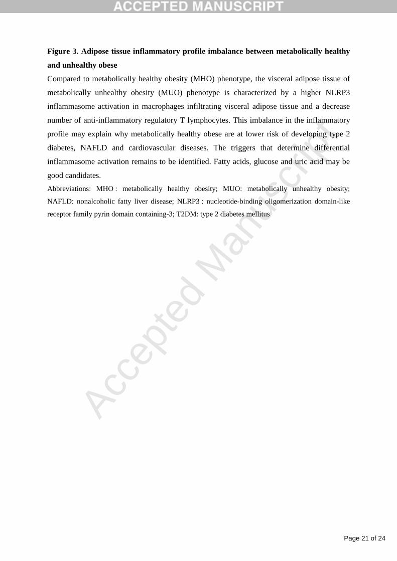
Figure 1. Adipose tissue inflammation in pathogenesis of metabolic syndrome and type 2
diabetes
Obesity induces adipocyte hypertrophy and changes in stromovascular cell composition with
their phenotypic activation to a pro-inflammatory state. Cells from the adaptive immune
system interact with adipose tissue macrophages to modify their activation state. In lean
adipose tissue, T helper type 2 and regulatory T lymphocytes promote a M2 macrophage
polarization, which maintain an anti-inflammatory state. Eosinophils may also induce a M2
macrophage polarization. In obesity and type 2 diabetes, adipose tissue is characterized by an
enrichment of macrophages and T lymphocytes with a shift from an anti-inflammatory to a
pro-inflammatory state. Cytotoxic CD8+, T helper type 1 and T helper type 17 lymphocytes
stimulate M1 macrophage polarization. Other cells, including B lymphocytes and mast cells,
are also increased in obese adipose tissue and may contribute to obesity-induced
inflammation. In obesity, the imbalance among immune cells results in production of
chemokines and pro-inflammatory cytokines, which promote systemic inflammation and
peripheral insulin resistance.
Abbreviations: IL: interleukin; TNFα: tumor necrosis factor alpha; T2DM: type 2 diabetes mellitus;
Treg : regulatory T lymphocytes.
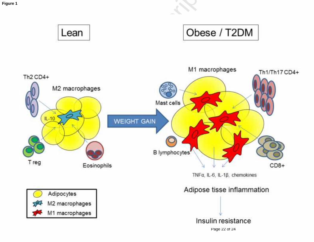
Figure 2. The NLRP3 inflammasome
NLRP3 inflammasome is an intracellular multiprotein complex formed through the
interaction of NLRP3, the protein adaptor ASC and the pro-caspase-1, leading to caspase-1
activation. One activated, caspase-1 cleaves the inactive precursor of IL-1β (pro-IL-1β) into
its biological active form which is secreted. NLRP3 activation requires a two-step process.
First or “priming” signal acts on TLR or cytokines receptors and converges on the activation
of NFκB pathway and transcriptional expression of inflammasome components, including
NLRP3 and pro-IL-1β. The second or “activating” signal is then able to directly induce
NLRP3 inflammasome formation and instigates caspase-1 activation. The second signal
includes various PAMPs and DAMPs.
Abbreviations: ASC: apoptosis-associated speck-like protein containing a caspase-recruitment
domain; DAMPs: danger-associated molecular pattern; IL-1β : interleukin-1β ; NFκB : nuclear factor-
kappa B ; NLRP3 : nucleotide-binding oligomerization domain-like receptor family pyrin domain
containing-3; PAMPs: pathogen-associated molecular patterns; TLR: toll-like receptor
Figure legend(s)
Page 21 of 24
Accep
ted
Man
uscr
ipt
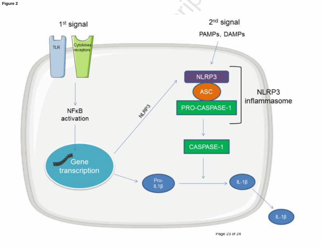
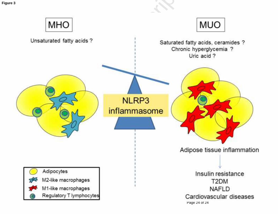
Figure 3. Adipose tissue inflammatory profile imbalance between metabolically healthy
and unhealthy obese
Compared to metabolically healthy obesity (MHO) phenotype, the visceral adipose tissue of
metabolically unhealthy obesity (MUO) phenotype is characterized by a higher NLRP3
inflammasome activation in macrophages infiltrating visceral adipose tissue and a decrease
number of anti-inflammatory regulatory T lymphocytes. This imbalance in the inflammatory
profile may explain why metabolically healthy obese are at lower risk of developing type 2
diabetes, NAFLD and cardiovascular diseases. The triggers that determine differential
inflammasome activation remains to be identified. Fatty acids, glucose and uric acid may be
good candidates.
Abbreviations: MHO : metabolically healthy obesity; MUO: metabolically unhealthy obesity;
NAFLD: nonalcoholic fatty liver disease; NLRP3 : nucleotide-binding oligomerization domain-like
receptor family pyrin domain containing-3; T2DM: type 2 diabetes mellitus
Page 22 of 24
Accep
ted
Man
uscr
ipt
Figure 1
Page 23 of 24
Accep
ted
Man
uscr
ipt
Figure 2
Page 24 of 24
Accep
ted
Man
uscr
ipt
Figure 3