Embed Size (px)
Citation preview
URTICARIA AND ANGIO-OEDEMA
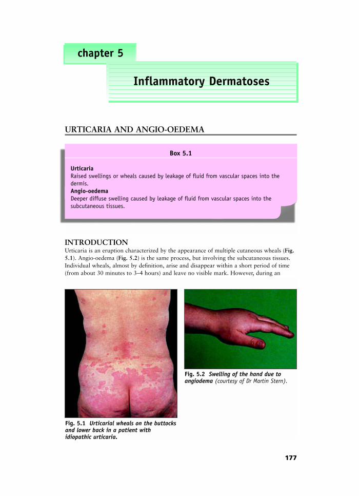
INTRODUCTIONUrticaria is an eruption characterized by the appearance of multiple cutaneous wheals (Fig.5.1). Angio-oedema (Fig. 5.2) is the same process, but involving the subcutaneous tissues.Individual wheals, almost by definition, arise and disappear within a short period of time(from about 30 minutes to 3–4 hours) and leave no visible mark. However, during an
177
chapter 5
Inflammatory Dermatoses
Box 5.1
UrticariaRaised swellings or wheals caused by leakage of fluid from vascular spaces into thedermis.Angio-oedemaDeeper diffuse swelling caused by leakage of fluid from vascular spaces into the subcutaneous tissues.
Fig. 5.1 Urticarial wheals on the buttocksand lower back in a patient with idiopathic urticaria.
Fig. 5.2 Swelling of the hand due toangiodema (courtesy of Dr Martin Stern).
attack of urticaria, crops of wheals continue to appear anywhere on the body surface. Insome patients, the whealing tendency lasts for a few days at most. In other patients, theprocess can continue for months or years, but there may be days free of whealing andothers where the problem is much more intense. Rarely, urticaria is part of a moregeneralized systemic reaction to injected, ingested, or inhaled proteins. This situation —known as anaphylaxis — may be associated with profound lowering of blood pressure anddeath.
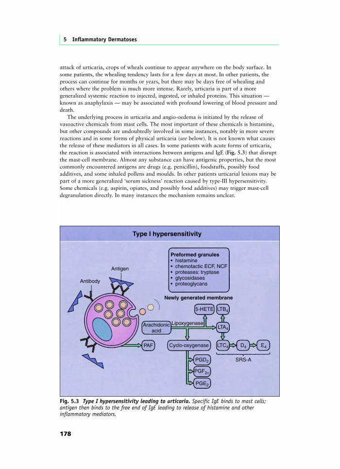
The underlying process in urticaria and angio-oedema is initiated by the release ofvasoactive chemicals from mast cells. The most important of these chemicals is histamine,but other compounds are undoubtedly involved in some instances, notably in more severereactions and in some forms of physical urticaria (see below). It is not known what causesthe release of these mediators in all cases. In some patients with acute forms of urticaria,the reaction is associated with interactions between antigens and IgE (Fig. 5.3) that disruptthe mast-cell membrane. Almost any substance can have antigenic properties, but the mostcommonly encountered antigens are drugs (e.g. penicillin), foodstuffs, possibly foodadditives, and some inhaled pollens and moulds. In other patients urticarial lesions may bepart of a more generalized ‘serum sickness’ reaction caused by type-III hypersensitivity.Some chemicals (e.g. aspirin, opiates, and possibly food additives) may trigger mast-celldegranulation directly. In many instances the mechanism remains unclear.
5 Inflammatory Dermatoses
178
Type I hypersensitivity
Preformed granules• histamine• chemotactic ECF, NCF• proteases: tryptase• glycosidases• proteoglycans
Antigen
Antibody
Arachidonicacid
PAF Cyclo-oxygenase
SRS-A
5-HETE
Newly generated membrane
LTB4
LTC4
PGD2
PGF2α
PGE2
D4 E4
LTA4Lipoxygenase
Fig. 5.3 Type I hypersensitivity leading to urticaria. Specific IgE binds to mast cells;antigen then binds to the free end of IgE leading to release of histamine and otherinflammatory mediators.
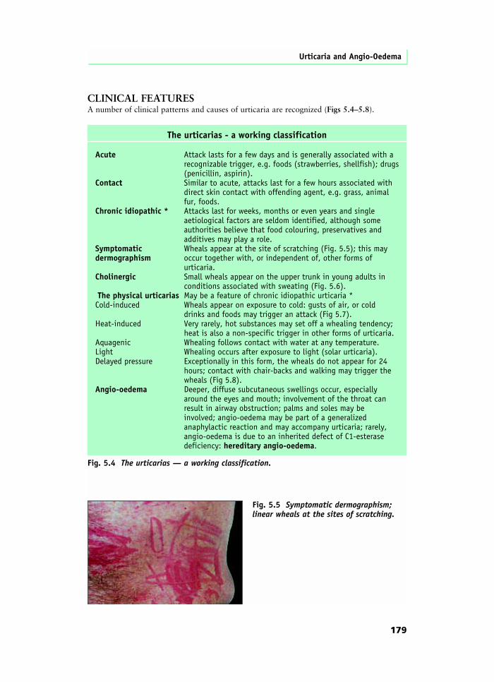
CLINICAL FEATURESA number of clinical patterns and causes of urticaria are recognized (Figs 5.4–5.8).
Urticaria and Angio-Oedema
179
The urticarias - a working classification
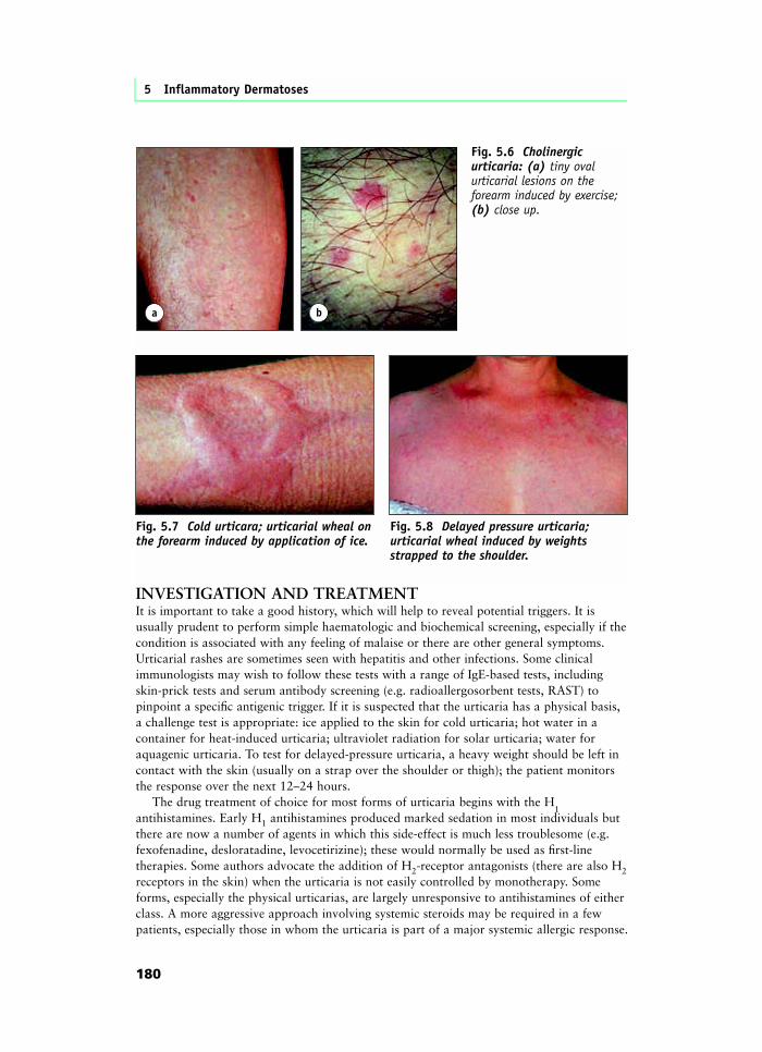
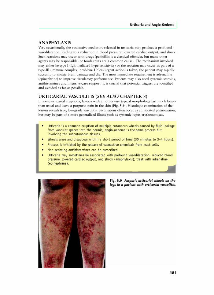
Acute Attack lasts for a few days and is generally associated with a recognizable trigger, e.g. foods (strawberries, shellfish); drugs (penicillin, aspirin).Contact Similar to acute, attacks last for a few hours associated with direct skin contact with offending agent, e.g. grass, animal fur, foods.Chronic idiopathic * Attacks last for weeks, months or even years and single aetiological factors are seldom identified, although some authorities believe that food colouring, preservatives and additives may play a role.Symptomatic Wheals appear at the site of scratching (Fig. 5.5); this maydermographism occur together with, or independent of, other forms of urticaria.Cholinergic Small wheals appear on the upper trunk in young adults in conditions associated with sweating (Fig. 5.6). The physical urticarias May be a feature of chronic idiopathic urticaria *Cold-induced Wheals appear on exposure to cold: gusts of air, or cold drinks and foods may trigger an attack (Fig 5.7).Heat-induced Very rarely, hot substances may set off a whealing tendency; heat is also a non-specific trigger in other forms of urticaria.Aquagenic Whealing follows contact with water at any temperature.Light Whealing occurs after exposure to light (solar urticaria).Delayed pressure Exceptionally in this form, the wheals do not appear for 24 hours; contact with chair-backs and walking may trigger the wheals (Fig 5.8).Angio-oedema Deeper, diffuse subcutaneous swellings occur, especially around the eyes and mouth; involvement of the throat can result in airway obstruction; palms and soles may be involved; angio-oedema may be part of a generalized anaphylactic reaction and may accompany urticaria; rarely, angio-oedema is due to an inherited defect of C1-esterase deficiency: hereditary angio-oedema.
Fig. 5.4 The urticarias — a working classification.
Fig. 5.5 Symptomatic dermographism;linear wheals at the sites of scratching.
INVESTIGATION AND TREATMENTIt is important to take a good history, which will help to reveal potential triggers. It isusually prudent to perform simple haematologic and biochemical screening, especially if thecondition is associated with any feeling of malaise or there are other general symptoms.Urticarial rashes are sometimes seen with hepatitis and other infections. Some clinicalimmunologists may wish to follow these tests with a range of IgE-based tests, includingskin-prick tests and serum antibody screening (e.g. radioallergosorbent tests, RAST) topinpoint a specific antigenic trigger. If it is suspected that the urticaria has a physical basis,a challenge test is appropriate: ice applied to the skin for cold urticaria; hot water in acontainer for heat-induced urticaria; ultraviolet radiation for solar urticaria; water foraquagenic urticaria. To test for delayed-pressure urticaria, a heavy weight should be left incontact with the skin (usually on a strap over the shoulder or thigh); the patient monitorsthe response over the next 12–24 hours.
The drug treatment of choice for most forms of urticaria begins with the H1
antihistamines. Early H1 antihistamines produced marked sedation in most individuals butthere are now a number of agents in which this side-effect is much less troublesome (e.g.fexofenadine, desloratadine, levocetirizine); these would normally be used as first-linetherapies. Some authors advocate the addition of H2-receptor antagonists (there are also H2receptors in the skin) when the urticaria is not easily controlled by monotherapy. Someforms, especially the physical urticarias, are largely unresponsive to antihistamines of eitherclass. A more aggressive approach involving systemic steroids may be required in a fewpatients, especially those in whom the urticaria is part of a major systemic allergic response.
5 Inflammatory Dermatoses
180
a
Fig. 5.6 Cholinergicurticaria: (a) tiny ovalurticarial lesions on theforearm induced by exercise;(b) close up.
b
Fig. 5.7 Cold urticara; urticarial wheal onthe forearm induced by application of ice.
Fig. 5.8 Delayed pressure urticaria;urticarial wheal induced by weightsstrapped to the shoulder.
ANAPHYLAXISVery occasionally, the vasoactive mediators released in urticaria may produce a profoundvasodilatation, leading to a reduction in blood pressure, lowered cardiac output, and shock.Such reactions may occur with drugs (penicillin is a classical offender, but many otheragents may be responsible) or foods (nuts are a common cause). The mechanism involvedmay either be type I (IgE-mediated hypersensitivity) or the reaction may occur as part of atype-III (immune complex) problem. Unless urgent action is taken, the patient may rapidlysuccumb to anoxic brain damage and die. The most immediate requirement is adrenaline(epinephrine) to improve circulatory performance. Patients may also need systemic steroids,antihistamines and intensive-care support. It is crucial that potential triggers are identifiedand avoided as far as possible.
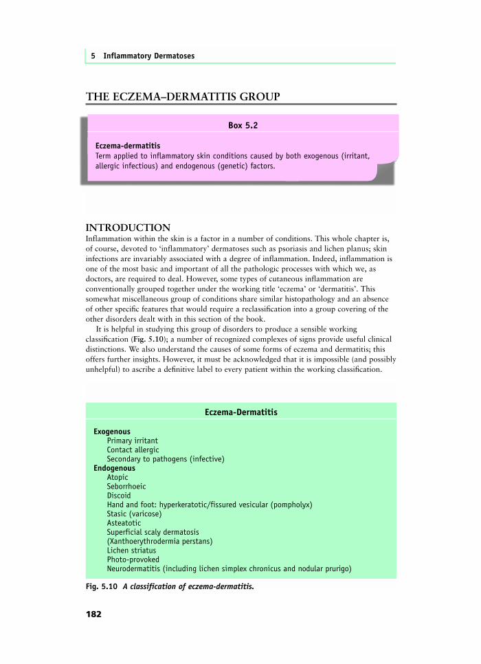
URTICARIAL VASCULITIS (SEE ALSO CHAPTER 8)In some urticarial eruptions, lesions with an otherwise typical morphology last much longerthan usual and leave a purpuric stain in the skin (Fig. 5.9). Histologic examination of thelesions reveals true, low-grade vasculitis. Such lesions often occur as an isolated phenomenon,but may be part of a more generalized illness such as systemic lupus erythematosus.
Urticaria and Angio-Oedema
181
• Urticaria is a common eruption of multiple cutaneous wheals caused by fluid leakage from vascular spaces into the dermis; angio-oedema is the same process but involving the subcutaneous tissues.
• Wheals arise and disappear within a short period of time (30 minutes to 3–4 hours).• Process is initiated by the release of vasoactive chemicals from mast cells.• Non-sedating antihistamines can be prescribed.• Urticaria may sometimes be associated with profound vasodilatation, reduced blood
pressure, lowered cardiac output, and shock (anaphylaxis); treat with adrenaline (epinephrine).
Fig. 5.9 Purpuric urticarial wheals on thelegs in a patient with urticarial vasculitis.
THE ECZEMA–DERMATITIS GROUP
INTRODUCTIONInflammation within the skin is a factor in a number of conditions. This whole chapter is,of course, devoted to ‘inflammatory’ dermatoses such as psoriasis and lichen planus; skininfections are invariably associated with a degree of inflammation. Indeed, inflammation isone of the most basic and important of all the pathologic processes with which we, asdoctors, are required to deal. However, some types of cutaneous inflammation areconventionally grouped together under the working title ‘eczema’ or ‘dermatitis’. Thissomewhat miscellaneous group of conditions share similar histopathology and an absenceof other specific features that would require a reclassification into a group covering of theother disorders dealt with in this section of the book.
It is helpful in studying this group of disorders to produce a sensible workingclassification (Fig. 5.10); a number of recognized complexes of signs provide useful clinicaldistinctions. We also understand the causes of some forms of eczema and dermatitis; thisoffers further insights. However, it must be acknowledged that it is impossible (and possiblyunhelpful) to ascribe a definitive label to every patient within the working classification.
5 Inflammatory Dermatoses
182
Box 5.2
Eczema-dermatitisTerm applied to inflammatory skin conditions caused by both exogenous (irritant, allergic infectious) and endogenous (genetic) factors.
Eczema-Dermatitis
Exogenous Primary irritant Contact allergic Secondary to pathogens (infective)Endogenous Atopic Seborrhoeic Discoid Hand and foot: hyperkeratotic/fissured vesicular (pompholyx) Stasic (varicose) Asteatotic Superficial scaly dermatosis (Xanthoerythrodermia perstans) Lichen striatus Photo-provoked Neurodermatitis (including lichen simplex chronicus and nodular prurigo)
Fig. 5.10 A classification of eczema-dermatitis.
It cannot be emphasized enough that nearly all eczema is caused by a combination ofboth endogenous (i.e. genetic) and exogenous factors. For example, atopic dermatitisclearly has a very significant genetic component (as shown by family involvement and theparticularly high degree of concordance for the disease between identical twins). However,it seems highly likely that something in the environment needs to activate this geneticpredisposition. The same is certainly true of irritant dermatitis, where the same exposure toirritants such as soaps or detergents will provoke a reaction much sooner in someone witha tendency to eczema than someone without. There may be several overlapping endogenousand exogenous factors at work.
CLINICAL FORMS OF ECZEMA–DERMATITIS
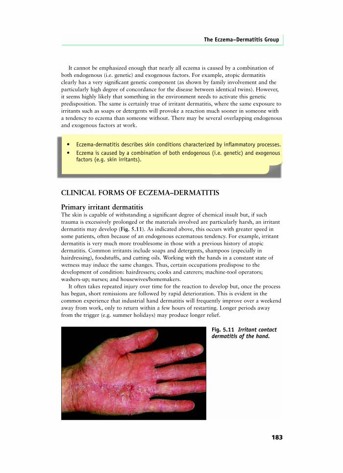
Primary irritant dermatitisThe skin is capable of withstanding a significant degree of chemical insult but, if suchtrauma is excessively prolonged or the materials involved are particularly harsh, an irritantdermatitis may develop (Fig. 5.11). As indicated above, this occurs with greater speed insome patients, often because of an endogenous eczematous tendency. For example, irritantdermatitis is very much more troublesome in those with a previous history of atopicdermatitis. Common irritants include soaps and detergents, shampoos (especially inhairdressing), foodstuffs, and cutting oils. Working with the hands in a constant state ofwetness may induce the same changes. Thus, certain occupations predispose to thedevelopment of condition: hairdressers; cooks and caterers; machine-tool operators;washers-up; nurses; and housewives/homemakers.
It often takes repeated injury over time for the reaction to develop but, once the processhas begun, short remissions are followed by rapid deterioration. This is evident in thecommon experience that industrial hand dermatitis will frequently improve over a weekendaway from work, only to return within a few hours of restarting. Longer periods awayfrom the trigger (e.g. summer holidays) may produce longer relief.
The Eczema–Dermatitis Group
183
• Eczema-dermatitis describes skin conditions characterized by inflammatory processes.• Eczema is caused by a combination of both endogenous (i.e. genetic) and exogenous
factors (e.g. skin irritants).
Fig. 5.11 Irritant contactdermatitis of the hand.
Investigation and treatment — The only permanent solution to severe irritant dermatitis isthe cessation of the provoking activity but, if this is impossible, some relief can sometimesbe obtained by the judicious use of topical corticosteroids to suppress inflammation, theliberal use of emollients and non-soap cleansers, and avoidance measures such as glovesand barrier creams.
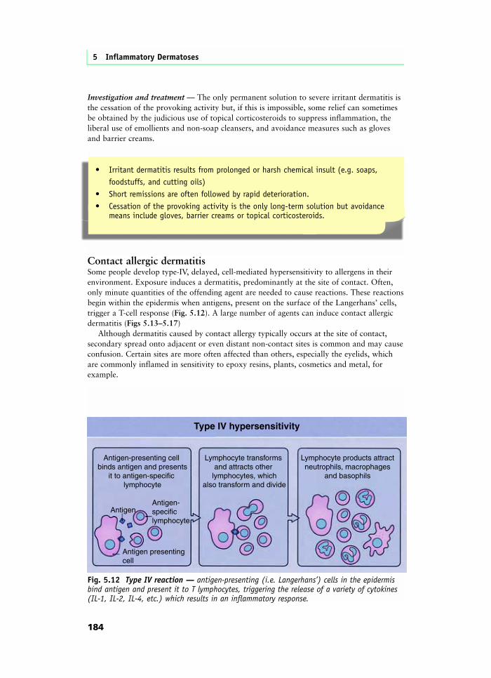
Contact allergic dermatitisSome people develop type-IV, delayed, cell-mediated hypersensitivity to allergens in theirenvironment. Exposure induces a dermatitis, predominantly at the site of contact. Often,only minute quantities of the offending agent are needed to cause reactions. These reactionsbegin within the epidermis when antigens, present on the surface of the Langerhans’ cells,trigger a T-cell response (Fig. 5.12). A large number of agents can induce contact allergicdermatitis (Figs 5.13–5.17)
Although dermatitis caused by contact allergy typically occurs at the site of contact,secondary spread onto adjacent or even distant non-contact sites is common and may causeconfusion. Certain sites are more often affected than others, especially the eyelids, whichare commonly inflamed in sensitivity to epoxy resins, plants, cosmetics and metal, forexample.
5 Inflammatory Dermatoses
184
• Irritant dermatitis results from prolonged or harsh chemical insult (e.g. soaps, foodstuffs, and cutting oils)
• Short remissions are often followed by rapid deterioration.• Cessation of the provoking activity is the only long-term solution but avoidance
means include gloves, barrier creams or topical corticosteroids.
Fig. 5.12 Type IV reaction — antigen-presenting (i.e. Langerhans’) cells in the epidermisbind antigen and present it to T lymphocytes, triggering the release of a variety of cytokines(IL-1, IL-2, IL-4, etc.) which results in an inflammatory response.
Type IV hypersensitivity
Lymphocyte products attractneutrophils, macrophages
and basophils
Antigen presenting cell
Antigen-specificlymphocyte
Lymphocyte transforms and attracts other
lymphocytes, whichalso transform and divide
Antigen-presenting cell binds antigen and presents
it to antigen-specific lymphocyte
Antigen
Any material that is volatile, or can be airborne in the form of dust, can give rise toairborne contact dermatitis. The pattern of involvement here is often of a diffuse dermatitisof the face, backs of hands, and other exposed areas, simulating a light-sensitive eczema.However, the classical light-spared areas are usually involved.Investigation and treatment — Investigation must begin with a careful history of exposureto potential sensitizers. It is important to establish a clear description of possible work anddomestic sources. This should include details of all tasks carried out, hobbies and leisurepursuits, and cosmetics, toiletries, and medicaments applied to the skin surface. The keyinvestigative technique is patch testing (Fig. 5.18), in which suspected offending agents areapplied to the surface of the skin for 48 hours before being removed; the site is thenexamined for evidence of allergic dermatitis (Fig. 5.19). A second examination after 72 or96 hours is also essential. It has been estimated that up to 30% of positive reactions will bemissed if this is not undertaken because some compounds produce later reactions (e.g.lanolin, neomycin). In most instances, a battery of common test allergens is used and thesemay be modified and adapted for local circumstances or for particular problem areas (e.g.medicaments) or occupations (e.g. hairdressing). It is also often useful to test materials
The Eczema–Dermatitis Group
185
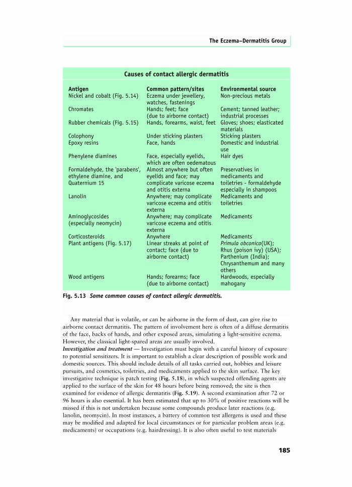
Causes of contact allergic dermatitis
Antigen Common pattern/sites Environmental sourceNickel and cobalt (Fig. 5.14) Eczema under jewellery, Non-precious metals watches, fasteningsChromates Hands; feet; face Cement; tanned leather; (due to airborne contact) industrial processesRubber chemicals (Fig. 5.15) Hands, forearms, waist, feet Gloves; shoes; elasticated materialsColophony Under sticking plasters Sticking plastersEpoxy resins Face, hands Domestic and industrial usePhenylene diamines Face, especially eyelids, Hair dyes which are often oedematousFormaldehyde, the 'parabens', Almost anywhere but often Preservatives inethylene diamine, and eyelids and face; may medicaments andQuaternium 15 complicate varicose eczema toiletries - formaldehyde and otitis externa especially in shampoosLanolin Anywhere; may complicate Medicaments and varicose eczema and otitis toiletries externaAminoglycosides Anywhere; may complicate Medicaments(especially neomycin) varicose eczema and otitis externa Corticosteroids Anywhere MedicamentsPlant antigens (Fig. 5.17) Linear streaks at point of Primula obconica(UK); contact; face (due to Rhus (poison ivy) (USA); airborne contact) Parthenium (India); Chrysanthemum and many othersWood antigens Hands; forearms; face Hardwoods, especially (due to airborne contact) mahogany
Fig. 5.13 Some common causes of contact allergic dermatitis.
5 Inflammatory Dermatoses
186
a
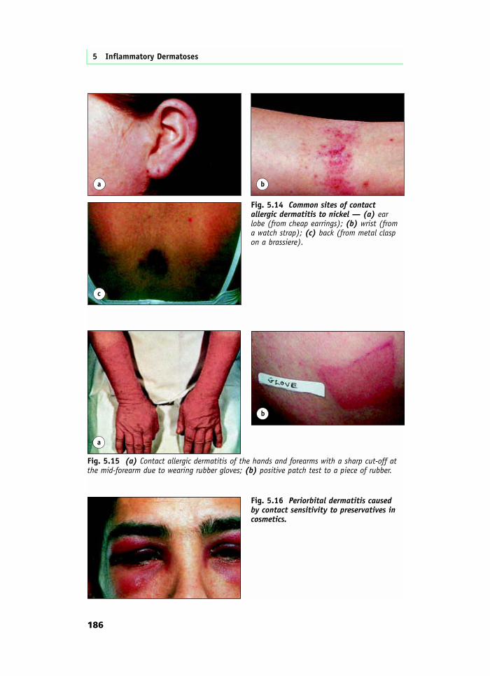
Fig. 5.15 (a) Contact allergic dermatitis of the hands and forearms with a sharp cut-off atthe mid-forearm due to wearing rubber gloves; (b) positive patch test to a piece of rubber.
b
a b
c
Fig. 5.14 Common sites of contactallergic dermatitis to nickel — (a) earlobe (from cheap earrings); (b) wrist (froma watch strap); (c) back (from metal claspon a brassiere).
Fig. 5.16 Periorbital dermatitis caused by contact sensitivity to preservatives incosmetics.
The Eczema–Dermatitis Group
187
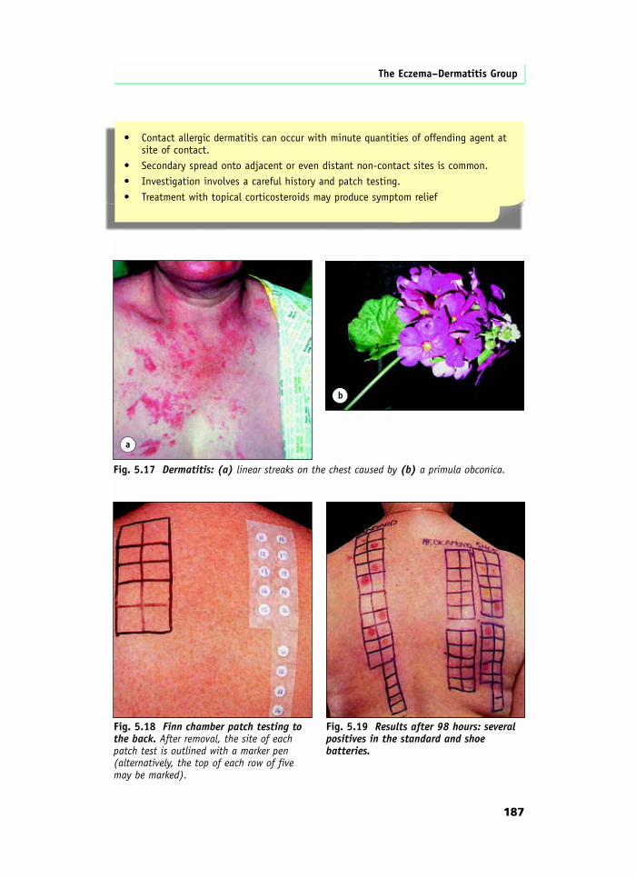
• Contact allergic dermatitis can occur with minute quantities of offending agent at site of contact.
• Secondary spread onto adjacent or even distant non-contact sites is common.• Investigation involves a careful history and patch testing.• Treatment with topical corticosteroids may produce symptom relief
a
Fig. 5.17 Dermatitis: (a) linear streaks on the chest caused by (b) a primula obconica.
b
Fig. 5.19 Results after 98 hours: severalpositives in the standard and shoebatteries.
Fig. 5.18 Finn chamber patch testing tothe back. After removal, the site of eachpatch test is outlined with a marker pen(alternatively, the top of each row of fivemay be marked).
5 Inflammatory Dermatoses
188
suspected by the patient of being responsible (e.g. make-ups, materials handled at work).However, care must be taken not to place highly irritant chemicals on the skin. If there isany doubt, a specialized test should be used.
It may also be useful to visit the patient’s place of work to see precisely what is involved.This may draw attention to tasks and contacts that were not apparent from the history alone.
Treatment of an acute attack with potent topical corticosteroids may produce significantsymptom relief; this could perhaps be combined with soaking in potassium permanganatesolution if there is a vesicular or bullous component (see also Dermatitis of the hands andfeet). However, it is essential to remove the causative agent from the environment as far aspossible.
Infective eczemaEczematous changes can be induced by invading organisms. The red, itchy rash that isassociated with ringworm (tinea) is nothing more than an inflammatory response to thepresence of fungal organisms in the skin (see Chapter 3). A bizarre form of this eczematousresponse to fungal infection is seen with the so-called dermatophytide, in which a widespreadeczema and pompholyx of hands and feet develop because of hypersensitivity to a fungalinfection, usually of one foot. Typical eczematous dermatitis also occurs commonly aroundareas of molluscum contagiosum, especially (but not exclusively) in children with an underlyingtendency to atopic dermatitis. Bacterial infections may occasionally produce similar changes.
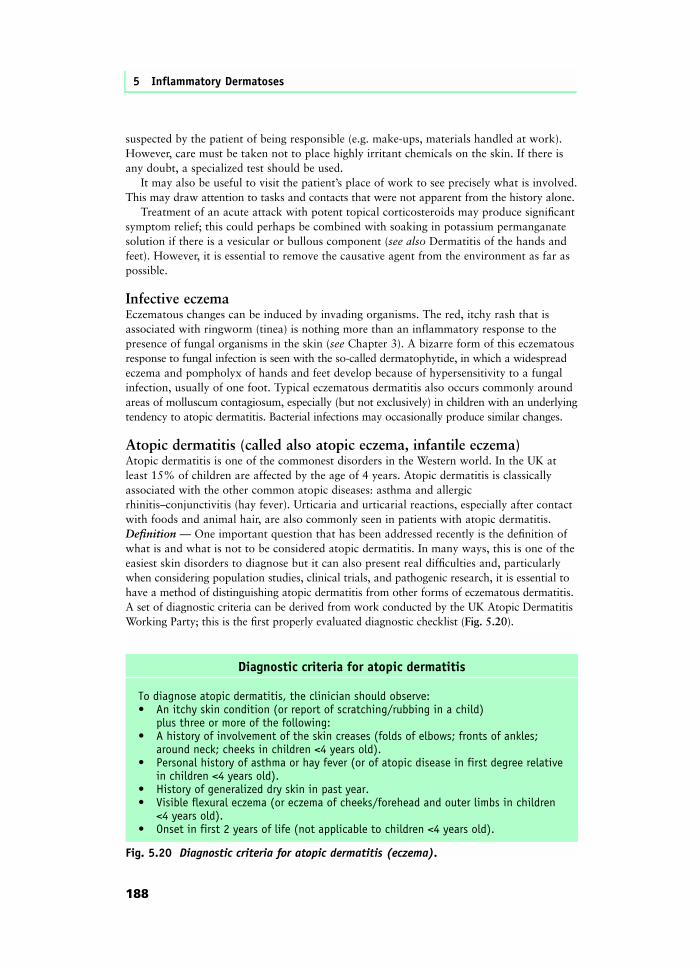
Atopic dermatitis (called also atopic eczema, infantile eczema)Atopic dermatitis is one of the commonest disorders in the Western world. In the UK atleast 15% of children are affected by the age of 4 years. Atopic dermatitis is classicallyassociated with the other common atopic diseases: asthma and allergicrhinitis–conjunctivitis (hay fever). Urticaria and urticarial reactions, especially after contactwith foods and animal hair, are also commonly seen in patients with atopic dermatitis.Definition — One important question that has been addressed recently is the definition ofwhat is and what is not to be considered atopic dermatitis. In many ways, this is one of theeasiest skin disorders to diagnose but it can also present real difficulties and, particularlywhen considering population studies, clinical trials, and pathogenic research, it is essential tohave a method of distinguishing atopic dermatitis from other forms of eczematous dermatitis.A set of diagnostic criteria can be derived from work conducted by the UK Atopic DermatitisWorking Party; this is the first properly evaluated diagnostic checklist (Fig. 5.20).
Diagnostic criteria for atopic dermatitis
To diagnose atopic dermatitis, the clinician should observe:• An itchy skin condition (or report of scratching/rubbing in a child) plus three or more of the following:• A history of involvement of the skin creases (folds of elbows; fronts of ankles; around neck; cheeks in children <4 years old).• Personal history of asthma or hay fever (or of atopic disease in first degree relative in children <4 years old).• History of generalized dry skin in past year.• Visible flexural eczema (or eczema of cheeks/forehead and outer limbs in children <4 years old).• Onset in first 2 years of life (not applicable to children <4 years old).
Fig. 5.20 Diagnostic criteria for atopic dermatitis (eczema).
Pathogenesis — It is clear that genetics are fundamentally important in atopic dermatitis.As indicated above, a family history of asthma, rhinitis, atopic dermatitis itself, and otheratopic phenomena is common in patients with the condition. Furthermore, a Danish twinstudy has demonstrated unequivocally that the risk of developing atopic dermatitis is muchhigher in monozygotic twins than in dizygotic twins or non-twin siblings. It also appearsfrom some studies that there may be a significant degree of specificity in the inheritance ofthe atopic disorders. However, it is also apparent that this condition, and the other atopicdisorders, are complex interactions between this genetic susceptibility and the environmentin which the individual lives. Atopic dermatitis is more common in higher socioeconomicgroups; in one community-based study in Leicester, we demonstrated that although atopicdermatitis is equally common among children of both (white) European and Asian families,a family history of atopic disease is significantly less frequent among Asians. Oneinterpretation of these findings is that genetic susceptibility exists equally in both groups,but that an element of the Western environment has triggered the development of atopicdermatitis.
It is highly likely that disordered immunologic function is fundamental in thishost–environment interaction. It is well known that the majority of patients with atopicdermatitis have high IgE levels and positive prick tests for allergens such as house dustmites, cat and other animal fur, dander, pollens, grasses, moulds, and some foods. However,it is also clear that the immunohistological features of the condition are more in keepingwith those of a type-IV hypersensitivity reaction than the type-I reaction classically seen inurticaria. Attempts have been made with some success to reproduce eczematous lesions bythe application of airborne allergens and house dust mite, and support for the possibleimportance of such allergens is beginning to emerge with therapeutic prophylactic andinterventional studies.
Two discoveries have been made about the clinically involved skin of patients withatopic dermatitis: epidermal Langerhans’ cells bind IgE; IgE was also found to be presenton the surface of antigen-presenting cells in the dermal infiltrate. This has led to thehypothesis that antigens absorbed through the skin might bind to allergen-specific IgE onthe surface of epidermal Langerhans’ cells, thereby inducing T-lymphocyte activation andan eczematous hypersensitivity response.
Further work also appears to indicate that T-cell proliferation in patients with atopicdermatitis results in the preferential expansion of a clone of T cells known as the T-helper 2subset. This type of work, together with a better understanding of the underlying moleculargenetics, will continue to refine our knowledge of the intricacies of the interactions takingplace in this disease.
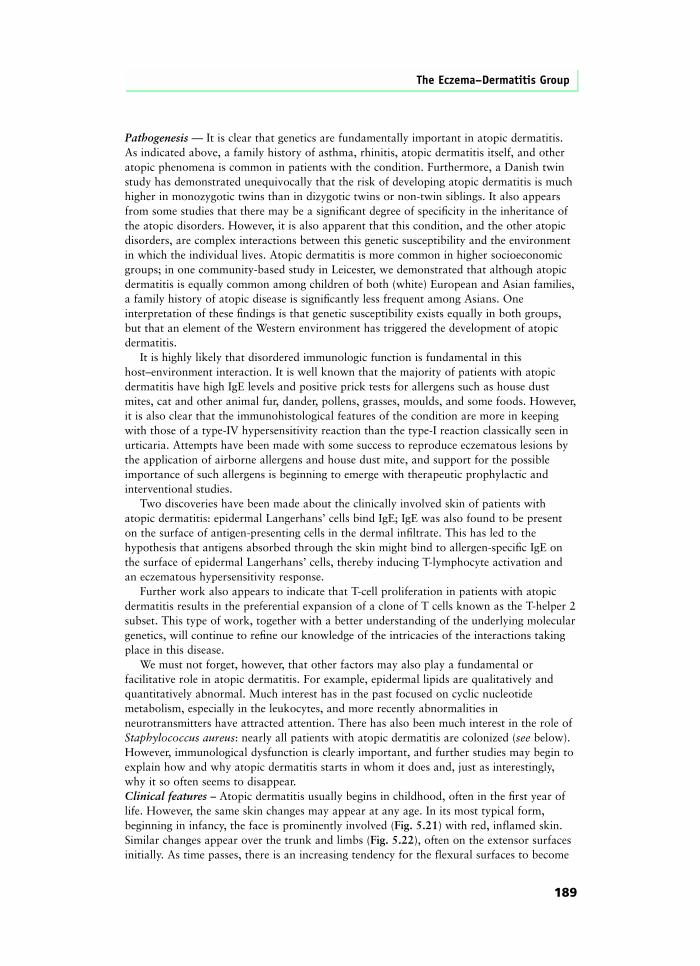
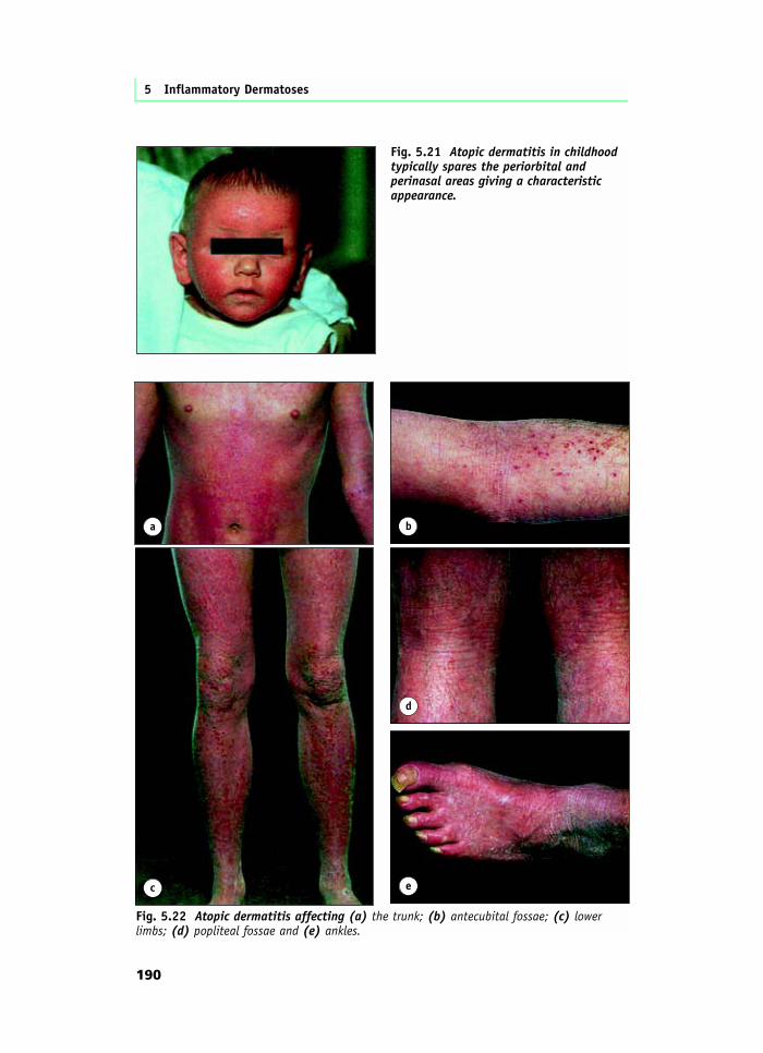
We must not forget, however, that other factors may also play a fundamental orfacilitative role in atopic dermatitis. For example, epidermal lipids are qualitatively andquantitatively abnormal. Much interest has in the past focused on cyclic nucleotidemetabolism, especially in the leukocytes, and more recently abnormalities inneurotransmitters have attracted attention. There has also been much interest in the role ofStaphylococcus aureus: nearly all patients with atopic dermatitis are colonized (see below).However, immunological dysfunction is clearly important, and further studies may begin toexplain how and why atopic dermatitis starts in whom it does and, just as interestingly,why it so often seems to disappear.Clinical features – Atopic dermatitis usually begins in childhood, often in the first year oflife. However, the same skin changes may appear at any age. In its most typical form,beginning in infancy, the face is prominently involved (Fig. 5.21) with red, inflamed skin.Similar changes appear over the trunk and limbs (Fig. 5.22), often on the extensor surfacesinitially. As time passes, there is an increasing tendency for the flexural surfaces to become
The Eczema–Dermatitis Group
189
5 Inflammatory Dermatoses
190
Fig. 5.21 Atopic dermatitis in childhoodtypically spares the periorbital andperinasal areas giving a characteristicappearance.
a b
c
Fig. 5.22 Atopic dermatitis affecting (a) the trunk; (b) antecubital fossae; (c) lowerlimbs; (d) popliteal fossae and (e) ankles.
d
e
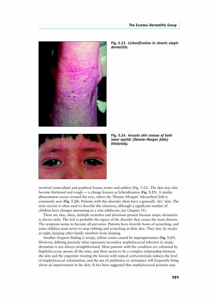
involved (antecubital and popliteal fossae; wrists and ankles) (Fig. 5.22). The skin may alsobecome thickened and rough — a change known as lichenification (Fig. 5.23). A similarphenomenon occurs around the eyes, where the ‘Dennie–Morgan’ infraorbital fold iscommonly seen (Fig. 5.24). Patients with this disorder often have a generally ‘dry’ skin. Theterm xerosis is often used to describe this situation, although a significant number ofchildren have changes amounting to a true ichthyosis (see Chapter 11).
There are also, often, multiple scratches and abrasions present because atopic dermatitisis always itchy. The itch is probably the aspect of the disorder that causes the most distress.The symptom seems to become all-pervasive. Patients have feverish bouts of scratching, andsome children seem never to stop rubbing and scratching at their skin. They may lie awakeat night, keeping other family members from sleeping.
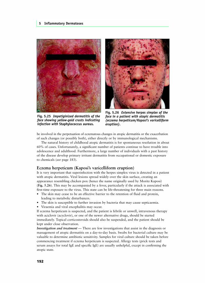
Another frequent finding is weepy, yellow crusts caused by impetiginization (Fig. 5.25).However, defining precisely what represents secondary staphylococcal infection in atopicdermatitis is not always straightforward. Most patients with the condition are colonized byStaphylococcus aureus all the time, and there seems to be a complex relationship betweenthe skin and the organism: treating the lesions with topical corticosteroids reduces the levelof staphylococcal colonization, and the use of antibiotics or antiseptics will frequently bringabout an improvement in the skin. It has been suggested that staphylococcal proteins may
The Eczema–Dermatitis Group
191
Fig. 5.23 Lichenification in chronic atopicdermatitis.
Fig. 5.24 Arcuate skin creases of bothlower eyelids (Dennie–Morgan folds)bilaterally.
be involved in the perpetuation of eczematous changes in atopic dermatitis or the exacerbationof such changes (or possibly both), either directly or by immunological mechanisms.
The natural history of childhood atopic dermatitis is for spontaneous resolution in about60% of cases. Unfortunately, a significant number of patients continue to have trouble intoadolescence and adulthood. Furthermore, a large number of individuals with a past historyof the disease develop primary irritant dermatitis from occupational or domestic exposureto chemicals (see page 183).
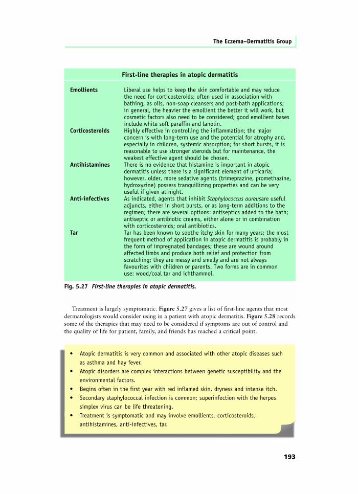
Eczema herpeticum (Kaposi’s varicelliform eruption)It is very important that superinfection with the herpes simplex virus is detected in a patientwith atopic dermatitis. Viral lesions spread widely over the skin surface, creating anappearance resembling chicken pox (hence the name originally used by Moritz Kaposi) (Fig. 5.26). This may be accompanied by a fever, particularly if the attack is associated withfirst-time exposure to the virus. This state can be life-threatening for three main reasons.• The skin may cease to be an effective barrier to the retention of fluid and protein,
leading to metabolic disturbances.• The skin is susceptible to further invasion by bacteria that may cause septicaemia.• Viraemia and viral encephalitis may occur.If eczema herpeticum is suspected, and the patient is febrile or unwell, intravenous therapywith aciclovir (acyclovir), or one of the newer alternative drugs, should be startedimmediately. Topical corticosteroids should also be suspended, and the patient should bekept under close observation.Investigation and treatment — There are few investigations that assist in the diagnosis ormanagement of atopic dermatitis on a day-to-day basis. Swabs for bacterial culture may bevaluable to determine antibiotic sensitivity. Samples for viral culture should be taken beforecommencing treatment if eczema herpeticum is suspected. Allergy tests (prick tests andserum assays for total IgE and specific IgE) are usually unhelpful, except in confirming theatopic state.
5 Inflammatory Dermatoses
192
Fig. 5.26 Extensive herpes simplex of theface in a patient with atopic dermatitis(eczema herpeticum/Kaposi’s varicelliformeruption).
Fig. 5.25 Impetiginized dermatitis of theface showing yellow-gold crusts indicatinginfection with Staphylococcus aureus.
Treatment is largely symptomatic. Figure 5.27 gives a list of first-line agents that mostdermatologists would consider using in a patient with atopic dermatitis. Figure 5.28 recordssome of the therapies that may need to be considered if symptoms are out of control andthe quality of life for patient, family, and friends has reached a critical point.
The Eczema–Dermatitis Group
193
First-line therapies in atopic dermatitis
Emollients Liberal use helps to keep the skin comfortable and may reduce the need for corticosteroids; often used in association with bathing, as oils, non-soap cleansers and post-bath applications; in general, the heavier the emollient the better it will work, but cosmetic factors also need to be considered; good emollient bases include white soft paraffin and lanolin.Corticosteroids Highly effective in controlling the inflammation; the major concern is with long-term use and the potential for atrophy and, especially in children, systemic absorption; for short bursts, it is reasonable to use stronger steroids but for maintenance, the weakest effective agent should be chosen.Antihistamines There is no evidence that histamine is important in atopic dermatitis unless there is a significant element of urticaria; however, older, more sedative agents (trimeprazine, promethazine, hydroxyzine) possess tranquillizing properties and can be very useful if given at night.Anti-infectives As indicated, agents that inhibit Staphylococcus aureusare useful adjuncts, either in short bursts, or as long-term additions to the regimen; there are several options: antiseptics added to the bath; antiseptic or antibiotic creams, either alone or in combination with corticosteroids; oral antibiotics.Tar Tar has been known to soothe itchy skin for many years; the most frequent method of application in atopic dermatitis is probably in the form of impregnated bandages; these are wound around affected limbs and produce both relief and protection from scratching; they are messy and smelly and are not always favourites with children or parents. Two forms are in common use: wood/coal tar and ichthammol.
Fig. 5.27 First-line therapies in atopic dermatitis.
• Atopic dermatitis is very common and associated with other atopic diseases such as asthma and hay fever.
• Atopic disorders are complex interactions between genetic susceptibility and the environmental factors.
• Begins often in the first year with red inflamed skin, dryness and intense itch.• Secondary staphylococcal infection is common; superinfection with the herpes
simplex virus can be life threatening.• Treatment is symptomatic and may involve emollients, corticosteroids,
antihistamines, anti-infectives, tar.
Seborrhoeic eczema/dermatitisThis is a very common clinical pattern of eczema seen in adults. There is a form of eczemaknown as infantile seborrhoeic dermatitis, which occurs (as the name implies) in infancy.The two are not directly related and must be considered separately. The infantile form isconsidered below.Clinical features — There are a number of unmistakeable features seen in classicalseborrhoeic dermatitis.Pathogenesis — Considered to be a purely endogenous disorder for many years, thepathogenesis of seborrhoeic dermatitis has at last begun to be unravelled. Although theprecise mechanisms involved have still to be elucidated, it seems clear that the yeastMalassezia/Pityrosporum orbiculare (Malassezia furfur) plays a major role in inducing andperpetuating the inflammation (see also Pityriasis/tinea versicolor and Pityrosporon
5 Inflammatory Dermatoses
194
Second-line therapies in atopic dermatitis
These modalities may need to be considered if measures outlined here fail to produce sufficient control or if, as in the case of topical corticosteroids, they may result in an unacceptable risk of side-effects:
Topical calcineurin inhibitors These agents provide an alternative to topical(tacrolimus, pimecrolimus) corticosteroids; they do not cause atrophy and are particularly useful for facial and flexural skin, and as an adjunct to conventional therapy: questions have been raised about possible carcinogenicity, but there is no evidence that this is an issue in clinical practice.Ultraviolet radiation Both ultraviolet B and PUVA have been shown to be effective in atopic dermatitis (for details, see Psoriasis).Dietary and other environmental Anecdotal reports of improvements of atopic alteration dermatitis with diet have led to several studies; a few show benefit, many do not; if all else fails it may be reasonable to try a restricted diet under careful supervision for a few weeks; house dust mite eradication has also been reported to have beneficial effects and may be worth considering.Evening primrose oil The evidence for any significant effect is thin.Immunosuppressives Systemic steroids and ACTH-like agents may occasionally have a role in producing short-term relief; azathioprine has its advocates and has been shown to be effective in two controlled tests; ciclosporin (cyclosporin) A is highly effective, if rather costly and potentially toxic.Chinese herbs There is some evidence that some mixtures of Chinese herbs may be effective in some patients; at this time, these agents have not been licensed.
There have been reports of toxicity (notably hepatitis) and some creams have been found to be adulterated with steroids.
Fig. 5.28 Second-line therapies in atopic dermatitis.
folliculitis, see Chapter 3). It is also of considerable interest that seborrhoeic dermatitis isone of the skin signs associated with HIV infection, particularly at the point at which CD4counts begin to fall.Investigation and treatment — There are no useful tests to aid diagnosis, but HIV infectionmust always be considered in any case where the condition appears to be particularly severeor resistant to treatment. Patients will often benefit from either a topical corticosteroidcream or a topical antifungal agent such as miconazole, clotrimazole, or ketoconazole.(Ketoconazole was the first agent to be investigated and the one that, in part, resulted in there-evaluation of the role of yeasts in this condition.) Sometimes a combination ofcorticosteroid cream and antifungal agent is helpful. Occasionally, severe seborrhoeicdermatitis requires oral treatment with an agent such as itraconazole.
The Eczema–Dermatitis Group
195
• Certain sites are prominently involved:
• Scalp. Mild scaling (or dandruff) represents one end of the clinical spectrum, with marked scaling and erythema at the other; seborrhoeic dermatitis is also one of the causes of the clinical change known as pityriasis amiantacea (Fig. 5.29; see also Psoriasis).
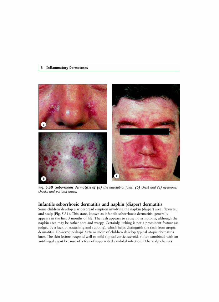
• Nasolabial folds (Fig. 5.30a).
• Upper chest (both front and back) (Fig. 5.30b).
• Behind the ears.
• Eyebrows (Fig. 5.30c).
• On the face the red, scaly, somewhat greasy-looking eruption is characteristic andmay spread out on to the cheeks (Fig. 5.30c).
• Some patients also suffer from an inflammation of the eyelids (blepharitis).
• Others develop a more flexural (intertriginous) form, with lesions in the axillae,groins, and areas where skin surfaces are apposed; these lesions merge, oftenindistinguishably, with the clinical appearance of flexural psoriasis (see page 208).
• Flexural seborrhoeic dermatitis may be mild but may also be extremely troublesome.
• Adult seborrhoeic dermatitis usually presents in adolescence or early adulthood and,although the severity may fluctuate, the tendency often persists throughout life.
Fig. 5.29 Pityriasis amiantacea — scaleenveloping the lower part of hairs inseborrhoeic dermatitis.
Infantile seborrhoeic dermatitis and napkin (diaper) dermatitisSome children develop a widespread eruption involving the napkin (diaper) area, flexures,and scalp (Fig. 5.31). This state, known as infantile seborrhoeic dermatitis, generallyappears in the first 3 months of life. The rash appears to cause no symptoms, although thenapkin area may be rather sore and weepy. Certainly, itching is not a prominent feature (asjudged by a lack of scratching and rubbing), which helps distinguish the rash from atopicdermatitis. However, perhaps 25% or more of children develop typical atopic dermatitislater. The skin lesions respond well to mild topical corticosteroids (often combined with anantifungal agent because of a fear of superadded candidal infection). The scalp changes
5 Inflammatory Dermatoses
a
Fig. 5.30 Seborrhoeic dermatitis of (a) the nasolabial folds; (b) chest and (c) eyebrows,cheeks and perioral areas.
cb
(known as cradle cap) often respond to the application of oils and gentle shampooing butmay require more aggressive therapy with salicylic acid.
Occasionally the skin lesions resemble psoriasis (see below), and the term napkinpsoriasis may be applied (Fig. 5.32).
There are also a number of other causes of inflammation in the napkin (diaper) area.• Primary irritant dermatitis (Fig. 5.33). This is generally thought to be caused by
faecal enzymes. The skin becomes sore and macerated. Exposure is curative butimpractical in most instances. Treatment usually involves attempts to keep the area asdry as possible and the use of emollients and barrier creams. At its most extreme,blisters may occur that resemble herpes (Fig. 5.34).
• Jacquet’s erythema. This is seen in the older child, who may be somewhat neglected.A strong smell of ammonia is usually present in napkins and over-clothing. Areas oferythema are accompanied, in severe forms, by punched-out ulcers (Fig. 5.35).
197


![Dermatoses Ocupacionais[1]](https://img.pdfslide.net/doc/110x75/55cf9be9550346d033a7d524/dermatoses-ocupacionais1.jpg)