Embed Size (px)
Citation preview

1
Influenceofageandsexonthelongitudinalrelaxationtime,T1,ofthelunginhealthynever-smokersSimonSIKindvall*1MS,SandraDiaz2MD,PhD,JonasSvensson3PhD,PerWollmer4MD,PhD,DariuszSlusarczyk2MD,LarsEOlsson1PhD
1. MedicalRadiationPhysics,TranslationalMedicine,LundUniversity,MalmöSweden
2. MedicalRadiology,TranslationalMedicine,LundUniversity,MalmöSweden3. Medicalimagingandphysiology,SkaneUniversityHospital,LundSweden4. ClinicalPhysiology,TranslationalMedicine,LundUniversity,MalmöSweden
*Correspondingauthor:SimonKindvallEmail:[email protected]: MedicalRadiationPhysicsLundUniversitySUSMalmö20502MalmöOffice:+4640-336734Fax:+4640-963185GrantSupport:ThisworkwassupportedbyAllmännaSjukhusetsiMalmöstiftelseförbekämpandeavcancerandStiftelsenförcancerforskningvidonkologiskaklinikenvidUniversitetsjukhusetMAS.RunningTitle:LungT1inhealthynever-smokers

2ABSTRACTPurpose:AsseveralstudieshaveprovidedevidencethatlungdiseaseaffectstheT1ofthehumanlung,ourpurposewastoinvestigatetheeffectofageontheT1-relaxationtimeinthelungsofhealthynever-smokers,includinggroupdifferencebetweensexes.Materialsandmethods:TheSnapshotFLASHpulsesequence(inversionrecoverywithmultiplegradientechoread-outs)wasusedtoquantifylungT1in30healthynever-smokingvolunteersat1.5Tesla.Measurementswereperformedunderbreathholdofatidalinspiration.Additionally,subjectsunderwentclinicalMRIandpulmonaryfunctiontests.AlinearregressionmodelofT1asafunctionofageandsexwastested.Results:TheslopeoflungT1attidalend-inspirationasafunctionofagewasstatisticallydifferentbetweenmalesandfemales(p<0.001).InalinearregressionmodelofT1asafunctionofageandsex,femaleshaveslopeof-4.1ms/year(95%CI=[-5.2,-3.0])atp<0.001,andmales-0.064ms/year(95%CI=[-1.2,1.1])atp=0.9,withawholemodelR2=0.83.Conclusion:TheobserveddependenciesoflungT1onageandsexarehereattributedtoapreviouslyreporteddifferenceinbloodT1betweensexes,andapreviouslyreporteddecreaseofpulmonarybloodvolumewithincreasingage.ThismayhaveimplicationsfortheinterpretationoflungT1measurementsinbothhealthyindividualsandpatients.
Keywords:Sexfactors,Lung,MagneticResonanceImaging,Agefactors,Spirometry

3
INTRODUCTIONDuringrecentyears,thenumberofapplicationsofmagneticresonanceimaging(MRI)ofthehumanlunghasgrown(1,2).OneMRImethodwhichhasgainedinterestforapplicationinthelungisquantificationofthewaterprotonlongitudinalrelaxationtimeT1(T1-mapping),andT1-mappingastheread-outforoxygenenhancedMRI,whichhasbeenshowntoreflectthelungdiffusingcapacity(3,4).T1-mappingandT1-mappingofoxygenenhancementinthelunghavebeenappliedtostudyhealthysubjects(5–9),diffuselungdisease(10),cysticfibrosis(11,12),asthma(13,14),pulmonaryhypertension(15),chroniclungallograftdysfunction(16)andinparticularchronicobstructivepulmonarydisease(COPD)(17–20).ConsideringthatCOPDaloneisthethirdlargestcauseofdeathglobally,surpassedonlybycardio-andcerebrovasculardisease(21),theinterestinthisnon-invasiveimagingtechniqueiseasytoappreciate.However,eventhoughlungT1hasalreadybeenusedasaread-outinclinicalstudiesofforexampleCOPD(20),nodetaileddatahavebeenpresentedforawell-definedhealthycohort.Theageinglungtissueofahealthyindividualischaracterizedbyincreasedcompliance(lesselasticrecoil)–presumablyduetochangesinconcentrationororientationofelasticfibers(22).AsimilarchangeinlungtissuecomplianceoccursinpulmonaryemphysemawhichhasalreadybeenshowntoyielddecreasedlungT1(10).SeveralstudiesofCOPD(17,19,20)havecomparedpatientsandcontrolgroups,whereseverediseaseisoftenassociatedwithahighage.IfT1iscorrelatedtoage,thiswillintroduceastatisticalbias,possiblyobscuringimportantfindings.Thus,thereareseveralreasonstostudyahealthycohort,evenlydistributedbetweensexesanddifferentagegroups.ThepurposeofthisstudyistoprovidebaselinelungT1valuesofhealthynever-smokers,maleandfemale,between20and70yearsofage,andspecificallyinvestigatetheeffectofageonthenativeT1-relaxationtimeinthelung,includinggroupdifferencesbetweensexes,usingapreviouslyestablishedmethodforT1-measurement.

4MATERIALSANDMETHODSWithapprovalfromtheRegionalEthicalReviewBoard,31never-smokinghealthysubjects(16male,15female)between20-70yearswererecruitedtoperformaclinicallungMRIexamination,wholelungT1measurementandpulmonaryfunctiontests(PFT).Never-smokersweredefinedaccordingtothreecriteria:a)neversmokeddailyformorethanamonth,b)smokingoccasionallylessthanonceamonth,andc)reporting0pack-yearsoflifetimetobaccouse.Subjectswereprovidedwithwritteninstructions,signedinformedconsent,reportedlifetimetobaccouse,pulmonaryhealthstatusandfilledoutaMRIsafetysheetpriortothevisit.ThePFTwaseithermadeimmediatelyaftertheMRIexamination,orthenextday.AllMRImeasurementsweremadeona1.5TeslaSiemensMagnetomAvantoFit(SIEMENSHealthcare,Erlangen,Germany),withan18channelbodycoilanda32channelspinematrix.
ClinicalImagesInordertoenablearadiologicalevaluationaclinicalprotocolprecededtheT1measurement.TheclinicalprotocolincludedtwofullcoveragecoronalHASTE(Half-FourierAcquisitionSingleshotTurboSpinEcho)acquisitionsduringend-inspirationbreathholdandduringfreebreathingrespectively,aswellasanaxialVIBEacquisitionduringend-inspirationbreathhold.Theimageswereexaminedbyclinicalradiologistswith10(S.D.)and5(D.S.)yearsofexperience.
T1-MeasurementsFollowingtheclinicalMRI,anumberofcoronalT1-mapscoveringtheentirelungvolumewerecollectedduringtidalend-inspirationbreathhold,resultingin8–12slices.AllT1measurementsweremadewiththeSnapshotFLASHpulsesequence(8)whichisbasedontheLookandLockersequence(23,24).Followingspininversion,severalgradientechoimagesarecollectedduringmagnetizationrecovery.Assumingsufficienttimeisleftbetweenmeasurementsandtheinversionpulseishomogeneous,theinitialmagnetization,steady-statemagnetizationandsignaldynamicscanbeusedtocalculatetheunbiasedT1.Imagingparameterswereasfollow:matrix128x64zerofilledto256x256;450mmsquarefieldofview;1.5cmslicethickness;TE=0.67ms;TR=3.0ms;andflipangle=7°.Anon-selectivehyperbolicsecantpulsewasusedforspininversion;andHanning-windowedsincpulseswith1.6sidelobeswereusedforread-out.OneT1measurementismadeineachslice;16gradientechoimageswithdifferentinversiontimesareusedinthecalculationoftheT1map;andthetotalmeasurementtimeis3.0secondsperslice.EachT1measurementwasprecededbyoralinstructionsforbreathholdafteratidalinspirationandsubjectsweregivenatleast10secondsoffreebreathinginbetweenmeasurements,whichisalsoassumedtobeenoughtorestoremagnetizationequilibriumforT1<2000ms.

5PulmonaryFunctionTestsPulmonaryfunctiontestswereperformedattheclinicalphysiologydepartment,usingaCarefusionMasterscreenPFTandBody/DiffusionandtheSentrySuiteversion2.7orhigher(CarefusionCorporation,SanDiego,CA).TheparametersrecordedinthePFTprotocolwere:height,weight,forcedexpiratoryvolumein1second(FEV1),functionalresidualcapacity(FRC),residualvolume(RV),totallungcapacity(TLC),vitalcapacity(VC),diffusioncapacityofthelungforcarbonmonoxide(DL,CO);andDL,COadjustedforalveolarvolume(kCO).
DataProcessingDataprocessingwasmadeinMatLabR2014b(MathWorks,Natick,MA).Thelungsweresegmentedin3Dusingaregiongrowingalgorithm(“regionGrowing”,DanielKeller,2011)or,incasethefirstalgorithmfails,aslicebyslicek-meansclusteringalgorithmwithfiveclusters.Eithersegmentationmethodwasappliedona3Dimagestack,containingthelastmagnitudeimagefromtheseriesofcollectedgradientechoes,followedbymanualremovalofmajorvessels.Segmentationswereconsideredsuccessfulwheneachsliceclearlyincludedthesignalmagnitudegradientrepresentingtheborderofthelung.TocalculateasingleT1-valueoftheentirelungvolume,aGaussiancurvewasfittedtothehistogramofallvoxelvaluesinthelungsegmentation(7,10).However,tospeedupthecalculationandidentifythelargestpeak,thewholehistogramwasthresholdedatthemeancountofallbins,resultinginaveryclearlydefinedpeakaspresentedin
Figure1.Inthedataprocessing,thestandarddeviation(SD)ofthefittedGaussiancurveisusedasameasureofintra-subjectT1variability.
StatisticalAnalysisStatisticalanalysiswasalsoperformedinMatLab.FortheincludedsubjectswithcompletePFT(n=24),linearmodelsontheformT1(x)=T1(0)+C*xasafunctionofage,sex,height,weightandPFTparametersweretestedforthewholegroup,andseparatelyforeachsex.Analysisofvariance(ANOVA)onthefitparameterswere

6performedtotestwhichslopes,C,weresignificantlydifferentbetweenthesexes.Thesubsequentanalysiswasdatadrivenandalllinearmodelsofpairsofvariableswithinteractionterms,T1(x*y)=T1(0)+Cx*x+Cy*y+Cx,y*x*y,weretested.Aselectionofstatisticallysignificantmodels(p<0.01duetomultipletesting)wereincludedintheresultsbasedonaposthocthresholdontheadjustedR2–value.Additionally,thePearsoncorrelationcoefficientsbetweenallpairsofindependentvariableswerecalculated.Sixsubjectscould,forlogisticalreasons,notperformPFTandcouldthusnotbeincludedintheaforementionedanalysis.TheyarehoweverincludedinthefinalmodelsT1(age),aswellasT1(age*sex),withatotalof30includedsubjects.Tohighlightthedetectedgroupdifference,atwosampleStudent’sT-testwasperformed,comparingthelungT1ofyoungwomen(age<40)toallothersubjects.
RESULTSAllsubjectswereabletocompletetheMRIexaminationsandT1-mapsofgoodvisualqualitywereproduced,representativeT1mapsinacentralsliceoffoursubjectsareprovidedin

7
Figure2.Onesubjectwasexcludedfromtheentirestudyduetosuspectedpathologicalfindingsintheclinicalimages,andsixsubjectswerenotabletoperformthePFTduetologisticalreasons.Thus,30subjectsareincludedinthefinalresultand24subjectsareincludedinthePFTanalysis.Segmentationsweresuccessfullyperformedonthe3Ddatasetsfromallsubjects.TheT1histogramsofallsubjectsweresimilarinshapeandconsistofaGaussianshaped

8peakwithatailonthelowT1-side–asingleexampleisprovidedin
Figure1whichwasrepresentativeofthegroup.TheSDoftheGaussiancurve(regardedasintrasubjectmeasurementuncertainty)wasnotsignificantlycorrelated(p>0.1)withanyoftheinterestingvariablessex,ageorlungT1,whichisaformalprerequisiteforlinearregressionanalysis.Thewidth(SD)oftheGaussianT1-peakafterthresholdingwas49+/-8ms(meanandSD)fortheentiregroup(n=30).PhysiologicaldataforthegroupwithcompletePFT(n=24)arepresentedinTable1.LinearregressionsofT1(x)asafunctionofallsingleparametersareprovidedinTable2,forbothsexesandforthewholegroup.Amongtheregressionsforasingleparameter,T1(RV)andT1(TLC)givethebestfitsforthewholegroupwithR2≥0.5andslopesof-106ms/l(95%CI[-146,-66])and-37.7ms/l(95%CI[-54,-21])respectively.OnlyT1(RV)andT1(age)yieldstatisticallydifferentslopesbetweensexes(p<0.01),theothermodelsdidnot(p>0.05).PearsoncorrelationcoefficientsbetweenindependentvariablesarepresentedinTable3.Asexpected,subjectlengthisassociatedwithalllungvolumes.RVishighlycorrelated,r=0.70,withage,weightismoderatelycorrelatedwithage,r=0.33.BothFEV1andkCOarenegativelycorrelatedwithage,withr=-0.37and-0.47respectively.ThelinearregressionRV(age)yieldsaslopeof0.0226liters/year(95%CI[0.01,0.03])atp<0.001forn=24.RegressionmodelsofT1(x*y)wastestedforallcombinationsofage,sex,height,weight,FEV1,TLC,VC,FRC,RV,DLCOandkCO.Testingthismanyvariableswithinteractions–mostofthemalsocorrelated–yieldseveralhigh(>0.5)R2-values.However,theonlymodelsfulfillingthecriteriaofadj.R2>0.7andp<0.01wereT1(age*sex),T1(RV*sex),T1(age*TLC)andT1(age*VC),whichindicatethattheydescribethevariationoflungT1inthestudiedsampletoahighdegree.ThemodelT1(age*sex)wasrobusttotheadditionofanyPFTvariables,orlengthorweight,asaconfounder,inthesensethattheadjusted95%confidenceintervalsforthemaleand

9femaleslopesneveroverlapped,andthattheslopeestimateschangedlessthan+/-1ms/y.ThelinearregressionmodelT1(age),alsoincludingthesubjectswithoutPFTdatawasstatisticallysignificantatp<0.01andexhibitedanegativeslopeof-2.3ms/year(95%CI[-3.9,-0.7])withR2=0.2(n=30).Thismeansthatthereisastatisticallysignificantnegativerelation,butthevariable“age”aloneisonlyaccountingfor20%ofthevariabilityinthedata.ThelinearmodelT1(age*sex)ispresentedin
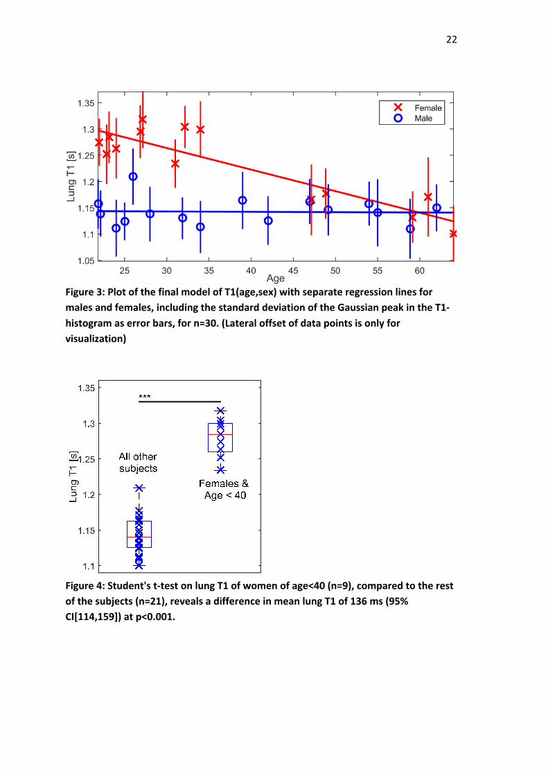
Figure3withparameterestimatesinTable4.Wholemodelissignificantatp<0.001(n=30)withadjustedr2=0.81andr2=0.83whichindicatesthatitaccountsformostofthevariationinthedata.TheslopeoflungT1attidalend-inspirationasafunctionofageforwomenwasfoundtobe-4.1ms/year(95%CI=[-5.2,-3.0])atp<0.001,and-0.064ms/year(95%CI=[-1.2,1.1])atp=0.9formen,whichindicatestherewasnocorrelationbetweenageandinspiredlungT1formen.TheslopeofT1asafunctionofagewasdifferentformenandwomenatp<0.001.
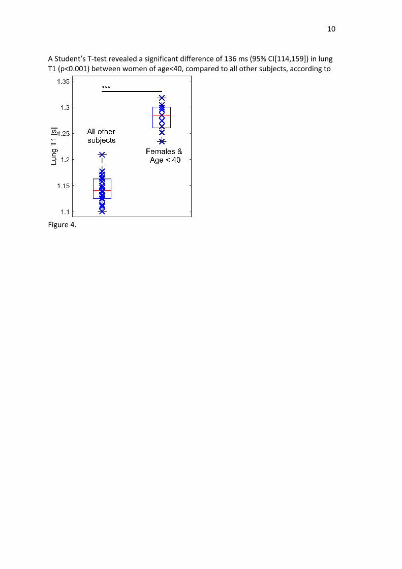
10AStudent’sT-testrevealedasignificantdifferenceof136ms(95%CI[114,159])inlungT1(p<0.001)betweenwomenofage<40,comparedtoallothersubjects,accordingto
Figure4.

11DISCUSSIONInthispaperwepresentevidencethatlungT1measuredduringtidalend-inspirationisnegativelycorrelatedwithageforwomen,butnotformen,inahealthynon-smokingpopulation.Overall,sexandagecombinedmakethebestfittotheT1datafromthestudiedgroup,followedbymodelsincludingTLC,VCandRV.However,asconfirmedbythepresentdataset,bothTLCandVCareverydependentonsex,andRVishighlydependentonage,whichmeansthatallmodelsT1(TLC*age),T1(VC*age),T1(sex*age)andT1(sex*RV)containessentiallythesameinformationasT1(age*sex).Themostinformativetwo-parameterinteractionmodeltodescribelungT1inhealthynever-smokersisconcludedtoincludeonlyageandsex,whichalsohasthehighestcorrelationvalueofthetestedmodelsinthisstudy.TheaverageT1atendtidalinspirationinthisstudywas1185ms(16male,14females,meanage38,TE=0.67ms)whichiswellinlinewiththemeanlungT1reportedinearlierpublicationsbyStadler:1199ms(8male,2female,meanage30,end-inspiration,TE=1.4ms)(7);Alamidi:1053ms(8male,4female,meanage63,TE=3ms,freebreathing,spinecho)(20)andRenne:1250ms(7male,5female,meanage28,TE0.8,end-inspiration)(25).ToexplainthepresentfindingsonemustconsidertheintrinsicT1ofhumanblood,aswellasthebloodcontentofthelung.Triphanetal.hasshownthatthemeasuredT1forlungatconventionalechotimesarelargelydeterminedbybloodT1(26).ItisthusreasonabletoassumethatbloodT1isamajordeterminantoflungT1attheTEofthepresentstudy(0.67ms).ArecentcardiacMRI-studybyPiechniketal.(2014)reportedfemaleleftventricularbloodT1onaverage86mshigherthaninmales,aswellasafemalespecificdecreaseinbloodT1of2ms/year,afteradjustingforhematocrit(27).ItwasalsoshownthatmalebloodT1increasesfrom30yearsofageupto65(27);thiscanbeexplainedbythemaindeterminantofbloodT1-hematocrit(28),whichislikelytodecreasewithfallingtestosteronelevels(29,30).OurowndatacanbeusedtoconfirmthegeneralizabilityofPiechnik’swork–acircularregionofinterestwasdrawnintheleftventricleoftheheartinthecollectedT1maps,andtheaverageT1ispresentedasafunctionofageandsexin

12
Figure5.Theslopesare-2.6ms/yearforfemales,and+1ms/yearformales,indicatingthatthefindingsofPiechniketal.alsoapplytothepresentstudysample.Theimplicationofthisisthatalmost50%oftheagedependencyinfemalelungT1(-4.3ms/year,presentstudy)canpotentiallybeattributedtobloodT1(-2.0ms/year,Piechniketal.).However,onewouldexpecttofindanincreaseinlungT1withageinmalesbasedpurelyonbloodT1,thereforethepulmonarybloodvolumemustalsobeconsidered.IthasbeenshownthattheinspiratorystateinfluenceslungT1,withtheend-inspirationT1onaveragebeing134mslowerthanatend-expiration(7).Itisalsoknownthatthevolumeadjustedpulmonarybloodfractionisloweratinspiration(31)andthatthelungparenchymaintrinsicT1islowerthanblood(26).ThedifferenceinlungT1atinspirationandexpirationmaythusbeattributedtodifferencesinpulmonarybloodvolume.Moreover,thereisalsosubstantialevidencethatpulmonarybloodvolumedecreaseswithage(32),which,inlightofthepreviouslymentionedfindings,wouldexplaintheobserveddecreaseinlungT1withageinourstudy.Theunderlyingexplanationfortheagedependencyofpulmonarybloodvolumemayinturnbeattributedtotheagedependentlossofelasticrecoilinthelung;leadingtoclosureofsmallairwaysatlargerlungvolumesandacorrespondingincreaseinRVwithage.Elderlysubjectswillthushavetidalbreathinginamoreinspiredstate(22).Inthecurrentsample(n=24),RVincreasedwith0.02liters/year,whichisverysimilartopreviouslyreportedvalues(0.022l/yformales,0.016l/yforfemales(33)).ThiswouldtheoreticallyyieldlowerpulmonarybloodfractionandalowerT1inelderlysubjects,asconsistentwithourfindings.Thus,combiningtheeffectsofbloodT1andpulmonarybloodvolumewecanexplainboththelargenegativeslopeoflungT1observedinwomen,andthenon-dependencybetweenlungT1andageobservedinmen;thisassumingthattheeffectsofbloodT1anddecreasingbloodvolumeareofapproximatelythesamemagnitude,butworkinginopposingdirectionsinmales.

13AdependenceoflungT1onpulmonarybloodvolumewouldhaveconsequencesfortheinterpretationofmeasurementsinpatients,aspulmonarybloodvolumeshowsregionalabnormalitiesinpatientswithinterstitiallungdisease(34,35),acuteandchronicpulmonaryoedema(36,37)andemphysema(32).ThisworkalsohighlightstheimportanceofmakingageandsexbalancedsubjectcohortsinstudiesinwhichT1isaread-outandnotgeneralizeresultstoapopulationbasedonalimitedgroup;suchasyoungmales.ResearchersutilizingT1-measurementsandOE-MRIshouldtakeintoaccountthesexdifferenceinbaselinelungT1intheinspiredstate,andmaybenefitfrommeasuringchangesinR1insteadofT1,sinceΔR1isnotdependentonbaselineT1.Alimitationofthepresentworkisthatmeasurementswereperformedoninspiratorybreathholdsonly–aretrospectivelygated,freebreathingprotocol,orseparatemeasurementsatinspirationandexpirationwouldhaveprovidedmoreindepthdataonthestudiedrelationships.Moreover,thesamplesizeofthepresentstudywaschosenfromapowercalculationbasedonanexpectedT1-agedependencyforbothsexes.Alargersubjectcohortwouldallowfurthermultiparametricanalysisonthedata.TousetheGaussiancurve’sstandarddeviationasameasureoftheintra-subjectT1variabilityisbasedontheexpectedheterogeneousdistributionofT1valuesinthesupineposition(38).ThestudywasdesignedtotesttheT1(age)relationship,andasexdependencywasquicklyestablished.ThestatisticalmethodsarenotusedtochooseapreferredmodelbutonlyanalyzethepreferredmodelT1(age*sex),sonomultiplecomparisonscorrectionhasbeenmadeonthepresentedp-values.Adjustingforseveralvariablesweredonetoelucidatepotentialconfounders,butthemainresult(i.e.thestatisticalsignificanceofthesexdependencyontheT1(age)relationship),andtheenfoldingdiscussion,wasnotaffectedbytheinclusionofadjustingvariables.Ifmoresubjectdatacanbepooled–forexampleinamulti-centerstudy–amoreadvanced,multiparametricmodelcanbeappliedtothedata,analyzingT1ofdifferentrespiratorystatesaswellastakingintoaccountseveralPFTparameters,whereRVmaybeanimportantconfounder.Inconclusion,wehaveprovidedevidencethatbaselinelungT1isdependentonageinwomen,butnotinmen.ThesefindingsarelikelyacombinedeffectofbloodT1andpulmonarybloodvolume,whicharebothdecreasingwithageinwomen,creatingaconcurrenteffectonT1.Formen,theeffectsofincreasingbloodT1anddecreasingpulmonarybloodvolumemaycanceleachother,toyieldanapparentnondependencybetweenageandT1.AdditionalstudiesareneededtofurtherdescribeindetailtheunderlyingdeterminantsoflungT1formenandwomen–lungT1maybeacomplexfunctionofbloodT1andbloodvolumeasdescribedhere,orinadditionbeafunctionoftheelasticrecoiloftheparenchyma.ThepresentworkunderlinesthatmuchisstilltobelearnedabouttheMRsignalgenerationinthehumanlung,andthatinterpretationoflungT1measurementsshouldincludeadiscussionaboutpulmonarybloodvolume.

14ACKNOWLEDGEMENTSWegreatlyappreciatethevaluablediscussionswithDrSimonTriphanandProfessorPeterJakobandtheirassiduousassistancewiththepulsesequence.WealsothankDrRasmusABååthandDrAldanaRossoforhelpwiththestatisticalanalysis.

15REFERENCES1.KauczorH-U,KreitnerKF:MRIofthepulmonaryparenchyma.EurRadiol1999;9:1755–1764.2.MillerGW,MuglerJP,SáRC,AltesTA,PriskGK,HopkinsSR:AdvancesinfunctionalandstructuralimagingofthehumanlungusingprotonMRI.NMRBiomed2014;27:1542–56.3.ArnoldJFT,FidlerF,WangT,PrachtED,SchmidtM,JakobPM:ImaginglungfunctionusingrapiddynamicacquisitionofT1-mapsduringoxygenenhancement.MAGMA2004;16:246–53.4.OhnoY,HatabuH,TakenakaD,VanCauterenM,FujiiM,SugimuraK:Dynamicoxygen-enhancedMRIreflectsdiffusingcapacityofthelung.MagnResonMed2002;47:1139–44.5.DeichmannR,HahnD,Haasea:FastT1mappingonawhole-bodyscanner.MagnResonMed1999;42:206–9.6.DeichmannR,HaaseA,HublandA:QuantificationofTlValuesbySNAPSHOT-FLASHNMRImaging.JMagnReson1992;96:608–612.7.StadlerA,JakobPM,GriswoldM,BarthM,BankierAa:T1mappingoftheentirelungparenchyma:Influenceoftherespiratoryphaseinhealthyindividuals.JMagnResonImaging2005;21:759–64.8.JakobPM,HillenbrandCM,WangT,SchultzG,HahnD,HaaseA:RapidQuantitativeLung1HT1Mapping.JMagnResonimaging2001;14:795–799.9.WangT,SchultzG,HebestreitH,HebestreitA,HahnD,JakobPM:Quantitativeperfusionmappingofthehumanlungusing1Hspinlabeling.JMagnResonImaging2003;18:260–5.10.StadlerA,JakobPM,GriswoldM,StiebellehnerL,BarthM,BankierAa:T1mappingoftheentirelungparenchyma:Influenceofrespiratoryphaseandcorrelationtolungfunctiontestresultsinpatientswithdiffuselungdisease.MagnResonMed2008;59:96–101.11.JakobPM,WangT,SchultzG,HebestreitH,HebestreitA,HahnD:Assessmentofhumanpulmonaryfunctionusingoxygen-enhancedT(1)imaginginpatientswithcysticfibrosis.MagnResonMed2004;51:1009–16.12.StadlerA,StiebellehnerL,JakobPM,etal.:Quantitativeando(2)enhancedMRIofthepathologiclung:findingsinemphysema,fibrosis,andcysticfibrosis.IntJBiomedImaging2007;2007:23624.13.OhnoY,KoyamaH,MatsumotoK,etal.:Oxygen-enhancedMRIvs.quantitativelyassessedthin-sectionCT:pulmonaryfunctionallossassessmentandclinicalstageclassificationofasthmatics.EurJRadiol2011;77:85–91.14.ZhangW-J,NivenRM,YoungSS,LiuY-Z,ParkerGJM,NaishJH:Dynamicoxygen-enhancedmagneticresonanceimagingofthelunginasthma—Initialexperience.EurJRadiol2015;84:318–326.

1615.MaxienD,DietrichO,ThiemeSF,etal.:Valueofoxygen-enhancedMRIofthelungsinpatientswithpulmonaryhypertension:aqualitativeandquantitativeapproach.JMagnResonImaging2012;35:86–94.16.RenneJ,LauermannP,HinrichsJB,etal.:ChronicLungAllograftDysfunction:Oxygen-enhancedT1-MappingMRImagingoftheLung.Radiology2015;276:266–73.17.OhnoY,KoyamaH,NogamiM,etal.:Dynamicoxygen-enhancedMRIversusquantitativeCT:pulmonaryfunctionallossassessmentandclinicalstageclassificationofsmoking-relatedCOPD.AJRAmJRoentgenol2008;190:8–10.18.MorganAR,ParkerGJM,RobertsC,etal.:Feasibilityassessmentofusingoxygen-enhancedmagneticresonanceimagingforevaluatingtheeffectofpharmacologicaltreatmentinCOPD.EurJRadiol2014;83:2093–101.19.JobstBJ,TriphanSMF,SedlaczekO,etal.:FunctionallungMRIinchronicobstructivepulmonarydisease:comparisonoft1mapping,oxygen-enhancedt1mappinganddynamiccontrastenhancedperfusion.PLoSOne2015;10:e0121520.20.AlamidiFD,MorganAR,HubbardCristinaccePL,etal.:COPDpatientshaveshortlungmagneticresonanceT1relaxationtime.JchronicObstrPulmDis2015;Inpress.21.BurneyPGJ,PatelJ,NewsonR,MinelliC,NaghaviM:GlobalandregionaltrendsinCOPDmortality,1990-2010.EurRespirJ2015;45:1239–47.22.WestJB:RespiratoryPhysiology:TheEssentials.9:thEditi.Baltimore:LippincottWilliams&Wilkins;2012.23.BrixG,SchadLR,DeimlingM,LorenzWJ:FastandpreciseT1imagingusingaTOMROPsequence.MagnResonImaging1990;8:351–356.24.LookDC:TimeSavinginMeasurementofNMRandEPRRelaxationTimes.RevSciInstrum1970;41:250.25.RenneJ,HinrichsJ,SchönfeldC,etal.:Oxygen-enhancedT1-mappingofthelung:ReproducibilityandImpactofdifferentgasdeliverymethods.ProcIntSocMagnResonMed2013;21:1497.26.TriphanSMF,JobstBJ,BreuerFA,etal.:EchotimedependenceofobservedT1inthehumanlung.JMagnResonImaging2015;42:610–616.27.PiechnikSK,FerreiraVM,LewandowskiAJ,etal.:NormalvariationofmagneticresonanceT1relaxationtimesinthehumanpopulationat1.5TusingShMOLLI.JCardiovascMagnReson2013;15:13.28.SpeesWM,YablonskiyDA,OswoodMC,AckermanJJH:WaterProtonMRPropertiesofHumanBloodat1.5Tesla :MagneticSusceptibility,T1,T2,T*.MagnResonMed2001;45:533–542.29.BachmanE,TravisonTG,BasariaS,etal.:TestosteroneInducesErythrocytosisviaIncreasedErythropoietinandSuppressedHepcidin:EvidenceforaNewErythropoietin/HemoglobinSetPoint.JournalsGerontolSerABiolSciMedSci2013;69:725–735.

1730.FeldmanHA,LongcopeC,DerbyCA,etal.:Agetrendsinthelevelofserumtestosteroneandotherhormonesinmiddle-agedmen:longitudinalresultsfromtheMassachusettsmaleagingstudy.JClinEndocrinolMetab2002;87:589–98.31.BrudinLH,RhodesCG,ValindSO,WollmerP,HughesJMB:RegionallungdensityandbloodvolumeinnonsmokingandsmokingsubjectsmeasuredbyPET.JApplPhysiol1987;63:1324–1334.32.MeinelFG,GraefA,SommerWH,ThierfelderKM,ReiserMF,JohnsonTRC:Influenceofvascularenhancement,ageandgenderonpulmonaryperfusedbloodvolumequantifiedbydual-energy-CTPA.EurJRadiol2013;82:1565–1570.33.StocksJ,QuanjerPH:Referencevaluesforresidualvolume,functionalresidualcapacityandtotallungcapacity:ATSWorkshoponLungVolumeMeasurementsOfficialStatementoftheEuropeanRespiratorySociety.InEurRespirJ.Volume8;1995:492–506.34.WollmerP,RhodesCG,HughesJMB:Regionalextravasculardensityandfractionalbloodvolumeofthelungininterstitialdisease.Thorax1984;39:286–293.35.HagspielKD,FlorsL,Housseinia.M,etal.:Pulmonarybloodvolumeimagingwithdual-energycomputedtomography:Spectrumoffindings.ClinRadiol2012;67:69–77.36.WollmerP,RhodesCG,DeanfieldJ,etal.:Regionalextravasculardensityofthelunginpatientswithacutepulmonaryedema.JApplPhysiol1987;63:1890–1895.37.WollmerP,RhodesCG,AllanRM,MaseriA,FazioF:Regionalextravascularlungdensityandfractionalpulmonarybloodvolumeinpatientswithchronicpulmonaryvenoushypertension.ClinPhysiol1983;3:241–256.38.NicholsMB,PaschalCB:Measurementoflongitudinal(T1)relaxationinthehumanlungat3.0Teslawithtissue-basedandregionalgradientanalyses.JMagnResonImaging2008;27:224–228.
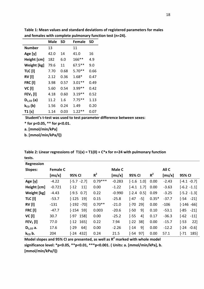
18Table1:Meanvaluesandstandarddeviationsofregisteredparametersformalesandfemaleswithcompletepulmonaryfunctiontest(n=24). Male SD Female SDNumber 13 11 Age[y] 42.0 14 41.0 16Height[cm] 182 6.0 166** 4.9Weight[kg] 79.6 11 67.5** 9.0TLC[l] 7.70 0.68 5.70** 0.66RV[l] 2.12 0.36 1.68* 0.47FRC[l] 3.98 0.57 3.01** 0.49VC[l] 5.60 0.54 3.99** 0.42FEV1[l] 4.18 0.60 3.19** 0.52DL,CO(a) 11.2 1.6 7.75** 1.13kCO(b) 1.56 0.24 1.49 0.20T1[s] 1.14 0.03 1.22** 0.07Student’st-testwasusedtotestparameterdifferencebetweensexes:*forp<0.05,**forp<0.01.a.[mmol/min/kPa]b.[mmol/min/kPa/l]) Table2:LinearregressionsofT1(x)=T1(0)+C*xforn=24withpulmonaryfunctiontests. RegressionSlopes:
FemaleC[ms/x] 95%CI R2
MaleC[ms/x] 95%CI R2
AllC[ms/x] 95%CI R2
Age[y] -4.22 [-5.7-2.7] 0.79*** -0.283 [-1.61.0] 0.00 -2.43 [-4.1-0.7] 0.25**Height[cm] -0.721 [-1211] 0.00 -1.22 [-4.11.7] 0.00 -3.63 [-6.2-1.1] 0.26**Weight[kg] -4.43 [-9.50.7] 0.22 -0.990 [-2.40.5] 0.09 -3.25 [-5.2-1.3] 0.33**TLC[l] -53.7 [-12519] 0.15 -25.8 [-47-5] 0.35* -37.7 [-54-21] 0.48***RV[l] -131 [-192-70] 0.70** -21.0 [-7029] 0.00 -106 [-146-66] 0.56***FRC[l] -47.7 [-15459] 0.003 -20.6 [-509] 0.10 -53.1 [-85-21] 0.32**VC[l] 30.7 [-97158] 0.00 -25.2 [-554] 0.17 -36.3 [-62-11] 0.25**FEV1[l] 77.0 [-12165] 0.22 7.94 [-2238] 0.00 -15.7 [-5322] 0.00DL,COa. 17.6 [-2964] 0.00 -2.26 [-149] 0.00 -12.2 [-24-0.6] 0.14*kCOb. 204 [-24432] 0.24 21.5 [-5497] 0.00 57.1 [-71185] 0.00Modelslopesand95%CIarepresented,aswellasR2markedwithwholemodelsignificancelevel:*p<0.05,**p<0.01,***p<0.001.(Units:a.[mmol/min/kPa],b.[mmol/min/kPa/l])

19Table3:Pearson’sCorrelationcoefficientforpairsofindependentvariables Age Length Weight TLC RV FRC VC FEV1 DL.COLength 0.08 Weight 0.33 0.68 TLC 0.18 0.90 0.63 RV 0.70 0.52 0.50 0.71 FRC 0.19 0.72 0.32 0.88 0.74 VC -0.12 0.90 0.55 0.94 0.45 0.78 FEV1 -0.37 0.71 0.31 0.68 0.12 0.56 0.84 DL.CO -0.18 0.74 0.45 0.76 0.32 0.63 0.82 0.59 kCO -0.47 0.01 0.00 -0.08 -0.36 -0.17 0.06 0.01 0.56Coefficients>0.5arebold. Table4:EstimatesoffinalmodelT1(age*sex)forn=30,with95%confidenceintervals. Estimate 95%CI pMale T1(0)[ms] 1145 [10981190] <1e-9Slope[ms/y] -0.064 [-1.21.1] 0.91Female T1(0)[ms] 1387 [13421430] <1e-9Slope[ms/y] -4.10 [-5.2-3.0] <1e-7

20
Figure1:HistogramofT1-valuesfromwholelungsegmentationfromamalesubject,representativeofthewholesubjectgroup.ThepeakofaGaussianfit,aftersubtractingathreshold-valuefromthewholehistogram,isdefinedastheT1ofthesubject.

21
Figure2:RepresentativeT1mapsfor(a)femaleage<30,(b)femaleage>50,(c)maleage<30,(d)maleage>50.ThecontrastisadjustedforT1=[0,1600]ms.
22
Figure3:PlotofthefinalmodelofT1(age,sex)withseparateregressionlinesformalesandfemales,includingthestandarddeviationoftheGaussianpeakintheT1-histogramaserrorbars,forn=30.(Lateraloffsetofdatapointsisonlyforvisualization)
Figure4:Student'st-testonlungT1ofwomenofage<40(n=9),comparedtotherestofthesubjects(n=21),revealsadifferenceinmeanlungT1of136ms(95%CI[114,159])atp<0.001.

23
Figure5:MeanT1incircledrawnintheleftventricleoftheheartasafunctionofageandsex.Althoughdataisnoisy(nocardiactriggeringisused)maleandfemalebloodT1exhibitdifferentslopesasafunctionofage(p=0.035).