Embed Size (px)
DESCRIPTION
Influence of Blood Flow on the Coagulation Cascade. Nina Marianne Andersen, Mads Peter Sørensen, Emil Sokoler Department of Mathematics (MAT), Techn. Univ. of Denmark Steen Ingwersen and Ole Hvilsted Olsen Biomodelling and Haemostasis Biochemistry, Novo Nordisk, Denmark. Content: - PowerPoint PPT Presentation
Citation preview
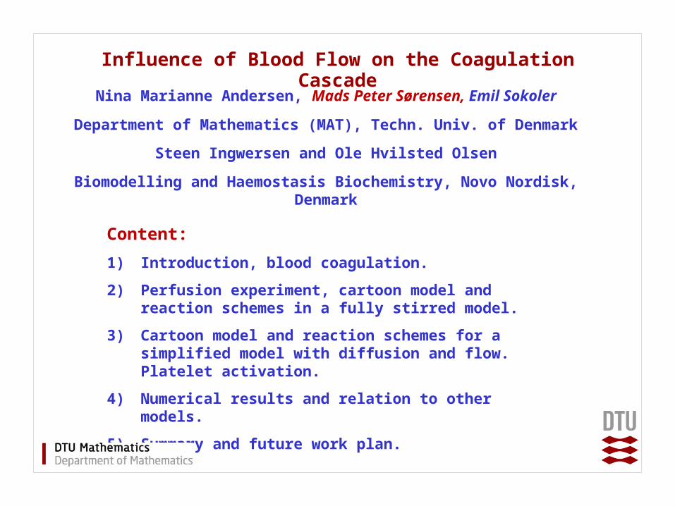
Influence of Blood Flow on the Coagulation Cascade
Nina Marianne Andersen, Mads Peter Sørensen, Emil Sokoler
Department of Mathematics (MAT), Techn. Univ. of Denmark
Steen Ingwersen and Ole Hvilsted Olsen
Biomodelling and Haemostasis Biochemistry, Novo Nordisk, Denmark
Content:
1) Introduction, blood coagulation.
2) Perfusion experiment, cartoon model and reaction schemes in a fully stirred model.
3) Cartoon model and reaction schemes for a simplified model with diffusion and flow. Platelet activation.
4) Numerical results and relation to other models.
5) Summary and future work plan.
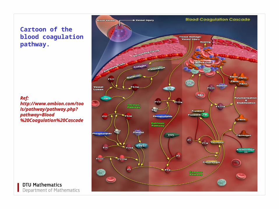
Ref: http://www.ambion.com/tools/pathway/pathway.php?pathway=Blood%20Coagulation%20Cascade
Cartoon of the blood coagulation pathway.
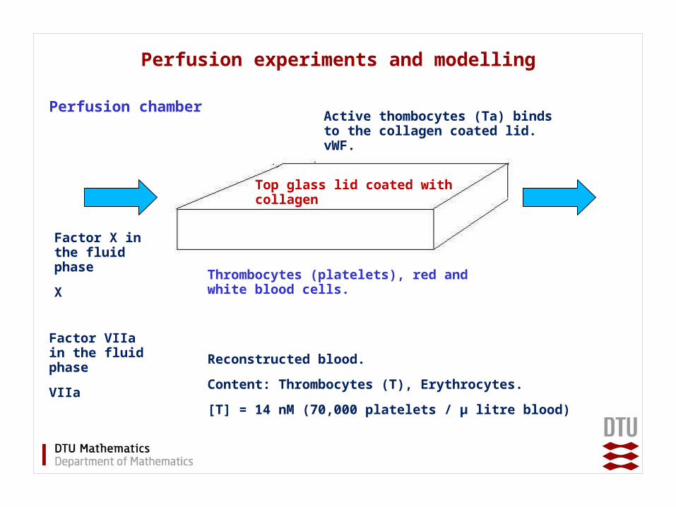
Perfusion experiments and modelling
Perfusion chamber
Top glass lid coated with collagen
Thrombocytes (platelets), red and white blood cells.
Factor X in the fluid phase
X
Factor VIIa in the fluid phase
VIIa
Active thombocytes (Ta) binds to the collagen coated lid. vWF.
Reconstructed blood.
Content: Thrombocytes (T), Erythrocytes.
[T] = 14 nM (70,000 platelets / μ litre blood)
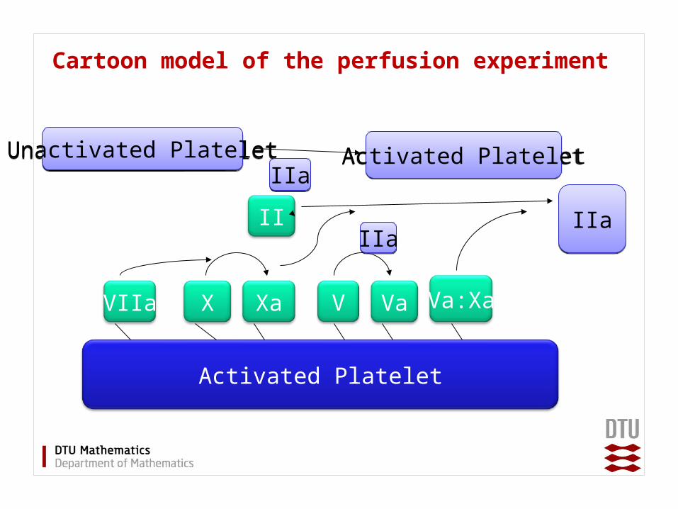
Cartoon model of the perfusion experiment
Activated Platelet
Va:XaVVIIa XaX Va
II IIaIIa
Unactivated PlateletUnactivated Platelet Activated PlateletActivated PlateletIIaIIa
IIaIIa
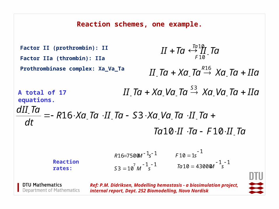
Reaction schemes, one example.
Ref: P.M. Didriksen, Modelling hemostasis - a biosimulation project, internal report, Dept. 252 Biomodelling, Novo Nordisk
TaIITaII 10Ta
10F
IIaTaXaTaXaTaII 16R
IIaTaVaXaTaVaXaTaII 3S
Factor II (prothrombin): II
Factor IIa (thrombin): IIa
Prothrombinase complex: Xa_Va_Ta
A total of 17 equations.
TaIITaVaXaSTaIITaXaRdtTadII
316
TaIIFTaIITa 1010
11750016 sMR
11103
7 sMS
1110
sF
114300010
sMTa
Reaction rates:
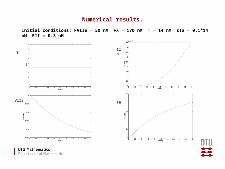
Numerical results.
T
VIIa Ta
IIa
Initial conditions: FVIIa = 50 nM FX = 170 nM T = 14 nM sTa = 0.1*14 nM FII = 0.3 nM
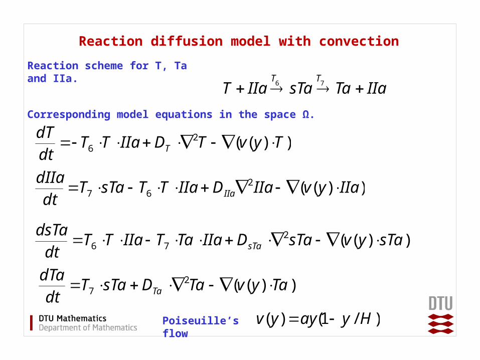
Reaction diffusion model with convection
Reaction scheme for T, Ta and IIa.
IIaTasTaIIaT 6T
7T
Corresponding model equations in the space Ω.
))((27 TayvTaDsTaT
dtdTa
Ta
))((26 TyvTDIIaTT
dtdT
T
))((276 sTayvsTaDIIaTaTIIaTT
dtdsTa
sTa
))((267 IIayvIIaDIIaTTsTaT
dtdIIa
IIa
Poiseuille’s flow
)/1()( Hyayyv
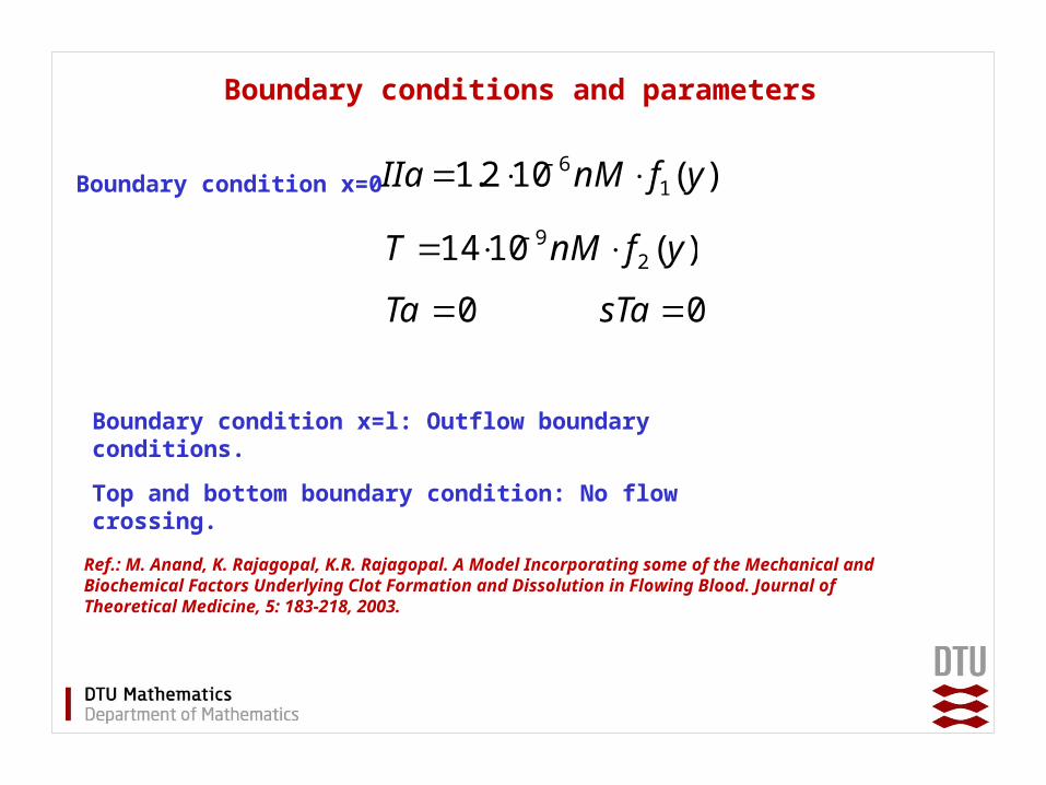
Boundary conditions and parameters
Boundary condition x=0 )(102.1 16 yfnMIIa
Ref.: M. Anand, K. Rajagopal, K.R. Rajagopal. A Model Incorporating some of the Mechanical and Biochemical Factors Underlying Clot Formation and Dissolution in Flowing Blood. Journal of Theoretical Medicine, 5: 183-218, 2003.
)(1014 29 yfnMT
0Ta 0sTa
Boundary condition x=l: Outflow boundary conditions.
Top and bottom boundary condition: No flow crossing.
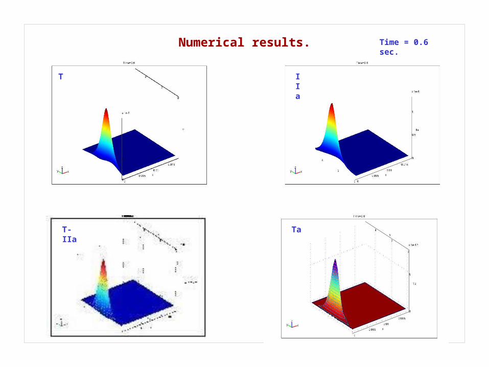
Numerical results. Time = 0.6 sec.
T-IIa
IIa
T
Ta
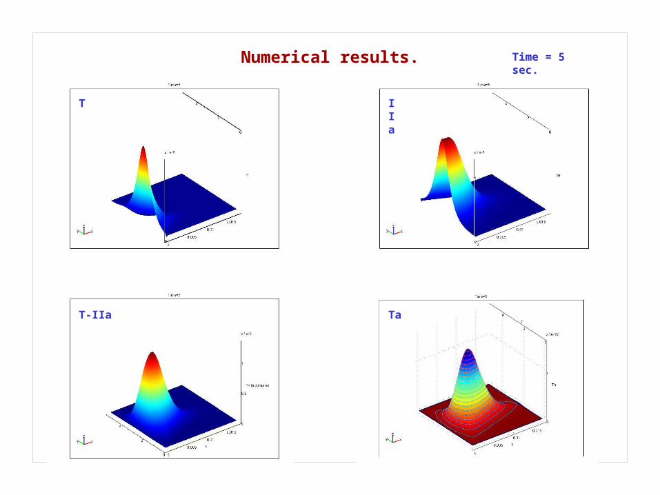
Numerical results. Time = 5 sec.
T IIa
T-IIa Ta
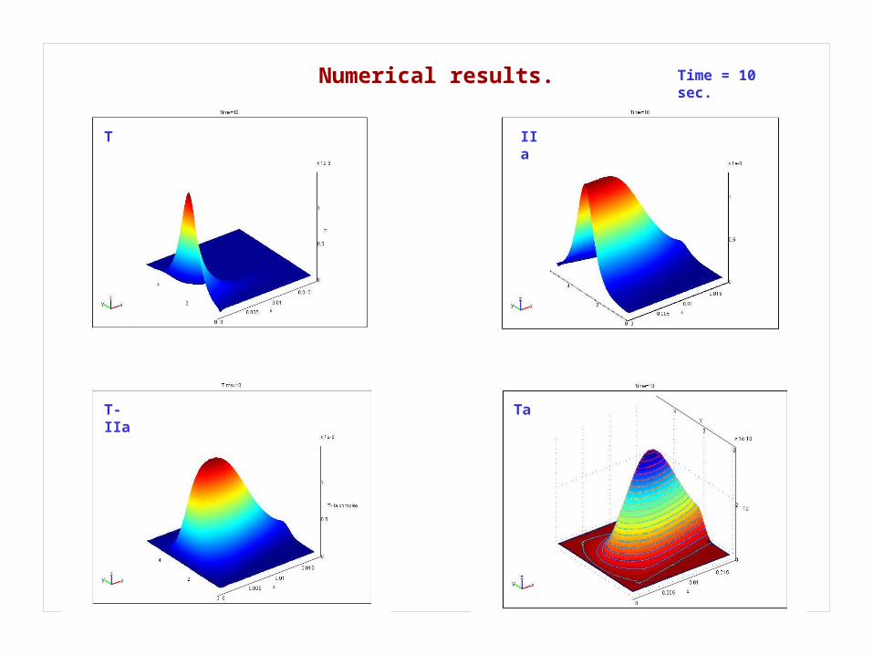
Numerical results. Time = 10 sec.
T-IIa
T IIa
Ta
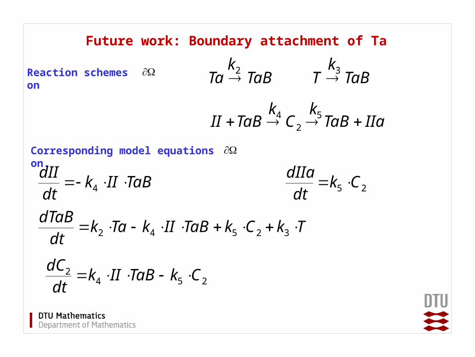
Future work: Boundary attachment of Ta
Reaction schemes on
Corresponding model equations on.
TaBTa TaBT
IIaTaBCTaBII 2
2k 3k
4k 5k
TaBIIkdtdII 4
25 CkdtdIIa
TkCkTaBIIkTakdtdTaB 32542
2542 CkTaBIIk
dtdC
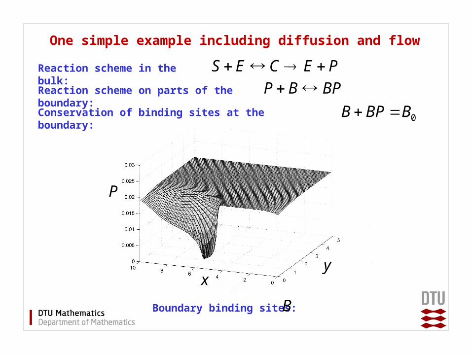
One simple example including diffusion and flow
xy
P
PECES Reaction scheme in the bulk: Reaction scheme on parts of the boundary:
BPBP
0BBPB Conservation of binding sites at the boundary:
Boundary binding sites: B
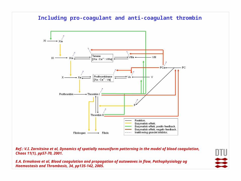
Including pro-coagulant and anti-coagulant thrombin
Ref.: V.I. Zarnitsina et al, Dynamics of spatially nonuniform patterning in the model of blood coagulation, Chaos 11(1), pp57-70, 2001.
E.A. Ermakova et al, Blood coagulation and propagation of autowaves in flow, Pathophysiology og Haemostasis and Thrombosis, 34, pp135-142, 2005.
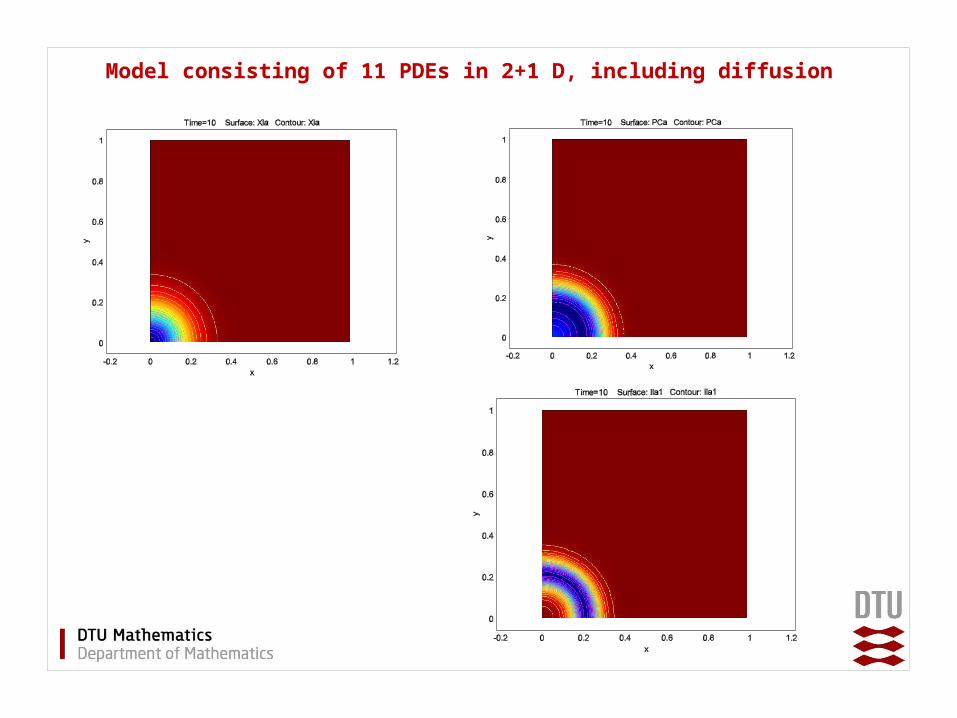
Model consisting of 11 PDEs in 2+1 D, including diffusion
Summary and future work
1. Modelling of perfusion experiment for blood coagulation.
2. Reduced PDE model including blood flow and diffusion.
3. Modelling of attachment of activated thrombocytes on collagen coated boundary.
4. Full PDE model.
5. Model of in vivo blood coagulation.
Simpel Model consisting of 3 PDEs, including diffusion