Embed Size (px)
Citation preview

Influence of Cellular Arachidonic Acid Levels on Phospholipid
Remodeling and CoA‐independent Transacylase Activity in Human
Monocytes and U937 Cells
Alma M. Astudillo, Gema Pérez‐Chacón, David Balgoma, Luis Gil‐de‐Gómez,
Violeta Ruipérez, Carlos Guijas, María A. Balboa, and Jesús Balsinde*
Instituto de Biología y Genética Molecular, Consejo Superior de Investigaciones
Científicas (CSIC), 47003 Valladolid, Spain
and Centro de Investigación Biomédica en Red de Diabetes y Enfermedades
Metabólicas Asociadas (CIBERDEM), 08036 Barcelona, Spain
*Corresponding author. Phone, +34‐983‐423‐062; Fax, +34‐983‐184‐800;
e‐mail: [email protected]
Keywords: Arachidonic Acid; Phospholipase A2; Free fatty acid; CoA‐independent
transacylase; Phospholipid Remodeling.
Running Title: Arachidonic Acid Remodeling in U937 Cells
Abbreviations used: AA, arachidonic acid; CoA‐IT, coenzyme A‐independent
transacylase, PC, choline glycerophospholipids; PE, ethanolamine glycerophospholipids,
PI, phosphatidylinositol; PMA, phorbol myristate acetate; TAG, triacylglycerol.
1
*ManuscriptClick here to view linked References
Abstract
The availability of free arachidonic acid (AA) constitutes a limiting step in the synthesis
of biologically active eicosanoids. Free AA levels in cells are regulated by a
deacylation/reacylation cycle of membrane phospholipids, the so‐called Lands cycle, as
well as by further remodeling reactions catalyzed by CoA‐independent transacylase. In
this work, we have comparatively investigated the process of AA incorporation into
and remodeling between the various phospholipid classes of human monocytes and
monocyte‐like U937 cells. AA incoporation into phospholipids was similar in both cell
types, but a marked difference in the rate of remodeling was appreciated. U937 cells
remodelled AA at a much faster rate than human monocytes. This difference was
found not to be related to the differentiation state of the U937 cells, but rather to the
low levels of esterified arachidonate found in U937 cells compared to human
monocytes. Incubating the U937 cells in AA‐rich media increased the cellular content
of this fatty acid and led to a substantial decrease of the rate of phospholipid AA
remodeling, which was due to reduced CoA‐independent transacylase activity.
Collectively, these findings provide the first evidence that cellular AA levels determine
the amount of CoA‐independent transacylase activity expressed by cells and provide
support to the notion that CoA‐IT is a major regulator of AA metabolism in human
monocytes.
2

1. Introduction
Arachidonic acid (AA) is an ω‐6 essential polyunsaturated fatty acid with key roles in
inflammation due to it being the precursor of a variety of compounds with potent pro‐
and anti‐inflammatory actions, the so‐called eicosanoids [1, 2]. Since the bulk of AA in
cells is found esterified in the sn‐2 position of membrane phospholipids, availability of
free AA is a limiting factor for eicosanoid synthesis [3, 4]
Control of free AA levels in cells is exquisitely regulated by two competing
reactions, namely, phospholipid deacylation by phospholipase A2 enzymes, and
reacylation into various phospholipid pools by acyltransferases [4]. Depending on the
activation state of the cell, one reaction dominates over the other, i.e. in resting cells,
reacylation dominates, and hence, the bulk of cellular AA is found in the esterified
form. In stimulated cells, the dominant reaction is the phospholipase A2‐mediated
deacylation, which results in a dramatic increase of free AA that is now available for
eicosanoid synthesis [4‐6].
The process of acylation/deacylation in membrane phospholipids is not the only
mechanism that regulates free AA levels in cells, as transacylation reactions between
phospholipids are also required to achieve the proper distribution of AA among
membrane phospholipid pools [3, 7]. These reactions are catalyzed by CoA‐
independent transacylase (CoA‐IT), an enzyme that transfers AA primarily from diacyl
PC species to ether‐linked species, in particular the PE plasmalogens [3, 7]. This
remodeling of AA within the various phospholipid species appears to be crucial for the
generation of eicosanoids during cellular stimulation, inasmuch as ether‐linked
3

phospholipids are thought to provide the bulk of free AA to be converted to
eicosanoids, and the rate of remodeling is accelerated several‐fold [8‐11].
In addition to cell activation, other factors may exist that modify the rate of
remodeling of phospholipids with AA. For example, a striking difference between
proliferating and non‐proliferating cells has repeatedly been observed [12‐15], and
blockade of the CoA‐IT‐dependent remodeling of proliferating cells promotes apoptotic
cell death [16‐19]. Despite numerous studies on CoA‐IT [3, 20‐25], purification and/or
cloning of the enzyme remains elusive, which has made it difficult to study in depth the
cellular regulation of this enzyme
The objective of this study was to understand better the biochemical features
of AA incorporation into, and remodeling within, phospholipid species of human
monocytes and monocyte‐like U937 cells, in particular with regard to the state of
cellular differentiation and intracellular content of esterified AA. The results presented
here suggest that the rate of phospholipid AA remodeling is strikingly dependent on
cellular content of this fatty acid.
4

2. Materials and Methods
Reagents ‐ RPMI 1640 medium was from Invitrogen Life Technologies (San Diego, CA).
[5,6,8,9,11,12,14,15‐3H]AA (specific activity 211 Ci/mmol) and radioactive substrates
for enzyme assays were from GE Healthcare (Alcobendas, Madrid, Spain). TLC plates
were from were from Scharlab (Barcelona, Spain). All other reagents were from Sigma.
Cell Isolation and Culture ‐ U937 cells were maintained in RPMI 1640 medium
supplemented with 10% (v/v) fetal calf serum, 2 mM L‐glutamine, penicillin (100
units/ml) and streptomycin (100 mg/ml). For experiments, the cells were incubated at
37°C in a humidified atmosphere of CO2/O2 (1:19) at a cell density of 0.5‐1 x 10⁶
cells/ml in 12‐well plastic culture dishes (Costar). Cell differentiation was induced by
adding PMA to a final concentration of 35 ng/ml for 24 h [26, 27]. U937 cell
differentiation was confirmed morphologically by light microscopy and by conversion
of cells to an adherent cell population. For preparation of cells loaded with unlabeled
AA, the cells were placed in serum‐free medium at a density of 0.5 x 10⁶ cells/ml and
exposed to 100 µM AA complexed with bovine serum albumin (ratio 5:1) for 20 h,
washed 3‐4 times with medium containing 0.5 mg/ml BSA, and placed in serum‐free
medium for subsequent experiments.
Human monocytes were obtained from buffy coats of healthy volunteer donors
obtained from the Centro de Hemoterapia y Hemodonación de Castilla y León
(Valladolid, Spain). Briefly, blood cells were diluted 1:1 with PBS, layered over a cushion
5
of Ficoll‐Paque and centrifuged at 750 x g during 30 min. The mononuclear cellular
layer was then recovered and washed three times with PBS, resuspended in RPMI
supplemented with 2 mM L‐glutamine and 40 mg/ml gentamicin and allowed to
adhere to plastic sterile dishes for 2 h. Non‐adherent cells were then removed by
extensively washing with PBS, and the remaining attached monocytes were used on
the next day. For all experiments, monocytes were cultured in a final volume of 2 ml in
serum‐free RPMI 1640 medium supplemented with 2 mM L‐glutamine and 40 μg/ml
gentamicin at 37°C in a humidified 5% CO2 atmosphere.
Measurement of [3H]AA Incorporation Into Cellular Phospholipids ‐ The cells were
placed in serum‐free medium for 60 min before exposure to exogenous [3H]AA (1 µM;
0.25 µCi/ml). At the indicated times, supernatants were removed and total cellular
lipids were extracted according to Bligh and Dyer [28], and separated by thin‐layer
chromatography with n‐hexane/diethyl ether/acetic acid (70:30:1, by vol.). For
separation of phospholipid classes, a mobile phase consisting of
chloroform/methanol/acetic acid/water (25:20:3:0.3, by vol.) was used.
Measurement of Phospholipid [3H]AA Remodeling ‐ For these experiments, the cells
were pulse‐labeled with [3H]AA (1 µM; 0.25 µCi/ml) for 30 min at 37 °C. The cells were
then washed four times with medium containing 0.5 mg/ml bovine serum albumin to
remove the unincorporated label. Afterward the cells were placed in serum‐free
medium and incubated at 37 °C for the indicated periods of time [14] The lipids were
extracted and separated as described above.
6

Gas Chromatography/Mass Spectrometry Analysis of Fatty Acid Methyl Esters ‐ 5‐10 x
106 cells were taken, washed once with PBS, and lipids extracted according to Bligh and
Dyer [28]. The resulting extract was transmethylated with 500 µl of 0.5 M KOH in
MeOH for 30 min at 37°C. One volume of 0.5 M HCl was added to neutralize and fatty
acid methyl esters were extracted twice with 2 volumes of n‐hexane. Analysis of fatty
acid methyl esters was carried out in an Agilent 6890N gas chromatograph coupled to
an Agilent 5975 mass‐selective detector (MSD) operated in electron impact mode (EI,
70 eV), equipped with an Agilent DB23 column (60 m x 0.25 mm I.D. x 0.15 µm film
thickness). 1 µl of sample was injected in splitless mode. Inlet temperature was
mantained at 250°C. Oven temperature was held at 50°C for 1 min, then increased to
175°C at intervals of 25°C per min, and to 230°C at intervals of 2.75°C per min. The final
temperature was maintained for 5 min, and the run time was 33 min. The mass
spectrometry transfer line was maintained at 250°C and the mass spectrometer
quadrupole and source at 150°C and 230°C respectively. Helium was used as a carrier
gas at a constant pressure of 180 kPa. Data acquisition was carried out both in scan
and selected ion monitoring (SIM) mode. Scan mode was used for compound
identification, comparing with authentical FAME standards and the NIST (National
Institute of Standards and Technology) MS library spectra. SIM mode was used for
quantitation, using 74 and 87 fragments for saturated, 83 for monounsaturated, 67
and 81 for diunsaturated and 79 and 91 for polyunsaturated fatty acid methyl esters. A
37‐component mixture from Supelco was used for calibration curves, and
nonadecanoic acid was used as an internal standard. Data analysis was carried out with
the Agilent G1701EA MSD Productivity Chemstation software, revision E.02.00.
7

Analysis of AA‐containing Phospholipids by Liquid Chromatography/Mass Spectrometry
‐ For lipid separation by high‐performace liquid chromatography, a binary pump Hitachi
LaChrom Elite® L‐2130 was used, together with a Hitachi Autosampler L‐2200 (Merck).
The liquid chromatography system was coupled on‐line to a Bruker esquire6000 ion‐
trap mass spectrometer (Bruker Daltonics, Bremen, Germany). In all cases the effluent
was split and 0.2 ml/min entered the electrospray interface of the mass spectrometer.
Nebulizer was set to 30 psi, dry gas to 8 l/min and dry temperature to 350°C. Analysis
of AA‐containing PI, PC and PE species was carried out exactly as described by Balgoma
et al. [11, 29].
Assay for CoA‐independent transacylase activity ‐ CoA‐independent transacylase
activity was measured following the procedure originally described by Venable et al.
[23], as modified by Balsinde et al. [30]. Briefly, the assay mixture was composed of
120 mM NaCl, 2 mM EGTA, 100 mM Tris‐HCl (pH 7.5), cell homogenate (up to 100 µg of
protein) and various concentrations of 1‐0‐[3H]hexadecyl‐2‐lyso‐sn‐glycero‐3‐
phosphocholine (lyso platelet‐activating factor, lysoPAF) as a substrate, in a final
volume of 0.2 ml. In this assay system, the lysophospholipid acceptor for the acylation
reaction (lysoPAF) is added to the assay mixture and the phospholipid donor is
provided by the homogenate. After incubation at 37°C for 5 min, the reaction was
stopped by the addition of 0.75 ml of chloroform/methanol (1:2). Chloroform (0.25 ml)
and water (0.25 ml) were added and the mixture was vortexed vigorously before
centrifugation at 1000 x g for 5 min. The organic phase was evaporated and
chromatographed on Silica gel G plates with chloroform/methanol/acetic acid/water
8

(50:25:8:4 by vol.) as the developing solvent. PC and lysoPAF were cut out of the plate
and assayed for radioactivity by liquid scintillation counting.
Other Assays ‐ Ca2+‐dependent phospholipase A2 activity was measured by using a
modification of the mammalian membrane assay described by Diez et al. [31]. Briefly,
cell homogenates were incubated for 1‐2 h at 37°C in 100 mM HEPES (pH 7.5)
containing 1.3 mM CaCl2 and 100,000 dpm of [3H]AA‐labeled membrane, used as
substrate, in a final volume of 0.15 ml. Prior to assay, the cell membrane substrate was
heated at 57°C for 5 min, in order to inactivate coenzyme A‐independent transacylase
activity [9]. The assay contained 25 μM bromoenol lactone to completely inhibit
endogenous Ca2+‐independent phospholipase A2 activity [30]. After lipid extraction,
free [3H]AA was separated by thin‐layer chromatography, using n‐hexane/ethyl
ether/acetic acid (70:30:1) as a mobile phase. For Ca2+‐independent PLA2 activity, the
cell homogenates were incubated for 2 h at 37°C in 100 mM Hepes (pH 7.5) containing
5 mM EDTA and 100 μM labeled phospholipid substrate (1‐palmitoyl‐2‐[3H]palmitoyl‐
glycero‐3‐phosphocholine, sp. act. 60 Ci/mmol, American Radiolabeled Chemicals, St.
Louis, MO) in a final volume of 150 μl. The phospholipid substrate was used in the form
of sonicated vesicles in buffer. The reactions were quenched by adding 3.75 volumes of
chloroform/methanol (1:2). After lipid extraction, free [3H]palmitic acid was separated
by thin‐layer chromatography, using n‐hexane/ethyl ether/acetic acid (70:30:1) as a
mobile phase. All of these assay conditions have been validated previously with regard
to time, homogenate protein and substrate concentration [14, 32‐37].
Arachidonoyl‐CoA synthetase activity was measured according to the procedure
described by Wilson et al. [38], as modified by Pérez‐Chacón et al. [39]. The cells were
9

homogenized, and 50 μg of cell extract was mixed with 20 mM MgCl2, 10 mM ATP, 1
mM CoA, 1 mM 2‐mercaptoethanol, 100 mM Tris‐HCl (pH 8) and [3H]AA (25‐150 μM),
and incubated at 37°C for 10 min. Reactions were stopped by adding 2.25 ml of 2‐
propanol/heptane/2 M sulfuric acid (40:10:1, by vol.). After the addition of 1.5 ml of
heptane and 1 ml of water, mixture was vortexed and centrifuged at 1000 x g for 5
min. The aqueous phase was collected, extracted twice with 2 ml of heptane
containing 4 mg/ml linoleic acid and finally analyzed for radioactivity by liquid
scintillation counting.
LysoPC:arachidonoyl‐CoA acyltransferase activity was determined according to
the procedure described by Lands et al. [40], as modified by Pérez‐Chacón et al. [39].
The cells were homogenized, and 50 μg of cell extract was mixed with 50 mM Tris‐HCl
(pH 7.5), 1 mM CoA, 10 mM ATP, 20 mM MgCl2, 1 mM 2‐mercaptoethanol, 50 µM
[3H]AA, and 50 µM lysoPC in a final volume of 150 μl. After a 20‐min incubation at 37°
C, the reactions were stopped by adding chloroform, and the lipids were extracted
according to Bligh and Dyer [28]. For separation of PC from lysoPC, a system of
choloform/methanol/28% ammonia (65:25:5, by vol.) was used as a mobile phase.
10

3. Results
When resting U937 promonocytic cells were exposed to exogenous AA (1 µM) the fatty
acid was rapidly incorporated into phospholipids but not into neutral lipids (Fig. 1A).
Analysis of the distribution of the AA into phospholipid species revealed that PC and PE
were the major species incorporating AA and that, after only a few minutes, the
amount of AA in PC decreased significantly in parallel to an increase in PE (Fig. 1B),
likely reflecting the remodeling action of CoA‐IT. From these data, it can be noted that
at approx. 0.25 h, the amount of AA in PE equaled that in PC. In order to make direct
comparisons between different conditions, we have defined the time at which the
amount of AA in PC is the same as that in PE as the remodeling time (rt). On the other
hand, levels of AA in PI remained fairly constant along the time course of the
experiment.
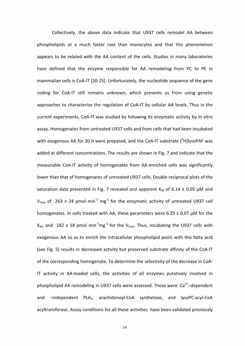
Comparative studies of AA incorporation and remodeling were carried out in
human monocytes under identical conditions as those depicted in Fig. 1. While
incorporation of exogenous AA into monocytes was generally similar to that in U937
cells (Fig. 2A), a striking difference was immediately noticed when the distribution of
AA in phospholipid classes was studied (Fig. 2B). In monocytes, the remodeling time
was much higher than in U937 cells, i.e. approx. 15 h. Thus, AA remodeling in human
U937 cells is extremely faster than in monocytes, despite the process occurring under
conditions where both types of cells incorporate similar amounts of exogenous AA into
phospholipids.
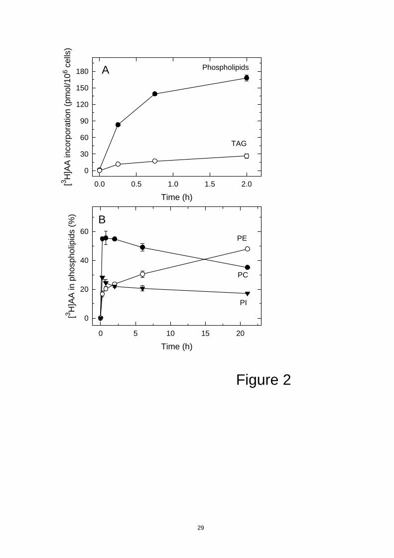
To further characterize the above findings, we used ion‐trap mass spectrometry
11

to determine the molecular phospholipid species to which exogenous AA initially
incorporated, i.e. a short time after addition of the fatty acid (30 min). For these
experiments, [2H]AA, not [3H]AA was used. From the data in Figs. 1 and 2, detection of
exogenous AA into various molecular species of all PI, PC and PE classes would be
expected in the U937 cells, the PE species arising primarily from transacylation from PC
species. In the case of human monocytes, appearance of exogenous AA into PI and PC
species would be expected, but little or no PE molecular species would be expected.
The results shown in Fig. 3 indicate that this is exactly what happened. Thus, in U937
cells some of the initial PC species for AA incorporation have lost the fatty acid, which
was likely transferred to various PE molecular species at 30 min, whereas such an
amount of time was not long enough to permit significant transfer from PC species to
PE species in human monocytes. From the data in Fig. 3, it is tempting to speculate
that PC(16:0/[2H]AA) and PC(18:2/[2H]AA) are two preferred donors of AA to PE
species, since they are present in human monocytes at 30 min but not in U937 cells. On
the other hand, PC(18:0/[2H]AA) was detected in U937 cells but still not in monocytes
at 30 min, despite this species constituting the major reservoir of endogenous AA
among PC species under equilibrium conditions [11]. The detection of
diarachidonoylated PC molecular species in monocytes but not in U937 cells has been
discussed elsewhere [29].
Phorbol esters such as PMA are very effective in inducing the differentiation of
U937 cells towards mature monocyte‐like cells [27]. To assess the effect of
differentiation on the processes of AA incorporation and remodeling, we incubated the
PMA‐differentiated U937 cells with [3H]AA, and the movement of the fatty acid
between phospholipids was followed at different times. The data, as shown in Fig. 4,
12
indicated that, although phospholipid AA incorporation was somewhat lower in the
PMA‐differentiated cells compared with the undifferentiated cells, the remodeling of
AA in the differentiated U937 cells was almost identical to that previously documented
for the fresh undifferentiated U937 cells (cf. Figs. 1 and 4). These data suggest that AA
remodeling in U937 cells is not influenced by the maturation degree of the cells. Thus
U937 cells remodel AA among phospholipids very rapidly regardless of whether or not
the cells are committed to differentiate.
A major difference between U937 cells and monocytes is that the former, as
cells in culture, are typically deficient in AA. Thus we studied whether the very fast
remodeling of U937 cells was somehow related to a diminished AA content. To this
end, we incubated the U937 cells with exogenous AA (100 µM, complexed with BSA
5:1) for 20 h, which raised the cellular AA content to levels similar to those found in
mature blood monocytes (Fig. 5). After the 20‐h incubation, the cells were pulse‐
chased with 1 µM [3H]AA and the movement of the fatty acid among phospholipids
was followed at different times. The amount of AA incorporated into phospholipids
was 200 ± 20 pmol per 106 cells at 120 min, which was similar to that found in U937
cells not loaded with AA or monocytes. Importantly however, the remodeling of AA
among phospholipids was much slower than that of U937 cells not loaded with AA,
becoming quite similar to that of monocytes. The remodeling time under these
conditions was 15 h (Fig. 6). In experiments utilizing differentiated U937 cells enriched
with AA, the remodeling of this fatty acid was essentially the same as that found in the
undifferentiated U937 cells enriched with AA (data not shown), thus providing further
evidence that this process is not influenced by the degree of maturation of the
phagocyte.
13
Collectively, the above data indicate that U937 cells remodel AA between
phospholipids at a much faster rate than monocytes and that this phenomenon
appears to be related with the AA content of the cells. Studies in many laboratories
have defined that the enzyme responsible for AA remodeling from PC to PE in
mammalian cells is CoA‐IT [20‐25]. Unfortunately, the nucleotide sequence of the gene
coding for CoA‐IT still remains unknown, which prevents us from using genetic
approaches to characterize the regulation of CoA‐IT by cellular AA levels. Thus in the
current experiments, CoA‐IT was studied by following its enzymatic activity by in vitro
assay. Homogenates from untreated U937 cells and from cells that had been incubated
with exogenous AA for 20 h were prepared, and the CoA‐IT substrate [3H]lysoPAF was
added at different concentrations. The results are shown in Fig. 7 and indicate that the
measurable CoA‐IT activity of homogenates from AA‐enriched cells was significantly
lower than that of homogenates of untreated U937 cells. Double reciprocal plots of the
saturation data presented in Fig. 7 revealed and apparent KM of 6.14 ± 0.05 µM and
Vmax of 263 ± 24 pmol min‐1 mg‐1 for the enzymatic activity of untreated U937 cell
homogenates. In cells treated with AA, these parameters were 6.25 ± 0.07 µM for the
KM, and 182 ± 18 pmol min‐1mg‐1 for the Vmax. Thus, incubating the U937 cells with
exogenous AA so as to enrich the intracellular phospholipid pools with this fatty acid
(see Fig. 5) results in decreased activity but preserved substrate affinity of the CoA‐IT
of the corresponding homogenate. To determine the selectivity of the decrease in CoA‐
IT activity in AA‐loaded cells, the activities of all enzymes putatively involved in
phospholipid AA remodeling in U937 cells were assessed. These were Ca2+–dependent
and –independent PLA2, arachidonoyl‐CoA synthetase, and lysoPC:acyl‐CoA
acyltransferase. Assay conditions for all these activities have been validated previously
14

for U937 cell homogenates [14, 30‐37, 39]. However, none of these activities was
found to appreciably change in homogenates after loading the cells with AA (data not
shown).
15

4. Discussion
In this study we have compared the incorporation and remodeling of AA within
membrane phospholipids of human U937 promonocyte‐like cells and human
peripheral blood monocytes. The importance of understanding cellular AA dynamics in
human phagocytic cells stems from the fact that AA and its oxygenated derivatives, the
eicosanoids, are key regulators of immunoinflammatory reactions. Remodeling of AA
between cellular phospholipids is important to achieve the appropriate distribution of
the fatty acid into the specific pools which will be acted upon by phospholipases A2 to
initiate eicosanoid biosynthesis [3, 4]. There is now strong evidence that CoA‐
dependent acyltransferase and CoA‐independent transacylases all participate
sequentially in the process of phospholipid AA remodeling [3, 4]. CoA‐dependent
transacylases mediate the initial incorporation of AA into phospholipids, which in turn
is followed by the action of CoA‐IT which redistributes the AA among phospholipid
classes. Importantly, inhibition of these pathways has been demonstrated to
profoundly alter cellular homeostasis and induce cell death [17‐19].
We demonstrate that U937 cells manifest a relatively high capacity to
incorporate AA into phospholipids that is quantitatively similar to that of blood
monocytes, and in both types of cells, import of AA into neutral lipids (TAG) is minor. In
both U937 cells and monocytes, AA is incorporated into PC and PI first, followed by a
transfer to PE at the expense of PC. Although this AA remodeling process is
qualitatively similar to that reported in other cell systems [3], what makes it
noteworthy in the U937 cells is that it is extremely rapid compared to that in
16

monocytes.
U937 cells can be induced to differentiate towards mature monocyte‐like cells
by exposing them to a variety of agents, including the phorbol ester PMA. When
exposed to PMA, U937 cells cease to divide, become adherent, form clumps and
express phenotypic properties of mature monocytes, including the expression of
surface antigens [27]. Importantly however, essentially the same exceedingly high
phospholipid AA remodeling of the undifferentiated U937 cells is also seen in PMA‐
differentiated cells. Hence, these results dissociate the differentiation‐induced changes
in lipid metabolism and the accompanying decreased need for synthesis of
phospholipids from AA trafficking between phospholipids, suggesting that these events
are regulated in an independent manner.
The amount of AA present in normal blood monocytes represents about 20% of
total cellular fatty acid (this study). In U937 cells however, AA represents roughly only
3%. Thus, the possibility arises that the remarkably high capacity of U937 cells to
remodel AA in phospholipids is related to their relative 'deficiency' in endogenous AA
as compared to normal monocytes. To investigate this hypothesis, we have prepared
U937 cells enriched in AA by treating them with fatty acid/albumin complex, so that
the intracellular amount of AA was similar to that of normal monocytes. When exposed
for a second time to exogenous AA, the AA‐enriched U937 cells incorporate the fatty
acid in a similar manner, both qualitatively and quantitatively, to both U937 cells or
monocytes. Importantly however, our results clearly show that in these AA‐enriched
cells, the phospholipid AA remodeling process is much slower than that of otherwise
untreated U937 cells, becoming similar to that of monocytes (remodeling time for AA‐
enriched U937 cells and monocytes being approx. 15 h).
17

With the recent cloning of mammalian Mg2+‐dependent phosphatidate
phosphatases (lipins) [41] and platelet‐activating factor acetyl transferase [42], CoA‐IT
remains as the last lipid signaling and remodeling enzyme whose sequence remains
unknown. Thus, currently the only manner to study the cell regulation of CoA‐IT is by
measuring its enzyme activity. By doing this, we have found that CoA‐IT activity levels
appear to be negatively regulated by the intracellular level of AA. Thus, in
homogenates from cells enriched in AA, CoA‐IT activity is significantly lower than in
homogenates from AA‐deficient cells. After replenishing the cells with AA, CoA‐IT
activity of homogenates from these cells displays a decreased Vmax with respect to
homogenates from cells untreated with AA, but the KM was found to be the same in
both cases. It is interesting to note here that extensive studies of the CoA‐IT activity
from broken neutrophils carried out by Winkler and colleagues have shown that such
an activity is not directly inhibited by phospholipid substrates or phospholipid products
[43]. Thus, the most straightforward explanation for our data is that enriching the cells
with AA may reduce the cellular mass of CoA‐IT and hence its measurable activity in
homogenates. This leads to the intriguing question of whether an AA‐containing
phospholipid that is present in the AA‐enriched cells but not in the otherwise
untreated cells may affect the expression levels of CoA‐IT. In this regard, recent studies
have highlighted the idea that particular phospholipid molecular species may exert
profound effects on gene expression by directly interacting with transcription factors
[44], and our recent lipidomic studies in human monocytes and U937 have unveiled
the existence of a very rapid turnover of particular AA‐containing species depending on
the activation state of the cell [10]. Collectively, the results reported here suggest that
the role of CoA‐IT in AA signaling may be more complex than merely regulating the
18

transfer of fatty acid between phospholipid pools, and suggest interesting implications
in terms of defining possible novel strategies to regulate lipid mediator formation by
controlling the cellular levels of AA .
Acknowledgements
We thank Montse Duque and Yolanda Sáez for expert technical asssistance. This work
was supported by the Spanish Ministry of Science and Innovation (grants BFU2007‐
67154, SAF2007‐60055, BFU2010‐18826, and SAF2010‐18831) and by the Regional
Government of Castile and León (Grant CSI09‐A08). A.M.A. was supported by a
predoctoral fellowship from the Regional Government of Castile and León. D.B. and
L.G.‐d.‐G. were supported by predoctoral fellowships from the Spanish Ministry of
Science and Innovation. CIBERDEM is an initiative of Instituto de Salud Carlos III.
19

References
1. C. D. Funk. Prostaglandins and leukotrienes: advances in eicosanoid biology.
Science 294 (2001) 1871‐1875
2. M. W. Buczynski, D. S. Dumlao, and E. A. Dennis. An integrated omics analysis of
eicosanoid biology. J. Lipid Res. 50 (2009) 1015‐1038
3. F. H. Chilton, A. N. Fonteh, M. E. Surette, M. Triggiani, and J. D. Winkler. Control
of arachidonate levels within inflammatory cells. Biochim. Biophys. Acta 1299
(1996) 1‐15
4. G. Pérez‐Chacón, A. M. Astudillo, D. Balgoma, M. A. Balboa, and J. Balsinde.
Control of free arachidonic acid levels by phospholipases A2 and
lysophospholipid acyltransferases. Biochim. Biophys. Acta 1791 (2009) 1103‐
1113
5. M. A. Balboa, and J. Balsinde. Oxidative stress and arachidonic acid
mobilization. Biochim. Biophys. Acta 1761 (2006) 385‐391
6. J. Balsinde, M. V. Winstead, and E. A. Dennis. Phospholipase A2 regulation of
arachidonic acid mobilization. FEBS Lett. 531 (2002) 2‐6
7. J. I. MacDonald, and H. Sprecher. Phospholipid fatty acid remodeling in
mammalian cells. Biochim Biophys Acta. 1084 (1991) 105‐121
8. A. N. Fonteh, and F. H. Chilton. Rapid remodeling of arachidonate from
phosphatidylcholine to phosphatidylethanolamine pools during mast cell
activation. J. Immunol. 148 (1992) 1784‐1791
9. J. D. Winkler, C‐M. Sung, L. Huang, and F. H. Chilton. CoA‐independent
transacylase activity is increased in human neutrophils after treatment with
20

tumor necrosis factor α. Biochim. Biophys. Acta 1215 (1994) 133–140
10. M. L. Nieto, M. E. Venable, S. A. Bauldry, D. G. Greene, M. Kenedy, D. A. Bass,
and R. L. Wykle. Evidence that hydrolysis of ethanolamine plasmalogens
triggers synthesis of platelet‐activating factor via a transacylation reaction. J.
Biol. Chem. 266 (1991) 18699‐18706
11. D. Balgoma, A. M. Astudillo, G. Pérez‐Chacón, O. Montero, M. A. Balboa, and J.
Balsinde. Markers of monocyte activation revealed by lipidomic profiling of
arachidonic acid‐containing phospholipids. J. Immunol. 184 (2010) 3857‐3865
12. J. Balsinde, S. E. Barbour, I. D. Bianco, and E. A. Dennis. Arachidonic acid
mobilization in P388D1 macrophages is controlled by two distinct Ca2+‐
dependent phospholipase A2 enzymes. Proc. Natl. Acad. Sci. U.S.A. 91 (1994)
11060‐11064
13. E. Boilard, and M. E. Surette. Anti‐CD3 and concanavalin A‐induced human T
cell proliferation is associated with an increased rate of arachidonate‐
phospholipid remodeling. Lack of involvement of group IV and group VI
phospholipase A2 in remodeling and increased susceptibility of proliferating T
cells to CoA‐independent transacyclase inhibitor‐induced apoptosis. J. Biol.
Chem. 276 (2001) 17568‐17575
14. J. Balsinde. Roles of various phospholipases A2 in providing lysophospholipid
acceptors for fatty acid phospholipid incorporation and remodelling. Biochem J.
364 (2002) 695‐702
15. M. E. Surette, and F. H. Chilton. The distribution and metabolism of
arachidonate‐containing phospholipids in cellular nuclei. Biochem. J. 330 (1998)
915‐921
21

16. J. Balsinde, R. Pérez, and M. A. Balboa. 2006. Calcium‐independent
phospholipase A2 and apoptosis. Biochim. Biophys. Acta 1761: 1344‐1350
17. M. E. Surette, A. N. Fonteh, C. Bernatchez, and F. H. Chilton. Perturbations in
the control of cellular arachidonic acid levels block cell growth and induce
apoptosis in HL‐60 cells. Carcinogenesis 20 (1999) 757‐763
18. A. J. Trimboli, B. M. Waite, G. Atsumi, A. N. Fonteh, A. M. Namen, C. E. Clay, T.
E. Kute, K. P. High, M. C. Willingham, and F. H. Chilton. Influence of coenzyme
A‐independent transacylase and cyclooxygenase inhibitors on the proliferation
of breast cancer cells. Cancer Res. 59 (1999) 6171‐6177
19. R. Pérez, X. Matabosch, A. Llebaria, M. A. Balboa, and J. Balsinde. Blockade of
arachidonic acid incorporation into phospholipids induces apoptosis in U937
promonocytic cells. J. Lipid Res. 47 (2006) 484‐491
20. R. M. Kramer, and D. Deykin. Arachidonoyl transacylase in human platelets. J.
Biol. Chem. 258 (1983) 13806‐13811
21. M. Robinson, M. L. Blank, and F. Snyder. Acylation of Lysophospholipids by
Rabbit Alveolar Macrophages. J. Biol. Chem. 260 (1985) 7889‐7895
22. Y. Uemura, T. Lee, and F. Snyder. A coenzyme A‐independent transacylase is
linked to the formation of platelet‐activating factor (PAF) by generating the
lyso‐PAF intermediate in the remodeling pathway. J. Biol. Chem. 266 (1991)
8268‐8272
23. M. E. Venable, M. L. Nieto, J. D. Schmitt, and R. L. Wykle. Conversion of 1‐O‐
[3H]Alkyl‐2‐arachidonoyl‐sn‐glycero‐3‐phosphorylcholine to lyso platelet‐
activating factor by the CoA‐independent transacylase in membrane fractions
of human neutrophils. J. Biol. Chem. 266 (1991) 18691‐18698
22

24. F. Snyder, T‐C Lee, and M. L. Blank. The role of metabolism platelet
transacylases in the of arachidonate and activating factor. Prog. Lipid Res. 31
(1992) 65‐86
25. F. H. Chilton, A. N. Fonteh, C‐M. Sung, D. M. B. Hickey, T. J. Trophy, R. J. Mayer,
L. A. Marshall, J. D. Heravi, and J. D. Winkler. Inhibitors of CoA‐independent
transacylase block the movement of arachidonate into 1‐ether‐linked
phospholipids of human netrophils. Biochemistry 34 (1995) 5403‐5410
26. J. Balsinde, and F. Mollinedo. Platelet‐activating factor synergizes with phorbol
myristate acetate in activating phospholipase D in the human promonocytic cell
line U937. Evidence for different mechanisms of activation. J. Biol. Chem. 266
(1991) 18726‐18730
27. J. Balsinde, and F. Mollinedo. Induction of the oxidative response and of
concanavalin A‐binding capacity in maturing human U937 cells. Biochim.
Biophys. Acta 1052 (1990) 90‐95
28. E. G. Bligh, and W. J. Dyer. A rapid method of total lipid extraction and
purification. Can. J. Biochem. Physiol. 37 (1959) 911‐917
29. D. Balgoma, O. Montero, M. A. Balboa, and J. Balsinde. Calcium‐independent
phospholipase A2‐mediated formation of 1,2‐diarachidonoyl‐
glycerophosphoinositol in monocytes. FEBS J. 275 (2008) 6180‐6191
30. J. Balsinde, S. E. Barbour, I. D. Bianco, and E. A. Dennis. Arachidonic acid
mobilization in P388D1 macrophages is controlled by two distinct Ca2+‐
dependent phospholipase A2 enzymes. Proc. Natl. Acad. Sci. U.S.A. 91 (1994)
11060‐11064
31. E. Diez, J. Balsinde, M. Aracil, and A. Schüller. Ethanol induces release of
23

arachidonic acid but not synthesis of eicosanoids in mouse peritoneal
macrophages. Biochim. Biophys. Acta 921 (1987) 82‐89
32. M. A. Balboa, and J. Balsinde. Involvement of calcium‐independent
phospholipase A2 in hydrogen peroxide‐induced accumulation of free fatty
acids in human U937 cells. J. Biol. Chem. 277 (2002) 40384‐40389
33. M. A. Balboa, Y. Sáez, and J. Balsinde. Calcium‐independent phospholipase A2 is
required for lysozyme secretion in U937 promonocytes. J. Immunol. 170 (2003)
5276‐5280
34. M. A. Balboa, R. Pérez, and J. Balsinde. Amplification mechanisms of
inflammation: paracrine stimulation of arachidonic acid mobilization by
secreted phospholipase A2 is regulated by cytosolic phospholipase A2‐derived
hydroperoxyeicosatetraenoic acid. J. Immunol. 171 (2003) 989‐994
35. L. Fuentes, R. Pérez, M. L. Nieto, J. Balsinde, and M. A. Balboa. Bromoenol
lactone promotes cell death by a mechanism involving phosphatidate
phosphohydrolase‐1 rather than calcium‐independent phospholipase A2. J. Biol.
Chem. 278 (2003) 44683‐44690
36. R. Pérez, R. Melero, M. A. Balboa, and J. Balsinde. Role of group VIA calcium‐
independent phospholipase A2 in arachidonic acid release, phospholipid fatty
acid incorporation, and apoptosis in U937 cells responding to hydrogen
peroxide. J. Biol. Chem. 279 (2004) 40385‐40391
37. M. A. Balboa, R. Pérez, and J. Balsinde. Calcium‐independent phospholipase A2
mediates proliferation of human promonocytic U937 cells. FEBS J. 275 (2008)
1915‐1924
24

38. D. B. Wilson, S. M. Prescott, and P. W. Majerus. Discovery of an arachidonoyl
coenzyme A synthetase in human platelets. J. Biol. Chem. 257 (1982) 3510‐3515
39. G. Pérez‐Chacón, A. M. Astudillo, V. Ruipérez, M. A. Balboa, and J. Balsinde.
Signaling role for lysophosphatidylcholine acyltransferase 3 in receptor‐
regulated arachidonic acid reacylation reactions in human monocytes. J.
Immunol. 184 (2010) 1071‐1078
40. W. E. Lands, M. Inoue, Y. Sugiura, and H. Okuyama. Selective incorporation of
polyunsaturated fatty acids into phosphatidylcholine by rat liver microsomes. J.
Biol. Chem. 257 (1982) 14968‐14972
41. G. M. Carman, and G. S. Han. Phosphatidic acid phosphatase, a key enzyme in
the regulation of lipid synthesis. J. Biol. Chem. 284 (2009) 2593‐2597
42. R. Morimoto, H. Shindou, Y. Oda, and T. Shimizu. Phosphorylation of
lysophosphatidylcholine acyltransferase 2 at Ser34 enhances platelet‐activating
factor production in endotoxin‐stimulated macrophages. J. Biol. Chem. 285
(2010) 29857‐29862
43. J. D. Winkler, C. M. Sung, C. F. Bennett, and F. H. Chilton. Characterization of
CoA‐independent transacylase activity in U937 cells. Biochim. Biophys. Acta
1081 (1991) 339‐346
44. M. V. Chakravarthy, I. J. Lodhi, L. Yin, R. R. Malapaka, H. E. Xu, J. Turk, and C. F.
Semenkovich. Identification of a physiologically relevant endogenous ligand for
PPARα in liver. Cell 138 (2009) 476‐488
25

Figure Legends
Figure 1. [3H]AA incorporation and remodeling into U937 cells. Cells were incubated
with 1 µM [3H]AA (0.25 µCi/ml) for the indicated times. Afterward total lipids were
extracted and [3H]AA incorporation in phospholipids ( ) and TAG ( ) (A) or remodeling
in PI ( ), PC ( ), and PE ( ) (B) was measured. Data are given as means ± SEM of at
least three independent determinations.
Figure 2. [3H]AA incorporation and remodeling into human monocytes. For [3H]AA
incorporation assay (A), cells were incubated with 1 µM [3H]AA (0.25 µCi/ml) for the
indicated times. Afterward total lipids were extracted and [3H]AA incorporation in
phospholipids ( ) and TAG ( ) was measured. For phospholipid AA remodeling (B), the
cells were exposed to 1 µM [3H]AA (0.25 µCi/ml) for 30 min, washed with medium
containing BSA and incubated in fresh medium for the indicated times, and the amount
of [3H]AA in PI ( ), PC ( ), and PE ( ) was measured. Data are given as means ± SEM
of at least three independent determinations.
Figure 3. Analysis of phospholipid species containing [2H]AA in U937 and human
monocytes. Cells were incubated with 1 µM [2H]AA for 30 min. Afterward total lipids
were extracted and [2H]AA‐containing glycerophospholipids were identified (blue
boxes) or not (yellow boxes) by HPLC/MS. Data are given as means ± SEM of at least
three independent determinations.
Figure 4. [3H]AA incorporation and remodeling into monocyte‐like U937 cells. Cells
previously incubated with 35 ng/ml PMA for 24 were exposed to 1 µM [3H]AA (0.25
µCi/ml) for the indicated times. Afterward total lipids were extracted and [3H]AA
incorporation in phospholipids ( ) and TAG ( )(figure 1A) or remodeling in PI ( ), PC
( ), and PE ( ) (figure 1B) was measured. Data are given as means ± SEM of at least
three independent determinations.
26

Figure 5. Total AA content in U937, human monocytes and AA‐enriched U937 cells.
Total lipids were extracted and transmethylated with 0.5 M KOH in MeOH. The
resulting fatty acid methyl esters were analyzed by GC/MS. Total mass of AA per mg
protein (white bars) or % of AA respect to total amount of fatty acid (grey bars) are
shown. Data are given as means ± SEM of at least three independent determinations.
Figure 6. [3H]AA incorporation and remodeling into AA‐enriched U937 cells. U937
incubated with medium containing AA 100 µM complexed with BSA (ratio 5:1) for 20 h
were washed and labeled with 1 µM [3H]AA (0.25 µCi/ml) for the indicated times.
Afterward total lipids were extracted and [3H]AA incorporation in PLs ( ) and TAG ( )
(A). For the remodeling assay (B), the cells were washed with medium containing BSA
after 30 min incubation with 1 µM [3H]AA (0.25 µCi/ml) and [3H]AA label was measured
in PI ( ), PC ( ), and PE ( ).Data are given as means ± SEM of at least three
independent determinations.
Figure 7. CoA‐IT activity is decreased in homogenates from AA‐enriched U937 cells.
Homogenates were prepared from untreated cells ( ) or AA‐enriched cells ( ). Data
are given as means ± SEM of at least three independent determinations.
27

Time (h)0 1 2 3 4 5
[3 H]A
A in
pho
spho
lipid
s (%
)
0
20
40
60
Time (h)0.0 0.5 1.0 1.5 2.0[3
H] A
A in
corp
orat
ion
(pm
ol/1
06 c
ells
)
0
50
100
150
200
250
Figure 1
PE
PC
PI
Phospholipids
TAG
A
B
28
Time (h)0 5 10 15 20
[3 H]A
A in
pho
spho
lipid
s (%
)
0
20
40
60
Time (h)0.0 0.5 1.0 1.5 2.0[3 H
]AA
inco
rpor
atio
n (p
mol
/106
cel
ls)
0
30
60
90
120
150
180
Figure 2
PE
PC
PI
Phospholipids
TAG
A
B
29
PI(16:0/[2H]AA)PI(18:1/[2H]AA)PI(18:0/[2H]AA)
PI([2H]AA/[2H]AA)
PE(P-16:0/[2H]AA)PE(P-18:1/[2H]AA)PE(P-18:0/[2H]AA)PE(18:1/[2H]AA)
PC(16:0/[2H]AA)PC(18:2/[2H]AA)PC(18:1/[2H]AA)PC(18:0/[2H]AA)
PC([2H]AA/[2H]AA)
PI(16:0/[2H]AA)PI(18:1/[2H]AA)PI(18:0/[2H]AA)
PI([2H]AA/[2H]AA)
PE(P-16:0/[2H]AA)PE(P-18:1/[2H]AA)PE(P-18:0/[2H]AA)PE(18:1/[2H]AA)
PC(16:0/[2H]AA)PC(18:2/[2H]AA)PC(18:1/[2H]AA)PC(18:0/[2H]AA)
PC([2H]AA/[2H]AA)
U937Mon.
Figure 3
30

Time (h)0 1 2 3 4 5
[3 H]A
A in
pho
spho
lipid
s (%
)
0
20
40
60
Time (h)0.0 0.5 1.0 1.5 2.0[3 H
] AA
inco
rpor
atio
n (p
mol
/106
cells
)
0
50
100
150
Figure 4
PE
PC
PI
Phospholipids
TAG
A
B
31

Monocytes U937 AA-enriched
Cel
lula
r AA
con
tent
(nm
ol/m
g)
0
50
100
150
200
Cel
lula
r AA
con
tent
(%)
0
10
20
30
40
50
U937
Figure 5
32

Time (h)0 5 10 15 20 25 30
[3 H]A
A in
pho
spho
lipid
s (%
)
0
20
40
60
Time (h)0.0 0.5 1.0 1.5 2.0[3 H
]AA
inco
rpor
atio
n (p
mol
/106
cel
ls)
0
30
60
90
120
150
180
210
Figure 6
PE
PC
PI
Phospholipids
TAG
A
B
33

[LysoPAF] (µM)0 5 10 15 20 25 30
CoA
-IT A
ctiv
ity (p
mol
min
-1 m
g-1)
0
50
100
150
200
250
Figure 7
34


![Research Article Modulation of Arachidonic Acid Metabolism ...downloads.hindawi.com/archive/2014/683508.pdf · metabolism of arachidonic acid to biologically active EETs [ ]. e three](https://img.pdfslide.net/doc/110x75/606ff9bcbd5c0d69301096c4/research-article-modulation-of-arachidonic-acid-metabolism-metabolism-of-arachidonic.jpg)