Embed Size (px)
Citation preview
137
Korean J Physiol PharmacolVol 15: 137-142, June, 2011DOI: 10.4196/kjpp.2011.15.3.137
ABBREVIATIONS: ELF-MF, extremely low frequency magnetic fields; GI, gastrointestinal.
Received April 20, 2011, Revised May 30, 2011, Accepted June 15, 2011
Corresponding to: Ji Hoon Jeong, Department of Pharmacology, College of Medicine, Chung-Ang University, 221, Heuksuk-dong, Dongjak-gu, Seoul 156-756, Korea. (Tel) 82-2-820-5688, (Fax) 82-2- 826-8752, (E-mail) [email protected]*The authors contribute equally to this paper and share the first authorship.
Influence of Exposure to Extremely Low Frequency Magnetic Field on Neuroendocrine Cells and Hormones in Stomach of Rats
Min Eui Hong1,*, Kyu Hyun Yoon3,*, Yoon Yang Jung1, Tae Jin Lee1, Eon Sub Park1, Uy Dong Sohn4, and Ji Hoon Jeong2
Departments of 1Pathology, 2Pharmacology, 3Internal Medicine, College of Medicine, 4Department of Pharmacology, College of Pharmacy, Chung-Ang University, Seoul 156-756, Korea
Extremely low frequency magnetic fields (ELF-MF) have the ability to produce a variety of behavioral and physiological changes in animals. The stomach, as the most sensitive part of the neuroendocrine organ of the gastrointestinal tract, is crucial for the initiation of a full stress response against all harmful stress. Thus, the purpose of this study was to examine whether ELF-MF stimuli induce changes in the activity of neuroendocrine cells, considering their involvement in endocrine or paracrine effect on surrounding cells. The exposure to ELF-MF (durations of 24 h and 1 or 2 weeks, 60 Hz fre-quency, 0.1 mT intensity) altered the distribution and occurrence of gastrin, ghrelin and somatostatin- positive endocrine cells in the stomach of rats. The change, however, in the secretion of those hormones into blood from endocrine cells did not appear significantly with ELF-MF exposure. Comparing with sham control, ELF-MF exposure for 1 and 2 week induced an increase in BaSO4 suspension propelling ratio of gastrointestinal tract, indicating that ELF-MF affects gastrointestinal motility. Our study revealed that ELF-MF exposure might influence the activity of endocrine cells, an important element of the intrinsic regulatory system in the digestive tract. The pathophysiological character of these changes and the mechanism responsible for neuroendocrine cell are still unclear and require further studies.
Key Words: ELF-MF, Gastrin, Somatostatin, Ghrelin
INTRODUCTION
The question of whether extremely low frequency mag-netic fields (ELF-MF) can affect biological systems has at-tracted attention for decades. Many field and laboratory in-vestigators have reported that ELF-MF originating from residentially proximate power line, household electrical wiring, medical devices, cellular phone and wireless com-munication, produce a variety of behavioral and physio-logical changes in animals [1,2]. Furthermore, fundamental research for ELF-MF suggested that changes by ELF-MF of neurotransmitter concentration, activity stimulation of numerous enzymes and hormones, stimulation of oxida-tion-reduction processes and cellular synthesis resulted in the biological effect of ELF-MF [3-6]. Although the data on the effects of EMF on human endo-crine system are scarce, experimental animal studies in-dicate that EMF may influence secretion of some hormones. Most of the results concentrate on influence of EMF on se-
cretion of hormone in brain. It is well known that exposure to ELF-MF may suppress the synthesis of the indoleamine hormone melatonin in the pineal gland of some species [7-9]. This effect of ELF-MF on pineal function is similar to that of light, which is the main environmental cue media-ting the response to photoperiod in mammals and birds. Moreover, the possible hormonal effects of ELF-MF on re-production and development, including gametogenesis, fer-tilization, implantation, embryogensis and endocrine sys-tem, have been studied [10-15]. In one previous study, ELF-MF exposure affected the level of thyroid hormone thy-roid [16,17]. Other study group reported that exposure to ELFMF induces a significant increase in the level of cortico-sterone in blood plasma and lead to impairment in discrim-ination between familiar and novel objects [18]. These re-sults might suggest that ELF-MF exposure may be an envi-ronmental stress factor by changing hormonal response. The behavioral consequence caused by the exposure to ELF-MF resembles the symptoms seen in biological re-sponse to exposure to uncontrolled stress. Stomach is specialized organ for food digestion by chem-ical secretion and physical motility. All these processes are regulated by the intrinsic neuroendocrine system, a group of cells dispersed among non-endocrine epithelial cells that specialized in the secretion of a variety of bioactive peptides to the blood or towards the neighboring cells. In various
138 ME Hong, et al
pathological states the number of neuroendocrine cells in the gastrointestinal (GI) tract undergoes some changes. The stomach, affected by broad stressful stimuli, might initiate a counter-response. One investigation suggests that the stomach, as the most sensitive part of the gastrointestinal tract and the largest neuroendocrine organ in the body, is crucial for the initiation of a full stress response against all harmful stress [19]. Some paper reported that MF might alter transcription and translation, including hsp70 and im-mediate early response genes myc, jun and fos [20-23]. The increased expression of stress expression in the presence of MF suggests that the stress-sensitive gastrointestinal tract may response to MF as an environmental stress. It is not yet clear, however, whether stress response by ELF-MF develop in gastric mucosa and how the length of ELF-EMF exposure affects stress adaptation in gastro-intestinal tract. The aim of our present work was, therefore, to study the consequences of the exposure to ELF-EMF (durations of 24 h and 1 or 2 weeks, 60 Hz frequency, 0.1 mT intensity) on gastric morphology and function in rats in connection with the development of chronic stress state. We examined the hormone changes associated with stom-ach function including ghrelin, somatostatin and gastrin. In order to assess ELF-MF stress-related endocrine changes in stomach, we counted the endocrine cell in stomach and measured the level of the respective hormone in blood.
METHODS
Animals
Male Sprague-Dawley rats (Samtaco Korea), 5∼6 weeks of age and weighing 200 g, were used in this study. The rat were housed for 1 week adaptation in a temperature- controlled room on a 12-h light/dark cycle and fed on a standard Purina rat chow diet before the experiment. Rats were continuously exposed to 60 Hz magnetic field for 7 days. This project was approved by the Institutional Animal Care and Use Committee of the Chung-Ang Medical Uni-versity.
Magnetic field exposure system
The 60 Hz magnetic field was produced by 1m-square Helmholz coil with widening embedded in an open woden rectangular frame. Each coil has 200 turns and was con-nected in a series connection to 220 V AC power supply via variable transformer. Each winding was split allowing the current to flow in the same sense through each half of the winding (Field aiding). We can modulate the in-tensity of magnetic field by the transformer. The magnetic field at the center of exposure system was measured by Gaussmeter (Lake shore Model 410), and we choose 0.1 mT for exposing intensity. The set of coils stood on the platform. We put the animal cage without material at the center.
Immunohistochemical staining
Expression of gastrin, ghrelin, and somatostain positive cell in gastric tissue were detected with the avidin-bio-tinylated horseradish peroxidase complex (DAKO LSAB, Los Angels, CA) according to the kit's instruction. Primary antibodies used were anti-gastrin (1:100; RB-1459, Fre-mont, CA), anti-ghrelin (1:200; SC10368, Santacruz, CA),
and anti-somatostatin (1:50; GTX71935, Parkway Irivine, CA). In short, paraffin-embedded gastric tissue specimes were consecutively sectioned into 4 μm thick slices. After deparaffinization in xyline and rehydration through de-creasing concentrations of ethanol, slides were immersed in citrate buffer (pH 6.0). After antigen retrieval using a microwave oven at 98oC for 15 min. The slides were in-cubated in 0.3% hydrogen peroxide for 30 min to block the endogeneous peroxidase activity. Then the sections were in-cubated with the primary antibody at room temperature for 30 min. The negative control staining slides were in-cubated in the absence of the primary antibody. The slides were washed and the chromogen was developed for 5 min with using liquid 3,3'-diaminobenzidine; the slides was then counter-stained with Mayer's hematoxylin, dehydrated, and mounted with Canada balsam for examination.
Counts of neuroendocrine cells (gastrin, ghrelin and somatostatin)
Cells with ghrelin, gastrin, and somatostatin expression searched and their topography was observed. The number of individual cells was determined per unit area (1 mm2) of each part of gastric mucosa (corpus and pyloric antrum). For the immunoreactive cell counts, all available slides were examined at a magnification of ×40 in the longitudinal sections of stomach. At least five sections were counted at a magnification of ×200 (Olympus BX51, 0.949 mm2). The cell count was expressed as the mean number of cells per visual field.
Determination of serum levels of gastrin, ghrelin, and somatostatin
The enzyme immunoassay kits for rat ghrelin and soma-tostatin were purchased from Phenix Pharmaceuticals (Burlingame, CA). The rat gastrin enzyme immunoassay kit was purchased from Assay Designs (Ann Arbor, MI). Procedures for the measurement of these peptides were per-formed in accordance with the manufacturer’s instructions.
Propelling test of gastrointestinal tract
For propelling test of gastrointestinal tract, 30 min after MF exposure, 0.5 mL of 25% BaSO4 suspended liquid was orally administered to the animals. All animals were sacri-ficed 20 min after the administration of 25% BaSO4, and the entire small intestine was removed. The small intestine from pylorus to the boundary of ileum and cecum was iso-lated and its length was "total length of small intestine". The length from pylorus to the foreland of 25% BaSO4 sus-pension was "BaSO4 suspension propelling length".
BaSO4 suspension propelling ratio=[BaSO4 suspension propelling length (cm)/total length of small intestine (cm)] ×100%
Statistical analysis
All data were presented as mean±standard deviation (SD). For endocrine cell densities, statistical comparisions for independent populations between the three groups were made and compared with one-way analysis of varience (ANOVA) and Student's t-test. Differences in values were considered significant if p<0.05.
Influence of ELF-MF on Neuroendocrine Cells in Stomach 139
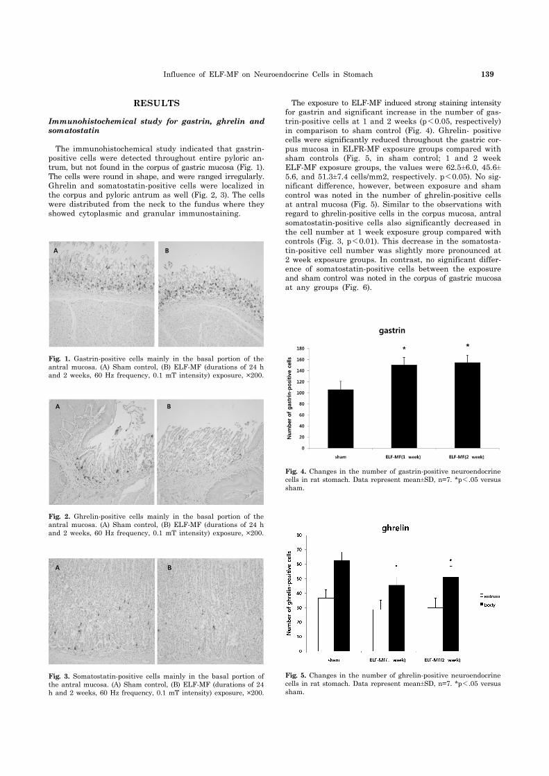
Fig. 4. Changes in the number of gastrin-positive neuroendocrine cells in rat stomach. Data represent mean±SD, n=7. *p<.05 versus sham.
Fig. 3. Somatostatin-positive cells mainly in the basal portion of the antral mucosa. (A) Sham control, (B) ELF-MF (durations of 24 h and 2 weeks, 60 Hz frequency, 0.1 mT intensity) exposure, ×200.
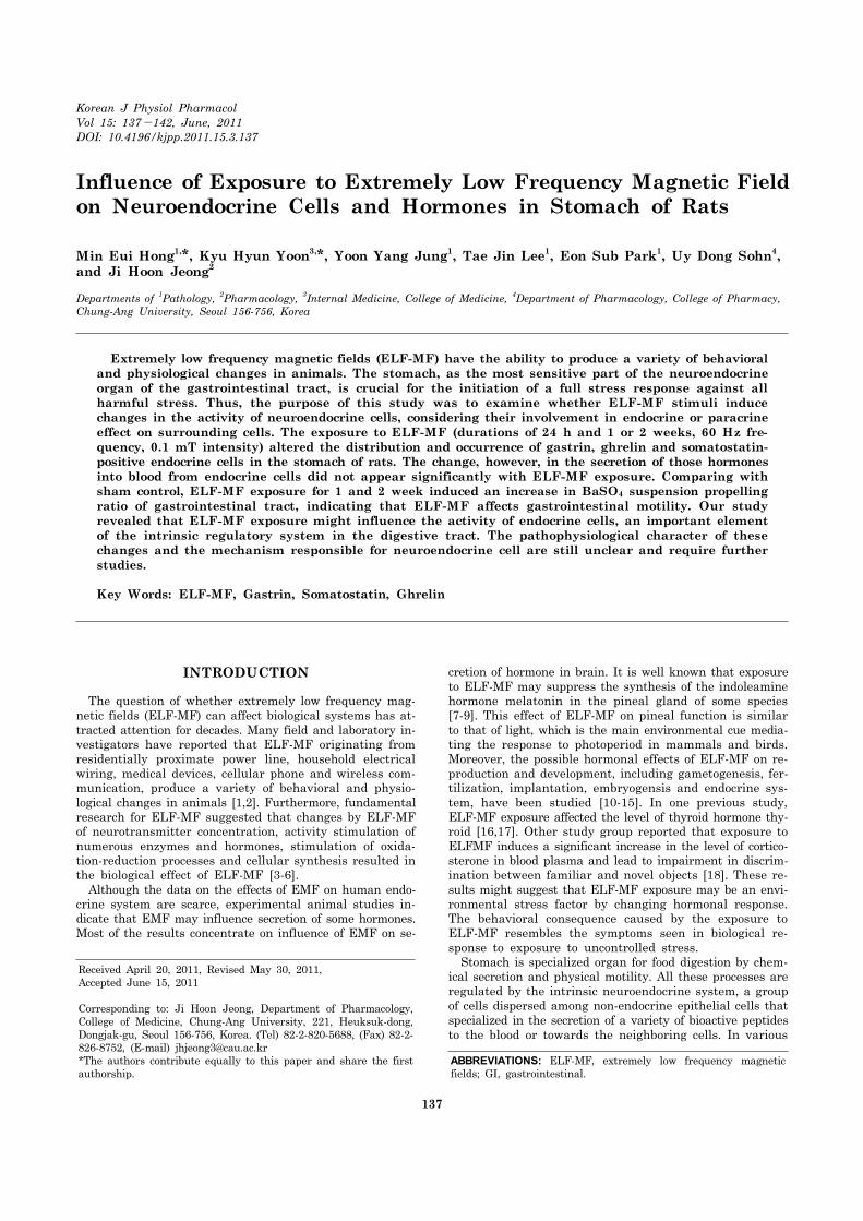
Fig. 1. Gastrin-positive cells mainly in the basal portion of the antral mucosa. (A) Sham control, (B) ELF-MF (durations of 24 h and 2 weeks, 60 Hz frequency, 0.1 mT intensity) exposure, ×200.
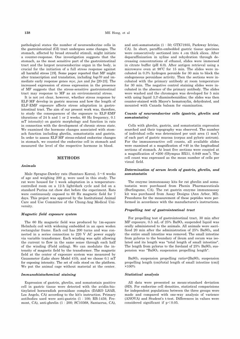
Fig. 2. Ghrelin-positive cells mainly in the basal portion of the antral mucosa. (A) Sham control, (B) ELF-MF (durations of 24 h and 2 weeks, 60 Hz frequency, 0.1 mT intensity) exposure, ×200.
Fig. 5. Changes in the number of ghrelin-positive neuroendocrine cells in rat stomach. Data represent mean±SD, n=7. *p<.05 versus sham.
RESULTS
Immunohistochemical study for gastrin, ghrelin and somatostatin
The immunohistochemical study indicated that gastrin- positive cells were detected throughout entire pyloric an-trum, but not found in the corpus of gastric mucosa (Fig. 1). The cells were round in shape, and were ranged irregularly. Ghrelin and somatostatin-positive cells were localized in the corpus and pyloric antrum as well (Fig. 2, 3). The cells were distributed from the neck to the fundus where they showed cytoplasmic and granular immunostaining.
The exposure to ELF-MF induced strong staining intensity for gastrin and significant increase in the number of gas-trin-positive cells at 1 and 2 weeks (p<0.05, respectively) in comparison to sham control (Fig. 4). Ghrelin- positive cells were significantly reduced throughout the gastric cor-pus mucosa in ELFR-MF exposure groups compared with sham controls (Fig. 5, in sham control; 1 and 2 week ELF-MF exposure groups, the values were 62.5±6.0, 45.6± 5.6, and 51.3±7.4 cells/mm2, respectively. p<0.05). No sig-nificant difference, however, between exposure and sham control was noted in the number of ghrelin-positive cells at antral mucosa (Fig. 5). Similar to the observations with regard to ghrelin-positive cells in the corpus mucosa, antral somatostatin-positive cells also significantly decreased in the cell number at 1 week exposure group compared with controls (Fig. 3, p<0.01). This decrease in the somatosta-tin-positive cell number was slightly more pronounced at 2 week exposure groups. In contrast, no significant differ-ence of somatostatin-positive cells between the exposure and sham control was noted in the corpus of gastric mucosa at any groups (Fig. 6).
140 ME Hong, et al
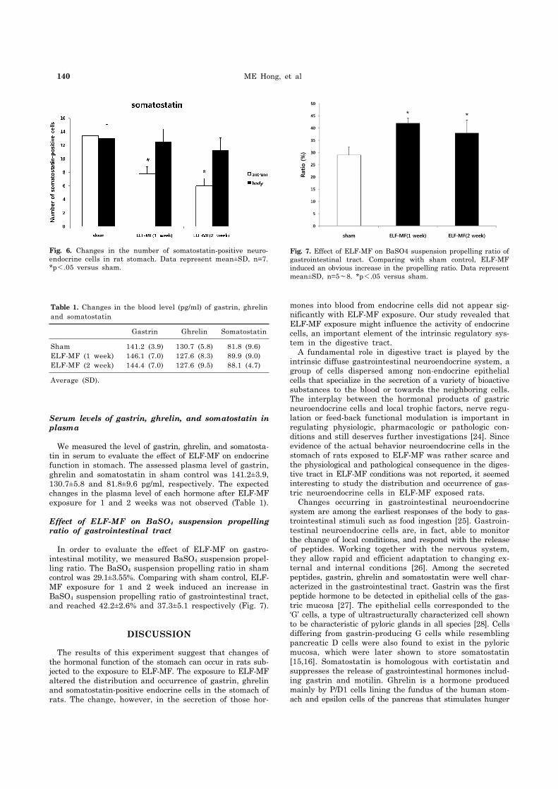
Fig. 6. Changes in the number of somatostatin-positive neuro-endocrine cells in rat stomach. Data represent mean±SD, n=7.*p<.05 versus sham.
Fig. 7. Effect of ELF-MF on BaSO4 suspension propelling ratio of gastrointestinal tract. Comparing with sham control, ELF-MF induced an obvious increase in the propelling ratio. Data represent mean±SD, n=5∼8. *p<.05 versus sham.
Table 1. Changes in the blood level (pg/ml) of gastrin, ghrelin and somatostatin
Gastrin Ghrelin Somatostatin
ShamELF-MF (1 week)ELF-MF (2 week)
141.2 (3.9)146.1 (7.0)144.4 (7.0)
130.7 (5.8)127.6 (8.3)127.6 (9.5)
81.8 (9.6)89.9 (9.0)88.1 (4.7)
Average (SD).
Serum levels of gastrin, ghrelin, and somatostatin in plasma
We measured the level of gastrin, ghrelin, and somatosta-tin in serum to evaluate the effect of ELF-MF on endocrine function in stomach. The assessed plasma level of gastrin, ghrelin and somatostatin in sham control was 141.2±3.9, 130.7±5.8 and 81.8±9.6 pg/ml, respectively. The expected changes in the plasma level of each hormone after ELF-MF exposure for 1 and 2 weeks was not observed (Table 1).
Effect of ELF-MF on BaSO4 suspension propelling ratio of gastrointestinal tract
In order to evaluate the effect of ELF-MF on gastro-intestinal motility, we measured BaSO4 suspension propel-ling ratio. The BaSO4 suspension propelling ratio in sham control was 29.1±3.55%. Comparing with sham control, ELF- MF exposure for 1 and 2 week induced an increase in BaSO4 suspension propelling ratio of gastrointestinal tract, and reached 42.2±2.6% and 37.3±5.1 respectively (Fig. 7).
DISCUSSION
The results of this experiment suggest that changes of the hormonal function of the stomach can occur in rats sub-jected to the exposure to ELF-MF. The exposure to ELF-MF altered the distribution and occurrence of gastrin, ghrelin and somatostatin-positive endocrine cells in the stomach of rats. The change, however, in the secretion of those hor-
mones into blood from endocrine cells did not appear sig-nificantly with ELF-MF exposure. Our study revealed that ELF-MF exposure might influence the activity of endocrine cells, an important element of the intrinsic regulatory sys-tem in the digestive tract. A fundamental role in digestive tract is played by the intrinsic diffuse gastrointestinal neuroendocrine system, a group of cells dispersed among non-endocrine epithelial cells that specialize in the secretion of a variety of bioactive substances to the blood or towards the neighboring cells. The interplay between the hormonal products of gastric neuroendocrine cells and local trophic factors, nerve regu-lation or feed-back functional modulation is important in regulating physiologic, pharmacologic or pathologic con-ditions and still deserves further investigations [24]. Since evidence of the actual behavior neuroendocrine cells in the stomach of rats exposed to ELF-MF was rather scarce and the physiological and pathological consequence in the diges-tive tract in ELF-MF conditions was not reported, it seemed interesting to study the distribution and occurrence of gas-tric neuroendocrine cells in ELF-MF exposed rats. Changes occurring in gastrointestinal neuroendocrine system are among the earliest responses of the body to gas-trointestinal stimuli such as food ingestion [25]. Gastroin-testinal neuroendocrine cells are, in fact, able to monitor the change of local conditions, and respond with the release of peptides. Working together with the nervous system, they allow rapid and efficient adaptation to changing ex-ternal and internal conditions [26]. Among the secreted peptides, gastrin, ghrelin and somatostatin were well char-acterized in the gastrointestinal tract. Gastrin was the first peptide hormone to be detected in epithelial cells of the gas-tric mucosa [27]. The epithelial cells corresponded to the ‘G’ cells, a type of ultrastructurally characterized cell shown to be characteristic of pyloric glands in all species [28]. Cells differing from gastrin-producing G cells while resembling pancreatic D cells were also found to exist in the pyloric mucosa, which were later shown to store somatostatin [15,16]. Somatostatin is homologous with cortistatin and suppresses the release of gastrointestinal hormones includ-ing gastrin and motilin. Ghrelin is a hormone produced mainly by P/D1 cells lining the fundus of the human stom-ach and epsilon cells of the pancreas that stimulates hunger
Influence of ELF-MF on Neuroendocrine Cells in Stomach 141
[29]. We found that ELF-MF induced a significant alteration in the number of endocrine cells in stomach, an increase in the number of G cells and interestingly, a decrease in D and P/D1 cells. A concomitant changes by ELF-MF in gastrin, somatostatin and ghrelin-positive cells in the an-tral mucosa (gastrin and somatostatin) or the corpus (ghrelin) of stomach may indicate that ELF-MF confer a stressful stimuli to the stomach. The previous studies of other investigators observed that ELF-MF was able to lead to a reversible change of ultrastructure of cells in the pan-creas [30]. The change was characterized by expansion of the Golgi apparatus, extension of rough endoplasmatic re-ticulum, mitochondrial swelling, expansion of b-granules and increase in number of empty vesicles in beta cells, oc-curred during the exposure [30]. The structure and function of pancreas is associated with a reciprocal relationship be-tween the activities of the gastric neuroendocrine cells. In addition, other studies evaluated the relationship between changes in proteolytic activity of pepsin and morphological characteristics of the gastric mucosa produced by non-thermal MF with the plane of polarization rotating in either right-handed or left-handed sense. The MF induced mor-phological changes in the gastric mucosa and gradual in-crease in the count of goblet cells resulted in mucoid trans-formation of glands. The surface epithelium underwent ex-foliation and secretary activity of glands was suppressed [31,32]. ELF-MF, a frequency of 50 Hz with a flux-density range of 0.3∼1.6 mT, was tested with regard to their influ-ence on cell proliferation, cyclic AMP-levels, and gap-junc-tion-mediated intercellular signalling and was found to in-duce periodic variations in cell-proliferation [33]. The re-sults of these studies are in accordance with the present data that ELF-MF may act as a stressful factor to alter the cell structure and function in digestive system. Although the mechanism of action of ELF-MF with bio-logical structures is up to now largely unknown, it has been proposed that the cell membrane must be the main target for interaction [34]. The present study has shown that the alteration of the number of endocrine cells by ELF-MF did not result in the change in the secretion of gastrin, somatostatin and ghrelin into blood. The data suggest that ELF-MF stimuli have a role in modulating paracrine effect in stomach rather than endocrine effect. Neuroendocrine cells transport their syn-thesized product through their long cytoplasmic processes and exert in this way paracrine effects [35]. It is also well known that neurohormone released from the neuroendo-crine cells in stomach may reach the connective tissue space of the lamina propia and exert paracrine effects also on oth-er endocrine cells to regulate acid secretion and gastro-intestinal motility [36,37]. One report proposes that long exposure of MF probably modulates the local release of en-dothelial neurohumoral and paracrine factors that act di-rectly on the tissue of the vascular wall, presumably by af-fecting ion channels or second messenger systems [38]. Although the mechanisms of action of ELF-MF on the proliferation or activation of gastric neuroendocrine cells re-mains further study, there is an increasing amount of in-formation pointing to a link between cell proliferation and ELF-MF exposure. The fact that EMF is capable of altering a gastric hormone-positive neuroendocrine cell might be of particular interest. Even if the influence of EMF on the se-cretion of the hormones into blood was found to be weak, EMF might serve as an stressful factor in stomach, which
generally lead to the paracrine effect on cells surrounding neuroendocrine cells. The pathophysiological character of these changes and the mechanism responsible for neuro-endocrine cell are still unclear and require further studies.
REFERENCES
1. Frey AH. Electromagnetic field interactions with biological systems. FASEB J. 1993;7:272-281.
2. Gould JL. Magnetic field sensitivity in animals. Annu Rev Physiol. 1984;46:585-598.
3. Janać B, Tovilović G, Tomić M, Prolić Z, Radenović L. Effect of continuous exposure to alternating magnetic field (50 Hz, 0.5 mT) on serotonin and dopamine receptors activity in rat brain. Gen Physiol Biophys. 2009;28:41-46.
4. Al-Akhras MA. Influence of 50 Hz magnetic field on sex hormones and body, uterine, and ovarian weights of adult female rats. Electromagn Biol Med. 2008;27:155-163.
5. Sakurai T, Satake A, Sumi S, Inoue K, Miyakoshi J. An extremely low frequency magnetic field attenuates insulin secretion from the insulinoma cell line, RIN-m. Bioelectromag-netics. 2004;25:160-166.
6. Jelenković A, Janać B, Pesić V, Jovanović DM, Vasiljević I, Prolić Z. Effects of extremely low-frequency magnetic field in the brain of rats. Brain Res Bull. 2006;68:355-360.
7. Rosen LA, Barber I, Lyle DB. A 0.5 G, 60 Hz magnetic field suppresses melatonin production in pinealocytes. Bioelectro-magnetics. 1998;19:123-127.
8. Yellon SM. Acute 60 Hz magnetic field exposure effects on the melatonin rhythm in the pineal gland and circulation of the adult Djungarian hamster. J Pineal Res. 1994;16:136-144.
9. Olcese J, Reuss S, Vollrath L. Evidence for the involvement of the visual system in mediating magnetic field effects on pineal melatonin synthesis in the rat. Brain Res. 1985;333: 382-384.
10. Al-Akhras MA, Darmani H, Elbetieha A. Influence of 50 Hz magnetic field on sex hormones and other fertility parameters of adult male rats. Bioelectromagnetics. 2006;27:127-131.
11. De Vita R, Cavallo D, Raganella L, Eleuteri P, Grollino MG, Calugi A. Effects of 50 Hz magnetic fields on mouse spermatogenesis monitored by flow cytometric analysis. Bioelectromagnetics. 1995;16:330-334.
12. Niehaus M, Brüggemeyer H, Behre HM, Lerchl A. Growth retardation, testicular stimulation, and increased melatonin synthesis by weak magnetic fields (50 Hz) in Djungarian hamsters, Phodopus sungorus. Biochem Biophys Res Commun. 1997;234:707-711.
13. Nakhilnitskaya ZN, Klimovskaya LD, Kuzmina ZF, Mastryu-kova VM, Smirnova NP, Strzhizhovsky AD, Cherkasov GV. Possible adaptation to strong magnetic fields. Acta Astronaut. 1983;10:159-161.
14. Forgács Z, Somosy Z, Kubinyi G, Sinay H, Bakos J, Thuróczy G, Surján A, Hudák A, Olajos F, Lázár P. Effects of whole-body 50-Hz magnetic field exposure on mouse Leydig cells. Scientific World Journal. 2004;4 Suppl 2:83-90.
15. Wilson BW, Matt KS, Morris JE, Sasser LB, Miller DL, Anderson LE. Effects of 60 Hz magnetic field exposure on the pineal and hypothalamic-pituitary-gonadal axis in the Siberian hamster (Phodopus sungorus). Bioelectromagnetics. 1999;20: 224-232.
16. Udintsev NA, Serebrov VIu, Tsyrov GI. Effect of an industrial frequency alternating magnetic field on the functional state of the thyroid gland and thyroxine absorption by the organs of rats. Biull Eksp Biol Med. 1978;86:544-546.
17. Burchard JF, Nguyen DH, Rodriguez M. Plasma concentrations of thyroxine in dairy cows exposed to 60 Hz electric and magnetic fields. Bioelectromagnetics. 2006;27:553-559.
18. Mostafa RM, Mostafa YM, Ennaceur A. Effects of exposure to extremely low-frequency magnetic field of 2 G intensity on memory and corticosterone level in rats. Physiol Behav.
142 ME Hong, et al
2002;76:589-595.19. Sikiric P, Petek M, Rucman R, Seiwerth S, Grabarevic Z,
Rotkvic I, Turkovic B, Jagic V, Mildner B, Duvnjak M, Lang N. A new gastric juice peptide, BPC. An overview of the stomach-stress-organoprotection hypothesis and beneficial effects of BPC. J Physiol Paris. 1993;87:313-327.
20. Lin H, Head M, Blank M, Han L, Jin M, Goodman R. Myc-mediated transactivation of HSP70 expression following exposure to magnetic fields. J Cell Biochem. 1998;69:181-188.
21. Lagroye I, Poncy JL. Influences of 50-Hz magnetic fields and ionizing radiation on c-jun and c-fos oncoproteins. Bioelectro-magnetics. 1998;19:112-116.
22. Miyakawa T, Yamada S, Harada S, Ishimori T, Yamamoto H, Hosono R. Exposure of Caenorhabditis elegans to extremely low frequency high magnetic fields induces stress responses. Bio-electromagnetics. 2001;22:333-339.
23. DiCarlo AL, Farrell JM, Litovitz TA. A simple experiment to study electromagnetic field effects: protection induced by short- term exposures to 60 Hz magnetic fields. Bioelectromagnetics. 1998;19:498-500.
24. Solcia E, Rindi G, Buffa R, Fiocca R, Capella C. Gastric endocrine cells: types, function and growth. Regul Pept. 2000; 93:31-35.
25. Dockray GJ. The G. W. Harris Prize Lecture. The gut endocrine system and its control. Exp Physiol. 1994;79:607-634.
26. Kwiecień S, Brzozowski T, Konturek SJ. Effects of reactive oxygen species action on gastric mucosa in various models of mucosal injury. J Physiol Pharmacol. 2002;53:39-50.
27. McGuigan JE. Gastric mucosal intracellular localization of gastrin by immunofluorescence. Gastroenterology. 1968;55:315- 327.
28. Schaffer K, McBride EW, Beinborn M, Kopin AS. Interspecies polymorphisms confer constitutive activity to the Mastomys cholecystokinin-B/gastrin receptor. J Biol Chem. 1998;273: 28779-28784.
29. Inui A, Asakawa A, Bowers CY, Mantovani G, Laviano A, Meguid MM, Fujimiya M. Ghrelin, appetite, and gastric motility: the emerging role of the stomach as an endocrine organ. FASEB J. 2004;18:439-456.
30. Laitl-Kobierska A, Cieślar G, Sieroń A, Grzybek H. Influence of alternating extremely low frequency ELF magnetic field on structure and function of pancreas in rats. Bioelectromagnetics. 2002;23:49-58.
31. Subbotina TI, Khadartsev AA, Yashin MA, Yashin AA. Effect of rotating electromagnetic fields on proteolytic activity of pepsin in rats. Bull Exp Biol Med. 2004;137:632-634.
32. Subbotina TI, Khadartsev AA, Yashin MA, Yashin AA. Regulation of proteolytic activity of pepsin in rats [correction of mice] by rotating electromagnetic field. Bull Exp Biol Med. 2005;139:316-318.
33. Schimmelpfeng J, Dertinger H. Action of a 50 Hz magnetic field on proliferation of cells in culture. Bioelectromagnetics. 1997;18:177-183.
34. Funk RH, Monsees TK. Effects of electromagnetic fields on cells: physiological and therapeutical approaches and molecular mechanisms of interaction. A review. Cells Tissues Organs. 2006;182:59-78.
35. Tzaneva MA. Ultrastructural immunohistochemical localization of gastrin, somatostatin and serotonin in endocrine cells of human antral gastric mucosa. Acta Histochem. 2003;105:191-201.
36. Yu PL, Fujimura M, Hayashi N, Nakamura T, Fujimiya M. Mechanisms in regulating the release of serotonin from the perfused rat stomach. Am J Physiol Gastrointest Liver Physiol. 2001;280:G1099-1105.
37. Ramkumar D, Schulze KS. Gastroduodenal motility. Curr Opin Gastroenterol. 2003;19:540-545.
38. Gmitrov J, Ohkubo C, Okano H. Effect of 0.25 T static magnetic field on microcirculation in rabbits. Bioelectromagnetics. 2002; 23:224-229.