Embed Size (px)
Citation preview

HEAD & FACE MEDICINE
Hellak et al. Head & Face Medicine (2015) 11:23 DOI 10.1186/s13005-015-0080-y
RESEARCH Open Access
Influence of maxillary advancement surgeryon skeletal and soft-tissue changes in thenose — a retrospective cone-beamcomputed tomography study
Andreas F. Hellak1*, Bernhard Kirsten2, Michael Schauseil1, Rolf Davids2, Wolfgang M. Kater2and Heike M. Korbmacher-Steiner1
Abstract
Objectives: Surgical correction of skeletal maxillary retroposition is often associated with changes in the morphologyof the nose. Unwanted alar flaring of the nose is observed in many cases. The aim of the present study was thereforeto investigate the influence of surgical advancement of the maxilla on changes in the soft-tissue morphology of thenose. Having a coefficient that allows prediction of change in the nasal width in Caucasian patients after surgery wouldbe helpful for treatment planning.
Materials and methods: All 33 patients included in this retrospective study were of Caucasian descent and hadskeletal Class III with maxillary retrognathia. They were all treated with maxillary advancement using a combination oforthodontic and maxillofacial surgery methods. Two cone-beam computed tomography (CBCT) datasets were availablefor all of the study's participants (16 female, 17 male; age 24.3 ± 10.4 years): the first CBCT imaging was obtained beforethe planned procedure (T0) and the second 14.1 ± 6.4 months postoperatively (T1). Morphological changes wererecorded three-dimensionally using computer-aided methods (Mimics (Materialise NV, Leuven/Belgium), Geomagic(Geomagics, Morrisville/USA)). Statistical analysis was carried out using SPSS 21 for Mac.
Results: The mean sagittal advancement of the maxilla was 5.58 mm. The width of the nose at the alar base (Alb)changed by a mean of + 2.59 mm (±1.26 mm) and at the ala (Al) by a mean of + 3.17 mm (±1.32 mm). Both of thesechanges were statistically highly significant (P = 0.000). The increase in the width of the nose corresponded toapproximately half of the maxillary advancement distance in over 80 % of the patients. The nasolabial angle declinedby an average of −6.65° (±7.71°).
Conclusions: Maxillary advancement correlates with a distinct morphological change in nasal width. This should betaken into account in the treatment approach and in the information provided to patients.
Keywords: Nasal changes, Orthognathic surgery, Retrognathia, CBCT superimposition, Three-dimensional analysis
IntroductionChanges in the position of the maxilla and/or mandibleare associated with corresponding changes in the softtissue overlying the bone [1]. After surgical correction ofmaxillary retrognathia with maxillary advancement orbimaxillary surgery, with maxillary advancement and
* Correspondence: [email protected] of Orthodontics, University Hospital, Georg-Voigt-Strasse 3,Marburg 35039, GermanyFull list of author information is available at the end of the article
© 2015 Hellak et al. This is an Open Access ar(http://creativecommons.org/licenses/by/4.0),provided the original work is properly creditedcreativecommons.org/publicdomain/zero/1.0/
mandibular setback, undesirable changes in the nosehave been observed in some cases. For many patients, adisturbing aesthetic appearance is the reason for under-going surgery, in addition to functional problems [2]. Ithas been clinically and scientifically proven that the ex-ternal nose undergoes changes in the context of surgicalrelocation of the maxilla [3]. This aspect should be ex-amined in greater detail, and it would be of interest toknow in what way advancement of the maxilla leads toalar flaring. Measurement of a coefficient capable of
ticle distributed under the terms of the Creative Commons Attribution Licensewhich permits unrestricted use, distribution, and reproduction in any medium,. The Creative Commons Public Domain Dedication waiver (http://) applies to the data made available in this article, unless otherwise stated.
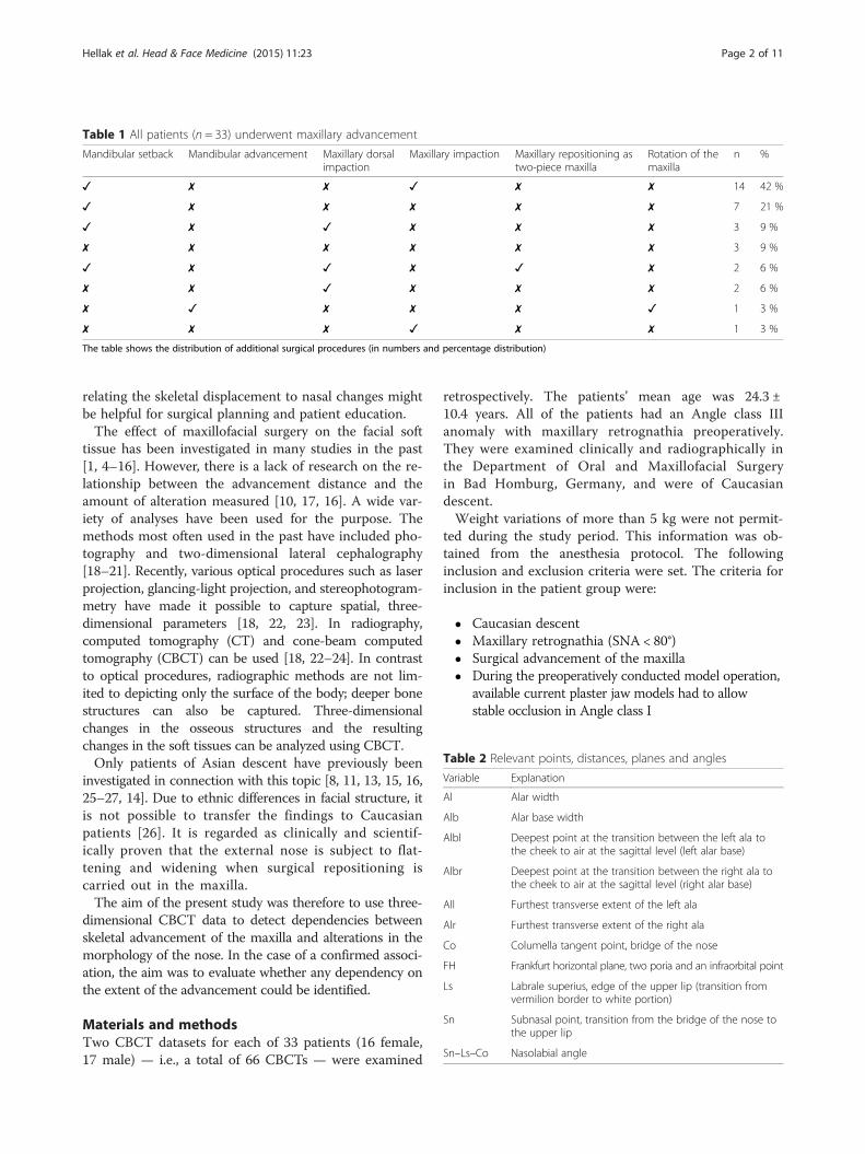
Table 1 All patients (n = 33) underwent maxillary advancement
Mandibular setback Mandibular advancement Maxillary dorsalimpaction
Maxillary impaction Maxillary repositioning astwo-piece maxilla
Rotation of themaxilla
n %
✓ ✗ ✗ ✓ ✗ ✗ 14 42 %
✓ ✗ ✗ ✗ ✗ ✗ 7 21 %
✓ ✗ ✓ ✗ ✗ ✗ 3 9 %
✗ ✗ ✗ ✗ ✗ ✗ 3 9 %
✓ ✗ ✓ ✗ ✓ ✗ 2 6 %
✗ ✗ ✓ ✗ ✗ ✗ 2 6 %
✗ ✓ ✗ ✗ ✗ ✓ 1 3 %
✗ ✗ ✗ ✓ ✗ ✗ 1 3 %
The table shows the distribution of additional surgical procedures (in numbers and percentage distribution)
Table 2 Relevant points, distances, planes and angles
Variable Explanation
Al Alar width
Alb Alar base width
Albl Deepest point at the transition between the left ala tothe cheek to air at the sagittal level (left alar base)
Albr Deepest point at the transition between the right ala tothe cheek to air at the sagittal level (right alar base)
All Furthest transverse extent of the left ala
Alr Furthest transverse extent of the right ala
Co Columella tangent point, bridge of the nose
FH Frankfurt horizontal plane, two poria and an infraorbital point
Ls Labrale superius, edge of the upper lip (transition fromvermilion border to white portion)
Sn Subnasal point, transition from the bridge of the nose tothe upper lip
Sn–Ls–Co Nasolabial angle
Hellak et al. Head & Face Medicine (2015) 11:23 Page 2 of 11
relating the skeletal displacement to nasal changes mightbe helpful for surgical planning and patient education.The effect of maxillofacial surgery on the facial soft
tissue has been investigated in many studies in the past[1, 4–16]. However, there is a lack of research on the re-lationship between the advancement distance and theamount of alteration measured [10, 17, 16]. A wide var-iety of analyses have been used for the purpose. Themethods most often used in the past have included pho-tography and two-dimensional lateral cephalography[18–21]. Recently, various optical procedures such as laserprojection, glancing-light projection, and stereophotogram-metry have made it possible to capture spatial, three-dimensional parameters [18, 22, 23]. In radiography,computed tomography (CT) and cone-beam computedtomography (CBCT) can be used [18, 22–24]. In contrastto optical procedures, radiographic methods are not lim-ited to depicting only the surface of the body; deeper bonestructures can also be captured. Three-dimensionalchanges in the osseous structures and the resultingchanges in the soft tissues can be analyzed using CBCT.Only patients of Asian descent have previously been
investigated in connection with this topic [8, 11, 13, 15, 16,25–27, 14]. Due to ethnic differences in facial structure, itis not possible to transfer the findings to Caucasianpatients [26]. It is regarded as clinically and scientif-ically proven that the external nose is subject to flat-tening and widening when surgical repositioning iscarried out in the maxilla.The aim of the present study was therefore to use three-
dimensional CBCT data to detect dependencies betweenskeletal advancement of the maxilla and alterations in themorphology of the nose. In the case of a confirmed associ-ation, the aim was to evaluate whether any dependency onthe extent of the advancement could be identified.
Materials and methodsTwo CBCT datasets for each of 33 patients (16 female,17 male) — i.e., a total of 66 CBCTs — were examined
retrospectively. The patients’ mean age was 24.3 ±10.4 years. All of the patients had an Angle class IIIanomaly with maxillary retrognathia preoperatively.They were examined clinically and radiographically inthe Department of Oral and Maxillofacial Surgeryin Bad Homburg, Germany, and were of Caucasiandescent.Weight variations of more than 5 kg were not permit-
ted during the study period. This information was ob-tained from the anesthesia protocol. The followinginclusion and exclusion criteria were set. The criteria forinclusion in the patient group were:
� Caucasian descent� Maxillary retrognathia (SNA < 80°)� Surgical advancement of the maxilla� During the preoperatively conducted model operation,
available current plaster jaw models had to allowstable occlusion in Angle class I
Hellak et al. Head & Face Medicine (2015) 11:23 Page 3 of 11
The criteria for exclusion from the patient group were:
� No maxillary retrognathia (SNA > 80°)� Not of Caucasian descent� Additional intraoperative augmentation of the midface� Craniofacial anomalies or syndromes, or any form of
cheilognathouranoschisis
Surgically, a Le Fort I osteotomy of the maxilla incombination with bridle sutures for the bases of the twoala was used [16, 17, 28, 29]. The Le Fort I osteotomymethod used by the surgeon (exclusively W.K.) is basedon the fracture line described by René Le Fort in 1901[30]. The osteotomy starts at the piriform aperture
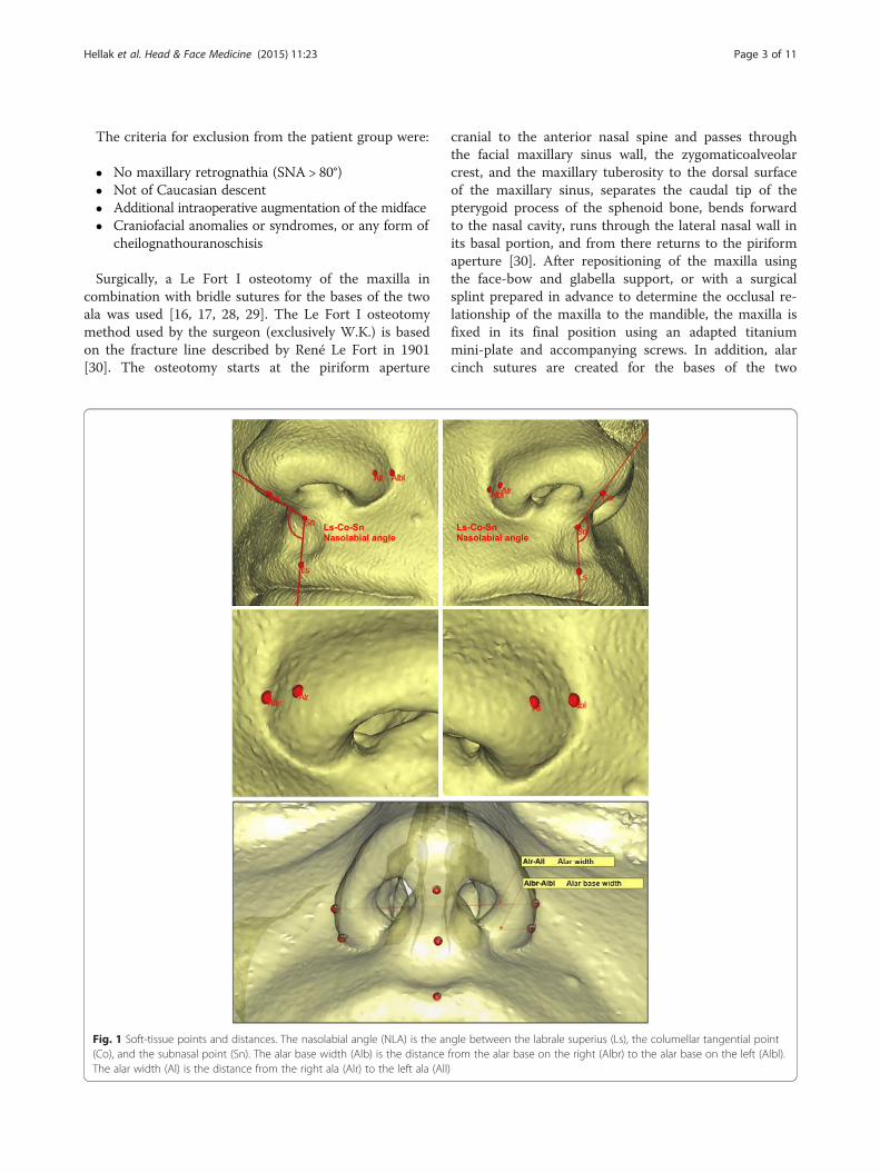
Fig. 1 Soft-tissue points and distances. The nasolabial angle (NLA) is the an(Co), and the subnasal point (Sn). The alar base width (Alb) is the distanceThe alar width (Al) is the distance from the right ala (Alr) to the left ala (All
cranial to the anterior nasal spine and passes throughthe facial maxillary sinus wall, the zygomaticoalveolarcrest, and the maxillary tuberosity to the dorsal surfaceof the maxillary sinus, separates the caudal tip of thepterygoid process of the sphenoid bone, bends forwardto the nasal cavity, runs through the lateral nasal wall inits basal portion, and from there returns to the piriformaperture [30]. After repositioning of the maxilla usingthe face-bow and glabella support, or with a surgicalsplint prepared in advance to determine the occlusal re-lationship of the maxilla to the mandible, the maxilla isfixed in its final position using an adapted titaniummini-plate and accompanying screws. In addition, alarcinch sutures are created for the bases of the two
gle between the labrale superius (Ls), the columellar tangential pointfrom the alar base on the right (Albr) to the alar base on the left (Albl).)
Hellak et al. Head & Face Medicine (2015) 11:23 Page 4 of 11
nostrils and attached with this technique at the anteriornasal spine [17, 3, 29, 28]All 33 patients underwent maxillary advancement. A
bimaxillary operation with maxillary advancement andmandibular setback was carried out in the majority ofthe patients. Table 1 presents an overview of the add-itional surgical procedures used and their frequencies.The first CBCT imaging procedure was carried out 2–
3 weeks before the planned procedure (T0). The secondimages were obtained after surgery (T1; 14.1 ±6.4 months postoperatively), but not before the com-pletion of soft-tissue healing. Completion of soft-tissuehealing was defined as 6 months after surgery, based onthe results of earlier studies [31, 32].Identical parameters were used for all CBCT imaging
procedures. All of the CBCT images were taken with aKaVo 3D eXam device (KaVo Dental Ltd., Bieberach/Riss, Germany). This CBCT device has a high-frequencyX-ray source with a constant potential of 120 kVp
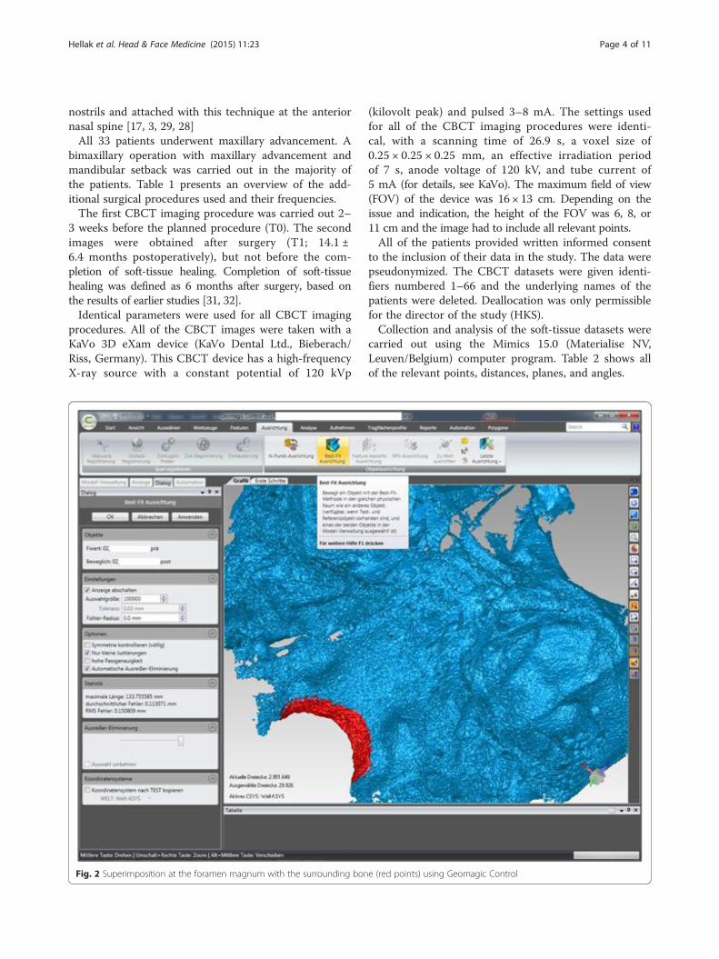
Fig. 2 Superimposition at the foramen magnum with the surrounding bon
(kilovolt peak) and pulsed 3–8 mA. The settings usedfor all of the CBCT imaging procedures were identi-cal, with a scanning time of 26.9 s, a voxel size of0.25 × 0.25 × 0.25 mm, an effective irradiation periodof 7 s, anode voltage of 120 kV, and tube current of5 mA (for details, see KaVo). The maximum field of view(FOV) of the device was 16 × 13 cm. Depending on theissue and indication, the height of the FOV was 6, 8, or11 cm and the image had to include all relevant points.All of the patients provided written informed consent
to the inclusion of their data in the study. The data werepseudonymized. The CBCT datasets were given identi-fiers numbered 1–66 and the underlying names of thepatients were deleted. Deallocation was only permissiblefor the director of the study (HKS).Collection and analysis of the soft-tissue datasets were
carried out using the Mimics 15.0 (Materialise NV,Leuven/Belgium) computer program. Table 2 shows allof the relevant points, distances, planes, and angles.
e (red points) using Geomagic Control
Hellak et al. Head & Face Medicine (2015) 11:23 Page 5 of 11
The nasolabial angle (NLA [33]), alar base width (Alb),and alar width (Al) were measured to assess changes inthe nasal soft tissues. The soft-tissue points shown inFig. 1 were used for measurements. The following dis-tances and angles were formed from the measurementpoints:
� Alar base width (Alb): from Albr to Albl: Alb distance� Alar width (Al): from All to Alr: Al distance� Nasolabial angle (NLA): angle between Ls to Co
and Sn
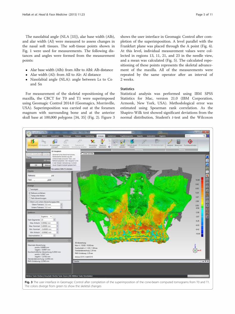
For measurement of the skeletal repositioning of themaxilla, the CBCT for T0 and T1 were superimposedusing Geomagic Control 2014.0 (Geomagics, Morrisville,USA). Superimposition was carried out at the foramenmagnum with surrounding bone and at the anteriorskull base at 100,000 polygons [34, 35] (Fig. 2). Figure 3
Fig. 3 The user interface in Geomagic Control after completion of the supThe colors diverge from green to show the skeletal changes
shows the user interface in Geomagic Control after com-pletion of the superimposition. A level parallel with theFrankfurt plane was placed through the A point (Fig. 4).At this level, individual measurement values were col-lected in regions 13, 11, 21, and 23 in the needle view,and a mean was calculated (Fig. 5). The calculated repo-sitioning of these points represents the skeletal advance-ment of the maxilla. All of the measurements wererepeated by the same operator after an interval of2 weeks.
StatisticsStatistical analysis was performed using IBM SPSSStatistics for Mac, version 21.0 (IBM Corporation,Armonk, New York, USA). Methodological error wasestimated using Spearman rank correlation. As theShapiro-Wilk test showed significant deviations from thenormal distribution, Student’s t-test and the Wilcoxon
erimposition of the cone-beam computed tomograms from T0 and T1.
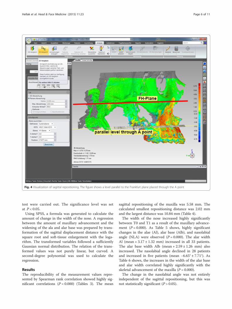
Fig. 4 Visualization of sagittal repositioning. The figure shows a level parallel to the Frankfurt plane placed through the A point
Hellak et al. Head & Face Medicine (2015) 11:23 Page 6 of 11
test were carried out. The significance level was setat P < 0.05.Using SPSS, a formula was generated to calculate the
amount of change in the width of the nose. A regressionbetween the amount of maxillary advancement and thewidening of the ala and alar base was prepared by trans-formation of the sagittal displacement distance with thesquare root and soft-tissue enlargement with the loga-rithm. The transformed variables followed a sufficientlyGaussian normal distribution. The relation of the trans-formed values was not purely linear, but curved. Asecond-degree polynomial was used to calculate theregression.
ResultsThe reproducibility of the measurement values repre-sented by Spearman rank correlation showed highly sig-nificant correlations (P = 0.000) (Tables 3). The mean
sagittal repositioning of the maxilla was 5.58 mm. Thecalculated smallest repositioning distance was 2.02 mmand the largest distance was 10.84 mm (Table 4).The width of the nose increased highly significantly
between T0 and T1 as a result of the maxillary advance-ment (P = 0.000). As Table 5 shows, highly significantchanges in the alae (Al), alar base (Alb), and nasolabialangle (NLA) were observed (P = 0.000). The alar widthAl (mean + 3.17 ± 1.32 mm) increased in all 33 patients.The alar base width Alb (mean + 2.59 ± 1.26 mm) alsoincreased. The nasolabial angle declined in 28 patientsand increased in five patients (mean −6.65° ± 7.71°). AsTable 6 shows, the increases in the width of the alar baseand alar width correlated highly significantly with theskeletal advancement of the maxilla (P = 0.000).The change in the nasolabial angle was not entirely
independent of the sagittal repositioning, but this wasnot statistically significant (P > 0.05).
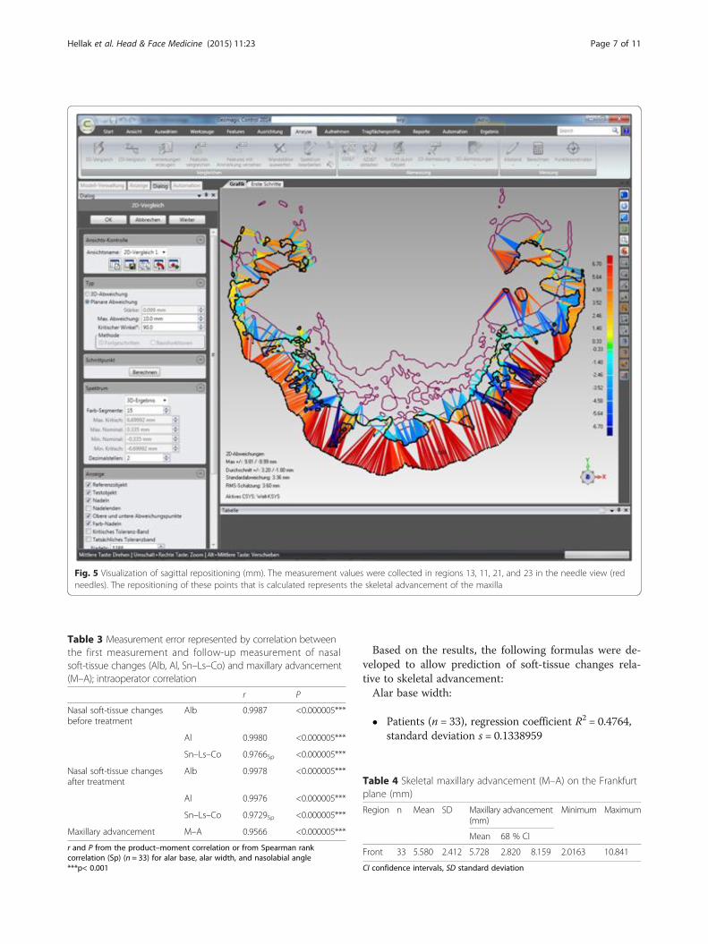
Fig. 5 Visualization of sagittal repositioning (mm). The measurement values were collected in regions 13, 11, 21, and 23 in the needle view (redneedles). The repositioning of these points that is calculated represents the skeletal advancement of the maxilla
Table 3 Measurement error represented by correlation betweenthe first measurement and follow-up measurement of nasalsoft-tissue changes (Alb, Al, Sn–Ls–Co) and maxillary advancement(M–A); intraoperator correlation
r P
Nasal soft-tissue changesbefore treatment
Alb 0.9987 <0.000005***
Al 0.9980 <0.000005***
Sn–Ls–Co 0.9766Sp <0.000005***
Nasal soft-tissue changesafter treatment
Alb 0.9978 <0.000005***
Al 0.9976 <0.000005***
Sn–Ls–Co 0.9729Sp <0.000005***
Maxillary advancement M–A 0.9566 <0.000005***
r and P from the product–moment correlation or from Spearman rankcorrelation (Sp) (n = 33) for alar base, alar width, and nasolabial angle***p< 0.001
Hellak et al. Head & Face Medicine (2015) 11:23 Page 7 of 11
Based on the results, the following formulas were de-veloped to allow prediction of soft-tissue changes rela-tive to skeletal advancement:Alar base width:
� Patients (n = 33), regression coefficient R2 = 0.4764,standard deviation s = 0.1338959
Table 4 Skeletal maxillary advancement (M–A) on the Frankfurtplane (mm)
Region n Mean SD Maxillary advancement(mm)
Minimum Maximum
Mean 68 % CI
Front 33 5.580 2.412 5.728 2.820 8.159 2.0163 10.841
CI confidence intervals, SD standard deviation

Table 5 Comparison of values at T0 and T1; P with Student’s t-test and the Wilcoxon test (W) (n = 33)
Mean SD Increase P
Mean SD
Alb Before treatment 33.665 2.632 3.171 1.322 <0.000005***
After treatment 36.837 2.651
Al Before treatment 35.459 2.912 2.588 1.255 <0.000005***
After treatment 38.047 2.851
Sn–Ls–Co Before treatment 102.992 14.388 −6.652 7.712 0.00002***W
After treatment 96.339 12.748
***p< 0.001
Hellak et al. Head & Face Medicine (2015) 11:23 Page 8 of 11
� Formula for y =ΔAlb in the course of x =maxillaryadvancement:log(ΔAlb) = 0.4558889 + 0.124167 × z +0.010571 × z2, where z = (√(maxillaryadvancement) – 2.305)/0.5255
Alar width:
� Patients (n = 33), regression coefficient R2 = 0.5281,standard deviation s = 0.1403169
� Formula for y =ΔAlin the course of x =maxillaryadvancement:
Tablwidthadvan
Increa
Alar b
Alar w
Nasol
Soft-timeasuSpearm***p<
log (ΔAl) = 0.3704371 + 0.1435941 ×z − 0.001474 × z2, where z = (√(maxillaryadvancement) – 2.305) / 0.5255
Table 7 Prediction of changes in alar base width
Maxillaryadvancement
Percentiles for increase in Alb (mm)
5.0 % 10.0 % 50.0 % 90.0 % 95.0 %
Advancement by 5 mm would thus lead to a meanwidening of the alar base of ≈ 2.8 mm. Ninety percent ofthe patients would have widening of between ≈ 1.7 mmand ≈ 4.6 mm (Table 7). As Table 8 shows, advancementof 5 mm would lead to a mean widening of the alaof ≈ 2.2 mm. Ninety percent of the patients could ex-pect widening of between ≈ 1.3 mm and ≈ 3.8 mm.
DiscussionInvestigations have been carried out since the 1980s toanalyze associations between changes occurring in thefacial soft tissue in connection with Le Fort I osteoto-mies in dysgnathia surgery. A correlation between sagit-tal repositioning of the maxilla and the amount of alar
e 6 Correlations of changes in the external nose (alar base, alar width, nasolabial angle) relative to skeletalcement of the maxilla
se in Region n rho P
ase width Front 33 0.6949 0.00001***
idth Front 33 0.7688 <0.000005***
abial angle Front 33 −0.3102 0.079
ssue parameters as the difference in means from T0 to T1; skeletalrements as means from T1 to T0 for maxillary advancement. P froman’s rank correlation
0.001
flaring was not identified at that time [36]. Preoperativeand postoperative lateral cephalograms or photographswere used for measurements in the early studies [10].However, these techniques did not allow any conclusionsto be drawn regarding correlations with the changingwidth of the nose [11].Three-dimensional measurement of hard-tissue models
obtained from CBCT and CT imaging is now increasinglybeing used, mainly for the planning of dental implants indental medicine [37]. The amount of correlation betweenthe initial measurements and control measurements ofthe soft tissue after 2 weeks is highly significant. Discus-sion is needed on mistakes during the reproducibility ofthese measurements. The reproducibility and reliabilityof different measurement points on lateral cephalogramshas been frequently investigated in the past and has alsobeen classified [38–40]. Similar findings were obtainedwith measurement points derived from lateral cephalo-grams created with CBCT [41]. The measurement pointscreated on three-dimensional surfaces using CBCT scansare thus regarded as being highly reliable [42–44].The sagittal movement of the maxilla was registered
on a plane parallel to the Frankfurt plane. In 2012,Daboul et al. reported that the reproducibility and reli-ability of the Frankfurt plane on 3D multiplanar re-formatting (MPR) images was excellent [45]. Similar
2 mm 1.13653 1.26409 1.88730 2.81777 3.13401
3 mm 1.29681 1.44235 2.15346 3.21515 3.57598
4 mm 1.46956 1.63449 2.44031 3.64343 4.05233
5 mm 1.65802 1.84409 2.75326 4.11066 4.57200
6 mm 1.86481 2.07410 3.09666 4.62337 5.14224
7 mm 2.09245 2.32728 3.47467 5.18774 5.76995
8 mm 2.34348 2.60649 3.89153 5.81012 6.46218
9 mm 2.62062 2.91473 4.35173 6.49721 7.22638
10 mm 2.92678 3.25524 4.86013 7.25626 8.07062
Approximate percentiles for Alb relative to maxillary advancement. A advancementof 5mm would lead to a mean widening of the alar base width of 2.8 mm
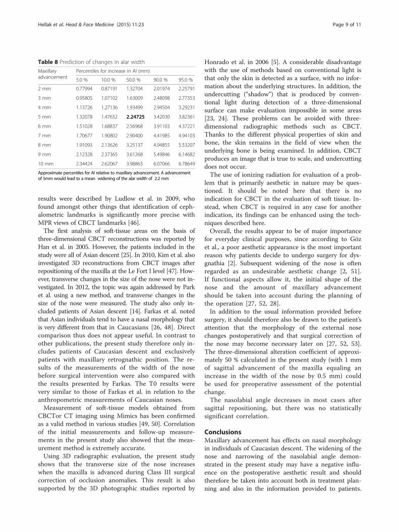
Table 8 Prediction of changes in alar width
Maxillaryadvancement
Percentiles for increase in Al (mm)
5.0 % 10.0 % 50.0 % 90.0 % 95.0 %
2 mm 0.77994 0.87191 1.32704 2.01974 2.25791
3 mm 0.95805 1.07102 1.63009 2.48098 2.77353
4 mm 1.13726 1.27136 1.93499 2.94504 3.29231
5 mm 1.32078 1.47652 2.24725 3.42030 3.82361
6 mm 1.51028 1.68837 2.56968 3.91103 4.37221
7 mm 1.70677 1.90802 2.90400 4.41985 4.94103
8 mm 1.91093 2.13626 3.25137 4.94855 5.53207
9 mm 2.12328 2.37365 3.61268 5.49846 6.14682
10 mm 2.34424 2.62067 3.98863 6.07066 6.78649
Approximate percentiles for Al relative to maxillary advancement. A advancementof 5mm would lead to a mean widening of the alar width of 2.2 mm
Hellak et al. Head & Face Medicine (2015) 11:23 Page 9 of 11
results were described by Ludlow et al. in 2009, whofound amongst other things that identification of ceph-alometric landmarks is significantly more precise withMPR views of CBCT landmarks [46].The first analysis of soft-tissue areas on the basis of
three-dimensional CBCT reconstructions was reported byHan et al. in 2005. However, the patients included in thestudy were all of Asian descent [25]. In 2010, Kim et al. alsoinvestigated 3D reconstructions from CBCT images afterrepositioning of the maxilla at the Le Fort I level [47]. How-ever, transverse changes in the size of the nose were not in-vestigated. In 2012, the topic was again addressed by Parket al. using a new method, and transverse changes in thesize of the nose were measured. The study also only in-cluded patients of Asian descent [14]. Farkas et al. notedthat Asian individuals tend to have a nasal morphology thatis very different from that in Caucasians [26, 48]. Directcomparison thus does not appear useful. In contrast toother publications, the present study therefore only in-cludes patients of Caucasian descent and exclusivelypatients with maxillary retrognathic position. The re-sults of the measurements of the width of the nosebefore surgical intervention were also compared withthe results presented by Farkas. The T0 results werevery similar to those of Farkas et al. in relation to theanthropometric measurements of Caucasian noses.Measurement of soft-tissue models obtained from
CBCTor CT imaging using Mimics has been confirmedas a valid method in various studies [49, 50]. Correlationof the initial measurements and follow-up measure-ments in the present study also showed that the meas-urement method is extremely accurate.Using 3D radiographic evaluation, the present study
shows that the transverse size of the nose increaseswhen the maxilla is advanced during Class III surgicalcorrection of occlusion anomalies. This result is alsosupported by the 3D photographic studies reported by
Honrado et al. in 2006 [5]. A considerable disadvantagewith the use of methods based on conventional light isthat only the skin is detected as a surface, with no infor-mation about the underlying structures. In addition, theundercutting (“shadow”) that is produced by conven-tional light during detection of a three-dimensionalsurface can make evaluation impossible in some areas[23, 24]. These problems can be avoided with three-dimensional radiographic methods such as CBCT.Thanks to the different physical properties of skin andbone, the skin remains in the field of view when theunderlying bone is being examined. In addition, CBCTproduces an image that is true to scale, and undercuttingdoes not occur.The use of ionizing radiation for evaluation of a prob-
lem that is primarily aesthetic in nature may be ques-tioned. It should be noted here that there is noindication for CBCT in the evaluation of soft tissue. In-stead, when CBCT is required in any case for anotherindication, its findings can be enhanced using the tech-niques described here.Overall, the results appear to be of major importance
for everyday clinical purposes, since according to Gözet al., a poor aesthetic appearance is the most importantreason why patients decide to undergo surgery for dys-gnathia [2]. Subsequent widening of the nose is oftenregarded as an undesirable aesthetic change [2, 51].If functional aspects allow it, the initial shape of thenose and the amount of maxillary advancementshould be taken into account during the planning ofthe operation [27, 52, 28].In addition to the usual information provided before
surgery, it should therefore also be drawn to the patient’sattention that the morphology of the external nosechanges postoperatively and that surgical correction ofthe nose may become necessary later on [27, 52, 53].The three-dimensional alteration coefficient of approxi-mately 50 % calculated in the present study (with 1 mmof sagittal advancement of the maxilla equaling anincrease in the width of the nose by 0.5 mm) couldbe used for preoperative assessment of the potentialchange.The nasolabial angle decreases in most cases after
sagittal repositioning, but there was no statisticallysignificant correlation.
ConclusionsMaxillary advancement has effects on nasal morphologyin individuals of Caucasian descent. The widening of thenose and narrowing of the nasolabial angle demon-strated in the present study may have a negative influ-ence on the postoperative aesthetic result and shouldtherefore be taken into account both in treatment plan-ning and also in the information provided to patients.
Hellak et al. Head & Face Medicine (2015) 11:23 Page 10 of 11
The correlation coefficient calculated between sagittaladvancement and soft-tissue changes in the nose maymake prediction easier.
Ethical considerationsThe study was compiled with the rules laid down by theDeclaration of Helsinki. It was explained to the patientsthat inclusion of their data in the study was voluntaryand that confidentiality and anonymity were guaranteed.They were also able to withdraw from the study at anytime before publication without needing to give any rea-son. Written informed consent was obtained from all ofthe participants.
AbbreviationsAl: Alar width; Alb: Alar base width; Albl: Alar base on the left; Albr: Alar baseon the right; All: Left ala; Alr: Right ala; CBCT: Cone-beam computedtomography; Co: Columellar tangential point; CT: Computed tomography;FOV: Field of view; Ls: Labrale superius; MPR: Multiplanar reformatting;NLA: Nasolabial angle; Sn: Subnasal point; SNA: Sella-nasion-A point angle.
Competing interestsThe authors declare that they have no competing interests.
Authors’ contributionsAH carried out the design and drafted the first manuscript. BK, MS, and RDcarried out the measurements and helped with the design and coordinationof the study and the drafting of the manuscript. WK performed the surgeryand helped with the coordination of the study. HKS helped with the studydesign, the translation into English and the illustrations. All of the authorsread and approved the final manuscript.
Author details1Department of Orthodontics, University Hospital, Georg-Voigt-Strasse 3,Marburg 35039, Germany. 2Private Practice, Bad Homburg, Germany.
Received: 21 October 2014 Accepted: 30 June 2015
References1. Baik HS, Kim SY. Facial soft-tissue changes in skeletal Class III orthognathic
surgery patients analyzed with 3-dimensional laser scanning. Am J OrthodDentofacial Orthop. 2010;138(2):167–78. doi:10.1016/j.ajodo.2010.02.022.
2. Göz G. Die Motivation bei kieferorthopädischen Operationen. Freiburg i. Br:Albert-Ludwigs-Universität Freiburg i. Br; 1981.
3. Guymon M, Crosby DR, Wolford LM. The alar base cinch suture to controlnasal width in maxillary osteotomies. Int J Adult Orthodon OrthognathSurg. 1988;3(2):89–95.
4. Choi JW, Lee JY, Oh TS, Kwon SM, Yang SJ, Koh KS. Frontal soft tissueanalysis using a 3 dimensional camera following two-jaw rotationalorthognathic surgery in skeletal class III patients. J CraniomaxillofacSurg. 2013. doi:10.1016/j.jcms.2013.05.004.
5. Honrado CP, Lee S, Bloomquist DS, Larrabee Jr WF. Quantitative assessmentof nasal changes after maxillomandibular surgery using a 3-dimensionaldigital imaging system. Arch Facial Plast Surg. 2006;8(1):26–35. doi:10.1001/archfaci.8.1.26.
6. Hwang DS, Kim YI, Park SB, Lee JY. Midfacial soft tissue changes afterleveling Le Fort I osteotomy with differential reduction. Cone-beamcomputed tomography volume superimposition. Angle Orthod.2012;82(3):424–31. doi:10.2319/052411-342.1.
7. Johnson BM, McNamara JA, Bandeen RL, Baccetti T. Changes in soft tissuenasal widths associated with rapid maxillary expansion in prepubertal andpostpubertal subjects. Angle Orthod. 2010;80(6):995–1001. doi:10.2319/033110-179.1.
8. Kim YI, Park SB, Son WS, Hwang DS. Midfacial soft-tissue changes afteradvancement of maxilla with Le Fort I osteotomy and mandibular setbacksurgery: comparison of conventional and high Le Fort I osteotomies by
superimposition of cone-beam computed tomography volumes. J OralMaxillofac Surg. 2011;69(6):e225–33. doi:10.1016/j.joms.2010.12.035.
9. Lee JY, Kim YI, Hwang DS, Park SB. Effect of setback Le Fort I osteotomy onmidfacial soft-tissue changes as evaluated by cone-beam computedtomography superimposition for cases of skeletal Class III malocclusion.Int J Oral Maxillofac Surg. 2013;42(6):790–5. doi:10.1016/j.ijom.2012.11.012.
10. Radney LJ, Jacobs JD. Soft-tissue changes associated with surgical totalmaxillary intrusion. Am J Orthod. 1981;80(2):191–212.
11. Mansour S, Burstone C, Legan H. An evaluation of soft-tissue changesresulting from Le Fort I maxillary surgery. Am J Orthod. 1983;84(1):37–47.
12. McCance AM, Moss JP, Fright WR, James DR, Linney AD. A threedimensional analysis of soft and hard tissue changes following bimaxillaryorthognathic surgery in skeletal III patients. Br J Oral Maxillofac Surg.1992;30(5):305–12.
13. Mommaerts MY, Lippens F, Abeloos JV, Neyt LF. Nasal profile changes aftermaxillary impaction and advancement surgery. J Oral Maxillofac Surg.2000;58(5):470–5. discussion 5–6.
14. Park SB, Yoon JK, Kim YI, Hwang DS, Cho BH, Son WS. The evaluation of thenasal morphologic changes after bimaxillary surgery in skeletal class IIImaloccusion by using the superimposition of cone-beam computedtomography (CBCT) volumes. J Craniomaxillofac Surg. 2012;40(4):e87–92.doi:10.1016/j.jcms.2011.05.008.
15. Ryckman MS, Harrison S, Oliver D, Sander C, Boryor AA, Hohmann AA, et al.Soft-tissue changes after maxillomandibular advancement surgery assessedwith cone-beam computed tomography. Am J Orthod Dentofacial Orthop.2010;137(4 Suppl):S86–93. doi:10.1016/j.ajodo.2009.03.041.
16. Westermark AH, Bystedt H, Von Konow L, Sallstrom KO. Nasolabialmorphology after Le Fort I osteotomies. Effect of alar base suture. Int JOral Maxillofac Surg. 1991;20(1):25–30.
17. Collins PC, Epker BN. The alar base cinch: a technique for prevention of alarbase flaring secondary to maxillary surgery. Oral Surg Oral Med Oral Pathol.1982;53(6):549–53.
18. Rustemeyer J, Martin A. Soft tissue response in orthognathic surgerypatients treated by bimaxillary osteotomy: cephalometry comparedwith 2-D photogrammetry. Oral Maxillofac Surg. 2013;17(1):33–41.doi:10.1007/s10006-012-0330-0.
19. Kajikawa Y. Changes in soft tissue profile after surgical correction of skeletalclass III malocclusion. J Oral Surg. 1979;37(3):167–74.
20. Lin SS, Kerr WJ. Soft and hard tissue changes in Class III patients treated bybimaxillary surgery. Eur J Orthod. 1998;20(1):25–33.
21. Kinzinger G, Frye L, Diedrich P. Class II treatment in adults: comparingcamouflage orthodontics, dentofacial orthopedics and orthognathicsurgery–a cephalometric study to evaluate various therapeutic effects.J Orofac Orthop. 2009;70(1):63–91. doi:10.1007/s00056-009-0821-2.
22. Verze L, Bianchi FA, Schellino E, Ramieri G. Soft tissue changes afterorthodontic surgical correction of jaws asymmetry evaluated by three-dimensional surface laser scanner. J Craniofac Surg. 2012;23(5):1448–52.doi:10.1097/SCS.0b013e31824e25fc.
23. Holberg C, Heine AK, Geis P, Schwenzer K, Rudzki-Janson I. Three-dimensionalsoft tissue prediction using finite elements. Part II: Clinical application. J OrofacOrthop. 2005;66(2):122–34. doi:10.1007/s00056-005-0422-7.
24. Holberg C, Schwenzer K, Rudzki-Janson I. Three-dimensional soft tissueprediction using finite elements. Part I: Implementation of a new procedure.J Orofac Orthop. 2005;66(2):110–21. doi:10.1007/s00056-005-0421-8.
25. Han SY, Baik HS, Kim KD, Yu HS. Facial soft tissue measuring analysis ofnormal occlusion using three-dimensional CT imaging. Korean J Orthod.2005;35(6):409–19.
26. Farkas LG, Katic MJ, Forrest CR, Alt KW, Bagic I, Baltadjiev G, et al.International anthropometric study of facial morphology in variousethnic groups/races. J Craniofac Surg. 2005;16(4):615–46.
27. Altman JI, Oeltjen JC. Nasal deformities associated with orthognathicsurgery: analysis, prevention, and correction. J Craniofac Surg.2007;18(4):734–9. doi:10.1097/SCS.0b013e3180684328.
28. Rauso R, Freda N, Curinga G, Del Pero C, Tartaro G. An alternative alar cinchsuture. Eplasty. 2010;10, e69.
29. Millard Jr DR. The alar cinch in the flat, flaring nose. Plast Reconstr Surg.1980;65(5):669–72.
30. Tessier P. The classic reprint: experimental study of fractures of the upperjaw. 3. Rene Le Fort, M.D., Lille, France. Plast Reconstr Surg. 1972;50(6):600–7.
31. Kau CH, Cronin A, Durning P, Zhurov AI, Sandham A, Richmond S. A newmethod for the 3D measurement of postoperative swelling following

Hellak et al. Head & Face Medicine (2015) 11:23 Page 11 of 11
orthognathic surgery. Orthod Craniofac Res. 2006;9(1):31–7. doi:10.1111/j.1601-6343.2006.00341.x.
32. Oh KM, Seo SK, Park JE, Sim HS, Cevidanes LH, Kim YJ, et al. Post-operativesoft tissue changes in patients with mandibular prognathism after bimaxillarysurgery. J Craniomaxillofac Surg. 2013;41(3):204–11. doi:10.1016/j.jcms.2012.09.001.
33. Legan HL, Burstone CJ. Soft tissue cephalometric analysis for orthognathicsurgery. J Oral Surg. 1980;38(10):744–51.
34. Cevidanes LH, Motta A, Proffit WR, Ackerman JL, Styner M. Cranial basesuperimposition for 3-dimensional evaluation of soft-tissue changes. Am JOrthod Dentofacial Orthop. 2010;137(4 Suppl):S120–9. doi:10.1016/j.ajodo.2009.04.021.
35. Gkantidis N, Halazonetis DJ. Morphological integration between the cranialbase and the face in children and adults. J Anat. 2011;218(4):426–38.doi:10.1111/j.1469-7580.2011.01346.x.
36. O'Ryan F, Schendel S. Nasal anatomy and maxillary surgery. I. Esthetic andanatomic principles. Int J Adult Orthodon Orthognath Surg. 1989;4(1):27–37.
37. Neugebauer J, Ritter L, Mischkowski R, Zoller JE. Three-dimensional diagnostics,planning and implementation in implantology. Int J Comput Dent.2006;9(4):307–19.
38. Baumrind S, Frantz RC. The reliability of head film measurements. 1.Landmark identification. Am J Orthod. 1971;60(2):111–27.
39. Miethke RR. Zur Lokalisationsgenauigkeit kephalometrischer Referenzpunkte.Prakt Kieferorthop. 1989;3:107–22.
40. Stabrun AE, Danielsen K. Precision in cephalometric landmark identification.Eur J Orthod. 1982;4(3):185–96.
41. Navarro Rde L, Oltramari-Navarro PV, Fernandes TM, Oliveira GF, Conti AC,Almeida MR, et al. Comparison of manual, digital and lateral CBCTcephalometric analyses. J Appl Oral Sci. 2013;21(2):167–76. doi:10.1590/1678-7757201302326.
42. Fourie Z, Damstra J, Gerrits PO, Ren Y. Accuracy and repeatability ofanthropometric facial measurements using cone beam computedtomography. Cleft Palate Craniofac J. 2011;48(5):623–30. doi:10.1597/10-076.
43. Naji P, Alsufyani NA, Lagravere MO. Reliability of anatomic structures aslandmarks in three-dimensional cephalometric analysis using CBCT. AngleOrthod. 2013. doi:10.2319/090413-652.1.
44. Oz U, Orhan K, Abe N. Comparison of linear and angular measurementsusing two-dimensional conventional methods and three-dimensional conebeam CT images reconstructed from a volumetric rendering programin vivo. Dentomaxillofac Radiol. 2011;40(8):492–500. doi:10.1259/dmfr/15644321.
45. Daboul A, Schwahn C, Schaffner G, Soehnel S, Samietz S, Aljaghsi A, et al.Reproducibility of Frankfort horizontal plane on 3D multi-planar reconstructedMR images. PLoS One. 2012;7(10), e48281. doi:10.1371/journal.pone.0048281.
46. Ludlow JB, Gubler M, Cevidanes L, Mol A. Precision of cephalometriclandmark identification: cone-beam computed tomography vs conventionalcephalometric views. Am J Orthod Dentofacial Orthop. 2009;136(3):312e1–10. doi:10.1016/j.ajodo.2008.12.018. discussion −3.
47. Kim YI, Kim JR, Park SB. Three-dimensional analysis of midfacial soft tissuechanges according to maxillary superior movement after horizontalosteotomy of the maxilla. J Craniofac Surg. 2010;21(5):1587–90.doi:10.1097/SCS.0b013e3181edc5c9.
48. Farkas LG, Phillips JH, Katic M. Anthropometric anatomical andmorphological nose widths in Canadian Caucasian adults. Can J PlastSurg. 1998;6(3):149–51.
49. Gorgulu S, Gokce SM, Olmez H, Sagdic D, Ors F. Nasal cavity volume changesafter rapid maxillary expansion in adolescents evaluated with 3-dimensionalsimulation and modeling programs. Am J Orthod Dentofacial Orthop.2011;140(5):633–40. doi:10.1016/j.ajodo.2010.12.020.
50. Shaw K, McIntyre G, Mossey P, Menhinick A, Thomson D. Validation ofconventional 2D lateral cephalometry using 3D cone beam CT. J Orthod.2013;40(1):22–8. doi:10.1179/1465313312Y.0000000009.
51. Honn M, Goz G. The ideal of facial beauty: a review. J Orofac Orthop.2007;68(1):6–16. doi:10.1007/s00056-007-0604-6.
52. Mitchell C, Oeltjen J, Panthaki Z, Thaller SR. Nasolabial aesthetics. J CraniofacSurg. 2007;18(4):756–65. doi:10.1097/scs.0b013e3180684360.
53. Rauso R, Tartaro G, Tozzi U, Colella G, Santagata M. Nasolabial changes aftermaxillary advancement. J Craniofac Surg. 2011;22(3):809–12. doi:10.1097/SCS.0b013e31820f3663.
Submit your next manuscript to BioMed Centraland take full advantage of:
• Convenient online submission
• Thorough peer review
• No space constraints or color figure charges
• Immediate publication on acceptance
• Inclusion in PubMed, CAS, Scopus and Google Scholar
• Research which is freely available for redistribution
Submit your manuscript at www.biomedcentral.com/submit