Embed Size (px)
Citation preview
1
REPUBLIC OF AZERBAIJAN
On the rights of the manuscript
INFLUENCE OF REPERFUSION SYNDROME ON SOME
LIVER FUNCTIONS AND THE ROLE OF BIOGENIC
ELEMENTS IN THEIR PATHOGENESIS
ABSTRACT
of the dissertation for the degree of Doctor of Philosophy
Speciality: 3243.01 – «Pathological physiology»
Field of science: «Medicine»
Applicant: Jala Rahman Gafarova
Baku – 2021
2
The work was performed at the Research Center of Azerbaijan
Medical University.
Scientific supervisor: Doctor of Medical Sciences, professor
-Galib Shalon Garayev
Official opponents:
Doctor of Medical Sciences, professor
-Khagigat Abdul Kadirova
Doctor of Medical Sciences
-Emil Almamed Isgenderov
Doctor of Philosophy in Medicine,
associate prof.
-Rafik Arshad Yusifli
Dissertation council FD 2.07 of Supreme Attestation Commission
under the President of the Republic of Azerbaijan operating at
Azerbaijan Medical University
Chairman of the Dissertation
council: Doctor of Medical Sciences, professor
______________ Sabir Jahan Aliyev
Scientific secretary of the
Dissertation council: Doctor of Biological Sciences
______________ Rena Anvar Jafarova
Chairman of the scientific
seminar: Doctor of Medical Sciences
______________ Fazil Ikram Alıyev
3
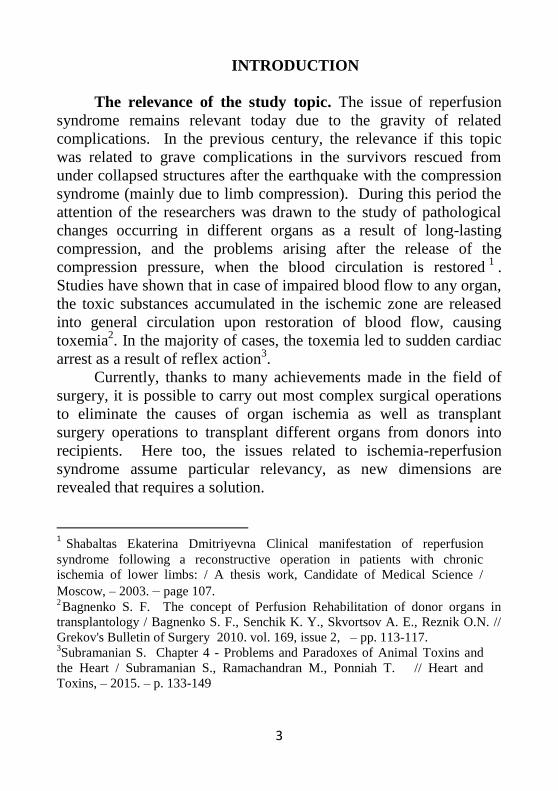
INTRODUCTION
The relevance of the study topic. The issue of reperfusion
syndrome remains relevant today due to the gravity of related
complications. In the previous century, the relevance if this topic
was related to grave complications in the survivors rescued from
under collapsed structures after the earthquake with the compression
syndrome (mainly due to limb compression). During this period the
attention of the researchers was drawn to the study of pathological
changes occurring in different organs as a result of long-lasting
compression, and the problems arising after the release of the
compression pressure, when the blood circulation is restored1
.
Studies have shown that in case of impaired blood flow to any organ,
the toxic substances accumulated in the ischemic zone are released
into general circulation upon restoration of blood flow, causing
toxemia2. In the majority of cases, the toxemia led to sudden cardiac
arrest as a result of reflex action3.
Currently, thanks to many achievements made in the field of
surgery, it is possible to carry out most complex surgical operations
to eliminate the causes of organ ischemia as well as transplant
surgery operations to transplant different organs from donors into
recipients. Here too, the issues related to ischemia-reperfusion
syndrome assume particular relevancy, as new dimensions are
revealed that requires a solution.
1Shabaltas Ekaterina Dmitriyevna Clinical manifestation of reperfusion
syndrome following a reconstructive operation in patients with chronic
ischemia of lower limbs: / A thesis work, Candidate of Medical Science /
Moscow, – 2003. – page 107. 2Bagnenko S. F. The concept of Perfusion Rehabilitation of donor organs in
transplantology / Bagnenko S. F., Senchik K. Y., Skvortsov A. E., Reznik O.N. //
Grekov's Bulletin of Surgery 2010. vol. 169, issue 2, – pp. 113-117. 3Subramanian S. Chapter 4 - Problems and Paradoxes of Animal Toxins and
the Heart / Subramanian S., Ramachandran M., Ponniah T. // Heart and
Toxins, – 2015. – p. 133-149
4
Owing to environmental degradation and unhealthy lifestyle
the number of patients suffering from liver damages of different
genesis incompatible with life is increasing year after year. Liver
transplantation in such cases often becomes the only life-saving
option. In terms of frequency, liver transplantation ranks next to the
combined number of transplantations of other organs4.
Toxic ischemia products along with functional derangements
and morphological abnormalities taking place during the
postoperative restoration of blood supply may lead to such severe
complications as graft rejection, development of inflammatory
processes, and liver tissue necrosis5
. Therefore, many scientific
studies today aim at the identification of underlying mechanisms of
such processes and the possibilities to reduce the pathological impact
of ischemic-reperfusion on the liver. It has been revealed that liver
reperfusion often causes severe damages to hepatocytes, capillary
and bile duct endothelium, and leads to postoperative graft rejection
in 10% of patients6.
Reperfusion syndrome occurring after an organ transplant
(liver, kidneys, heart) has its peculiar pathogenic characteristics. The
research has shown that the graft for transplantation undergoes
different changes as due to donor organ ischemia. As a result of such
changes cell membranes' integrity is compromised and lipid
4Qarayev G.Ş. Impact of additional sanitation of abdominal cavity with
superoxide dismutase at the terminal phase of peritonitis on endogenous
intoxication / G.Sh., GarayevA.Sh., Gasimov N.O. Guliyev //– Baku: Azerbaijan
Medical Journal, – 2013. issue 2, – pp. 84-88.5Qafarova M. E. Aggregation, disaggregation and deformability of erythrocytes in
the rat models of ischaemic stroke / Qafarova M. E., Naumova G.M., Qulyayev
M.V. et al. // Regional blood circulation and microcirculation, – 2015. vol. 14,
issue 2, (54). – pp. 63-69.6Qarayev G.Ş. On endotoxicosis and mechanisms that lead to its development /
Qarayev G.Ş., Nazaraliyeva I. I., Ismailov Y. B. ant et al.// – "Sağlamlıq"
magazine, – Baku: – 2010. issue 8, – pp. 175-179.
5
peroxidation processes are activated. The toxic intermediary
products formed during this process along with the edema they cause
simultaneously activate reabsorption and become a factor leading to
endogenous intoxication78
.
Thus, from literary sources it becomes clear that there were
carried out large-scale researches to study the pathophysiological
aspects of endotoxicosis, which revealed that the toxic substances
formed as a result of ischemia in the reperfusion period reach all
organs and tissues through blood circulation, causing pathologies in
such organs and tissues. Cellular breakdowns lead to the release of
many biologically active compounds. Among these substances, there
are such biogenic elements as cobalt, iron, manganese, copper, tin,
etc. Presumably, by entering different metabolic systems these
substances may provoke pathological processes leading to
reperfusion syndrome. However, in spite of this state of studies and
acquired so far knowledge, many issues related to changes in
ischemic organs and associated metabolic disorders in them yet
remain open.
Considering the significance of this problem, study of the
fermentative and protein synthesis functions of liver depending on
the duration of ischemia and reperfusion, as well as elucidation of the
role of the number of macro- and microelements in their
pathogenesis were thought to be worthy.
The present study is meant to study the impact of reperfusion
syndrome on enzymological and protein-synthesis functions of liver
7Noritaka Sano. Relationship between histologic features and outcomes of carotid
revascularization for radiation-induced stenosis / Noritaka Sano, TetsuSatow,
Daisuke Maruyama [et al.] // Journal of Vascular Surgery,– 2015. – v. 62(2), – p.
370-377. 8Qarayev G.Ş. On endotoxicosis and mechanisms that lead to its development /
Qarayev G.Ş., Nazaraliyeva I. I., Ismailov Y. B. ant et al.// – "Sağlamlıq"
magazine, – Baku: – 2010. issue 8, – pp. 175-179.
6
and identify the role played by certain biogenic elements in the
pathogenesis of such changes.
Objectives of the study are to: 1. identify the enzymatic changes taking place in the
antioxidant defense system and degree of lipid peroxidation in the
blood of animals against the background of ischemia and reperfusion
of differing durations.
2. identify the changes in enzymological and protein-synthesis
functions of liver.
3. in the blood of animals, to determine the enzyme markers of
liver damage.
4. study the level of non-enzyme liver damage markers in the
blood.
5. determine quantitative and qualitative changes in the
composition of blood macro- and micronutrients
6. identify the link between the changes in the composition of
blood macro- and micronutrients and duration of ischemic-
reperfusion periods.
Research methods:
-experimental modeling of ischemia-reperfusion;
-biochemical research;
-statistical analysis.
Main points of the Thesis to be defended:
1.Oxidative stress develops against the background of liver
ischemia model with periods of ischemia lasting 10, 20 and 30
minutes, the intensity of which is positively related to ischemia
duration. The activity of such enzymes as catalase and peroxidase is
reduced along with general antioxidant status deterioration. A time-
dependent increase in the content of primary and secondary lipid
peroxidation products is observed against the background of
ischemia and enzymatic activity reduction. After releasing the
pressure on the vessels and restoration of blood flow (the reperfusion
period) the lipid peroxidation products continue to accumulate,
which is related to general toxemia.
7
2. Liver's protein synthesis function deteriorates as a result of
ischemic-reperfusion damages to hepatocytes. Blood total protein
and albumin count is reduced, while the globulin and fibrinogen
content are increased.
3. During the ischemic-reperfusion period the level of such
enzyme and non-enzyme markers of liver damage as aspartate
aminotransaminase (AST), alanine aminotransferase (ALT), total
lactate dehydrogenase (LDG), creatine phosphokinase (CPK), γ-
glutamyltransferase (GTF), total bilirubin, C--reactive protein is
increased dramatically along with decrease in the level of urea and
uric acid.
4. Concentrations of sodium, chlorine, iron, calcium and
manganese in the blood of the animals decrease during ischemia and
continues to decrease further during the reperfusion period.A
relationship is observed between the blood concentrations of macro-
and micronutrients and albumin levels. The decrease in the level of
albumin is accompanied by a decrease in the content of iron, calcium
and manganese ions in the blood. Reciprocally, the content of
potassium in the blood of the animals increases. All the above-
mentioned changes are intensified with an increased duration of
ischemic exposure.
Academic novelty
- the changes in the concentration of blood bioelements against
the background of a hepatic ischemia model were identified
- the relationship between blood element and protein
composition and gravity of intoxication was established
- a correlation relationship was established between the blood
phosphorus, calcium and iron contents, and duration of ischemic-
reperfusion periods
Practical implications of the study.
- The results obtained be of crucial importance to the
development of preventive measures to reduce toxemia during
ischemic-reperfusion.
- the established interaction mechanisms of interaction between
the examined indicators may have a theoretical value in terms of
8
elucidating some issues associated with the pathogenesis of
reperfusion syndrome.
Approbation of the Thesis. Certain provisions of the Thesis
have been reported and discussed at the following events: 17th
International Conference on European Science and Technology,
Munich, 2017, II International Conference on Biology and Medical
Sciences, Austria, 2017, conferences held by Azerbaijan Medical
University Research and Development Centre (2016, 2017),
Scientific Research of the SCO Countries: Synergy and Integration,
Beijing, China 2019, workshop held by the Approbation Committee
under the Dissertation Council VD 03.013 of Azerbaijan Medical
University (Baku, 2018), the meeting at the Workshop under the
Dissertation Council FD 2.07 of the Higher Attestation Commission
under the President of Azerbaijan Republic operated by Azerbaijan
Medical University (Baku, 2021).
Organization in which the Thesis carried out
The research was carried out on the basis of theScientific
Research Center of the Azerbaijan Medical University.
Published papers. The Thesis's main content was represented
to date through 4 articles published in the magazines recommended
by the Higher Attestation Commission (HAC) of Azerbaijan and 3
articles in the magazines recommended by HACs of Ukraine and
Russia as well as among the theses included in the digests of the 17th
International Conference on European Science and Technology,
Munich, 2017, II International Conference on Biology and Medical
Sciences, Austria, December 14, 2017, Scientific Research of the
SCO Countries: Synergy and Integration, Beijing, China 2019. These
publications have been included in such largest international citation
bases as Web of Knowledge, Agris, Russian Science Citation Index.
The relationship of the study to the plan of scientific and
research activities in the field of medical sciences.This Thesis is a
part of the scientific research plan for 2011-2015 developed by
Azerbaijan Medical University Research and Development Centre on
the theme “Qaraciyər köçürməsində reperfusion sindromun nəticələri
və ona uyğunlaşma reaksiyasının tənzimlənməsi”. (Impact of
9
reperfusion syndrome on liver transplant and regulation of adaptation
reactions). State registration number: 01 11 40 91.
Volume and structure of the Thesis.The Thesis is presented
as a computer document containing 173 pages. The text appears in
the following order: Introduction (7286), Literature Review (33364),
Research Materials and Methods (3417), 2 chapters on the study:
chapter III (32583), chapter IV (76842), Findings (54532),
Conclusions (2191), List of References (43741), List of
abbreviations (282). The Paper is completed with 18 tables and
illustrated by 25 diagrams.
MATERIALS AND METHODS
Thestudy was conducted at the premises of Azerbaijan Medical
University Research and Development Centre. The experiments were
carried out on 72 white outbred rats weighing 170 - 210 grams.
In line with the goals and objectives of the study, the animals
were divided into the following groups:
1st group – the Intact Group consisting of 6 white rats. In
animals of this group, the test biochemical parameters in the intact
state were determined. In the rats of the groups from 2 to 4 liver
ischemia was modeled.
The 2nd group consisting of 18 rats was divided into 3
subgroups (6 rats per each subgroup). The animals in the 1st, 2nd
and 3rd ischemia modeled subgroups were subjected to 10, 20 and
30 minutes of ischemia accordingly, following which they were
sacrificed by decapitation, and the blood and liver tissue samples
were obtained immediately for biochemical tests.
The 3rd group containing 24 rats was subjected to ischemic
liver modeling for 10 minutes followed by reperfusion, while the 4th
group (24 animals) underwent liver ischemia for 20 minutes.
The animals in these groups were divided into 4 subgroups (6
rats in each of the subgroups). The animals in 4 subgroups were
subjected to reperfusion for the following periods: 1st subgroup – 1
hour; 2nd subgroup – 2 hours; 3rd subgroup – 24 hours; and the 4th
subgroup – for 72 hours.
10
Ischemia and liver reperfusion modeling method. The
modeling was carried out in accordance with the European
Committee on Bioethics recommendations on the humane treatment
of experimental animals and the requirements of the Helsinki
declaration concerning the humane treatment of vertebrate animals
used in scientific experiments. As required by these declarations, we
injected 1 ml of Calypsol solution intramuscularly in experimental
animals to anesthetize the procedures. After induction of general
anesthesia, the abdominal cavity of the animals was opened along the
upper midline of the abdomen. By this we obtained a clearly visible
area in the portahepatis site. Upon administration of 2 ml procaine
hydrochloride solution into the hepatic arterial bed, the arterial bed of
the artery branching off to the right lobe of liver was opened and a
ligature was applied under it. After performing this procedure on the
animals, we were able to create a model of ischemia by tightening
the ligature, and by relaxing - a model of reperfusion.
Blood determinations. We determined lipid peroxidation
products - concentration of diene conjugates by the A. M.
Goryachkovckiy (1998) method and malondialdehyde by the method
developed by L.I. Andreeva et al. (1988).9,10
Other biochemical counts (total protein, albumin, globulin, uric
acid (UA), urea, creatinine, total bilirubin (TB), C-reactive protein
contents; the activity of the enzymes AST, ALT, LDG, KPK, γ-
glutamyltransferase (GTF) were performed using "HUMAN" reagent
kits on BIOSCREEN MS 2000 microanalyzer (USA). Determination
of macro- and micronutrients were also carried out using standard
reagent kits "HUMAN" reagent kits on BIOSCREEN MS 2000
microanalyzer (USA). The determined bioelements consisted of
macronutrients constituting more than 0.001% of body weight - Na,
9Qoryachkovskiy A.M. KlinicheskayaKhimia/ Odessa - Astroprint. – 1998. 364
pages. 10
Andreyeva L. I. Modification of the method for determining lipid peroxides in
the tests performed using thiobarbituric acid / Andreeva L.I., KozhemyakinYa.A.,
Kushkin A.A. // Laboratornoedelo, – 1988. issue 11, – pp.41-43.
11
K, Ca, P, Cl and trace elements constituting less than 0.001% of
body weight - Fe, Cu, Mn, Zn.
The obtained digital data were statistically processed by the
methods of variance analysis (U-Mann-Whitney) employing the
statistical functions of MS EXCEL-2016.
RESULTS AND DISCUSSION
We determined such markers in liver homogenates and blood
of the animals as the content of lipid peroxidation products,
antioxidant and other enzymes, proteins, inorganic macro- and
micronutrients and other markers, allowing us to judge the functional
state of the liver in experimental liver ischemia followed by
reperfusion. Then, the contents of indicators determined in blood and
homogenates of animal liver against the background of ischemia
were designated as initial values [1,2].
Determination of the content of lipid peroxidation products in
liver homogenates [6] showed that compared to the intact group
values, the concentration of diene conjugates (DC) and
malondialdehyde (MDA) was increased against the background of:
ischemia for: 10 minutes – DC by 9,1% and MDA by 10,3%; 20
minutes – DC by 24.8% and MDA by 39.1%; 30 minutes – DC by
36.7% and MDA by 82.1%. As can be seen from the above, an
abrupt activation of lipid peroxidation takes place against the
background of the ischemia model, and also, the blood content of DC
and MDA is directly dependent on the increases in the duration of
the ischemia period.
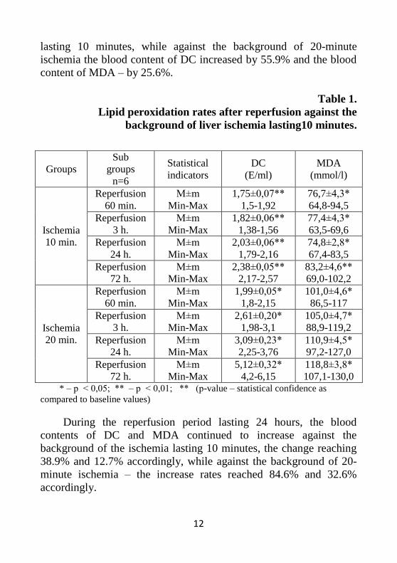
The periods of reperfusion (see: Table 1) were characterized by
the following dynamics of changes (as compared to baseline values):
during the 60-minute reperfusion the blood content of DC and MDA
increased by 19.8% and 15.7% accordingly against the background
of the ischemia lasting 10 minutes, while the increases in these
indicators against the background of 20-minute ischemia were 19.0%
and 20.7% accordingly During the reperfusion period lasting 3 hours
the blood content of DC was increased by 24.4%, and the blood
content of MDA – by 16.7% against the background of the ischemia
12
lasting 10 minutes, while against the background of 20-minute
ischemia the blood content of DC increased by 55.9% and the blood
content of MDA – by 25.6%.
Table 1.
Lipid peroxidation rates after reperfusion against the
background of liver ischemia lasting10 minutes.
* – р ˂ 0,05; ** – р ˂ 0,01; ** (р-value – statistical confidence as
compared to baseline values)
During the reperfusion period lasting 24 hours, the blood
contents of DC and MDA continued to increase against the
background of the ischemia lasting 10 minutes, the change reaching
38.9% and 12.7% accordingly, while against the background of 20-
minute ischemia – the increase rates reached 84.6% and 32.6%
accordingly.
Groups
Sub
groups
n=6
Statistical
indicators
DC
(E/ml)
MDA
(mmol/l)
Ischemia
10 min.
Reperfusion
60 min.
M±m
Min-Max
1,75±0,07**
1,5-1,92
76,7±4,3*
64,8-94,5
Reperfusion
3 h.
M±m
Min-Max
1,82±0,06**
1,38-1,56
77,4±4,3*
63,5-69,6
Reperfusion
24 h.
M±m
Min-Max
2,03±0,06**
1,79-2,16
74,8±2,8*
67,4-83,5
Reperfusion
72 h.
M±m
Min-Max
2,38±0,05**
2,17-2,57
83,2±4,6**
69,0-102,2
Ischemia
20 min.
Reperfusion
60 min.
M±m
Min-Max
1,99±0,05*
1,8-2,15
101,0±4,6*
86,5-117
Reperfusion
3 h.
M±m
Min-Max
2,61±0,20*
1,98-3,1
105,0±4,7*
88,9-119,2
Reperfusion
24 h.
M±m
Min-Max
3,09±0,23*
2,25-3,76
110,9±4,5*
97,2-127,0
Reperfusion
72 h.
M±m
Min-Max
5,12±0,32*
4,2-6,15
118,8±3,8*
107,1-130,0
13
During the reperfusion period lasting 72 hours, the blood
content of DC and MDA increased even more against the
background of the ischemia lasting 10 minutes, the change reaching
62.9%, and 25.4% accordingly.
The state of the body's antioxidant defense system (ADS) plays
an important role.
In our experiment, we determined the blood contents and
activity of such ADS enzymes as catalase and peroxidase along with
the total antioxidant activity index.Catalase activity decreased
against the background of ischemia for 10 minutes, falling from
0.305 ± 0.008 mmol/l (the intact state) to 0.255 ± 0.010 mmol/l,
declining by 16.4%. The changes are reliable at р˂0.01. Catalase
activity against the background of ischemia for 20 minutes catalase
activity was 0.240 ± 0,011 mmol/l, decreasing by 21.3% (р˂0.01).
Catalase activity decreased against the background of ischemia for
30 minutes falling down to 0.20 ± 0.013 mmol/l, declining by
34.4% compared to intact indicators (p˂0.01).
Peroxidase activity decreased against the background of
ischemia for 10 minutes, falling from 80.8 ± 0.6 mmol/l (the intact
state) to 0.255 ± 0.010 mmol/l, declining by 5.6%. This indicator
decreased against the background of ischemia for 20 minutes, falling
down to 73.9 ± 2.4 mmol/l, declining by 14.7% compared to intact
indicators (p˂0.01). As the duration of ischemia extended, reaching
30 minutes, the peroxidase activity continued to decrease, falling
down to 58.7 ± 2.6 mmol/l, declining by 31.4% compared to intact
indicators (p˂0.01).
At that, the total antioxidant activity index dropped as follows:
in the 2nd group, against the background of ischemia for 10 minutes,
it dropped from 37.8 ± 1.1% (the intact state) to 30.8 ± 0.8%. Thus,
the decrease in this indicator in the 2nd group amounted to 18.5%
(р˂0.01). In the 3rd group, against the background of ischemia for 20
minutes, the total antioxidant activity dropped by 26.9%, falling
down to 27.7±1.6% (р˂0.01). In the 4th group, against the
background of ischemia for 30 minutes, compared to the intact state,
14
the decrease in the total antioxidant activity amounted to 54.6%,
while the blood content of antioxidants was 58,7±1,2% (р˂0.01).
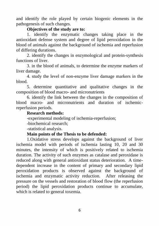
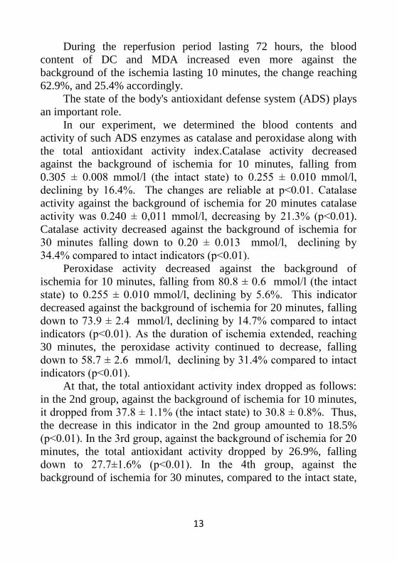
The analysis of the obtained results is presented in Diagram 3.2,
which shows that as the duration of ischemia increases, the activity
of catalase and peroxidase enzymes decreases, as well as the overall
antioxidant activity. Notably, the most pronounced decrease is
observed in total antioxidant activity. Changes in all defined
indicators possessed statistical reliability.
Diagram 1. Changes in the state of the antioxidant defense
system of the animal body depending on the duration of liver
ischemia (the intact state is taken as 100%).
(translation of the Diagram: the intact state;– ischemia for 10
minutes; – ischemia for 20 minutes; - ischemia for 30 minutes; -
catalase;– peroxidase; – total antioxidant activity)
Thus, the study demonstrated that lipid peroxidation is activated
against the background of liver ischemia, which is further confirmed
by an increase in the level of DC and MDA in the liver. In parallel,
the activity of such ADS enzymes as catalase, peroxidase along with
the general antioxidant activity decrease against the background of
ischemia lasting: 10 minutes — by 16, 5.6 and 18.5%; 20 minutes –
100
83,6 78,7
65,6
100 94,4
86,3
68,6
100
81,5 73,1
45,4
0
20
40
60
80
100
120
intact condition ischemia 10 min ischemia 20 min ischemia 30 min
catalase peroxidase total antioxidant activity
15
by 21.3, 13.7 and 26.9% respectively. Lipid peroxidation activation
against the background of ischemia was accompanied by changes in
all investigated blood parameters of the animals [6].
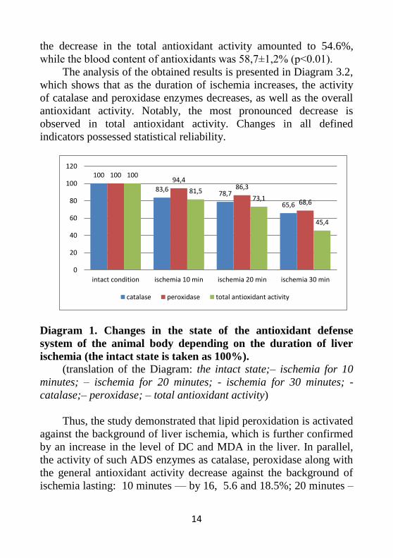
The concentration of the following markers of liver damage
against the background of ischemia for 10 minutes increased as
follows [7] (Fig.1): ALT – by 3.0%, AST – by 7.8%, total LDG –
by 1.2%, KPK – by 12.0%, γ - GTP – by 7.7%, while the
concentrations of: urea – decreased by 12.8%, creatinine – increased
by 19.9%, uric acid – decreased by 3.4%, total bilirubin – increased
by 28.0%, and C-reactive protein –by 35.0%.
The protein concentrations [3, 8] changed as follows: total
protein content – decreased by 0.9%, albumins – by 1.5%; globulins
– remained unchanged, and fibrinogen– increased by 33.3%.
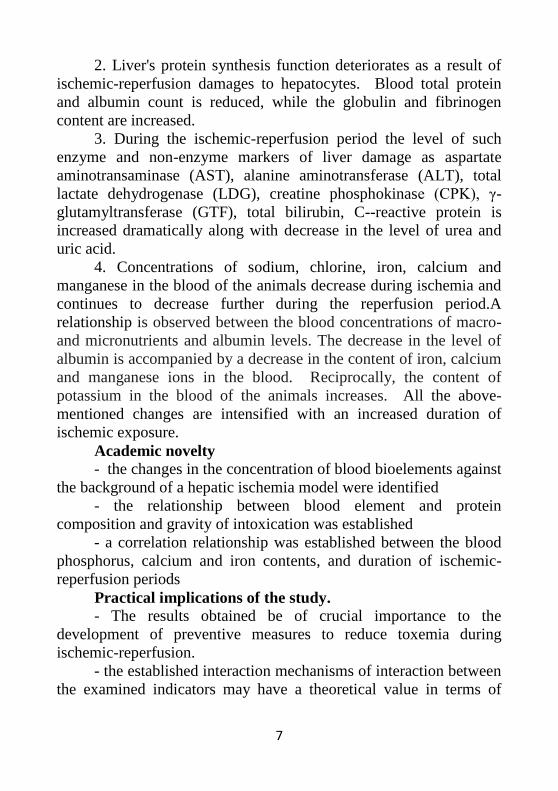
Diagram. Changes in the blood contents of liver damage
markers of the experimental animals
( r-n – reperfusion; hours; minutes; ischemia; ischemia 1 and r-n 1
– the 2nd group indicators; ischemia 2 and r-n 2 – the 3rd group
indicators; ischemia 3 and r-n 3 – the 4th group indicators; ALT;
AST; LDG; KPK; γ-GTP)
0 200 400 600 800
ischemia 1
ischemia 2
ischemia 3
r-n 60 min 1
r-n 60 min 2
r-n 3 hours 1
r-n 3 hours 2
r-n 24 hours 1
r-n 24 hours 2
r-n 72 hours 1
r-n 72 hours 2
ALT
AST
LDH
KPK
ɣ-GTP
16
We determined the concentrations of iron, calcium, manganese,
potassium, magnesium, chlorine and phosphorus ions in the blood
serum of the experimental animals [9]. These are the most important
blood bioelements, and their insufficiency may interfere with the
functions of all organs and systems, lead to cell destabilization, and
result in cellular damage in some cases. The pathological processes
taking place in cells can upset this balance, leading to further
aggravation of the pathological processes in the body, while also so-
called "positive feedback" (destructive in many cases) process is
commenced. The results of our study indicate that against the
background of ischemia for 10 minutes, the concentration of iron
ions fell from 23.4 ± 3.0 mmol/l (the intact state) to 22.9 ±
3.1mmol/l (1st group), declining by 1.9% (р˃0.05); while against the
background of ischemia for 20 minutes, this indicator decreased
further, falling down to 22.6 ± 3.1mmol/l, declining by 3.2%
(р˃0.05); against the background of ischemia for 30 minutes
concentration of iron ions continued to decrease, falling down to 19.3
± 3.3 mmol/l, declining by 14.7% (р˃0.05).
Against the background of ischemia for 10 minutes, the
concentration of calcium ions fell from 2.383±0.060 mmol/l (the
intact state) to 2.150 ± 0. 076mmol/l (1st group), declining by 9.8%
(р<0.05); while against the background of ischemia for 20 minutes,
this indicator decreased further, falling down to 1.80 ± 0.106
mmol/l, declining by 24.5% (р<0.01); against the background of
ischemia for 30 minutes concentration continued to decrease, falling
down to 1.517±0.135 mmol/l, declining by 36.4% (р<0.01).
Against the background of ischemia for 10 minutes, the
concentration of manganese ions fell from 0.867±0.064 mmol/l (the
intact state) to 0.832 ± 0.057 mmol/l, declining by 4% (р˃0.05);
while against the background of ischemia for 20 minutes, this
indicator decreased further, falling down to 0.797 ± 0.061 mmol/l,
declining by 8.1% (р˃0.05); against the background of ischemia
extending up to 30 minutes, the concentration of manganese ions
continued to decrease, falling down to 0.762 ± 0.065 mmol/l. In
17
percentage terms, compared to the intact state, the concentration of
manganese ions in this group decreased by 12.1% (р˃0.05).
Against the background of ischemia for 10 minutes, the
concentration of potassium ions fell from 4.23 ± 0.28 mmol/l (the
intact state) to 4.23 ± 0.28 mmol/l, declining by 6.3% (р˃0.05);
while against the background of ischemia for 20 minutes, this
indicator increased up to 4.87 ± 0.24 mmol/l, rising by 15.0%
(р>0.05); against the background of ischemia extending up to 30
minutes, it continued to increase, reaching 5.70 ± 0.135 mmol/l,
rising by 34.6% (р˂0.01) as compared to the intact state.
Against the background of ischemia for 10 minutes, the
concentration of sodium ions fell from 139.8 ± 1.5 mmol/l (the
intact state) to 137.0 ±1.8 mmol/l, declining by 2% (р˃0.05); while
against the background of ischemia for 20 minutes, this indicator
decreased further, falling down to 130.7±1,8 mmol/l, declining by
6.6% (р<0.01); against the background of ischemia extending up to
30 minutes, the concentration of sodium ions continued to decrease,
falling down to 123.7±2.3 mmol/l, declining by 11.6% (р˂0.01).
Concentration of chlorine ions decreased against the
background of ischemia for 10 minutes, falling from 99.5±1.3
mmol/l (the intact state) to 92.6±1.6 mmol/l (1st group). declining
by 3.4% (р˃0,05) in comparison with the intact state. Against the
background of ischemia for 20 minutes, the concentration of chlorine
ions fell down to 92.7±1.7 mmol/l, declining by 6.9% (р˂0.01).
Against the background of ischemia extending up to 30 minutes, the
concentration of chlorine ions continued to decrease, now falling
down to 89.2±2.0 mmol/l, declining by 10.4% (р˂0.01) as compared
to intact state indicators.
The phosphorus ion concentrations varied ambiguously. Thus, it
increased against the background of ischemia for 10 minutes, rising
from 1.092±0.077 mmol/l (the intact state) up to 1.285±0.136
mmol/l, increasing by 17.7% (р˃0.05). Against the background of
ischemia for 20 minutes, the concentration of phosphorus ions
continued to increase and reached 1.338±0.143 mmol/l, rising by
22.6% (р˃0.05) as compared to the intact state. However, a further
18
increase of ischemia duration resulted in a gradual decrease of the
phosphorus ions concentration in the blood of the experimental
animals, and against the background of for 30 minutes, it reached
1.212±0.158.
Comparative analysis of the obtained data showed that against
the background of ischemia there is an increase in potassium and
phosphorus ions concentrations during ischemia lasting 10 minutes
and 20 minutes. Increased duration of ischemia up to 30 minutes
further increased the concentration of potassium ions, while the
concentration of phosphorus ions began to decrease, although still
exceeding the intact state value by 11%.
Concentrations of iron, calcium, manganese, sodium and
chlorine ions during ischemia of 10, 20 and 30 minutes saw a gradual
decline. The decrease in the concentration of iron ions was not
statistically confirmed. The decrease in the concentration of calcium
ions was considerable and statistically significant at all periods. The
decrease in concentration of manganese ions was not statistically
confirmed. The decrease in the concentration of sodium and chlorine
ions was not confirmed statistically, while the decrease accelerated
significantly and statistically confirmed with increased duration of
ischemia.
The examined indicators were changing as follows during the
reperfusion against the background of ischemia for 10 minutes (as
compared to the baseline values): against the background of
reperfusion for 60 minutes concentrations of: ALT – increased by
13.6%, AST – increased by 17.3%, LDG – increased by 4.4%, KPK
– increased by 30.0%, γ-GTP – increased by 7.2%, creatinine –
increased by 13..39%, total bilirubin – by 45.6%, and C-reactive
protein –by 133.3%, while the concentrations of: urea by 24.3%,
uric acid by 0.6%. The total protein and albumin content decreased
by 8.9% and 14.6% accordingly, and globulins and fibrinogen
increased by 14.0% and by 87.5% accordingly.
The concentration of sodium ions during this period decreased
by 3.8%, potassium ions increased by 18.5%, chlorine ions decreased
19
by 3.5%, iron ions decreased by 12.5%, calcium ions - by 14.7%,
manganese ions - by 4.2%, and phosphorus ions increased by 10.9%.
Concentrations of liver damage markers against the background
of reperfusion for 3 hours continued to increase: ALT – increased by
31.0%, AST – increased by 52.5%, LDG – increased by 18.8%, KPK
– increased by 96.4%, γ-GTP – by 30.5%, while the concentrations
of: urea – increased by 9.3%, creatinine – by 27.8%, total bilirubin
– by 74.4%, and C-reactive protein –by 285.2%. A decrease was
observed only in uric acid concentrations, amounting to 10.2. The
total protein and albumin content decreased by 17.8% and 22.8%
accordingly, globulins and fibrinogen increased by 27.3% and by
108.3% accordingly. Determination of macro- and micronutrients in
the blood showed that the concentration of sodium ions decreased by
18.1%, potassium ions increased by 47.5%, chlorine ions decreased
by 6.9%, iron ions decreased by 14.5%, calcium ions - by 29.5%,
manganese ions - by 9%, and phosphorus ions increased by 17.6%.
The observed dynamics of the changes against the background
of 24 hours reperfusion were as follows: concentrations of ALT –
increased by 55.0%, AST – increased by 88.3%, LDG – increased by
27.5%, KPK – increased by 140.7%, γ-GTP – by 38.7%. The
concentrations of: urea decreased by 22.5%, creatinine increased by
40.9%, uric acid decreased by 21.5%, total bilirubin increased by
124.4%, and C-reactive protein – by 396.3%. The total protein and
albumin content decreased by 22.8% and 29.5% accordingly,
globulins and fibrinogen increased by 42.03% and by 162.5%
accordingly. The concentration of sodium ions during this period
decreased by 28.1%, potassium ions increased by 62.2%, chlorine
ions decreased by 18.0%, iron ions decreased by 19.3%, calcium ions
- by 46.5%, manganese ions - by 13.2%, and phosphorus ions
increased by 14.4%.
Concentrations of liver damage markers against the background
of reperfusion for 72 hours continued to increase: ALT – increased
by 76.4%, AST – increased by 121.8%, LDG– increased by 40.1%,
KPK – increased by 194.3%, γ-GTP – by 56.2%, while the
concentrations of: urea – decreased by 47.9%, creatinine – increased
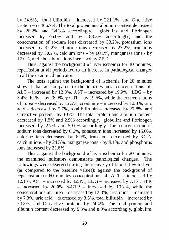
20
by 24.6%, total bilirubin – increased by 221.1%, and C-reactive
protein –by 466.7%. The total protein and albumin content decreased
by 26.2% and 34.3% accordingly, globulins and fibrinogen
increased by 46.0% and by 183.3% accordingly; and the
concentration of sodium ions decreased by 33.2%, potassium ions
increased by 92.2%, chlorine ions decreased by 27.2%, iron ions
decreased by 30.2%, calcium ions - by 60.5%, manganese ions - by
17.0%, and phosphorus ions increased by 7.5%.
Thus, against the background of liver ischemia for 10 minutes,
reperfusion at all periods led to an increase in pathological changes
in all the examined indicators.
The tests against the background of ischemia for 20 minutes
showed that as compared to the intact values, concentrations of:
ALT – increased by 12.8%, AST – increased by 19.9%, LDG – by
3.4%, KPK – by 28.0%, γ-GTP – by 19.6%, while the concentrations
of: urea – decreased by 12.5%, creatinine – increased by 12.3%, uric
acid – decreased by 9.7%, total bilirubin – increased by 27.8%, and
C-reactive protein –by 105%. The total protein and albumin content
decreased by 1.8% and 2.9% accordingly, globulins and fibrinogen
increased by 2.7% and 50.0% accordingly The concentration of
sodium ions decreased by 6.6%, potassium ions increased by 15.0%,
chlorine ions decreased by 6.9%, iron ions decreased by 3.2%,
calcium ions - by 24.5%, manganese ions - by 8.1%, and phosphorus
ions increased by 22.6%.
Thus, against the background of liver ischemia for 20 minutes,
the examined indicators demonstrate pathological changes. The
followings were observed during the recovery of blood flow to liver
(as compared to the baseline values): against the background of
reperfusion for 60 minutes concentrations of: ALT – increased by
12.1%, AST – increased by 12.1%, LDG – increased by 7.1%, KPK
– increased by 20.0%, γ-GTP – increased by 10.2%, while the
concentrations of: urea – decreased by 12.8%, creatinine – increased
by 7.3%, uric acid – decreased by 8.5%, total bilirubin – increased by
20.8%, and C-reactive protein –by 24.4%. The total protein and
albumin content decreased by 5.3% and 8.0% accordingly, globulins
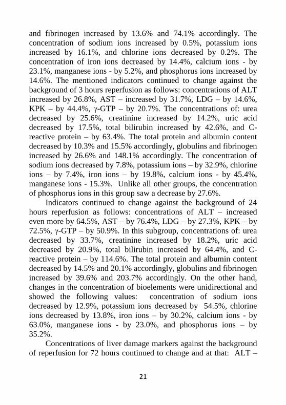
21
and fibrinogen increased by 13.6% and 74.1% accordingly. The
concentration of sodium ions increased by 0.5%, potassium ions
increased by 16.1%, and chlorine ions decreased by 0.2%. The
concentration of iron ions decreased by 14.4%, calcium ions - by
23.1%, manganese ions - by 5.2%, and phosphorus ions increased by
14.6%. The mentioned indicators continued to change against the
background of 3 hours reperfusion as follows: concentrations of ALT
increased by 26.8%, AST – increased by 31.7%, LDG – by 14.6%,
KPK – by 44.4%, γ-GTP – by 20.7%. The concentrations of: urea
decreased by 25.6%, creatinine increased by 14.2%, uric acid
decreased by 17.5%, total bilirubin increased by 42.6%, and C-
reactive protein – by 63.4%. The total protein and albumin content
decreased by 10.3% and 15.5% accordingly, globulins and fibrinogen
increased by 26.6% and 148.1% accordingly. The concentration of
sodium ions decreased by 7.8%, potassium ions – by 32.9%, chlorine
ions – by 7.4%, iron ions – by 19.8%, calcium ions - by 45.4%,
manganese ions - 15.3%. Unlike all other groups, the concentration
of phosphorus ions in this group saw a decrease by 27.6%.
Indicators continued to change against the background of 24
hours reperfusion as follows: concentrations of ALT – increased
even more by 64.5%, AST – by 76.4%, LDG – by 27.3%, KPK – by
72.5%, γ-GTP – by 50.9%. In this subgroup, concentrations of: urea
decreased by 33.7%, creatinine increased by 18.2%, uric acid
decreased by 20.9%, total bilirubin increased by 64.4%, and C-
reactive protein – by 114.6%. The total protein and albumin content
decreased by 14.5% and 20.1% accordingly, globulins and fibrinogen
increased by 39.6% and 203.7% accordingly. On the other hand,
changes in the concentration of bioelements were unidirectional and
showed the following values: concentration of sodium ions
decreased by 12.9%, potassium ions decreased by 54.5%, chlorine
ions decreased by 13.8%, iron ions – by 30.2%, calcium ions - by
63.0%, manganese ions - by 23.0%, and phosphorus ions – by
35.2%.
Concentrations of liver damage markers against the background
of reperfusion for 72 hours continued to change and at that: ALT –
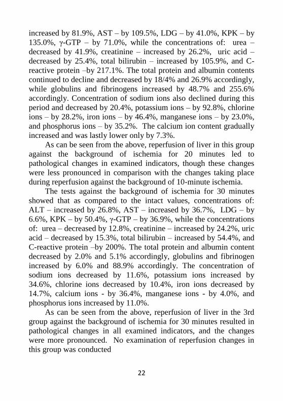
22
increased by 81.9%, AST – by 109.5%, LDG – by 41.0%, KPK – by
135.0%, γ-GTP – by 71.0%, while the concentrations of: urea –
decreased by 41.9%, creatinine – increased by 26.2%, uric acid –
decreased by 25.4%, total bilirubin – increased by 105.9%, and C-
reactive protein –by 217.1%. The total protein and albumin contents
continued to decline and decreased by 18/4% and 26.9% accordingly,
while globulins and fibrinogens increased by 48.7% and 255.6%
accordingly. Concentration of sodium ions also declined during this
period and decreased by 20.4%, potassium ions – by 92.8%, chlorine
ions – by 28.2%, iron ions – by 46.4%, manganese ions – by 23.0%,
and phosphorus ions – by 35.2%. The calcium ion content gradually
increased and was lastly lower only by 7.3%.
As can be seen from the above, reperfusion of liver in this group
against the background of ischemia for 20 minutes led to
pathological changes in examined indicators, though these changes
were less pronounced in comparison with the changes taking place
during reperfusion against the background of 10-minute ischemia.
The tests against the background of ischemia for 30 minutes
showed that as compared to the intact values, concentrations of:
ALT – increased by 26.8%, AST – increased by 36.7%, LDG – by
6.6%, KPK – by 50.4%, γ-GTP – by 36.9%, while the concentrations
of: urea – decreased by 12.8%, creatinine – increased by 24.2%, uric
acid – decreased by 15.3%, total bilirubin – increased by 54.4%, and
C-reactive protein –by 200%. The total protein and albumin content
decreased by 2.0% and 5.1% accordingly, globulins and fibrinogen
increased by 6.0% and 88.9% accordingly. The concentration of
sodium ions decreased by 11.6%, potassium ions increased by
34.6%, chlorine ions decreased by 10.4%, iron ions decreased by
14.7%, calcium ions - by 36.4%, manganese ions - by 4.0%, and
phosphorus ions increased by 11.0%.
As can be seen from the above, reperfusion of liver in the 3rd
group against the background of ischemia for 30 minutes resulted in
pathological changes in all examined indicators, and the changes
were more pronounced. No examination of reperfusion changes in
this group was conducted
23
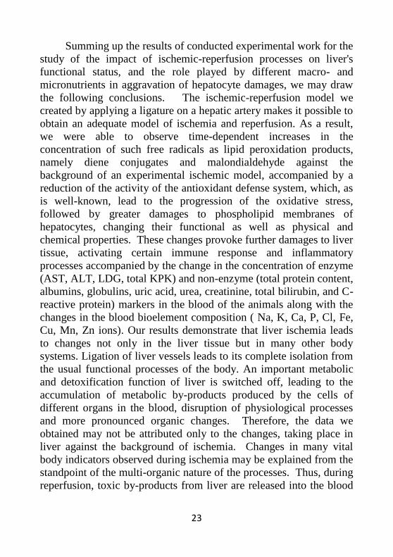
Summing up the results of conducted experimental work for the
study of the impact of ischemic-reperfusion processes on liver's
functional status, and the role played by different macro- and
micronutrients in aggravation of hepatocyte damages, we may draw
the following conclusions. The ischemic-reperfusion model we
created by applying a ligature on a hepatic artery makes it possible to
obtain an adequate model of ischemia and reperfusion. As a result,
we were able to observe time-dependent increases in the
concentration of such free radicals as lipid peroxidation products,
namely diene conjugates and malondialdehyde against the
background of an experimental ischemic model, accompanied by a
reduction of the activity of the antioxidant defense system, which, as
is well-known, lead to the progression of the oxidative stress,
followed by greater damages to phospholipid membranes of
hepatocytes, changing their functional as well as physical and
chemical properties. These changes provoke further damages to liver
tissue, activating certain immune response and inflammatory
processes accompanied by the change in the concentration of enzyme
(AST, ALT, LDG, total KPK) and non-enzyme (total protein content,
albumins, globulins, uric acid, urea, creatinine, total bilirubin, and C-
reactive protein) markers in the blood of the animals along with the
changes in the blood bioelement composition ( Na, K, Ca, P, Cl, Fe,
Cu, Mn, Zn ions). Our results demonstrate that liver ischemia leads
to changes not only in the liver tissue but in many other body
systems. Ligation of liver vessels leads to its complete isolation from
the usual functional processes of the body. An important metabolic
and detoxification function of liver is switched off, leading to the
accumulation of metabolic by-products produced by the cells of
different organs in the blood, disruption of physiological processes
and more pronounced organic changes. Therefore, the data we
obtained may not be attributed only to the changes, taking place in
liver against the background of ischemia. Changes in many vital
body indicators observed during ischemia may be explained from the
standpoint of the multi-organic nature of the processes. Thus, during
reperfusion, toxic by-products from liver are released into the blood
24
circulation, including the lipid peroxidation products, cytokines etc.
which exert a damaging impact on the body tissues, amplifying the
positive feedback reaction of the body. On the other hand, the toxic
metabolic by-products, accumulating in the blood, enter liver and
inflict further damages to liver tissues. This explains the increasingly
pronounced nature of lipid peroxidation and other indicators
observed in our experiments during reperfusion after the ischemia for
10, 20 and 30 minutes. During ischemia for 10, 20 and 30 minutes, a
change in protein content – a small one in percentage terms (0.9%,
.8% and 2.0% accordingly), but very important for body functioning
– takes place. As previously shown, the functional capabilities of
hepatocytes are not restored during reperfusion, rather, their
destruction aggravates due to the presence of toxic products in blood,
formed during the isolation of liver from the body's vital systems.
Consequently, the reduction of total protein content accelerates
gradually and after 72 hours of reperfusion, the protein lack in the
reached 26.9% and 19.9% in the 2nd and 3rd groups accordingly.As
could be expected, the content of the albumin fraction decreases and
the content of the globulin fraction increases, the level of fibrinogen
increases against the background of general intoxication and the
inflammatory process during all periods of ischemia and reperfusion.
At that, these changes against the background of 20-minute ischemia
in the 3rd group were more pronounced than the changes against the
background of 10-minute ischemia in the 2nd group. As the results
show, the concentration of such enzymes as AST, ALT, LDG, KPK
and γ-GTP increases as increases the duration of ischemia.
Hepatocyte deterioration is clearly reflected by the changes in the
blood concentrations of urea, creatinine, total bilirubin and C-
reactive protein, where such changes continue to occur after
reperfusion. Changes in the concentration of macro- and
micronutrients both during ischemia and all reperfusion periods
conform with the pathology of overall changes taking place during
the respective examination periods and fall in line with the data from
literary sources. Also, it should be noted that during the reperfusion
period following the ischemia for 20 minutes said changes in
25
examined indicators are less intense. This paradox may be explained
by the active involvement of switched-on adaptive body mechanisms
during ischemia for 20 minutes.
CONCLUSIONS
1. One of the factors conditioning the pathological changes
taking place in the blood of the experimental animals during the 10-,
20- and 30-minute periods of ischemia created by applying a ligature
on arteries vessels of liver is linked to the reduction of the activity of
such antioxidant enzymes, as catalase and peroxidase, and general
antioxidant status. A time-dependent increase in the content of
primary and secondary lipid peroxidation products is observed
against the background of ischemia and enzymatic activity reduction.
After releasing the pressure on the vessels and restoration of blood
flow, the lipid peroxidation products continue to accumulate, which
is related to general toxemia.
2. Liver's protein synthesis function deteriorates as a result of
reperfusion damages to hepatocytes. A particularly harmful impact
on the body state of the animals is exerted by the reduction of blood
albumin concentration, taking into account their biological functions.
Decreases in the total level of plasma proteins cause the disruption of
water-salt metabolism, hyperkalemia, cellular acidosis, increased
toxicity of some metabolic products, which are normally blocked by
albumins, and transferred to their destination points.
3. Deterioration of hepatocytes during the ischemic-reperfusion
period is reflected by the dramatic increases in the level of such
enzyme and non-enzyme markers of liver damage as AST, ALT,
LDG, KPK,γ-GTP, total bilirubin, C--reactive protein, along with a
decrease in the level of urea and uric acid.
4. Concentrations of sodium, chlorine, iron, calcium and
manganese in the blood of the animals decrease during ischemia and
continues to decrease further during the reperfusion period. A
relationship is observed between the blood concentrations of macro-
and micronutrients and albumin levels. The decrease in the level of
26
albumin is accompanied by a decrease in the content of iron, calcium
and manganese ions in the blood. Reciprocally, the content of
potassium in the blood of the animals increases. All the above-
mentioned changes are intensified with increased duration of
ischemic exposure.
5. There is observed a link between the blood phosphorus and
calcium levels and duration of ischemia periods. Increasing the
duration of ischemia up to 20 minutes led to a decrease in blood
phosphorus and an increase in blood calcium concentration during
reperfusion. Whereas an increase in the levels of phosphorus and
calcium ions was observed during reperfusion against the
background of ischemia for 10 minutes.
6. The defensive and adaptive functions of the body are
switched on beginning from the ischemia for 20 minutes, which
explains the moderate course of toxemia and reperfusion syndrome
in the group of the experimental animals during reperfusion against
the background of ischemia for 20 minutes.
27
LIST OF PUBLISHED WORKS
1. Gafarova J.R. Some aspects of the pathological and
biochemical processes taking place during ischemia // "Sağlamlıq"
magazine, 2010, issue 4, – p. 179-182.
2. Gafarova J.R., Khalilova V.Q, Guliyeva S.V. Reperfusion
damages to organs // "Azərbayjan tababatinin muasir nailiyyatlari"
magazine, 2015, issue 2, –p. 20-23.
3. Determination of the amounts of serum bioelements in
experimental animals against the background of a hepatic ischemia
model // "Azərbayjan tababatinin muasir nailiyyatlari" magazine,
2017, issue 4, –p. 111-114
4. Gafarova J.R., Garayev G. Sh. Jafarova R. E. Toxic
substances formed during ischemia and their impact on the body //
"Sağlamlıq" magazine, 2017, issue 4, – p. 7-13.
5. Gafarova J. R., Garayev H. Sh., Jafarova R. E. Reperfusion
syndrome in liver transplantation and mechanisms of its development
// Ukrainian medical magazine "Chasopis", Kiyev, Ukraine, 2017,
issue 1, – p. 77-80
6. Garayev H. Sh., Gafarova J. R., Jafarova R. E. Determination
of the intensity of lipid peroxidation in liver tissues against the
background of experimental ischemia-reperfusion / Digest of the
17th International Conference on European Science and Technology
Conference, Munich, 2017, –p. 91-96
7. Garayev H. Sh., Gafarova J. R., Jafarova R. E. Study of
changes in the activity of the liver antioxidant defense system against
the background of experimental ischemic-reperfusion / Digest of II
International Conference on Biology and Medical Sciences, Austria,
December 14, 2017; –p. 42-47
8. Gafarova J.R., Garayev G. Sh. Jafarova R. E. Changes in
blood protein composition during ischemia-reperfusion syndrome
modeled by impaired liver blood flow // Vestnik Rossiyskoy
Voenno-Meditsinskoy Akademii, 2018, issue 2 (62), –p. 110-114
9. Gafarova J.R., Jafarova R.A. Changing the balance of micro
and macro elements of blood against the background of an
28
experimental model of liver ischemia-reperfusion / Scientific
Research of the SCO Countries: Synergy and Integration, Beijing,
China 2019, – p.110-115

29

30
31
The defense will be held on ______ __________ ___________
at _______ at the meeting of the Dissertation council FD 2.07
of Supreme Attestation Commission under the President of the
Republic of Azerbaijan operating at Azerbaijan Medical University.
Address: AZ-1022, Baku, ave. A.Kasumzade, 14
Dissertation is accessible at the Azerbaijan Medical University
Library.
Electronic versions of dissertation and its abstract are available on
the official website of the Azerbaijan Medical Univeristy
(https://amu.edu.az/).
Abstract was sent to the required addresses on ___ _______ ______
32
Signed for print: 25.05.2021
Paper format: A5
Volume: 37506
Number of hard copies: 20