Embed Size (px)
Citation preview
RESEARCH ARTICLE Open Access
Influence of temperature on development,reproduction and regeneration in theflatworm model organism, MacrostomumlignanoJakub Wudarski1, Kirill Ustyantsev2, Lisa Glazenburg1 and Eugene Berezikov1*
Abstract
Background: The free-living marine flatworm Macrostomum lignano is a powerful model organism for use in studyingmechanisms of regeneration and stem cell regulation due to its combination of biological and experimental properties,including the availability of transgenesis methods, which is unique among flatworm models. However, due to itsrelatively recent introduction in research, many aspects of this animal’s biology remain unknown. One such question isthe influence of culture temperature on Macrostomum biology.
Results: We systematically investigated how different culture temperatures affect development time, reproduction rate,regeneration, heat shock response, and gene knockdown efficiency by RNA interference (RNAi) in M. lignano. We usedmarker transgenic lines to accurately measure the regeneration endpoint, and to establish the stress responsethreshold for temperature shock. We found that compared to the culture temperature of 20 °C commonly used for M.lignano, temperatures of 25 °C–30 °C substantially increase the speed of development and regeneration, lead to fastermanifestation of RNAi phenotypes, and increase reproduction rate without detectable negative consequences for theanimal, while temperatures above 30 °C elicit a heat shock response.
Conclusions: We show that altering temperature conditions can be used to reduce the time required to establish M.lignano cultures, perform RNAi experiments, store important lines, and optimize microinjection procedures fortransgenesis. These findings will help to optimize the design of experiments in M. lignano, and thus facilitate futureresearch using this model organism.
Keywords: Temperature, Heat shock response, Flatworms, Macrostomum lignano, Fertility, Regeneration
BackgroundFlatworms (Platyhelminthes) are a large phylum in theanimal kingdom (Metazoa), many of which exhibit thecapacity to regenerate lost tissues and body parts [1].This regenerative ability has long attracted the interestsof scientists, and the free-living planarian flatworms(Tricladida) Schmidtea mediterranea and Dugesia japon-ica in particular have been studied extensively andyielded numerous insights into the mechanisms under-lying regeneration [2–5]. More recently, a non-planarian
flatworm model Macrostomum lignano (Macrostopmor-pha) has been introduced into the regeneration researcharena, offering an attractive combination of experimentaland biological features [6, 7]. Macrostomum lignano is afree-living marine flatworm capable of regeneration an-terior to the brain and posterior to the pharynx [8].Similar to other flatworms, regeneration in M. lignano ismade possible by stem cells called neoblasts [9]. It is asmall and transparent animal that is easy to culture inlaboratory conditions. These features, together with therecently reported genome and transcriptome assemblies[10, 11] and the development of a robust transgenesismethod [12] make M. lignano a versatile model organ-ism for research on stem cells and regeneration [7].
* Correspondence: [email protected] Research Institute for the Biology of Ageing, University ofGroningen, University Medical Center Groningen, Antonius Deusinglaan 1,9713AV, Groningen, The NetherlandsFull list of author information is available at the end of the article
© The Author(s). 2019 Open Access This article is distributed under the terms of the Creative Commons Attribution 4.0International License (http://creativecommons.org/licenses/by/4.0/), which permits unrestricted use, distribution, andreproduction in any medium, provided you give appropriate credit to the original author(s) and the source, provide a link tothe Creative Commons license, and indicate if changes were made. The Creative Commons Public Domain Dedication waiver(http://creativecommons.org/publicdomain/zero/1.0/) applies to the data made available in this article, unless otherwise stated.
Wudarski et al. Zoological Letters (2019) 5:7 https://doi.org/10.1186/s40851-019-0122-6
Macrostomum lignano is a non-self-fertilizing herm-aphrodite with a short generation time of 2–3 weeks[13]. When cultured in standard laboratory conditions,animals lay approximately one egg per day. Embryonicdevelopment takes five days, and hatchlings reach adult-hood in about two weeks. The laid eggs are fertilized,relatively large (100 μm) and follow the archoophoranmode of development [13]; i.e., they have a large,yolk-rich oocyte instead of separate yolk cells that supplya small oocyte. These properties of the eggs make thema good target for delivery of external agents, such asDNA, RNA and protein, by means of microinjection.The possibility to introduce foreign genetic material
and modify the genome of an animal is a highlysought-after experimental property, and an integral partof the genomic toolkit in model organisms commonlyused for genetic research, such as the nematode Caenor-habditis elegans, fruit fly Drosophila melanogaster, yeast,and mouse, as it broadens the range of usable experi-mental approaches and greatly improves the chances ofdeciphering biological phenomena of interest. We re-cently demonstrated that microinjection of DNA intosingle-cell stage embryos can be used to generate trans-genic M. lignano animals [12]. The technique is efficientand robust, and stable transgenic lines can be obtainedwithin three-to-four months, including F1 and F2crosses. However, from the experimental perspective, itwould be advantageous if the time required to generatetransgenic animals could be shortened. Manipulation ofculture temperature conditions is one way to approachthis challenge.Temperature is one of the most important factors in-
fluencing most biological processes. The overall range oftemperatures that support active life on Earth stretchesfrom as low as − 1.8 °C in polar regions to around 113 °Cfor thermophilic archaea at the other extreme [14, 15].Most animals have specific temperature ranges that areoptimal to their growth; this is because even small alter-ations in the temperature can lead to significant changesin animal metabolism. If these changes are sustainablefor the organism, the increase in the temperature usuallyleads to an increase in the speed of physiological pro-cesses. The most common way of showing this relation-ship is using the temperature coefficient Q10 = (Rate 2 /Rate 1)10/(Temperature 2 − Temperature 1), which compares therates of a process at a given temperature with the rate attemperature increased by 10 °C [16]. The most popularway of using the Q10 index is by measuring oxygen con-sumption [17], but it may also be applied to various dif-ferent measurements, such as electric organ discharge[18] or locomotor performance [19].All living organisms respond to changes in environmental
temperature. Perturbations in temperature will usually trig-ger the heat shock response pathway, which is an ancient,
universal mechanism based on specialized chaperone mole-cules called heat shock proteins, or Hsps [20]. These pro-teins can help other proteins to fold correctly, repairdamaged proteins, or degrade them. They can also be agood indicator of stress that an organism undergoes [21].Here we present how temperature affects development,
growth, fertility, and regeneration capabilities of Macrosto-mum lignano. We tested the stress response to elevatedtemperatures by measuring the activity of the Heat shock20 (Hsp20) gene using qRT-PCR and Hsp20::mScarlettransgene expression. Furthermore, we measured thehatching speed of M. lignano eggs when incubated at dif-ferent temperatures, as well as the number of offspringproduced in these conditions. We also investigated howchanges in temperature influence regeneration speed inM. lignano, and the speed of the development of pheno-types upon gene knockdown by RNA interference (RNAi).Our findings establish optimal conditions for M. lignanocultures and will help inform future research using thismodel organism, particularly for creating transgenic ani-mals and performing RNAi experiments.
Materials and methodsStrainsThe DV1 inbred M. lignano line used in this study hasbeen described previously [10, 22, 23]. The NL10 andNL22 lines were previously established in our laboratory[12]. Animals were cultured under laboratory conditionsin plastic Petri dishes (Greiner), filled with nutrient-enriched artificial seawater (Guillard’s f/2 medium).Worms were fed ad libitum on the unicellular diatomNitzschia curvilineata (Heterokontophyta, Bacillariophy-ceae) (SAG, Göttingen, Germany). Conditions in the cli-mate chambers were set at 20 °C, 25 °C, 30 °C and 35 °Cwith constant aeration, and a 14 h/10 h day/night cycle.The heat shock sensor construct KU#49 was created
using a previously described double-promoter vector ap-proach [12]. The promoter region of M. lignano hsp20homolog gene (Mlig005128.g2) was cloned using primers5′-GGATGGATCCTCATTTATAAGCGTACCGTACT-3′and 5′-TTATAAGCTTCATGCTGTTGTTGACTGGCGTA-3′ to drive expression of mScarlet-I red fluorescentprotein, while elongation factor 1 alpha (EFA) promoterdriving expression of NeonGreen fluorescent protein wasused for the selection of transgenic animals. Two hundredthirty-five single-cell eggs were injected with the KU#49plasmid as previously described [12], but without radiationtreatment. Hatchlings were selected based on the presenceof green fluorescence, and a stable transgenic line NL28was established.
Egg hatchingTwenty single-cell stage eggs per temperature conditionwere picked and transferred to single wells in a 6-well
Wudarski et al. Zoological Letters (2019) 5:7 Page 2 of 10
plate. They were monitored daily, and hatched worms wereimmediately removed from the test well. Each temperaturecondition was tested independently in triplicate.
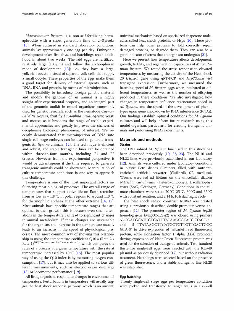
Heat shockTo measure expression levels of the Mlig-hsp20 gene byqRT-PCR, 50 worms of the same age were selected foreach of the three replicates. Animals were incubated fortwo hours at different temperatures (20 °C, 25 °C, 33 °C,34 °C and 35 °C) and two more hours at 20 °C, beforeRNA extraction (RNeasy, Qiagen). Quantitative RT-PCRwas done using the Light Cycler 480 (Roche) with5′-CGAAGATGTCACTGAGGTCAAG-3′ and 5′-GCGCCTGCAGTAGAAGAAT-3′ as primers and GAPDH,COX and EIF as reference target genes, as previously de-scribed [24]. Analysis of the results was performed usingthe qBase+ software (Biogazelle).To monitor heat shock response using Hsp20::mScarlet
transgene, NL28 transgenic animals were incubated for 2h at different temperatures (20 °C, 25 °C to 35 °C with 1 °Cinterval, and 37 °C), followed by 22 h at 20 °C, and thenimaged using Zeiss Axio Zoom V16 microscope with anHRm digital camera and Zeiss filter sets 38HE (FITC) and43HE (DsRed). Images were analyzed using ImageJ soft-ware. Images were converted to 8-bits, the area of theworm was selected, and the median value of the signalwas measured and visualized using box plot. Graphs weregenerated using the ggplot2 package for R.
ReproductionThree sets of 20 freshly hatched worms were selected perline and temperature condition. They were kept in theselected temperature until the end of the experiment, a totalduration of five weeks. Twice per week, the worms weretransferred to new plates with fresh food, and the old plates,where animals laid eggs in the preceding time interval, wereincubated until all eggs were hatched, after which the hatch-lings were counted. The approach of counting hatchlings asa proxy to the number of laid eggs rather than the directcounting of eggs was chosen because it is easier and morereliable to count hatchlings than to count egg clumps.
RegenerationTwelve NL22 worms expressing GFP marker in testes[12] were used per condition and cut above the testes.The worms were then placed in 12-well plates with freshdiatom and monitored daily. The days of the first ap-pearance of GFP signal in the testes and in the seminalvesicle were noted and used to measure the time re-quired for regeneration.
RNA interferenceFor the knockdown of Mlig-ddx39 gene, 15 worms pertemperature condition (20 °C, 25 °C and 30 °C) were
selected and treated with Mlig-ddx39 dsRNA fragmentsas previously described [11]. The morphology and viabil-ity of the worms was monitored on a daily basis and anyabnormalities were noted. GFP dsRNA was used as anegative control. For measuring the efficiency ofMlig-ddx-39 knockdown, qRT-PCR was performed inthe same way as described above for the Mlig-hsp20gene but with Mlig-ddx39 primers 5′-ACCCAGAGCTGCTGGACTAT-3′ and 5′-GTAGGAGCCCTTGTGACCTG-3′. For the knockdown of Mlig-sperm1 gene,twenty worms per temperature condition were treatedwith Mlig-sperm1 dsRNA fragments as previouslydescribed [25], and the number of animals with the en-larged testes were counted on day 4 of the treatment.
ResultsEstablishing the temperature rangeCommonly used laboratory conditions for Macrostomumlignano cultures are as follows: temperature of 20 °C, hu-midity 60%, and light/dark cycle of 14 h/10 h. These con-ditions were chosen mainly because they are optimal forthe growth of the diatom Nitzschia curvilineata, whichis the main food source for the worms [26]. To assessthe temperature conditions that can be used in the ex-periment, we first established the temperature range inwhich the worms survive. While freezing the wormsproved to be lethal, they could survive when kept at 4 °Cfor at least two weeks. However, because the diatomdoes not grow in these conditions, we decided to ex-clude 4 °C from further experiments. Other temperaturesbelow 20 °C were also excluded from the experiment,since the primary objective of the study was to find con-ditions that accelerate growth and development. On theother side of the temperature spectrum, the worms dis-solve when kept at 42 °C for two hours, and died afterone week of culture at 37 °C. Therefore, we have decidedto use 20 °C, 25 °C, 30 °C and 35 °C as our experimentalconditions to study long-term temperature effects on M.lignano (Fig. 1).
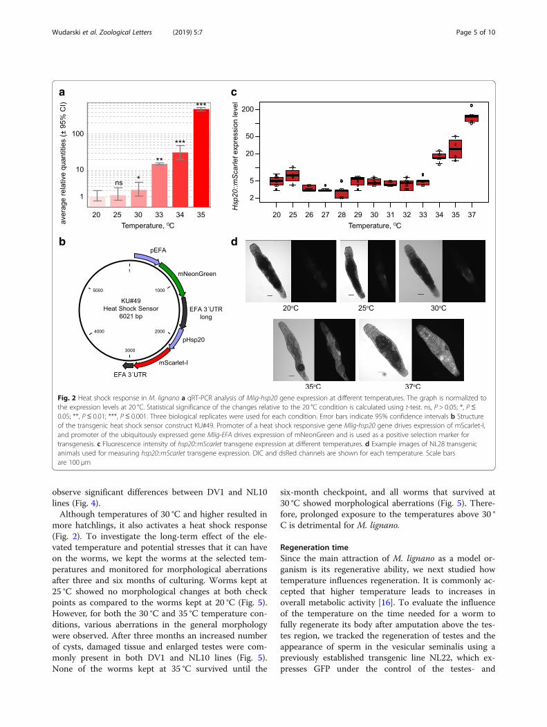
Heat shock responseTo investigate which temperatures induce stress re-sponse in the worms, we monitored the activity of theheat shock 20 promoter (Mlig-hsp20). First, we per-formed a quantitative RT-PCR to measure the expres-sion level of Mlig-hsp20 at 20 °C, 25 °C, 30 °C, 33 °C, 34 °C and 35 °C. There was no significant difference in theexpression level of Hsp20 between 20 °C and 25 °C(Fig. 2a). However, a small (2-fold), but significant (P =0.027, t-test), increase in the expression was observed at30 °C compared to 20 °C (Fig. 2a). More than a ten-foldraise in the expression level was observed at 33 °C,which increased to more than 100-fold at the highesttested temperature of 35 °C (Fig. 2a). In the second test
Wudarski et al. Zoological Letters (2019) 5:7 Page 3 of 10
we created a transgenic line expressing mScarlet-I proteinunder the control of the Mlig-hsp20 promoter (Fig. 2b)and measured the level of fluorescence 24 h after a 2-h in-cubation at different temperatures ranging from 20 °C to37 °C (Fig. 2c, d). More than two- and ten-fold increase inthe fluorescence was observed at 34 °C and 37 °C respect-ively (Fig. 2c).
Speed of embryonic developmentAccording to Morris et al. [13], it takes around 120 h(five days) for Macrostomum eggs to fully develop
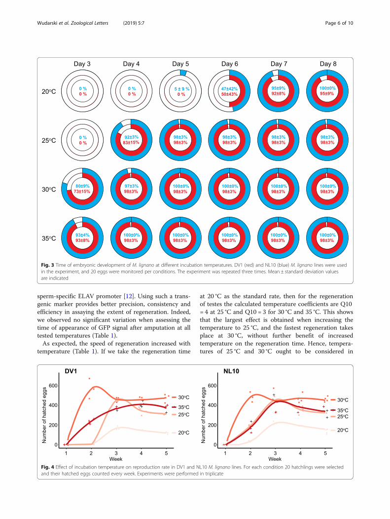
when eggs are kept at 20 °C. To investigate how thetemperature affects the speed of embryonic develop-ment and hatching, we picked freshly laid embryosand monitored their development until hatching atdifferent temperatures. To investigate potential differ-ences due to genetic background, we used two M. lig-nano lines that are currently used in a majority ofstudies on M. lignano, the DV1 and NL10 lines.These lines are independently derived from wild-typepopulations from the same geographical location anddiffer by a whole-chromosome duplication but other-wise behave very similarly under laboratory conditions[12]. We first studied the effect of low temperature.At 4 °C the development of the eggs is arrested andthey can be stored for at least one month, and re-sume their development once returned to higher tem-peratures. Next, we studied how quickly the eggsdevelop at temperatures between 20 °C and 35 °C. Asshown in Fig. 3, when kept at standard conditions(20 °C) the eggs started to hatch after six days. In-crease in the temperature resulted in proportionallyfaster embryonic development and earlier hatching,which was two times faster at 35 °C compared to 20 °C and took only three days. Of note, about 10% ofeggs remain unhatched after eight days of incubationat 20 °C, compared to less than 5% at higher tempera-tures, suggesting that even the highest testedtemperature of 35 °C does not have detrimental ef-fects on the survival of embryos.
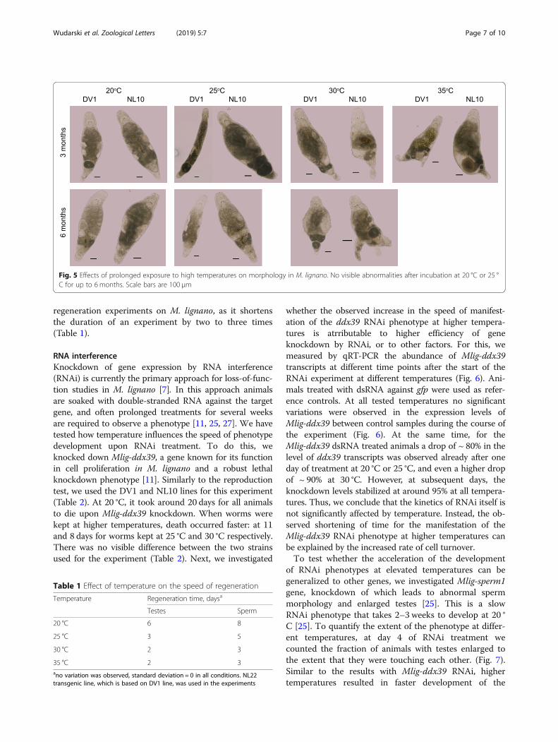
ReproductionThe reproduction rate is a very important factor for amodel organism, since animals with shorter generationtime enable faster generation of data in genetic experi-ments. In addition, if animals produce large numbers ofoffspring, the generated data will, in most cases, havehigher statistical power.To assess the temperature impact on M. lignano
reproduction rate we have compared the number of off-spring generated over the course of five weeks by wormskept at different temperature conditions. The experi-ment was started with hatchlings to incorporate postem-bryonic development in the study, and both DV1 andNL10 line were used. It took the hatchlings of both linesthree weeks to grow and produce first progeny at 20 °C,while at 25 °C hatchlings were observed in two weeks forNL10 line but not for DV1. At 30 °C and 35 °C both linesproduced progeny already after two weeks (Fig. 4). Fromthree weeks onwards, the number of hatchlings pro-duced per week increased from below 200 at 20 °C tomore than 300 in temperatures above 20 °C; hatchlingnumbers were highest for worms kept at 30 °C. This wastrue for both genetic backgrounds, and we did not
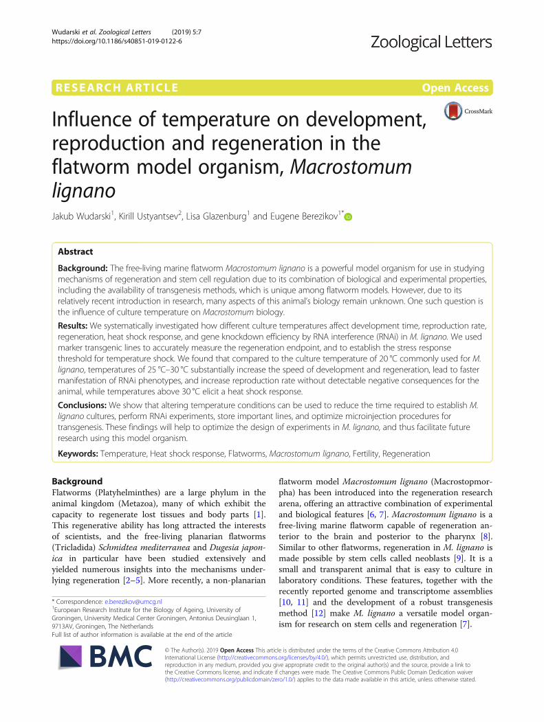
Fig. 1 Design of the study. Embryos and animals of two wild-typestrains, DV1 and NL10, were cultured at a range of temperaturesfrom 20 °C to 35 °C and development time, reproduction,regeneration time, heat shock response and efficiency of geneknockdown by RNA interference were measured
Wudarski et al. Zoological Letters (2019) 5:7 Page 4 of 10
observe significant differences between DV1 and NL10lines (Fig. 4).Although temperatures of 30 °C and higher resulted in
more hatchlings, it also activates a heat shock response(Fig. 2). To investigate the long-term effect of the ele-vated temperature and potential stresses that it can haveon the worms, we kept the worms at the selected tem-peratures and monitored for morphological aberrationsafter three and six months of culturing. Worms kept at25 °C showed no morphological changes at both checkpoints as compared to the worms kept at 20 °C (Fig. 5).However, for both the 30 °C and 35 °C temperature con-ditions, various aberrations in the general morphologywere observed. After three months an increased numberof cysts, damaged tissue and enlarged testes were com-monly present in both DV1 and NL10 lines (Fig. 5).None of the worms kept at 35 °C survived until the
six-month checkpoint, and all worms that survived at30 °C showed morphological aberrations (Fig. 5). There-fore, prolonged exposure to the temperatures above 30 °C is detrimental for M. lignano.
Regeneration timeSince the main attraction of M. lignano as a model or-ganism is its regenerative ability, we next studied howtemperature influences regeneration. It is commonly ac-cepted that higher temperature leads to increases inoverall metabolic activity [16]. To evaluate the influenceof the temperature on the time needed for a worm tofully regenerate its body after amputation above the tes-tes region, we tracked the regeneration of testes and theappearance of sperm in the vesicular seminalis using apreviously established transgenic line NL22, which ex-presses GFP under the control of the testes- and
a c
b d
Fig. 2 Heat shock response in M. lignano a qRT-PCR analysis of Mlig-hsp20 gene expression at different temperatures. The graph is normalized tothe expression levels at 20 °C. Statistical significance of the changes relative to the 20 °C condition is calculated using t-test. ns, P > 0.05; *, P ≤0.05; **, P ≤ 0.01; ***, P ≤ 0.001. Three biological replicates were used for each condition. Error bars indicate 95% confidence intervals b Structureof the transgenic heat shock sensor construct KU#49. Promoter of a heat shock responsive gene Mlig-hsp20 gene drives expression of mScarlet-I,and promoter of the ubiquitously expressed gene Mlig-EFA drives expression of mNeonGreen and is used as a positive selection marker fortransgenesis. c Fluorescence intensity of hsp20::mScarlet transgene expression at different temperatures. d Example images of NL28 transgenicanimals used for measuring hsp20::mScarlet transgene expression. DIC and dsRed channels are shown for each temperature. Scale barsare 100 μm
Wudarski et al. Zoological Letters (2019) 5:7 Page 5 of 10
sperm-specific ELAV promoter [12]. Using such a trans-genic marker provides better precision, consistency andefficiency in assaying the extent of regeneration. Indeed,we observed no significant variation when assessing thetime of appearance of GFP signal after amputation at alltested temperatures (Table 1).As expected, the speed of regeneration increased with
temperature (Table 1). If we take the regeneration time
at 20 °C as the standard rate, then for the regenerationof testes the calculated temperature coefficients are Q10= 4 at 25 °C and Q10 = 3 for 30 °C and 35 °C. This showsthat the largest effect is obtained when increasing thetemperature to 25 °C, and the fastest regeneration takesplace at 30 °C, without further benefit of increasedtemperature on the regeneration time. Hence, tempera-tures of 25 °C and 30 °C ought to be considered in
Fig. 3 Time of embryonic development of M. lignano at different incubation temperatures. DV1 (red) and NL10 (blue) M. lignano lines were usedin the experiment, and 20 eggs were monitored per conditions. The experiment was repeated three times. Mean ± standard deviation valuesare indicated
Fig. 4 Effect of incubation temperature on reproduction rate in DV1 and NL10 M. lignano lines. For each condition 20 hatchlings were selectedand their hatched eggs counted every week. Experiments were performed in triplicate
Wudarski et al. Zoological Letters (2019) 5:7 Page 6 of 10
regeneration experiments on M. lignano, as it shortensthe duration of an experiment by two to three times(Table 1).
RNA interferenceKnockdown of gene expression by RNA interference(RNAi) is currently the primary approach for loss-of-func-tion studies in M. lignano [7]. In this approach animalsare soaked with double-stranded RNA against the targetgene, and often prolonged treatments for several weeksare required to observe a phenotype [11, 25, 27]. We havetested how temperature influences the speed of phenotypedevelopment upon RNAi treatment. To do this, weknocked down Mlig-ddx39, a gene known for its functionin cell proliferation in M. lignano and a robust lethalknockdown phenotype [11]. Similarly to the reproductiontest, we used the DV1 and NL10 lines for this experiment(Table 2). At 20 °C, it took around 20 days for all animalsto die upon Mlig-ddx39 knockdown. When worms werekept at higher temperatures, death occurred faster: at 11and 8 days for worms kept at 25 °C and 30 °C respectively.There was no visible difference between the two strainsused for the experiment (Table 2). Next, we investigated
whether the observed increase in the speed of manifest-ation of the ddx39 RNAi phenotype at higher tempera-tures is atrributable to higher efficiency of geneknockdown by RNAi, or to other factors. For this, wemeasured by qRT-PCR the abundance of Mlig-ddx39transcripts at different time points after the start of theRNAi experiment at different temperatures (Fig. 6). Ani-mals treated with dsRNA against gfp were used as refer-ence controls. At all tested temperatures no significantvariations were observed in the expression levels ofMlig-ddx39 between control samples during the course ofthe experiment (Fig. 6). At the same time, for theMlig-ddx39 dsRNA treated animals a drop of ~ 80% in thelevel of ddx39 transcripts was observed already after oneday of treatment at 20 °C or 25 °C, and even a higher dropof ~ 90% at 30 °C. However, at subsequent days, theknockdown levels stabilized at around 95% at all tempera-tures. Thus, we conclude that the kinetics of RNAi itself isnot significantly affected by temperature. Instead, the ob-served shortening of time for the manifestation of theMlig-ddx39 RNAi phenotype at higher temperatures canbe explained by the increased rate of cell turnover.To test whether the acceleration of the development
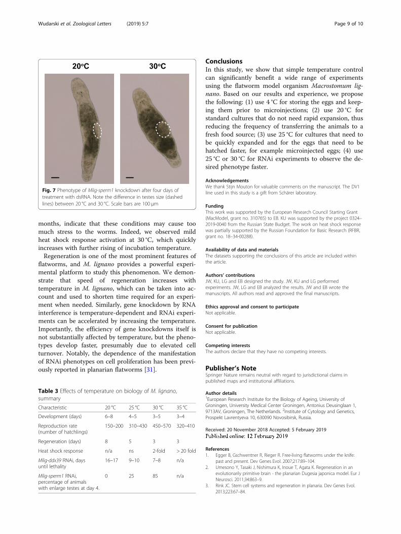
of RNAi phenotypes at elevated temperatures can begeneralized to other genes, we investigated Mlig-sperm1gene, knockdown of which leads to abnormal spermmorphology and enlarged testes [25]. This is a slowRNAi phenotype that takes 2–3 weeks to develop at 20 °C [25]. To quantify the extent of the phenotype at differ-ent temperatures, at day 4 of RNAi treatment wecounted the fraction of animals with testes enlarged tothe extent that they were touching each other. (Fig. 7).Similar to the results with Mlig-ddx39 RNAi, highertemperatures resulted in faster development of the
Fig. 5 Effects of prolonged exposure to high temperatures on morphology in M. lignano. No visible abnormalities after incubation at 20 °C or 25 °C for up to 6 months. Scale bars are 100 μm
Table 1 Effect of temperature on the speed of regeneration
Temperature Regeneration time, daysa
Testes Sperm
20 °C 6 8
25 °C 3 5
30 °C 2 3
35 °C 2 3ano variation was observed, standard deviation = 0 in all conditions. NL22transgenic line, which is based on DV1 line, was used in the experiments
Wudarski et al. Zoological Letters (2019) 5:7 Page 7 of 10
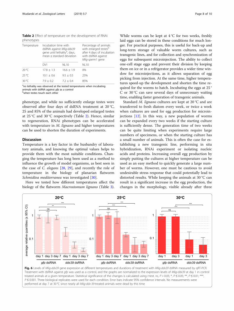
phenotype, and while no sufficiently enlarge testes wereobserved after four days of dsRNA treatment at 20 °C,25 and 85% of the animals had developed enlarged testesat 25 °C and 30 °C respectively (Table 2). Hence, similarto regeneration, RNAi phenotypes can be acceleratedwith temperature in M. lignano and higher temperaturescan be used to shorten the duration of experiments.
DiscussionTemperature is a key factor in the husbandry of labora-tory animals, and knowing the optimal values helps toprovide them with the most suitable conditions. Chan-ging the temperature has long been used as a method toinfluence the growth of model organisms, as best seen inthe case of C. elegans [28, 29], and recently the role oftemperature in the biology of planarian flatwormSchmidtea mediterranea was investigated [30].Here we tested how different temperatures affect the
biology of the flatworm Macrostomum lignano (Table 3).
While worms can be kept at 4 °C for two weeks, freshlylaid eggs can be stored in these conditions for much lon-ger. For practical purposes, this is useful for back-up andlong-term storage of valuable worm cultures, such astransgenic lines, and for collection and synchronization ofeggs for subsequent microinjection. The ability to collectone-cell stage eggs and prevent their division by keepingthem on ice or in a refrigerator provides a wider time win-dow for microinjections, as it allows separation of eggpicking from injection. At the same time, higher tempera-tures speed-up the development and shorten the time re-quired for the worms to hatch. Incubating the eggs at 25 °C or 30 °C can save several days of unnecessary waitingtime, enabling faster generation of transgenic animals.Standard M. lignano cultures are kept at 20 °C and are
transferred to fresh diatom every week, or twice a weekwhen cultures are used for egg production for microin-jections [12]. In this way, a new population of wormscan be expanded every two weeks if the starting cultureis sufficiently dense. The generation time of two weekscan be quite limiting when experiments require largenumbers of specimens, or when the starting culture hasa small number of animals. This is often the case for es-tablishing a new transgenic line, performing in situhybridization, RNAi experiment or isolating nucleicacids and proteins. Increasing overall egg production bysimply putting the cultures at higher temperature can beused as an easy method to quickly generate a large num-ber of worms. However, one must be cautious to avoidundesirable stress response that could potentially lead todistorted results. While keeping the animals at 30 °C canresult in a significant increase in the egg production, thechanges in the morphology, visible already after three
Table 2 Effect of temperature on the development of RNAiphenotypes
Temperature Incubation time withdsRNA against Mlig-ddx39gene until lethalitya, days,mean ± standard deviation
Percentage of animalswith enlarged testesb
after 4 days of incubationwith dsRNA againstMlig-sperm1 gene
DV1 NL10 NL10
20 °C 17.8 ± 1.3 16.6 ± 1.9 0%
25 °C 10.1 ± 0.6 9.5 ± 0.5 25%
30 °C 7.9 ± 0.2 7.2 ± 0.4 85%ano lethality was observed at the tested temperatures when incubatinganimals with dsRNA against gfp as a controlbwhen testes touch each other
Fig. 6 Levels of Mlig-ddx39 gene expression at different temperatures and durations of treatment with Mlig-ddx39 dsRNA measured by qRT-PCR.Treatment with dsRNA against gfp was used as a control, and the graphs are normalized to the expression levels of Mlig-ddx39 at day 1 in controltreated animals at a given temperature. Statistical significance of the changes is calculated using t-test. ns, P > 0.05; *, P ≤ 0.05; **, P ≤ 0.01; ***,P ≤ 0.001. Three biological replicates were used for each condition. Error bars indicate 95% confidence intervals. No measurements wereperformed at day 7 at 30 °C since nearly all Mlig-ddx-39-treated animals were dead by this time
Wudarski et al. Zoological Letters (2019) 5:7 Page 8 of 10
months, indicate that these conditions may cause toomuch stress to the worms. Indeed, we observed mildheat shock response activation at 30 °C, which quicklyincreases with further rising of incubation temperature.Regeneration is one of the most prominent features of
flatworms, and M. lignano provides a powerful experi-mental platform to study this phenomenon. We demon-strate that speed of regeneration increases withtemperature in M. lignano, which can be taken into ac-count and used to shorten time required for an experi-ment when needed. Similarly, gene knockdown by RNAinterference is temperature-dependent and RNAi experi-ments can be accelerated by increasing the temperature.Importantly, the efficiency of gene knockdowns itself isnot substantially affected by temperature, but the pheno-types develop faster, presumably due to elevated cellturnover. Notably, the dependence of the manifestationof RNAi phenotypes on cell proliferation has been previ-ously reported in planarian flatworms [31].
ConclusionsIn this study, we show that simple temperature controlcan significantly benefit a wide range of experimentsusing the flatworm model organism Macrostomum lig-nano. Based on our results and experience, we proposethe following: (1) use 4 °C for storing the eggs and keep-ing them prior to microinjections; (2) use 20 °C forstandard cultures that do not need rapid expansion, thusreducing the frequency of transferring the animals to afresh food source; (3) use 25 °C for cultures that need tobe quickly expanded and for the eggs that need to behatched faster, for example microinjected eggs; (4) use25 °C or 30 °C for RNAi experiments to observe the de-sired phenotype faster.
AcknowledgementsWe thank Stijn Mouton for valuable comments on the manuscript. The DV1line used in this study is a gift from Schärer laboratory.
FundingThis work was supported by the European Research Council Starting Grant(MacModel, grant no. 310765) to EB. KU was supported by the project 0324–2019-0040 from the Russian State Budget. The work on heat shock responsewas partially supported by the Russian Foundation for Basic Research (RFBR,grant no. 18–34-00288).
Availability of data and materialsThe datasets supporting the conclusions of this article are included withinthe article.
Authors’ contributionsJW, KU, LG and EB designed the study. JW, KU and LG performedexperiments. JW, LG and EB analyzed the results. JW and EB wrote themanuscripts. All authors read and approved the final manuscripts.
Ethics approval and consent to participateNot applicable.
Consent for publicationNot applicable.
Competing interestsThe authors declare that they have no competing interests.
Publisher’s NoteSpringer Nature remains neutral with regard to jurisdictional claims inpublished maps and institutional affiliations.
Author details1European Research Institute for the Biology of Ageing, University ofGroningen, University Medical Center Groningen, Antonius Deusinglaan 1,9713AV, Groningen, The Netherlands. 2Institute of Cytology and Genetics,Prospekt Lavrentyeva 10, 630090 Novosibirsk, Russia.
Received: 20 November 2018 Accepted: 5 February 2019
References1. Egger B, Gschwentner R, Rieger R. Free-living flatworms under the knife:
past and present. Dev Genes Evol. 2007;217:89–104.2. Umesono Y, Tasaki J, Nishimura K, Inoue T, Agata K. Regeneration in an
evolutionarily primitive brain - the planarian Dugesia japonica model. Eur JNeurosci. 2011;34:863–9.
3. Rink JC. Stem cell systems and regeneration in planaria. Dev Genes Evol.2013;223:67–84.
Fig. 7 Phenotype of Mlig-sperm1 knockdown after four days oftreatment with dsRNA. Note the difference in testes size (dashedlines) between 20 °C and 30 °C. Scale bars are 100 μm
Table 3 Effects of temperature on biology of M. lignano,summary
Characteristic 20 °C 25 °C 30 °C 35 °C
Development (days) 6–8 4–5 3–5 3–4
Reproduction rate(number of hatchlings)
150–200 310–430 450–570 320–410
Regeneration (days) 8 5 3 3
Heat shock response n/a ns 2-fold > 20 fold
Mlig-ddx39 RNAi, daysuntil lethality
16–17 9–10 7–8 n/a
Mlig-sperm1 RNAi,percentage of animalswith enlarge testes at day 4.
0 25 85 n/a
Wudarski et al. Zoological Letters (2019) 5:7 Page 9 of 10
4. Dattani A, Sridhar D, Aziz AA. Planarian flatworms as a new model systemfor understanding the epigenetic regulation of stem cell pluripotency anddifferentiation. Semin Cell Dev Biol. 2018;S1084–9521(17):30441–X.
5. Cebrià F, Adell T, Saló E. Rebuilding a planarian: from early signaling to finalshape. Int J Dev Biol. 2018;62:537–50.
6. Ladurner P, Schärer L, Salvenmoser W, Rieger RM. A new model organismamong the lower Bilateria and the use of digital microscopy in taxonomy ofmeiobenthic Platyhelminthes: Macrostomum lignano, n. Sp. (Rhabditophora,Macrostomorpha). J Zool Syst Evol Res. 2005;43:114–26.
7. Mouton S, Wudarski J, Grudniewska M, Berezikov E. The regenerativeflatworm Macrostomum lignano, a model organism with high experimentalpotential. Int J Dev Biol. 2018;62:551–8.
8. Egger B, Ladurner P, Nimeth K, Gschwentner R, Rieger R. The regenerationcapacity of the flatworm Macrostomum lignano - on repeated regeneration,rejuvenation, and the minimal size needed for regeneration. Dev GenesEvol. 2006;216:565–77.
9. Nimeth KT, Egger B, Rieger R, Salvenmoser W, Peter R, Gschwentner R.Regeneration in Macrostomum lignano (Platyhelminthes): cellular dynamicsin the neoblast stem cell system. Cell Tissue Res. 2007;327:637–46.
10. Wasik K, Gurtowski J, Zhou X, Ramos OM, Delás MJ, Battistoni G, et al.Genome and transcriptome of the regeneration-competent flatworm.Macrostomum lignano Proc Natl Acad Sci. 2015;112:12462–7.
11. Grudniewska M, Mouton S, Simanov D, Beltman F, Grelling M, De Mulder K,et al. Transcriptional signatures of somatic neoblasts and germline cells inMacrostomum lignano. Elife [Internet]. 2016;5:e20607.
12. Wudarski J, Simanov D, Ustyantsev K, de Mulder K, Grelling M, Grudniewska M, etal. Efficient transgenesis and annotated genome sequence of the regenerativeflatworm model Macrostomum lignano. Nat Commun. 2017;8:2120.
13. Morris J, Nallur R, Ladurner P, Egger B, Rieger R, Hartenstein V. Theembryonic development of the flatworm Macrostomum sp. Dev GenesEvol. 2004;214:220–39.
14. Schmidt-Nielsen K. Animal physiology: adaptation and environment:Cambridge University Press; 1997.
15. Stetter KO. Hyperthermophiles in the history of life. Philos Trans R Soc B BiolSci. 2006;361:1837–43.
16. Cossins A. Temperature biology of animals. Springer Science & BusinessMedia; 2012.
17. Nespolo RF. Lardies M a, Bozinovic F. Intrapopulational variation in the standardmetabolic rate of insects: repeatability, thermal dependence and sensitivity (Q10)of oxygen consumption in a cricket. J Exp Biol. 2003;206:4309–15.
18. Dunlap KD, Smith GT, Yekta A. Temperature dependence ofElectrocommunication signals and their underlying neural rhythms in theweakly electric fish. Apteronotus leptorhynchus. 2000;2000:152–62.
19. Navas CA, James RS, Wakeling JM, Kemp KM, Johnston IA. An integrativestudy of the temperature dependence of whole animal and muscleperformance during jumping and swimming in the frog Rana temporaria. JComp Physiol B. 1999;169:588–96.
20. Richter K, Haslbeck M, Buchner J. The heat shock response: life on the vergeof death. Mol Cell. 2010;40:253–66.
21. Jolly C. Role of the heat shock response and molecular chaperones inoncogenesis and cell death. J Natl Cancer Inst. 2000;92:1564–72.
22. Janicke T, Marie-Orleach L, De Mulder K, Berezikov E, Ladurner P, VizosoDBBDB, et al. Sex allocation adjustment to mating group size in asimultaneous hermaphrodite. Evolution. 2013;67:3233–42.
23. Zadesenets KS, Vizoso DB, Schlatter A, Konopatskaia ID, Berezikov E, SchärerL, et al. Evidence for karyotype polymorphism in the free-living flatworm,macrostomum lignano, a model organism for evolutionary anddevelopmental biology. PLoS One. 2016;11:e0164915.
24. Mouton S, Grudniewska M, Glazenburg L, Guryev V, Berezikov E. Resilienceto aging in the regeneration-capable flatworm Macrostomum lignano.Aging Cell. 2018:e12739.
25. Grudniewska M, Mouton S, Grelling M, Wolters AHG, Kuipers J, GiepmansBNG, et al. A novel flatworm-specific gene implicated in reproduction inMacrostomum lignano. Sci Rep. 2018;8:3192.
26. Rieger R, Gehlen M, Haszprunar G, Holmlund M, Legniti A, Salvenmoser W,et al. Laboratory cultures of marine Macrostomida (Turbellaria). FortschrZool. 1988;36.
27. De Mulder K, Pfister D, Kuales G, Egger B, Salvenmoser W, Willems M, et al.Stem cells are differentially regulated during development, regenerationand homeostasis in flatworms. Dev Biol. 2009;334:198–212.
28. Hu PJ. Dauer. WromBook. 2007;1–19.
29. Sulston J and JH. Methods, The nematode C. elegans. Wood WB, editor.Cold Spring Harbor Laboratory Press, Cold Spring Harbor, NY; 1988.
30. Hammoudi N, Torre C, Ghigo E, Drancourt M. Temperature affects thebiology of Schmidtea mediterranea. Sci Rep. 2018;8:14934.
31. Takano T, Pulvers JN, Inoue T, Tarui H, Sakamoto H, Agata K, et al.Regeneration-dependent conditional gene knockdown (Readyknock) inplanarian: demonstration of requirement for Djsnap-25 expression in thebrain for negative phototactic behavior. Dev Growth Differ [Internet]. 2007;49:383–94.
Wudarski et al. Zoological Letters (2019) 5:7 Page 10 of 10


![Influence of temperature on development, reproduction and ... · free-living marine flatworm capable of regeneration an-terior to the brain and posterior to the pharynx [8]. Similar](https://img.pdfslide.net/doc/110x75/60068708c824ef6e46780f43/influence-of-temperature-on-development-reproduction-and-free-living-marine.jpg)