Embed Size (px)
Citation preview
37
Infratemporal fossa abscess (IFA) can be misdiagnosed due to its rarity. The primary cause of IFA is an odontogenic infection originating from the mandibular molars.
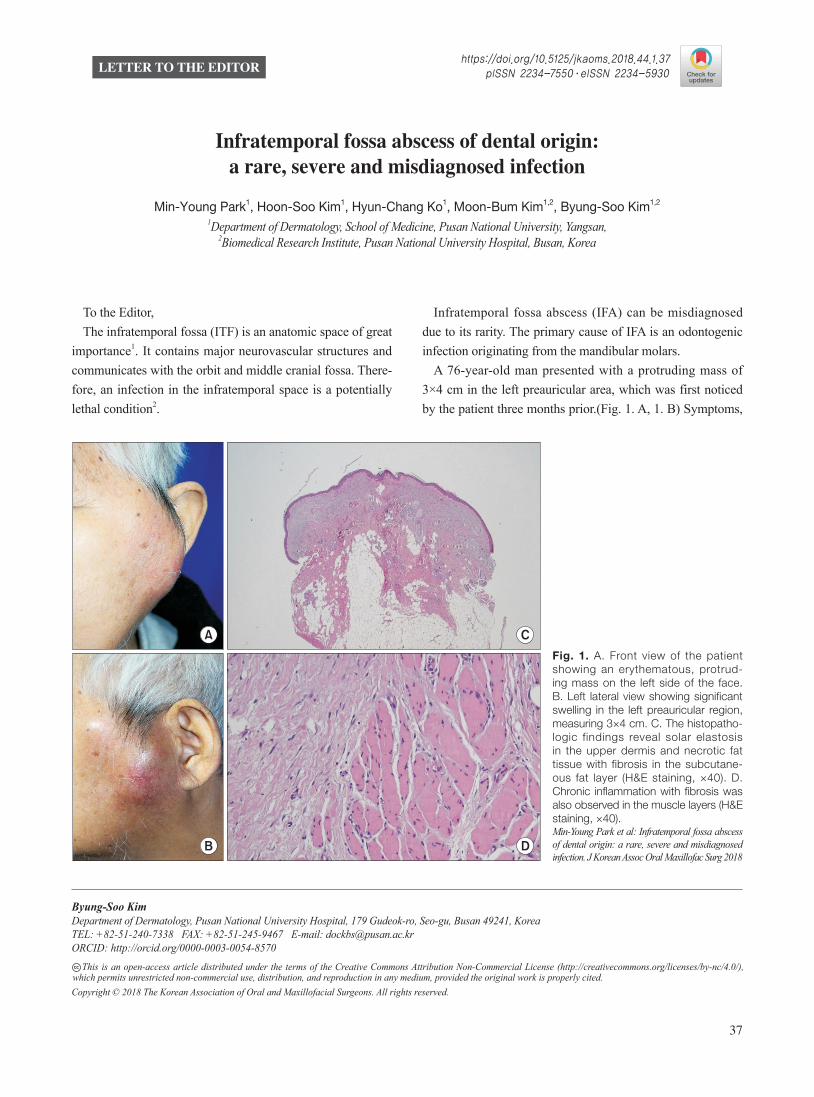
A 76-year-old man presented with a protruding mass of 3×4 cm in the left preauricular area, which was first noticed by the patient three months prior.(Fig. 1. A, 1. B) Symptoms,
To the Editor,The infratemporal fossa (ITF) is an anatomic space of great
importance1. It contains major neurovascular structures and communicates with the orbit and middle cranial fossa. There-fore, an infection in the infratemporal space is a potentially lethal condition2.
LETTER TO THE EDITOR
Byung-Soo KimDepartment of Dermatology, Pusan National University Hospital, 179 Gudeok-ro, Seo-gu, Busan 49241, KoreaTEL: +82-51-240-7338 FAX: +82-51-245-9467 E-mail: [email protected]: http://orcid.org/0000-0003-0054-8570 This is an open-access article distributed under the terms of the Creative Commons Attribution Non-Commercial License (http://creativecommons.org/licenses/by-nc/4.0/), which permits unrestricted non-commercial use, distribution, and reproduction in any medium, provided the original work is properly cited.CC
Infratemporal fossa abscess of dental origin: a rare, severe and misdiagnosed infection
Min-Young Park1, Hoon-Soo Kim1, Hyun-Chang Ko1, Moon-Bum Kim1,2, Byung-Soo Kim1,2
1Department of Dermatology, School of Medicine, Pusan National University, Yangsan, 2Biomedical Research Institute, Pusan National University Hospital, Busan, Korea
Copyright © 2018 The Korean Association of Oral and Maxillofacial Surgeons. All rights reserved.
https://doi.org/10.5125/jkaoms.2018.44.1.37pISSN 2234-7550·eISSN 2234-5930
A
B
C
D
Fig. 1. A. Front view of the patient showing an erythematous, protrud-ing mass on the left side of the face. B. Left lateral view showing significant swelling in the left preauricular region, measuring 3×4 cm. C. The histopatho-logic findings reveal solar elastosis in the upper dermis and necrotic fat tissue with fibrosis in the subcutane-ous fat layer (H&E staining, ×40). D. Chronic inflammation with fibrosis was also observed in the muscle layers (H&E staining, ×40).Min-Young Park et al: Infratemporal fossa abscess of dental origin: a rare, severe and misdiagnosed infection. J Korean Assoc Oral Maxillofac Surg 2018
J Korean Assoc Oral Maxillofac Surg 2018;44:37-39
38
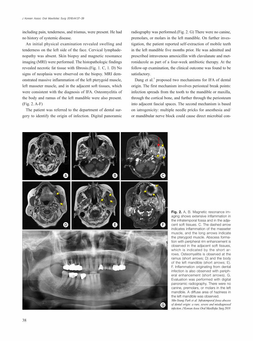
radiography was performed.(Fig. 2. G) There were no canine, premolars, or molars in the left mandible. On further inves-tigation, the patient reported self-extraction of mobile teeth in the left mandible five months prior. He was admitted and prescribed intravenous amoxicillin with clavulanate and met-ronidazole as part of a four-week antibiotic therapy. At the follow-up examination, the clinical outcome was found to be satisfactory.
Dang et al.3 proposed two mechanisms for IFA of dental origin. The first mechanism involves periosteal break points: infection spreads from the tooth to the mandible or maxilla, through the cortical bone, and further through the periosteum into adjacent fascial spaces. The second mechanism is based on iatrogenicity: multiple needle pricks for anesthesia and/or mandibular nerve block could cause direct microbial con-
including pain, tenderness, and trismus, were present. He had no history of systemic disease.
An initial physical examination revealed swelling and tenderness on the left side of the face. Cervical lymphade-nopathy was absent. Skin biopsy and magnetic resonance imaging (MRI) were performed. The histopathologic findings revealed necrotic fat tissue with fibrosis.(Fig. 1. C, 1. D) No signs of neoplasia were observed on the biopsy. MRI dem-onstrated massive inflammation of the left pterygoid muscle, left masseter muscle, and in the adjacent soft tissues, which were consistent with the diagnosis of IFA. Osteomyelitis of the body and ramus of the left mandible were also present.(Fig. 2. A-F)
The patient was referred to the department of dental sur-gery to identify the origin of infection. Digital panoramic
A B C
D E F
G
Fig. 2. A, B. Magnetic resonance im-aging shows extensive inflammation in the infratemporal fossa and in the adja-cent soft tissues. C. The dashed arrow indicates inflammation of the masseter muscle, and the long arrows indicate the pterygoid muscle. Abscess forma-tion with peripheral rim enhancement is observed in the adjacent soft tissues, which is indicated by the short ar-rows. Osteomyelitis is observed at the ramus (short arrows; D) and the body of the left mandible (short arrows; E). F. Inflammation originating from dental infection is also observed with periph-eral enhancement (short arrows). G. Evaluation was performed with digital panoramic radiography. There were no canine, premolars, or molars in the left mandible. A diffuse area of haziness in the left mandible was observed.Min-Young Park et al: Infratemporal fossa abscess of dental origin: a rare, severe and misdiagnosed infection. J Korean Assoc Oral Maxillofac Surg 2018
Infratemporal fossa abscess of dental origin
39
tamination of the ITF. IFA is rare and its clinical diagnosis tends to be challeng-
ing due to its non-specific symptoms. The diagnosis of IFA mainly relies on a high index of suspicion.
Plain radiographs, including panoramic views, may identi-fy the source of infection and soft tissue swelling. Computed tomography or MRI usually yield diagnostic findings; how-ever, it may be difficult to distinguish between an abscess and a tumour4. In agreement with most reported cases, microbial culture did not result in isolation of a particular organism. In addition to the polymicrobial nature of odontogenic infec-tions, this observation might be attributed to the use of sever-al antibiotics that were prescribed before the final diagnosis5.
IFA has potentially fatal complications. The infection might spread through the pterygoid plexus to the cavernous sinus or through the valveless ophthalmic veins into the orbit, which may result in intracranial and orbital complications. Thus, en-suring minimal treatment time is crucial for the diagnosis and successful management of IFA.
Here, we report a rare case of IFA secondary to odontogen-ic infection. A thorough medical and dental history, as well as oral, facial, and systemic examination, is required. This infor-mation, together with radiographic and histopathologic find-ings and knowledge of the anatomical structures involved, can aid timely diagnosis and appropriate management of ITF infections.
ORCID
Min-Young Park, http://orcid.org/0000-0002-4880-8514Hoon-Soo Kim, http://orcid.org/0000-0002-5378-6292Hyun-Chang Ko, http://orcid.org/0000-0002-6055-4190Moon-Bum Kim, http://orcid.org/0000-0001-7032-2548Byung-Soo Kim, http://orcid.org/0000-0003-0054-8570
Authors’ Contributions
M.Y.P. and B.S.K. participated in data collection and wrote the manuscript. M.Y.P., H.S.K., H.C.K., and M.B.K. partici-pated in the study design and performed the statistical analy-sis. M.Y.P. and B.S.K. participated in the study design and coordination and helped to draft the manuscript. All authors read and approved the final manuscript.
Consent for Publishing Photographs
Written informed consent was obtained from the patients for publication of this letter and accompanying images.
Conflict of Interest
No potential conflict of interest relevant to this article was reported.
References
1. Miloro M, Ghali GE, Larsen PE, Waite PD. Peterson’s principles of oral and maxillofacial surgery. 5th ed. Hamilton: BC Decker; 2008:318-24.
2. Zhang C, Tang Y, Zheng M, Yang J, Zhu G, Zhou H, et al. Maxil-lofacial space infection experience in West China: a retrospective study of 212 cases. Int J Infect Dis 2010;14:e414-7.
3. Dang NP, Barthélémy I, Pavier Y, Picard M, Delbet-Dupas C. In-fratemporal fossa abscess of dental origin: a rare, severe, and mis-diagnosed infection. J Craniofac Surg 2016;27:e221-2.
4. Diacono MS, Wass AR. Infratemporal and temporal fossa abscess complicating dental extraction. J Accid Emerg Med 1998;15:59-61.
5. Huang TT, Tseng FY, Liu TC, Hsu CJ, Chen YS. Deep neck in-fection in diabetic patients: comparison of clinical picture and outcomes with nondiabetic patients. Otolaryngol Head Neck Surg 2005;132:943-7.