Embed Size (px)
Citation preview

Ast
hm
adia
gnosi
sand
treatm
ent
Inhaled steroids are associated with reducedlung function decline in subjects with asthmawith elevated total IgE
Roberto de Marco, PhD,a Alessandro Marcon, MSc,a Deborah Jarvis, FFPHM,b Simone
Accordini, MSc,a Massimiliano Bugiani, MD,c Lucia Cazzoletti, MSc,a Isa Cerveri, MD,d
Angelo Corsico, MD, PhD,d David Gislason, MD,e Amund Gulsvik, MD, PhD,f Rain Jogi,
PhD,g Jesus Martınez-Moratalla, MD,h Isabelle Pin, MD,i and Christer Janson, MD,j
on behalf of the ECRHS Therapy Group Verona, Turin, and Pavia, Italy, London, United
Kingdom, Reykjavik, Iceland, Bergen, Norway, Tartu, Estonia, Albacete, Spain, Grenoble, France,
and Uppsala, Sweden
611
Background: Few studies have investigated the long-term
association between inhaled corticosteroids (ICSs) and lung
function decline in asthma.
Objective: To evaluate whether prolonged treatment with ICSs
is associated with FEV1 decline in adults with asthma.
Methods: An international cohort of 667 subjects with asthma
(20-44 years old) was identified in the European Community
Respiratory Health Survey (1991-1993) and followed up from
1999 to 2002. Spirometry was performed on both occasions.
FEV1 decline was analyzed according to age, sex, height, body
mass index, total IgE, time of ICS use, and smoking, while
adjusting for potential confounders.
Results: As ICS use increased, the decline in FEV1 was lower
(P trend 5 .025): on average, decline passed from 34 mL/y in
nonusers (half of the sample) to 20 mL/y in subjects treated for
48 months or more (18%). When adjusting for all covariates,
there was an interaction (P 5 .02) between ICS use and total
IgE: in subjects with high (>100 kU/L) IgE, ICS use for 4 years
or more was associated with a lower FEV1 decline (23 mL/y;
From athe University of Verona, Department of Medicine and Public Health,
Unit of Epidemiology and Medical Statistics; bthe Respiratory Epidemiol-
ogy and Public Health Group, National Heart and Lung Institute, Imperial
College, London; cthe Unit of Pneumology, Consorzio Provinciale Antitu-
bercolare, Azienda Sanitaria Locale 4 Piemonte, Turin; dthe Division of
Respiratory Diseases, Istituto di Ricovero e Cura a Carattere Scientifico
San Matteo Hospital, University of Pavia; ethe Department of Allergy, Res-
piratory Medicine and Sleep, Landspitali University Hospital, Reykjavik;fthe Department of Thoracic Medicine, Haukeland University Hospital,
University of Bergen; gTartu University Hospital, Clinics, Lung Clinic;hServicio de Neumologıa del Complejo Hospitalario Universitario de Alba-
cete, Servicio de Salud de Castilla-La Mancha; ithe Department of Pediatrics
and Institut National de la Sante et de la Recherche Medicale U578, Centre
Hospitalier Universitarie de Grenoble; and jthe Department of Medical Sci-
ences, Respiratory Medicine and Allergology, Uppsala University.
The coordination of the ECRHS II was supported by the European Commission
as part of their Quality of Life program.
Disclosure of potential conflict of interest: The authors have declared that they
have no conflict of interest.
Received for publication August 1, 2006; revised November 3, 2006; accepted
for publication November 6, 2006.
Available online January 31, 2007.
Reprint requests: Roberto de Marco, PhD, Unit of Epidemiology and Medical
Statistics, Department of Medicine and Public Health, Universita degli Studi
di Verona, c/o Istituti Biologici II, Strada Le Grazie 8, 37134 Verona, Italy.
E-mail: [email protected].
0091-6749/$32.00
� 2007 American Academy of Allergy, Asthma & Immunology
doi:10.1016/j.jaci.2006.11.696
95% CI, 8-38 compared with nonusers). This association
was not seen in those with lower IgE.
Conclusion: Although confirming a beneficial long-term
association between ICSs and lung function in asthma, our
study suggests that subjects with high IgE could maximally
benefit from a prolonged ICS treatment.
Clinical implications: This study adds further evidence to the
beneficial effect of inhaled steroids on lung function in asthma;
future studies will clarify whether calibrating the corticosteroid
dose according to the level of total IgE is a feasible approach
in asthma management. (J Allergy Clin Immunol
2007;119:611-7.)
Key words: Asthma, lung function decline, inhaled corticosteroids,
total IgE, eosinophils, prospective cohort study, FEV1 decline,
European Community Respiratory Health Survey, ECRHS
In patients with asthma, the decline in lung function isaccelerated compared with subjects without asthma1 and,in more severe asthma, it may result in an irreversible air-flow obstruction.2,3 The rapid decline in lung function andthe airflow obstruction may stem from structural changes(airway remodeling) or functional changes that may ormay not be associated with the underlying chronic inflam-mation.4-8
Inhaled corticosteroids (ICSs) are the mainstay ofasthma treatment,9 and many short-term clinical trialshave demonstrated their efficacy in decreasing airway in-flammation, improving lung function, and reducing symp-toms and airway hyperresponsiveness.10-13 However,there is limited evidence that ICSs are able to slowdown the progressive loss of lung function over time inasthma, and also that ICSs can prevent or revert structuralchanges in the lungs.14-16 This is mainly because of thedifficulty in following up patients in clinical trials overmany years. Therefore, observational studies make animportant contribution to assess the effectiveness of thelong-term use of ICSs.
Recently, 2 observational follow-up studies have beenpublished,17,18 both reporting a beneficial effect of thelong-term use of ICSs on lung function decline. Langeet al18 reported that treatment with ICSs was associatedwith an 18 mL/y reduction in the rate of decline in FEV1
compared with no use of ICSs, and Dijkstra et al17 found
J ALLERGY CLIN IMMUNOL
MARCH 2007
612 de Marco et al
Asth
ma
dia
gnosis
and
treatm
ent
Abbreviations usedATS: American Thoracic Society
BMI: Body mass index
ECRHS: European Community Respiratory Health Survey
ED: Emergency department
ICS: Inhaled corticosteroid
IQR: Interquartile range
LABA: Long-acting b2-agonist
a similar effect only in nonsmoking men. However, bothsurveys investigated relatively small samples of selectedindividuals.
The aim of this study was to evaluate whetherprolonged use of ICSs is associated with a reduced long-term decline in lung function in a large, international,population-based cohort of subjects with asthma, followedup for 9 years in the European Community RespiratoryHealth Survey (ECRHS).
METHODS
Study design
The ECRHS is an international multicenter study of asthma. The
first survey19 was performed from 1991 to 1993 on random commu-
nity-based samples of adults age 20 to 44 years. Each participant was
sent a brief questionnaire (stage 1) and, from those who responded, a
20% random sample was invited to undergo a more detailed clinical
examination (stage 2). In addition, a symptomatic sample consisting
of those who reported symptoms of waking with shortness of breath
or asthma attacks in the last 12 months or who were using asthma
medication in stage 1 was also studied. The ECRHS II20 was a fol-
low-up study of all participants in stage 2 of the ECRHS I, performed
from 1999 to 2002 (the full protocol can be found at www.ecrhs.org).
Subjects answered a standardized questionnaire administered by
trained interviewers and underwent lung function and blood tests.
Quality control procedures are fully described in the study proto-
cols.19,20 The current study includes data from 26 centers that took
part in the ECRHS II. Ethical approval was obtained for each center
from the appropriate institutional or regional ethics committee, and
written consent was obtained for each participant.
Subjects and definitions
All individuals with current asthma identified in the ECRHS I
(1991-1993) who had performed spirometry according to the
American Thoracic Society (ATS) criteria for reproducibility21 and
who participated in the ECRHS II were eligible for this analysis.
Current asthma was defined as having reported, in the ECRHS I,
asthma confirmed by a doctor and having had asthmalike symptoms
(wheeze; nocturnal chest tightness; attacks of breathlessness after
activity, at rest, or at nighttime; asthma attacks), and/or having
used inhaled/oral medicines because of breathing problems during
the last 12 months.
In the ECRHS I, 1348 subjects with current asthma had their lung
function measured according to the ATS criteria (700 from the
random and 648 from the symptomatic sample). Although there were
no differences in age, sex, smoking habits, FEV1, and IgE levels at
baseline (ECRHS I) in random and symptomatic subjects, the former
had a slightly longer duration of the disease (17.8 vs 16.2 years) and a
lower percentage of manual workers (25% vs 35%) and of people re-
porting exposure to vapors, gas, dust, or fumes in the workplace (44%
vs 50%). Of these 1348 individuals, 860 (64%) attended the second
study (1999-2002) and were therefore eligible for the analysis.
In the ECRHS II (1999-2002), some eligible subjects did not
repeat spirometry (135 subjects) or had their lung function measured
in disagreement with the ATS criteria (32 subjects) and were
excluded from the analysis. Among those who had performed the
lung function test, some had used inhaled long-acting b2-agonists
(LABAs) in the 12 hours before the test. Because the bronchodilating
effect of LABAs can persist for 8 to 12 hours after use, these subjects
were also excluded (26 subjects). Finally, 667 subjects were included
in the analysis (311 from the random and 356 from the symptomatic
sample).
Decline in lung function
The maximum FEV1 of as many as 5 technically acceptable blows
was recorded, both at baseline and at the end of the follow-up, accord-
ing to the ATS criteria for reproducibility.21 The predicted value
of FEV1 was calculated on the basis of sex, age, and height,22 and
the FEV1 % predicted was obtained (100 * measured FEV1/predicted
FEV1).
For each subject, the average change in lung function during the
follow-up (in mL/y) was computed as the difference between FEV1
measured in the ECRHS I and II, divided by the individual duration
of the follow-up (ie, a positive value represents decline).
Clinical and questionnaire data
In both surveys, the height, weight, and serum total IgE level of the
participants were measured. For each subject, detailed information
was collected by questionnaire about sex, age, smoking habits
(smoking status, number of cigarettes smoked per day, age at which
they started, age at which they gave up if they did), occupation,
exposure to occupational risk (if a subject had ever been exposed to
vapors, dust, gas, or fumes in the working environment), age at first
asthma attack, family asthma (if a subject reported that his/her mother
or father had ever had asthma), and hospitalizations and/or emer-
gency department (ED) visits (if a subject had ever spent 1 night in the
hospital, and/or visited a casualty department or ED) because of
breathing problems.
Asthma duration was estimated as the difference between the age
of the subject at the ECRHS I interview and the age when the first
asthma attack occurred. Body mass index (BMI) was computed
dividing weight by height squared (kg/m2). Lifetime pack-years were
calculated combining information on smoking habits obtained in the
ECRHS I and II.23
At baseline, subjects performed a methacholine challenge test24;
however, bronchial hyperresponsiveness was not considered in the
main analysis because about 20% of the subjects had not performed
the challenge test, and the exclusion criteria for the test were associ-
ated with baseline FEV1 (eg, FEV1 < 70% predicted, FEV1 < 1.5 L).
Use of inhaled steroids
In both surveys, the participants were asked whether they had used
ICSs in the last 12 months, and the type/brand of steroid and the type
of inhaler employed over the last year were recorded. In the ECRHS
II, quantitative information was collected about ICS use during the
follow-up (how many months per year, how many years since the last
survey a subject had been on ICSs). The data on ICSs were combined
to calculate the cumulative time of treatment during the follow-up.
Subjects with asthma were stratified according to the time of steroid
use into (1) nonusers, (2) people who had used ICSs for <8.7 months
(1st tertile of time of ICS use distribution among users), (3) �8.7
months but <48 months (2nd tertile), and (4) �48 months.
J ALLERGY CLIN IMMUNOL
VOLUME 119, NUMBER 3
de Marco et al 613
Ast
hm
adia
gnosi
sand
treatm
ent
Statistics
Data were summarized as percentages or means (with SDs), with
95% CIs. Median with interquartile range (IQR) was used for
asymmetrical variables. x2 Test, Student t test, ANOVA, and Wil-
coxon test were used to test differences, where appropriate, using a
significance level of .05.
To assess the association between FEV1 decline (dependent vari-
able) and the cumulative time of ICS use, a 2-level random intercept
regression model was fitted to the data, with level 1 units (subjects)
nested into level 2 units (centers crossed by type of sample: random
or symptomatic). The independent variables included in the model
were sex, age, height, baseline BMI, and smoking habits (lifetime
pack-years); the model also adjusted for a set of potential confounders
measured at baseline—that is, occupation (in a manual job, nonman-
ual job, or other), occupational risk, duration of asthma, family
asthma, total IgE, and previous hospitalizations/ED visits for breath-
ing problems. When evaluating the interaction between time of ICS
use and total IgE, the latter was used as a dichotomous variable
(�100 kU/L; >100 kU/L).25
The statistical analysis was performed using STATA software,
release 8.2 (Stata Corp, College Station, Tex).
RESULTS
Comparison between includedand excluded subjects
Table I describes the main baseline features of the sub-jects included and not included in the analysis. The latterare either nonparticipants in the ECRHS II, or participantswhose FEV1 was unavailable, not in agreement with theATS criteria, or measured within 12 hours after the last ad-ministration of a LABA. Subjects included in the analysiswere slightly older and had a lower percentage of currentsmokers (30% vs 39%) and of manual workers than sub-jects who were excluded. On average, among the individ-uals included in the analysis, the follow-up time was 9years (range, 6-11 years).
TABLE I. Baseline (ECRHS I) characteristics of excluded
and included subjects; data provided as means (SDs)
or percentages (%) unless stated otherwise*
Excluded Included P value
Subjects (N) 681 667 —
Sex (% female) 54.6 55.0 .88
Age (y) 32.3 (7.2) 33.9 (7.2) .0001
BMI (kg/m2) 23.9 (4.3) 24.2 (4.5) .31
Smoking habits* (pack-years) 2.0 (10.0) 0.5 (9.0) .046
Occupation
(% nonmanual) 36.4 48.0 <.0001
(% manual) 31.3 29.1
(% other) 32.3 22.9
Occupational exposure (%) 46.6 47.6 .71
Duration of asthma* (y) 16.2 (16.5) 15.6 (19.1) .60
Family asthma (%) 28.5 26.2 .36
Total IgE (log transformation) 1.98 (0.71) 1.91 (0.68) .053
Hospitalizations/ED visits (%) 36.3 35.9 .88
ICS� (% users) 41.6 43.4 .60
FEV1 (L) 3.4 (0.9) 3.4 (0.8) .50
*Median (interquartile range); P values of nonparametric test reported.
�Over the period of the last 12 months.
Use of inhaled steroids
The cumulative time of ICS use could not be evaluatedfor 31 individuals (<5%) because some data were missing.Out of the remaining 636 subjects, 297 (47%) had neverbeen on ICSs, whereas 339 (53%) had used ICSs duringthe follow-up. Among ICS users, the median cumulativetime of treatment was 1.4 (IQR, 6.3) years, with nosignificant difference between men and women (P 5 .33).Among people who had used ICSs for �4 years, themedian time of use was 8.2 (IQR, 2.0) years.
The steroid that was most commonly used duringthe year before the follow-up visit (ECRHS II) was bude-sonide (51%), followed by beclomethasone (27%), andfluticasone (20%); the majority of subjects used dry-powder inhalers (65%), 1/3 used metered-dose inhalers,and only 1% used nebulizers.
Characteristics of steroid usersand nonsteroid users
At baseline, on average, subjects with asthma treatedwith ICSs during the follow-up (Table II) had a worse lungfunction, higher levels of total IgE, a shorter duration ofasthma, and a greater prevalence of family asthma andof hospitalizations/ED visits than nonsteroid users.Steroid users were more likely to be women and had ahigher baseline BMI than nonsteroid users, and they also
TABLE II. Baseline (ECRHS I) characteristics (and lifetime
pack-years smoked) of the subjects with asthma, stratified
according to the use of inhaled corticosteroids during the
follow-up; data provided as means (SDs) or percentages (%)
unless stated otherwise*
ICSs
Nonusers Users P value
Subjects, N (%) 297 (46.7) 339 (53.3) —
Sex (% female) 45.4 63.7 <.001
Age (y) 33.7 (7.1) 34.1 (7.3) .44
BMI (kg/m2) 23.6 (3.6) 24.5 (4.9) .001
Smoking habits* (lifetime
pack-years)
1.6 (15.6) 0 (10.0) .013
Occupation
(% nonmanual) 47.5 49.0 .48
(% manual) 28.0 30.4
(% other) 24.5 20.6
Occupational exposure (%) 47.6 48.4 .85
Duration of asthma* (y) 18.7 (19.1) 13.5 (17.9) .001
Family asthma (%) 19.3 31.1 .001
Total IgE� (log
transformation)
1.84 (0.66) 1.96 (0.68) .033
Hospitalizations/ED
visits (%)
28.6 42.9 <.0001
FEV1 (L) 3.7 (0.8) 3.2 (0.8) <.0001
FEV1 % predicted� 101.6 (12.6) 95.0 (17.9) <.0001
*Median (interquartile range); P values of nonparametric test reported.
�Predicted values of FEV1 were calculated on the basis of sex, age,
and height.
�Corresponding to a geometric mean of 69.6 kU/L in nonusers and of
91.9 kU/L in users.
J ALLERGY CLIN IMMUNOL
MARCH 2007
614 de Marco et al
Asth
ma
dia
gnosis
and
treatm
ent
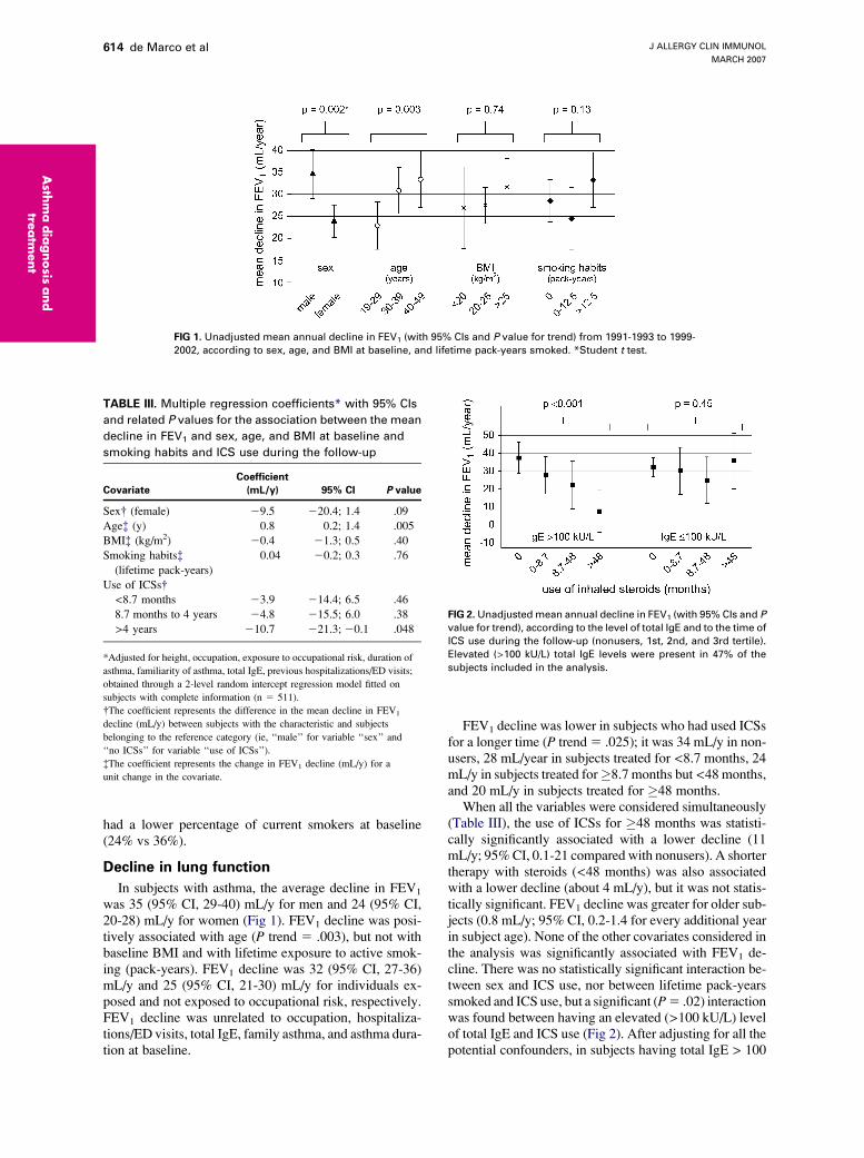
FIG 1. Unadjusted mean annual decline in FEV1 (with 95% CIs and P value for trend) from 1991-1993 to 1999-
2002, according to sex, age, and BMI at baseline, and lifetime pack-years smoked. *Student t test.
had a lower percentage of current smokers at baseline(24% vs 36%).
Decline in lung function
In subjects with asthma, the average decline in FEV1
was 35 (95% CI, 29-40) mL/y for men and 24 (95% CI,20-28) mL/y for women (Fig 1). FEV1 decline was posi-tively associated with age (P trend 5 .003), but not withbaseline BMI and with lifetime exposure to active smok-ing (pack-years). FEV1 decline was 32 (95% CI, 27-36)mL/y and 25 (95% CI, 21-30) mL/y for individuals ex-posed and not exposed to occupational risk, respectively.FEV1 decline was unrelated to occupation, hospitaliza-tions/ED visits, total IgE, family asthma, and asthma dura-tion at baseline.
TABLE III. Multiple regression coefficients* with 95% CIs
and related P values for the association between the mean
decline in FEV1 and sex, age, and BMI at baseline and
smoking habits and ICS use during the follow-up
Covariate
Coefficient
(mL/y) 95% CI P value
Sex� (female) 29.5 220.4; 1.4 .09
Age� (y) 0.8 0.2; 1.4 .005
BMI� (kg/m2) 20.4 21.3; 0.5 .40
Smoking habits�(lifetime pack-years)
0.04 20.2; 0.3 .76
Use of ICSs�<8.7 months 23.9 214.4; 6.5 .46
8.7 months to 4 years 24.8 215.5; 6.0 .38
>4 years 210.7 221.3; 20.1 .048
*Adjusted for height, occupation, exposure to occupational risk, duration of
asthma, familiarity of asthma, total IgE, previous hospitalizations/ED visits;
obtained through a 2-level random intercept regression model fitted on
subjects with complete information (n 5 511).
�The coefficient represents the difference in the mean decline in FEV1
decline (mL/y) between subjects with the characteristic and subjects
belonging to the reference category (ie, ‘‘male’’ for variable ‘‘sex’’ and
‘‘no ICSs’’ for variable ‘‘use of ICSs’’).
�The coefficient represents the change in FEV1 decline (mL/y) for a
unit change in the covariate.
FEV1 decline was lower in subjects who had used ICSsfor a longer time (P trend 5 .025); it was 34 mL/y in non-users, 28 mL/year in subjects treated for <8.7 months, 24mL/y in subjects treated for�8.7 months but <48 months,and 20 mL/y in subjects treated for �48 months.
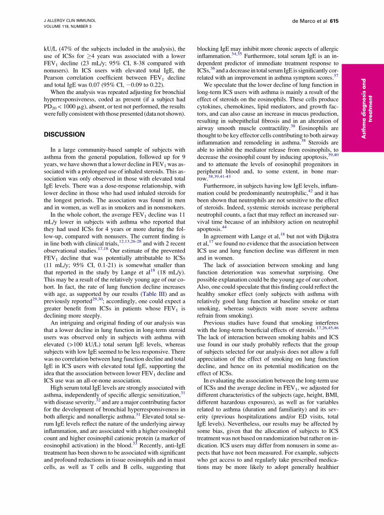
When all the variables were considered simultaneously(Table III), the use of ICSs for �48 months was statisti-cally significantly associated with a lower decline (11mL/y; 95% CI, 0.1-21 compared with nonusers). A shortertherapy with steroids (<48 months) was also associatedwith a lower decline (about 4 mL/y), but it was not statis-tically significant. FEV1 decline was greater for older sub-jects (0.8 mL/y; 95% CI, 0.2-1.4 for every additional yearin subject age). None of the other covariates considered inthe analysis was significantly associated with FEV1 de-cline. There was no statistically significant interaction be-tween sex and ICS use, nor between lifetime pack-yearssmoked and ICS use, but a significant (P 5 .02) interactionwas found between having an elevated (>100 kU/L) levelof total IgE and ICS use (Fig 2). After adjusting for all thepotential confounders, in subjects having total IgE > 100
FIG 2. Unadjusted mean annual decline in FEV1 (with 95% CIs and P
value for trend), according to the level of total IgE and to the time of
ICS use during the follow-up (nonusers, 1st, 2nd, and 3rd tertile).
Elevated (>100 kU/L) total IgE levels were present in 47% of the
subjects included in the analysis.
J ALLERGY CLIN IMMUNOL
VOLUME 119, NUMBER 3
de Marco et al 615
Ast
hm
adia
gnosi
sand
treatm
ent
kU/L (47% of the subjects included in the analysis), theuse of ICSs for �4 years was associated with a lowerFEV1 decline (23 mL/y; 95% CI, 8-38 compared withnonusers). In ICS users with elevated total IgE, thePearson correlation coefficient between FEV1 declineand total IgE was 0.07 (95% CI, 20.09 to 0.22).
When the analysis was repeated adjusting for bronchialhyperresponsiveness, coded as present (if a subject hadPD20 < 1000 mg), absent, or test not performed, the resultswere fully consistent with those presented (data not shown).
DISCUSSION
In a large community-based sample of subjects withasthma from the general population, followed up for 9years, we have shown that a lower decline in FEV1 was as-sociated with a prolonged use of inhaled steroids. This as-sociation was only observed in those with elevated totalIgE levels. There was a dose-response relationship, withlower decline in those who had used inhaled steroids forthe longest periods. The association was found in menand in women, as well as in smokers and in nonsmokers.
In the whole cohort, the average FEV1 decline was 11mL/y lower in subjects with asthma who reported thatthey had used ICSs for 4 years or more during the fol-low-up, compared with nonusers. The current finding isin line both with clinical trials,12,13,26-28 and with 2 recentobservational studies.17,18 Our estimate of the preventedFEV1 decline that was potentially attributable to ICSs(11 mL/y; 95% CI, 0.1-21) is somewhat smaller thanthat reported in the study by Lange et al18 (18 mL/y).This may be a result of the relatively young age of our co-hort. In fact, the rate of lung function decline increaseswith age, as supported by our results (Table III) and aspreviously reported29,30; accordingly, one could expect agreater benefit from ICSs in patients whose FEV1 isdeclining more steeply.
An intriguing and original finding of our analysis wasthat a lower decline in lung function in long-term steroidusers was observed only in subjects with asthma withelevated (>100 kU/L) total serum IgE levels, whereassubjects with low IgE seemed to be less responsive. Therewas no correlation between lung function decline and totalIgE in ICS users with elevated total IgE, supporting theidea that the association between lower FEV1 decline andICS use was an all-or-none association.
High serum total IgE levels are strongly associated withasthma, independently of specific allergic sensitization,31
with disease severity,32 and are a major contributing factorfor the development of bronchial hyperresponsiveness inboth allergic and nonallergic asthma.31 Elevated total se-rum IgE levels reflect the nature of the underlying airwayinflammation, and are associated with a higher eosinophilcount and higher eosinophil cationic protein (a marker ofeosinophil activation) in the blood.33 Recently, anti-IgEtreatment has been shown to be associated with significantand profound reductions in tissue eosinophils and in mastcells, as well as T cells and B cells, suggesting that
blocking IgE may inhibit more chronic aspects of allergicinflammation.34,35 Furthermore, total serum IgE is an in-dependent predictor of immediate treatment response toICSs,36 and a decrease in total serum IgE is significantly cor-related with an improvement in asthma symptom scores.37
We speculate that the lower decline of lung function inlong-term ICS users with asthma is mainly a result of theeffect of steroids on the eosinophils. These cells producecytokines, chemokines, lipid mediators, and growth fac-tors, and can also cause an increase in mucus production,resulting in subepithelial fibrosis and in an alteration ofairway smooth muscle contractility.38 Eosinophils arethought to be key effector cells contributing to both airwayinflammation and remodeling in asthma.38 Steroids areable to inhibit the mediator release from eosinophils, todecrease the eosinophil count by inducing apoptosis,39,40
and to attenuate the levels of eosinophil progenitors inperipheral blood and, to some extent, in bone mar-row.38,39,41-43
Furthermore, in subjects having low IgE levels, inflam-mation could be predominantly neutrophilic,42 and it hasbeen shown that neutrophils are not sensitive to the effectof steroids. Indeed, systemic steroids increase peripheralneutrophil counts, a fact that may reflect an increased sur-vival time because of an inhibitory action on neutrophilapoptosis.44
In agreement with Lange et al,18 but not with Dijkstraet al,17 we found no evidence that the association betweenICS use and lung function decline was different in menand in women.
The lack of association between smoking and lungfunction deterioration was somewhat surprising. Onepossible explanation could be the young age of our cohort.Also, one could speculate that this finding could reflect thehealthy smoker effect (only subjects with asthma withrelatively good lung function at baseline smoke or startsmoking, whereas subjects with more severe asthmarefrain from smoking).
Previous studies have found that smoking interfereswith the long-term beneficial effects of steroids.17,26,45,46
The lack of interaction between smoking habits and ICSuse found in our study probably reflects that the groupof subjects selected for our analysis does not allow a fullappreciation of the effect of smoking on lung functiondecline, and hence on its potential modification on theeffect of ICSs.
In evaluating the association between the long-term useof ICSs and the average decline in FEV1, we adjusted fordifferent characteristics of the subjects (age, height, BMI,different hazardous exposures), as well as for variablesrelated to asthma (duration and familiarity) and its sev-erity (previous hospitalizations and/or ED visits, totalIgE levels). Nevertheless, our results may be affected bysome bias, given that the allocation of subjects to ICStreatment was not based on randomization but rather on in-dication. ICS users may differ from nonusers in some as-pects that have not been measured. For example, subjectswho get access to and regularly take prescribed medica-tions may be more likely to adopt generally healthier
J ALLERGY CLIN IMMUNOL
MARCH 2007
616 de Marco et al
Asth
ma
dia
gnosis
and
treatm
ent
lifestyles (such as healthy diet and exercise). High dietaryintake of antioxidants has been associated with lung func-tion,47 but we have no information on this for our sample.On the other hand, one might expect subjects with moresevere disease to be prescribed more medication. At base-line, the treated group had poorer lung function, higherrates of hospitalization for respiratory diseases, and higherprevalence of family asthma than the untreated group.Therefore, we might expect them to have a steeper de-crease in lung function than subjects who did not receivetreatment (the horse-racing effect).48,49 If this is true, wehave underestimated the association between ICS useand FEV1 decline.
A potential limitation of the current study is that a self-reported doctor diagnosis of asthma was used to identifyindividuals with asthma. Although this definition may beopen to some degree of misclassification, it has beenproven to be highly specific,50 so that only milder or undi-agnosed asthmatics would not have been included in ourstudy.
Compared with other recent longitudinal studies inves-tigating the association between ICSs and lung functiondecline,17,18 our study has some points of strength. First,the ECRHS sample was selected from the general pop-ulation in an international setting. Second, in the currentstudy, a cumulative time of ICS use was estimated foreach individual in the follow-up period. Finally, the indi-viduals who had used inhaled LABAs in the 12 hours be-fore lung function testing were excluded, thus reducing therisk of a bias because of the residual bronchodilating effectof these drugs.
In conclusion, the longitudinal analysis of the declineof lung function in a large, international, population-based sample of subjects with asthma supports thehypothesis that the long-term use of ICSs might preventthe deterioration of lung function in individuals havingelevated levels of total serum IgE, whereas lung functioncould be less influenced by steroids in subjects withlower levels of IgE. This finding is not to be interpretedas an advise to prescribe ICSs only in subjects withasthma and elevated IgE levels. In fact, we investigatedonly the effect of a prolonged use of ICSs on lungfunction deterioration, whereas ICSs have been clearlyshown to have favorable effects on several short-termoutcomes. Moreover, a long-term use of ICSs has beendemonstrated to reduce the risk of both asthma-relateddeath51 and hospitalization.52 Our findings underline theimportance of total IgE as a feature of asthma, not onlybecause it helps to predict its severity32 and prognosis,but also because it might influence decisions on long-term anti-inflammatory treatment. Further investigationsare needed to clarify whether calibrating the corticoste-roid dose according to the level of IgE is a feasible ap-proach in asthma management.
We thank the ECRHS Coordinating Centre (London), the Project
Management Group, and the Study Group for their assistance (for a
list of principal participants in ECRHS, see this article’s Online
Repository at www.jacionline.org).
REFERENCES
1. Peat JK, Woolcock AJ, Cullen K. Rate of decline of lung function in
subjects with asthma. Eur J Respir Dis 1987;70:171-9.
2. Lange P, Parner J, Vestbo J, Schnohr P, Jensen G. A 15-year follow-up
study of ventilatory function in adults with asthma. N Engl J Med 1998;
339:1194-200.
3. Ulrik CS. Outcome of asthma: longitudinal changes in lung function. Eur
Respir J 1999;13:904-18.
4. Homer RJ, Elias JA. Airway remodeling in asthma: therapeutic implica-
tions of mechanisms. Physiology (Bethesda) 2005;20:28-35.
5. Jeffery PK. Remodeling in asthma and chronic obstructive lung disease.
Am J Respir Crit Care Med 2001;164:S28-38.
6. Macklem PT. A theoretical analysis of the effect of airway smooth mus-
cle load on airway narrowing. Am J Respir Crit Care Med 1996;153:
83-9.
7. Mitchell RW, Ruhlmann E, Magnussen H, Leff AR, Rabe KF. Passive
sensitization of human bronchi augments smooth muscle shortening
velocity and capacity. Am J Physiol 1994;267:L218-22.
8. Villanove X, Marthan R, Tunon de Lara JM, Johnson PR, Savineau JP,
McKay KO, et al. Sensitization decreases relaxation in human isolated
airways. Am Rev Respir Dis 1993;148:107-12.
9. National Heart Lung and Blood Institute. Global initiative for asthma.
Global strategy for asthma management and prevention. NHLBI/WHO
workshop report. Bethesda (Md): National Institutes of Health; 1995.
Update 2005. NIH publication #02-3659.
10. Barnes PJ. Inhaled glucocorticoids for asthma. N Engl J Med 1995;332:
868-75.
11. Suissa S, Ernst P. Inhaled corticosteroids: impact on asthma morbidity
and mortality. J Allergy Clin Immunol 2001;107:937-44.
12. Adams NP, Bestall JC, Lasserson TJ, Jones PW. Inhaled fluticasone ver-
sus placebo for chronic asthma in adults and children. Cochrane Database
Syst Rev 2005;2:CD003135.
13. Adams NP, Bestall JB, Malouf R, Lasserson TJ, Jones PW. Inhaled
beclomethasone versus placebo for chronic asthma. Cochrane Database
Syst Rev 2005;1:CD002738.
14. Jeffery PK. Remodeling and inflammation of bronchi in asthma and
chronic obstructive pulmonary disease. Proc Am Thorac Soc 2004;1:
176-83.
15. Laitinen A, Altraja A, Kampe M, Linden M, Virtanen I, Laitinen LA.
Tenascin is increased in airway basement membrane of asthmatics and
decreased by an inhaled steroid. Am J Respir Crit Care Med 1997;156:
951-8.
16. White SR, Dorscheid DR. Corticosteroid-induced apoptosis of airway
epithelium: a potential mechanism for chronic airway epithelial damage
in asthma. Chest 2002;122(suppl 6):278S-84S.
17. Dijkstra A, Vonk JM, Jongepier H, Koppelman GH, Schouten JP,
ten Hacken NH, et al. Lung function decline in asthma: association
with inhaled corticosteroids, smoking and sex. Thorax 2006;61:
105-10.
18. Lange P, Scharling H, Ulrik CS, Vestbo J. Inhaled corticosteroids and de-
cline of lung function in community residents with asthma. Thorax 2006;
61:100-4.
19. Burney PG, Luczynska C, Chinn S, Jarvis D. The European Community
Respiratory Health Survey. Eur Respir J 1994;7:954-60.
20. European Community Respiratory Health Survey II Steering Committee.
The European Community Respiratory Health Survey II. Eur Respir J
2002;20:1071-9.
21. American Thoracic Society. Standardization of spirometry, 1994 update.
Am J Respir Crit Care Med 1995;152:1107-36.
22. Quanjer PH, Tammeling GJ, Cotes JE, Pedersen OF, Peslin R, Yernault
JC. Lung volumes and forced ventilatory flows. Report Working Party
Standardization of Lung Function Tests, European Community for Steel
and Coal. Official statement of the European Respiratory Society. Eur
Respir J Suppl 1993;16:5-40.
23. Chinn S, Jarvis D, Melotti R, Luczynska C, Ackermann-Liebrich U,
Anto JM, et al. Smoking cessation, lung function, and weight gain: a
follow-up study. Lancet 2005;365:1629-35.
24. Chinn S, Jarvis D, Luczynska CM, Ackermann-Liebrich U, Anto JM,
Cerveri I, et al. An increase in bronchial responsiveness is associated
with continuing or restarting smoking. Am J Respir Crit Care Med
2005;172:956-61.
J ALLERGY CLIN IMMUNOL
VOLUME 119, NUMBER 3
de Marco et al 617
Ast
hm
adia
gnosi
sand
treatm
ent
25. de Marco R, Pattaro C, Locatelli F, Svanes C, ECRHS Study Group. In-
fluence of early life exposures on incidence and remission of asthma
throughout life. J Allergy Clin Immunol 2004;113:845-52.
26. Kerstjens HA, Brand PL, Hughes MD, Robinson NJ, Postma DS, Sluiter
HJ, et al. A comparison of bronchodilator therapy with or without inhaled
corticosteroid therapy for obstructive airways disease. Dutch Chronic
Non-Specific Lung Disease Study Group. N Engl J Med 1992;327:
1413-9.
27. Haahtela T, Jarvinen M, Kava T, Kiviranta K, Koskinen S, Lehtonen K,
et al. Comparison of a beta 2-agonist, terbutaline, with an inhaled corti-
costeroid, budesonide, in newly detected asthma. N Engl J Med 1991;
325:388-92.
28. Pauwels RA, Pedersen S, Busse WW, Tan WC, Chen YZ, Ohlsson SV,
et al, START Investigators Group. Early intervention with budesonide in
mild persistent asthma: a randomised, double-blind trial. Lancet 2003;
361:1071-6.
29. Burrows B, Lebowitz MD, Camilli AE, Knudson RJ. Longitudinal
changes in forced expiratory volume in one second in adults: methodo-
logic considerations and findings in healthy nonsmokers. Am Rev Respir
Dis 1986;133:974-80.
30. Kupczyk M, Kuprys I, Gorski P, Kuna P. Long-term deterioration of
lung function in asthmatic outpatients. Respiration 2004;71:233-40.
31. Beeh KM, Ksoll M, Buhl R. Elevation of total serum immunoglobulin E
is associated with asthma in nonallergic individuals. Eur Respir J 2000;
16:609-14.
32. de Marco R, Marcon A, Jarvis D, Accordini S, Almar E, Bugiani M, et al,
European Community Respiratory Health Survey Therapy Group. Prog-
nostic factors of asthma severity: a 9-year international prospective
cohort study. J Allergy Clin Immunol 2006;117:1249-56.
33. Bjornsson E, Janson C, Hakansson L, Enander I, Venge P, Boman G. Se-
rum eosinophil cationic protein in relation to bronchial asthma in a young
Swedish population. Allergy 1994;49:730-6.
34. Djukanovic R, Wilson SJ, Kraft M, Jarjour NN, Steel M, Chung KF,
et al. Effects of treatment with anti-immunoglobulin E antibody omalizu-
mab on airway inflammation in allergic asthma. Am J Respir Crit Care
Med 2004;170:583-93.
35. Holgate ST, Djukanovic R, Casale T, Bousquet J. Anti-immunoglobulin
E treatment with omalizumab in allergic diseases: an update on anti-in-
flammatory activity and clinical efficacy. Clin Exp Allergy 2005;35:
408-16.
36. Kerstjens HA, Overbeek SE, Schouten JP, Brand PL, Postma DS.
Airways hyperresponsiveness, bronchodilator response, allergy and
smoking predict improvement in FEV1 during long-term inhaled cortico-
steroid treatment. Dutch CNSLD Study Group. Eur Respir J 1993;6:
868-76.
37. Ohrui T, Funayama T, Sekizawa K, Yamaya M, Sasaki H. Effects of in-
haled beclomethasone dipropionate on serum IgE levels and clinical
symptoms in atopic asthma. Clin Exp Allergy 1999;29:357-61.
38. Pascual RM, Peters SP. Airway remodeling contributes to the progress-
ive loss of lung function in asthma: an overview. J Allergy Clin Immunol
2005;116:477-86.
39. Barnes PJ. Molecular mechanisms of corticosteroids in allergic diseases.
Allergy 2001;56:928-36.
40. Barnes PJ, Adcock IM. How do corticosteroids work in asthma? Ann
Intern Med 2003;139:359-70.
41. Baatjes AJ, Sehmi R, Saito H, Cyr MM, Dorman SC, Inman MD, et al.
Anti-allergic therapies: effects on eosinophil progenitors. Pharmacol
Ther 2002;95:63-72.
42. Amin K, Ludviksdottir D, Janson C, Nettelbladt O, Bjornsson E, Room-
ans GM, et al. Inflammation and structural changes in the airways of pa-
tients with atopic and nonatopic asthma. BHR Group. Am J Respir Crit
Care Med 2000;162:2295-301.
43. Belvisi MG. Regulation of inflammatory cell function by corticosteroids.
Proc Am Thorac Soc 2004;1:207-14.
44. Meagher LC, Cousin JM, Seckl JR, Haslett C. Opposing effects of glu-
cocorticoids on the rate of apoptosis in neutrophilic and eosinophilic
granulocytes. J Immunol 1996;156:4422-8.
45. Chalmers GW, Macleod KJ, Little SA, Thomson LJ, McSharry CP,
Thomson NC. Influence of cigarette smoking on inhaled corticosteroid
treatment in mild asthma. Thorax 2002;57:226-30.
46. Pedersen B, Dahl R, Karlstrom R, Peterson CG, Venge P. Eosinophil
and neutrophil activity in asthma in a one-year trial with inhaled budeso-
nide: the impact of smoking. Am J Respir Crit Care Med 1996;153:
1519-29.
47. Guenegou A, Leynaert B, Pin I, Le Moel G, Zureik M, Neukirch F. Se-
rum carotenoids, vitamins A and E, and 8 year lung function decline in a
general population. Thorax 2006;61:320-6.
48. Fletcher CM, Peto R, Tinker C, Speizer FE. The natural history of
chronic obstructive lung disease in working men in London. New
York: Oxford University Press; 1976.
49. Peto R. The horse-racing effect. Lancet 1981;2:467-8.
50. de Marco R, Cerveri I, Bugiani M, Ferrari M, Verlato G. An undetected
burden of asthma in Italy: the relationship between clinical and epidemi-
ological diagnosis of asthma. Eur Respir J 1998;11:599-605.
51. Suissa S, Ernst P, Benayoun S, Baltzan M, Cai B. Low-dose inhaled cor-
ticosteroids and the prevention of death from asthma. N Engl J Med
2000;343:332-6.
52. Suissa S, Ernst P, Kezouh A. Regular use of inhaled corticosteroids and
the long term prevention of hospitalization for asthma. Thorax 2002;57:
880-4.