Embed Size (px)
Citation preview
Inherited Metabolic Diseases
Georg F. HoffmannJohannes ZschockeWilliam L. Nyhan (Eds.)
Inherited Metabolic Diseases
A Clinical Approach
ISBN: 978-3-540-74722-2 e-ISBN: 978-3-540-74723-9
DOI: 10.1007/978-3-540-74723-9
Springer Heidelberg Dordrecht London New York
Library of Congress Control Number: 2009931335
© Springer-Verlag Berlin Heidelberg 2010
This work is subject to copyright. All rights are reserved, whether the whole or part of the material is concerned, specifi cally the rights of translation, reprinting, reuse of illustrations, recitation, broadcasting, reproduction on microfi lm or in any other way, and storage in data banks. Duplication of this publication or parts thereof is permitted only under the provisions of the German Copyright Law of September 9, 1965, in its current version, and permission for use must always be obtained from Springer. Violations are liable to prosecution under the German Copyright Law.
The use of general descriptive names, registered names, trademarks, etc. in this publication does not imply, even in the absence of a specifi c statement, that such names are exempt from the relevant protective laws and regulations and therefore free for general use.
Product liability: The publishers cannot guarantee the accuracy of any information about dosage and appli-cation contained in this book. In every individual case the user must check such information by consulting the relevant literature.
Cover design: eStudio Calamar, Figueres/Berlin
Printed on acid-free paper
Springer is part of Springer Science+Business Media (www.springer.com)
Prof. Dr. Georg F. HoffmannRuprecht-Karls-UniversityUniversity Children's HospitalIm Neuenheimer Feld 43069120 [email protected]
Johannes Zschocke, Dr.med.habil., PhD, Professor of Human GeneticsDivisions of Human Geneticsand Clinical GeneticsMedical University InnsbruckSchöpfstr. 416020 [email protected]
William L. Nyhan, MD, PhD, ProfessorUniversity of California, San DiegoSchool of MedicineDepartment of Pediatrics9500 Gilman DriveLa Jolla, CA [email protected]

v
Dedication
To our patients and their families
vii
The fi eld of inherited metabolic diseases has changed from a limited group of rare, untreatable, often fatal disorders to an important cause of acutely life-threatening but increasingly treatable illness. Unchanged is the orphan nature of these disorders with mostly relatively nonspecifi c initial clinical manifestations.
The patient does not come to the physician with the diagnosis; the patient comes with a history, symptoms, and signs. This book starts with those and proceeds logi-cally through algorithms from questions to answers. Special emphasis is placed on acutely presenting disorders and emergency situations. The rationale of the approaches presented in this book are based on extensive, collective clinical experience. To utilize as broad an experience as possible, its concept has been extended from a pocket-size book written jointly by fi ve colleagues to a textbook combining the experience of over 20 expert metabolic physicians. It is now imbedded in the environment of Springer Pediatric Metabolic Medicine in addition to the disease-based approach in Inborn Metabolic Diseases edited by John Fernandes and colleagues as well the series edited by Nenad Blau and colleagues on specifi c biochemical diagnostics, laboratory methods, and treatment.
A system and symptom-based approach to inherited metabolic diseases should help colleagues from different specialties to diagnose their patients and to come to an optimal program of therapy. For metabolic and genetic specialists, this book is designed as a quick reference for what may be (even for the specialist) infrequently encountered presentations.
Heidelberg, Germany Georg F. HoffmannInnsbruck, Austria Johannes ZschockeSan Diego, California, USA William L. Nyhan
Preface
ix
Part A Introduction to Inborn Errors of Metabolism . . . . . . . . . . . . . . . . . 1
A1 Disorders of Intermediary Metabolism . . . . . . . . . . . . . . . . . . . . . . . . . 3Johannes Zschocke
A2 Disorders of the Biosynthesis and Breakdown of Complex Molecules. . . . . . . . . . . . . . . . . . . . . . . . . . 7Johannes Zschocke
A3 Neurotransmitter Defects and Related Disorders . . . . . . . . . . . . . . . . . 11Georg F. Hoffmann
Part B Approach to the Patient with Metabolic Disease . . . . . . . . . . . . . . . 13
B1 When to Suspect Metabolic Disease . . . . . . . . . . . . . . . . . . . . . . . . . . . . 15William L. Nyhan
B2 Metabolic Emergencies . . . . . . . . . . . . . . . . . . . . . . . . . . . . . . . . . . . . . . 25William L. Nyhan
B3 Patient Care and Treatment. . . . . . . . . . . . . . . . . . . . . . . . . . . . . . . . . . . 61William L. Nyhan and Georg F. Hoffmann
B4 Anesthesia and Metabolic Disease . . . . . . . . . . . . . . . . . . . . . . . . . . . . . . 63William L. Nyhan
Part C Organ Systems in Metabolic Disease . . . . . . . . . . . . . . . . . . . . . . . . 67
C1 Approach to the Patient with Cardiovascular Disease . . . . . . . . . . . . . 69Joachim Kreuder and Stephen G. Kahler
C2 Liver Disease . . . . . . . . . . . . . . . . . . . . . . . . . . . . . . . . . . . . . . . . . . . . . . . 89Georg F. Hoffmann and Guido Engelmann
Contents
x Contents
C3 Gastrointestinal and General Abdominal Symptoms . . . . . . . . . . . . . . 109Stephen G. Kahler
C4 Kidney Disease and Electrolyte Disturbances . . . . . . . . . . . . . . . . . . . . 117William L. Nyhan
C5 Neurological Disease . . . . . . . . . . . . . . . . . . . . . . . . . . . . . . . . . . . . . . . . . 127Angels García-Cazorla, Nicole I. Wolf, and Georg F. Hoffmann
C6 Metabolic Myopathies. . . . . . . . . . . . . . . . . . . . . . . . . . . . . . . . . . . . . . . . 161Stephen G. Kahler
C7 Psychiatric Disease . . . . . . . . . . . . . . . . . . . . . . . . . . . . . . . . . . . . . . . . . . 177Ertan Mayatepek
C8 Eye Disorders . . . . . . . . . . . . . . . . . . . . . . . . . . . . . . . . . . . . . . . . . . . . . . 181Alberto Burlina and Alessandro P. Burlina
C9 Skin and Hair Disorders. . . . . . . . . . . . . . . . . . . . . . . . . . . . . . . . . . . . . . 197Carlo Dionisi-Vici, May El Hachem, and Enrico Bertini
C10 Physical Abnormalities in Metabolic Diseases . . . . . . . . . . . . . . . . . . . . 219Ute Moog, Johannes Zschocke and Stephanie Grünewald
C11 Hematological Disorders . . . . . . . . . . . . . . . . . . . . . . . . . . . . . . . . . . . . . 233Ellen Crushell and Joe T R Clarke
C12 Immunological Problems . . . . . . . . . . . . . . . . . . . . . . . . . . . . . . . . . . . . . 243Ertan Mayatepek
Part D Investigations for Metabolic Diseases . . . . . . . . . . . . . . . . . . . . . . . . 249
D1 Newborn Screening for Inherited Metabolic Disease . . . . . . . . . . . . . . 251Piero Rinaldo and Dietrich Matern
D2 Biochemical Studies . . . . . . . . . . . . . . . . . . . . . . . . . . . . . . . . . . . . . . . . . 263Kenneth M. Gibson and Cornelis Jakobs
D3 Enzymes, Metabolic Pathways, Flux Control Analysis, and the Enzymology of Specifi c Groups of Inherited Metabolic Diseases . . . . . . . . . . . . . . . . . . . . . . . . . . . . . . . . . . . . . . . . . . 283Ronald J. A. Wanders, Ben J. H. M. Poorthuis, and Richard J. T. Rodenburg
Contents xi
D4 DNA Studies. . . . . . . . . . . . . . . . . . . . . . . . . . . . . . . . . . . . . . . . . . . . . . . . 305Johannes Zschocke
D5 Pathology − Biopsy . . . . . . . . . . . . . . . . . . . . . . . . . . . . . . . . . . . . . . . . . . 311Hans H. Goebel
D6 Suspected Mitochondrial Disorder . . . . . . . . . . . . . . . . . . . . . . . . . . . . . 325Nicole I. Wolf
D7 Postmortem Investigations . . . . . . . . . . . . . . . . . . . . . . . . . . . . . . . . . . . . 335Piero Rinaldo
D8 Function Tests . . . . . . . . . . . . . . . . . . . . . . . . . . . . . . . . . . . . . . . . . . . . . . 339Johannes Zschocke
D9 Family Issues, Carrier Tests, and Prenatal Diagnosis . . . . . . . . . . . . . . 357Johannes Zschocke
Part E Appendix . . . . . . . . . . . . . . . . . . . . . . . . . . . . . . . . . . . . . . . . . . . . . . . 361
E1 Differential Diagnosis of Clinical and Biochemical Phenotypes. . . . . . 363
E2 Reference Books . . . . . . . . . . . . . . . . . . . . . . . . . . . . . . . . . . . . . . . . . . . . 375
E3 Internet Resources . . . . . . . . . . . . . . . . . . . . . . . . . . . . . . . . . . . . . . . . . . 377
Index . . . . . . . . . . . . . . . . . . . . . . . . . . . . . . . . . . . . . . . . . . . . . . . . . . . . . . . . . . 379
xiii
Contributors
Enrico Bertini Ospedale Pediatrico Bambino Gesù, Piazza S. Onofrio, 4, 00165 Rome, Italy, [email protected]
Alberto Burlina Department of Pediatrics, Division of Metabolic Disorders, University Hospital, Via Giustiniani 3, 35128 Padova, Italy [email protected]
Alessandro P. Burlina Department of Neuroscience, Neurological Clinic, University of Padova, Via Giustiniani 5, 35128 Padova, Italy, [email protected]
Ellen Crushell 26 Wainsfort Grove, Terenure, Dublin 6 W, Ireland, [email protected]
Joe T. R. Clarke Division of Clinical Genetics, Hospital for Sick Children, 555 University Avenue, Toronto, Ontario, M5G 1X8, Canada, [email protected]
Carlo Dionisi-Vici Division of Metabolism, Bambino Gesu Hospital, Piazza S. Onofrio 4, 00165 Rome, Italy, [email protected]
Guido Engelmann University Children’s Hospital, Ruprecht-Karls-UniversityIm , Neuenheimer Feld 430, D-69120 Heidelberg, Germany, e-mail: [email protected]
Angels García-Cazorla Servicio de Neurologia, Hospital Sant Joan de Deu, Passeig Sant Joan de Deu 2, 08950 Esplugues, Barcelona, Spain, [email protected]
Kenneth M. Gibson Department of Biological Sciences, Michigan Technological University, Dow 740, ESE 1400, Townsend Drive, Houghton, MI 49931, [email protected]
Hans H. Goebel Department of Neuropathology, Mainz University Medical Center, Langenbeckstraβe 1, 55131 Mainz, Germany, [email protected]
Stephanie Grünewald Great Ormond Street Hospital, London WC1N 3JHUnited Kingdom, [email protected]
May El Hachem Ospedale Pediatrico Bambino Gesù, Piazza S. Onofrio, 4, 00165 Rome, Italy, [email protected]

Part
Introduction to Inborn Errors of Metabolism
A
Georg F. Hoffmann University Children’s Hospital, Ruprecht-Karls-University, Im Neuenheimer Feld 430, 69120 Heidelberg, Germany, [email protected]
Cornelis Jakobs Department of Clinical Chemistry, Metabolic Unit, VU University Medical Center, De Boelelaan 1117, 1081 HV Amsterdam, The Netherlands C. [email protected]
Stephen G. Kahler Department of Pediatrics, Division of Clinical Genetics, University of Arkansas for Medical Sciences, 4301 W. Markham Street, Little Rock, AR 72205, USA, [email protected]
Division of Clinical Genetics, Slot 512–22, Arkansas Children‘s Hospital, 800 Marshall Street, Little Rock, AR 72202–3591, USA
Joachim Kreuder Department Pediatric Cardiology, University Children’s Hospital, Feulgenstraβe 12, 35385 Giessen, Germany, [email protected]
Dietrich Matern Biochemical Genetics Laboratory, Department of Laboratory Medicine and Pathology, Mayo Clinic, 200 First Street S. W., Rochester, MN 55905, USA, [email protected]
Ertan Mayatepek Department of General Pediatrics, University Children’s Hospital, Moorenstrasse 5, 40225 Düsseldorf, [email protected]
Ute Moog Institute of Human Genetics, Ruprecht-Karls-University, Im Neuenheimer Feld 366, D-69120 Heidelberg, Germany, [email protected]
William L. Nyhan Department of Pediatrics, University of California, UCSD School of Medicine, 9500 Gilman Drive, La Jolla, CA 92093, USA, [email protected]
Ben J. H. M. Poorthuis Department of Medical Biochemistry, K1–252, Academic Medical Centre, University of Amsterdam, Meibergdreef 9, 1105 AZ Amsterdam, The Netherlands, B. J. [email protected]
Piero Rinaldo Biochemical Genetics Laboratory, Mayo Clinic College of Medicine, 200 First Street S. W., Rochester, MN 55905, USA, [email protected]
Richard J. T. Rodenburg Laboratory for Pediatrics and Neurology, University Medical Centre St Radboud, Nijmegen, The Netherlands, R. [email protected]
Ronald J. A. Wanders Laboratory of Genetic Metabolic Diseases, Academic Medical Center, University of Amsterdam, Meibergdreef 9, 1105 AZ Amsterdam, The Netherlands, [email protected]
Nicole I. Wolf Department of Child Neurology, VU Medisch Centrum, 1007 MB Amsterdam, The Netherlands, [email protected]
Johannes Zschocke Divisions of Human Genetics and Clinical Genetics, Medical University Innsbruck, Schöpfstr. 41,6020 Innsbruck, Austria, [email protected]
xiv Contributors
3G. F. Hoffmann et al. (eds.), Inherited Metabolic Diseases,DOI: 10.1007/978-3-540-74723-9_A1, © Springer-Verlag Berlin Heidelberg 2010
Disorders of Intermediary Metabolism
Johannes Zschocke
A1
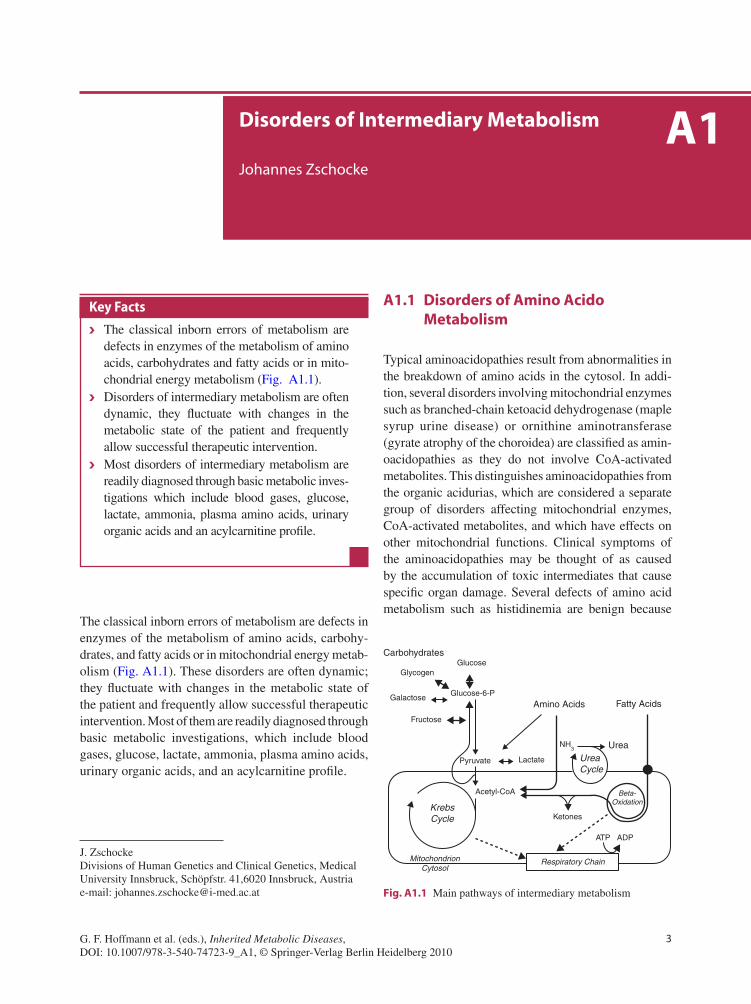
The classical inborn errors of metabolism are defects in enzymes of the metabolism of amino acids, carbohy-drates, and fatty acids or in mitochondrial energy metab-olism (Fig. A1.1). These disorders are often dynamic; they fl uctuate with changes in the metabolic state of the patient and frequently allow successful therapeutic intervention. Most of them are readily diagnosed through basic metabolic investigations, which include blood gases, glucose, lactate, ammonia, plasma amino acids, urinary organic acids, and an acylcarnitine profi le.
A1.1 Disorders of Amino Acido Metabolism
Typical aminoacidopathies result from abnormalities in the breakdown of amino acids in the cytosol. In addi-tion, several disorders involving mitochondrial enzymes such as branched-chain ketoacid dehydrogenase (maple syrup urine disease) or ornithine aminotransferase (gyrate atrophy of the choroidea) are classifi ed as amin-oacidopathies as they do not involve CoA-activated metabolites. This distinguishes aminoacidopathies from the organic acidurias, which are considered a separate group of disorders affecting mitochondrial enzymes, CoA-activated metabolites, and which have effects on other mitochondrial functions. Clinical symptoms of the aminoacidopathies may be thought of as caused by the accumulation of toxic intermediates that cause specifi c organ damage. Several defects of amino acid metabolism such as histidinemia are benign because
J. ZschockeDivisions of Human Genetics and Clinical Genetics, Medical University Innsbruck, Schöpfstr. 41,6020 Innsbruck, Austriae-mail: [email protected] Fig. A1.1 Main pathways of intermediary metabolism
CarbohydratesGlucose
Glucose-6-P
Glycogen
GalactoseAmino Acids
Fructose
LactatePyruvate
Fatty Acids
UreaNH3
UreaCycle
Beta-Oxidation
Ketones
ATP ADP
Respiratory ChainMitochondrionCytosol
KrebsCycle
Acetyl-CoA
Key Facts
The classical inborn errors of metabolism are ›defects in enzymes of the metabolism of amino acids, carbohydrates and fatty acids or in mito-chondrial energy metabolism (Fig. A1.1).Disorders of intermediary metabolism are often ›dynamic, they fl uctuate with changes in the metabolic state of the patient and frequently allow successful therapeutic intervention. Most disorders of intermediary metabolism are ›readily diagnosed through basic metabolic inves-tigations which include blood gases, glucose, lactate, ammonia, plasma amino acids, urinary organic acids and an acylcarnitine profi le.
4 J. Zschocke
the metabolites that accumulate are not toxic. The pathogenetic relevance of an inborn error of amino acid metabolism is not always easy to ascertain as clinical symptoms observed in the child may be coincidental or the reason for performing the analysis in the fi rst place. Aminoacidopathies are diagnosed through the analysis of plasma (or urinary) concentrations of amino acids and sometimes of urinary organic acids. A majority is treatable through dietary restriction of protein and of the amino acid involved in the defective pathway and by the avoidance or prompt treatment of catabolic states that lead to the breakdown of large amounts of protein. Another therapeutic strategy that has been suc-cessful in hepatorenal tyrosinemia is the inhibition of a biochemical step before the actual genetic defi ciency, thereby changing a harmful disease into a more benign amino acid accumulation without the accumulation of the more damaging substances downstream.
A1.2 Organic Acidurias
The classical organic acidurias are defi ciencies of enzymes in the mitochondrial metabolism of CoA-activated carboxylic acids, most of which are derived from amino acid breakdown. In this way, they are dis-tinguished from disorders of fatty acid oxidation, which not only involve CoA esters but also present dif-ferent diagnostic and therapeutic challenges. The term organic acidurias is preferred to the alternative term organic acidemias as they are most often detected by analysis of the urine. Biochemically, some of the reac-tions impaired in the organic acidurias are parallel to the dehydrogenase, hydratase, or ketothiolase reac-tions of the mitochondrial b-oxidation cycle. Clinical features are caused not only by the accumulation of toxic intermediates but also by a disturbance of mito-chondrial energy metabolism and carnitine homeosta-sis; they may include encephalopathy and episodic metabolic acidosis. Organic acidurias are diagnosed through the analysis of organic acids in the urine or acylcarnitines in the blood. Treatment is similar to that of the aminoacidopathies and involves the dietary restriction of the relevant amino acid(s) and the avoid-ance of protein catabolism. However, as the defective enzymes are distant (more downstream) from the respective amino acids, restriction may not lead to a stoichiometric reduction of pathological metabolites, although it does in methylmalonic aciduria. Unexpected
fl uctuations occur and complete return to normal inter-mediary metabolism is usually impossible. Supplemen-tation with carnitine and sometimes other substances such as glycine (e.g., to form isovalerylglycine in isovaleric aciduria) are very useful adjuncts to the treatment.
Disorders of biotin metabolism are included among the organic acidurias. Biotin is a cofactor of the mito-chondrial carboxylases and a defi ciency of biotini-dase or holocarboxylase synthetase leads to multiple carboxylase defi ciency. It is also usually diagnosed through urinary organic acid analysis. Biotinidase enzyme analysis of dried blood spots has been included into programs of neonatal screening as it is well treated with biotin supplementation.
A1.3 Disorders of Ammonia Detoxifi cation
The breakdown of protein produces large amounts of nitrogen in the form of ammonia that is highly neurotoxic but is normally converted to urea and excreted in the urine. Defects in enzymes of the urea cycle and other disorders of ammonia detoxifi ca-tion present clinically with encephalopathy and other symptoms of hyperammonemia. Metabolic investigations should include analysis of the amino acids in plasma and urine and urinary orotic acid. Treatment requires the reduction of protein intake in conjunction with the supplementation of essential amino acids, the avoidance of catabolic states and the administration of benzoate or phenylacetate/phe-nylbutyrate, which remove nitrogen in the form of alternative conjugates of amino acids such as gly-cine and glutamine.
A1.4 Disorders of Fatty Acid Oxidation and Ketogenesis
Mitochondrial fatty acid oxidation is required for the provision of energy during fasting, either through com-plete oxidation or through production of ketones in the liver that then serve as an alternative energy source for the brain. Disorders in this pathway typically present as hypoketotic hypoglycemia precipitated by
A1 Disorders of Intermediary Metabolism 5
fasting, leading to coma or convulsions. In addition, some disorders cause severe hepatopathy and (cardio-)myopathy, probably as results of the accumulation of toxic metabolites. The diagnosis is best reached in the acute situation through the analysis of free fatty acids and the ketone bodies 3-hydroxybutyrate and acetoac-etate as well as the acylcarnitine profi le and urinary organic acids. The diagnosis may be missed if samples are obtained in the normal interval between episodes or after the patient has been treated with intravenous glucose. Treatment consists of avoidance of fasting. Carnitine supplementation is mostly unessessary and must be carefully balanced in some defects, particu-larly those that cause cardiomyopathy or hepatopathy.
A1.5 Disorders of Carbohydrate Metabolism and Transport
The disorders in this group display a relatively wide range of clinical features and may cause clinical symp-toms because of toxicity, defi ciency of energy, hypo-glycemia, or storage.
Disorders of Galactose and Fructose Metabolism• : Defects in the cytosolic metabolism of galactose and fructose for glycolysis cause disease through accumulation of pathogenic metabolites. Children with galactosemia and fructosemia typically develop evidence of severe damage to the liver and/or kid-ney after dietary intake of lactose (milk, milk prod-ucts) or fructose (fruit, sucrose), respectively. Treatment requires the elimination of the intake of galactose or fructose.Disorders of Gluconeogenesis and Glycogen • Storage: Typical metabolic features are hypoglyce-mia after relatively short periods of fasting and lactic acidemia. There may be variable organ dysfunction, most frequently hepatopathy. Glycogen storage leads to hepatic enlargement, which in infancy may be massive. In some disorders such as glycogenosis type III there are elevations of the transaminases and creatine phosphate kinase, and there may be clinical myopathy. Treatment includes frequent meals, corn-starch supplementation, or continuous overnight tube feeding to avoid hypo glycemia.Disorders of Carbohydrate Transport• : There are a number of different glucose and other carbohy-drate carriers, and clinical symptoms differ greatly
depending on the tissue localization of the individ-ual defect. Symptoms are frequently gastrointesti-nal or renal but also include the central nervous system (defi cient glucose transport across the blood/brain barrier).
A1.6 Mitochondrial Disorders
Disorders of energy metabolism (usually summarized as mitochondrial disorders although enzymes defi cient, e.g., in organic acidurias or fatty acid oxidation defects are also located in the mitochondrion) include genetic defects of the pyruvate dehydrogenase complex, the Krebs cycle and the respiratory electron transport chain, comprising the fi nal pathways of substrate breakdown and the production of ATP. Mitochondrial disorders manifest clinically with symptoms and signs of energy defi ciency and a highly variable pattern of organ dys-functions. In many cases, there are lactic acidemia and progressive neurodegenerative disease. Periods of meta-bolic stress such as intercurrent infections may trigger a deterioration of the patient’s condition. The diagnostic work-up may be diffi cult and should include frequent measurements of blood lactate levels, CSF lactate, plasma amino acids and alanine, and often a search for mutations in mitochondrial DNA. Repeated, careful examinations of organ functions as well as imaging are essential. Treatment options are limited but usually include various vitamins and cofactors such as ribofl a-vin, coenzyme Q, or thiamine. Heterozygous mutations in the genes of some Krebs cycle enzymes (e.g., fumarase) cause inherited cancer predisposition syndromes.
A1.7 Disorders of Cobalamine and Folate Metabolism
Genetically determined or nutritional defi ciencies of vitamin cofactors may affect various metabolic path-ways and cause a wide range of clinical symptoms. They can frequently be satisfactorily treated by sup-plementation of the defi cient substance. Of particular importance in intermediary metabolism are cobalamin (vitamin B
12) and folate which are essential for cyto-
solic methyl group transfer. The cellular methylation reactions require methyl group transfer from serine to S-adenosylmethionine involving the folate cycle,
6 J. Zschocke
cobalamin (vitamin B12
) and the methionine–homo-cysteine cycle. A disturbance in this pathway may be caused by methylcobalamin defi ciency, a disturbance of the folate cycle, or by defi cient remethylation of homocysteine to methionine. Most disorders of coba-lamin metabolism as well as nutritional defi ciency of vitamin B
12 cause methylmalonic aciduria. Clinically,
disorders of cytosolic methyl group transfer cause an encephaloneuropathy, often with additional hemato-logical problems such as megaloblastic anemia and thromb embolic complications of hyperhomocysteine-mia. The diagnosis involves the analysis of urinary organic acids, plasma amino acids (homocysteine), and levels of folate and cobalamin. Treatment includes supplementation of cobalamin and folate, in some situ-ations with addition of betain and methionine.
A1.8 Disorders of Amino Acid Transport
Defi ciencies in the intestinal and/or renal transport of amino acids may be nonsymptomatic or cause symp-toms because of defi cient absorption of essential amino acids (e.g., tryptophan in Hartnup disease) or because of increased urinary concentration of unsoluble amino acids which causes nephrolithiasis (e.g., cystein in cystinuria). These disorders are diagnosed by the quan-titative analysis of amino acids in plasma and urine. Treatment depends on the clinical picture. Defi ciency of essential amino acids is treated by supplementation with large amounts of these compounds, or in the case of tryptophan defi ciency, supply of the cofactor nico-tinic acid that is normally synthesized from tryptophan. Renal calculi in cystinuria can be prevented by treat-ment with a chelating agent such as penicillamine, which forms mixed disulfi des with cysteine, and cal-culi once formed can be resorbed if they have not incorporated too much calcium.
A1.9 Disorders of Peptide Metabolism
The tripeptide • glutathione and the gammaglutamyl cycle have multiple functions in cellular metabolism, ranging from amino acid transport across membranes
to detoxifi cation of peroxides. Defi ciencies may cause neurological and hematological as well as met-abolic problems. Investigations should include the determination of organic acids in the urine and gluta-thione in various body fl uids. Treatment is largely symptomatic; certain drugs should be avoided.Defective breakdown of • dipeptides of histidine such as homocarnosine or carnosine may be found in patients with mental retardation, although the caus-ative relationship is not always proven. Ulcers of the skin, particularly of the legs are seen in proli-dase defi ciency. Investigations should include amino acid and peptide analysis of the urine. Treatment is symptomatic; some individuals with disorders of dipeptide metabolism are asymptom-atic and do not require treatment.
A1.10 Disorders of the Transport or Utilization of Copper, Iron, and Zinc
Disorders of copper metabolism• : Wilson disease causes a chronic hepatopathy and symptoms of cen-tral nervous dysfunction, while patients with Menke disease suffer from neurological problems in con-junction with abnormalities of hair, connective tis-sue, and bones. Diagnosis involves the analysis of copper and coeruloplasmin in serum, urine, and liver tissue. Treatment in Wilson disease is aimed at reducing copper load, while copper should be par-enterally substituted in Menke disease.Disorders of iron metabolism• : Patients affected with such disorders may present with iron-defi cient ane-mia, e.g., due to insuffi cient intestinal absorption of iron, or with iron overload and liver dysfunction as in hemochromatosis. Secondary iron overload may be observed in some hemolytic anemias. Treatment is directed at substitution or removal of iron.Disorders of zinc metabolism• : Acrodermatitis enteropathica is characterized by chronic skin prob-lems, alopecia, and central nervous symptoms. It is diagnosed through reduced levels of zinc and alka-line phosphatase and is treated with supplementa-tion of zinc.
7G. F. Hoffmann et al. (eds.), Inherited Metabolic Diseases,DOI: 10.1007/978-3-540-74723-9_A2, © Springer-Verlag Berlin Heidelberg 2010
Disorders of the Biosynthesis and Breakdown of Complex Molecules
Johannes Zschocke
A2
Disorders in this group typically show slowly progres-sive clinical symptoms and are less likely to cause acute metabolic crises. They are not usually recog-nized by basic metabolic analyses but require specifi c investigations for their diagnosis.
A2.1 Disorders of Purine and Pyrimidine Metabolism
Defi ciencies in enzymes required for the biosynthe-sis or breakdown of purines and pyrimidines cause neuromuscular abnormalities, nephrolithiasis, gouty arthritis, or anemia and immune dysfunction. They may be recognized through increased or reduced uri-nary urea in relation to creatinine, urine microscopy, or
specifi cally through the analysis of urinary purines and pyrimidines. Some metabolites of pyrimidine break-down are only recognized by urinary organic acid analysis. Nephrolithiasis may be treated or prevented by allopurinol. A high fl uid intake is helpful. Some disorders of pyrimidine metabolism, notably orotic aciduria and overactivity of 5' nucleotidase (nucleotide depletion syndrome), are treatable with uridine or tri-acetyluridine. There is no effective treatment for most of the primarily neurological manifestations of disor-ders of purine metabolism.
A2.2 Lysosomal Storage Disorders
Lysosomes contain a number of hydrolases required for the intracellular breakdown of large lipid and mucopoly-saccharide molecules. If one of these enzymes is defi -cient, its substrate accumulates and causes enlargement and/or functional impairment of the organ system. Clinical features include progressive neurological dete-rioration, dysmorphic features, and organomegaly. There is usually no metabolic decompensation, although acute symptoms (e.g., severe pain) is a major feature in some conditions. Investigations include careful roentgeno-graphic examination of the skeleton for dysostosis mul-tiplex, analysis of leukocytes and other cells for vacuoles, and assessment of parenchymatous organs. The urine may be investigated for abnormal glycosaminoglycans and oligosaccharides; specifi c enzyme studies are usu-ally required to make the exact diagnosis. For most dis-orders there is no specifi c therapy yet, although enzyme replacement therapy or bone marrow transplantation has been shown benefi cial in several disorders.
Key Facts
Disorders of the biosynthesis and breakdown ›of complex molecules typically show slowly progressive clinical symptoms and are less likely to cause acute metabolic crises. Disorders in this group are not usually recogn- ›ised by basic metabolic analyses but require specifi c investigations for their diagnosis.
J. ZschockeDivisions of Human Genetics and Clinical Genetics, Medical University Innsbruck, Schöpfstr. 41,6020 Innsbruck, Austriae-mail: [email protected]
8 J. Zschocke
Mucopolysaccharidoses (MPS)• , affected children typically develop progressive dysmorphic features, hepatomegaly and psychomotor retardation or regression. They are usually recognized through the analysis of urine for glycosaminoglycans.Oligosaccharidoses• may resemble the MPS, but many show more severe neurological symptoms and are more frequently symptomatic at birth (nonim-mune hydrops fetalis). The diagnosis is made through the demonstration of abnormal oligosaccharide pat-terns in the urine or enzyme analyses.Sphingolipidoses• and lipid storage disorders usu-ally present with progressive neurological deterio-ration. Hepatomegaly may be present, skeletal deformities and dysmorphic features are rare. Other presentation patterns are found particularly in Fabry disease (pain and paresthesias) and nonneuronop-athic Gaucher disease (hematoma, anemia, massive splenomegaly, and abdominal/bone pain). The spe-cifi c diagnosis usually requires enzyme analysis. The neuronal ceroid lipofuscinoses are usually sus-pected by electron microscopy and confi rmed by mutation analysis. Mucolipidoses combine clinical features of the mucopolysaccharidoses and sphin-golipidoses and may refl ect the defi ciency of sev-eral lysosomal enzymes as a consequence of defective enzyme processing.Lysosomal transport defects:• Cystinosis causes neph-ropathy and dysfunction of other organs including the thyroid gland and the eyes; it is diagnosed on the basis of increased cystine content of leucocytes. Sialic acid storage disease causes progressive en cephaloneuropathy; it is recognized through ele-vated free sialic acid in the urine. Both of these disor-ders result from defective transport out of lysosomes. Cystinosis is treated with oral cysteamine, cysteam-ine eye drops, and renal transplantation.
A2.3 Peroxisomal Disorders
The biochemical roles of peroxisomes are very diverse. Peroxisomal defects usually cause severe, progressive multisystem disorders.
Defects of • peroxisome biogenesis or the activation and b-oxidation of long-chain fatty acids cause pro-gressive neurological disease, structural abnormali-ties as in Zellweger syndrome, and abnormalities in hepatic, intestinal, or adrenal function. They are
usually recognized through the analysis of very long-chain fatty acids in blood or cultured fi bro-blasts. There is no effective treatment.Refsum disease is a defect in the metabolism of • exogenous phytanic acid. It causes slowly progres-sive neurological, visual, and auditory abnormali-ties, and often does not present until adulthood. It is diagnosed through the quantifi cation of serum phy-tanic acid and is treatable by a diet restricted of phytanic acid.Defects of • ether-phospholipid biosynthesis cause rhizomelic chondrodysplasia punctata characterized by proximal shortening of the limbs in addition to neurological and other manifestations. It is diag-nosed through quantifi cation of plasmalogens in erythrocytes. There is no effective treatment.Catalase defi ciency is the only known defect of the • detoxifi cation of oxygen radicals. It causes chronic ulcers in the oral mucosal membranes.Primary hyperoxaluria type I is the only known • defect of glyoxylate metabolism; it causes nephro-lithiasis and nephrocalcinosis. It is recognizable by organic acid or HPLC analysis for oxalate and gly-oxylate. It has been treated by transplantation of liver and kidney.
A2.4 Disorders of the Metabolism of Isoprenoids and Sterols
Isoprenoids and sterols are essential in many cellular and developmental processes. Most defects of their synthesis are caused by enzyme defi ciencies in the postsqualene portion of the pathway. Only mevalonic aciduria and hyperimmunoglobulinemia D syndrome, both due to mevalonate kinase defi ciency, are found in the proximal part of the pathway.
Mevalonate kinase defi ciency is the only known • defect of isoprenoid biosynthesis. It causes dysmor-phic features, failure to thrive, mental retardation, and recurrent febrile crises. An attenuated variant causes hyper-IgD syndrome. Treatment is symptomatic.Defects of • sterol biosynthesis cause various struc-tural abnormalities including the dysmorphic fea-tures of the Smith–Lemli–Opitz syndrome and mental retardation. Diagnosis involves plasma sterol analysis. In Smith–Lemli–Opitz syndrome, specifi c treatment by cholesterol supplementation has been of limited success.
A2 Disorders of the Biosynthesis and Breakdown of Complex Molecules 9
A2.5 Disorders of Bile Acid and Bilirubine Metabolism, Inherited Cholestasis and Porphyrias
Genetic defects of • bile acid biosynthesis cause symptoms either through bile acid defi ciency or through deposition of precursors. The former causes progressive cholestasis and malabsorption, while the precursors can lead to progressive neuro-logical dysfunction and xanthomas. The bile acid biosynthetic pathway is located partly in the per-oxisomes and is affected by peroxisomal disorders. Diagnosis involves the analysis of urinary bile acids. Treatment with bile acids is effective in the bile acid defi ciency states and to down-regulate bile acid biosynthesis.Hem is metabolized to bilirubin and excreted • together with bile acids in the urine. Genetic defects may involve specifi c enzymes or mechanisms of transport into the bile ducts. They cause indirect or direct hyperbilirubinemia. Specifi c treatment strate-gies have been developed for some disorders.Porphyrias are disorders of hem biosynthesis, fre-• quently inherited as autosomal dominant traits. Neurotoxic metabolites accumulate in defi ciencies affecting the fi rst few steps of the pathway and typi-cally cause intermittent acute symptoms such as abdominal pain triggered by various factors, in par-ticular induction of hem-containing enzymes. Porphyrins accumulating in more distal enzyme defi -ciencies are associated with photosensitivity and der-matologial symptoms. The diagnosis involves analysis of porphyrins and porphyrin precursors in urine, feces, or erythrocytes. Management entails the avoidance of precipitating factors.
A2.6 Congenital Disorders of Glycosylation (CDG)
Many proteins including enzymes, transport and mem-brane proteins, as well as hormones require glycosyla-tion in the Golgi apparatus or endoplasmatic reticulum to render them functional glycoproteins. A defi ciency
of one of the more than 40 different enzymes involved in glycosylation leads to a wide range of structural abnormalities and disturbances of physiological functions. A disorder from the CDG group should be considered in all patients with unclear multisystem or neurological disorder. The diagnosis in N-glycosylation disorders is usually made by isoelectric focussing of transferrin in serum. There is no effective treatment for most disor-ders of this group.
A2.7 Disorders of Lipoprotein Metabolism
Many disorders of lipoprotein metabolism cause clini-cal symptoms through the deposition of lipid in tissues and premature atherosclerosis. Others cause gastroin-testinal or peripheral neurological problems. They are recognized by quantifi cation of cholesterol and trig-lycerides and through lipoprotein electrophoresis. Many disorders are open to dietary or pharmacological therapy.
Elevated blood cholesterol levels in • hypercholester-olemias and mixed hyperlipidemias cause lipid deposition in the form of xanthomas and xan-thelasma. They lead to complications of premature atherosclerosis, especially myocardial infarction and cerebrovascular disease. Therapeutic options include diet, drugs, and lipid apharesis.Hypertriglyceridemia• may be caused by genetic disorders that affect the utilization of chylomicrons and very low-density lipoproteins. They may cause failure to thrive and abdominal symptoms, and sometimes severe pancreatitis. These disorders require stringent restriction of dietary fat.Genetic disorders affecting • HDL metabolism cause a variety of clinical manifestations including pre-mature atherosclerosis, neuropathy, nephropathy, and corneal clouding. Therapy is symptomatic.Genetic disorders in which there are • reduced LDL cholesterol and triglycerides lead to symp-toms of fat malabsorption. They are treated by restriction of fat and supplementation with fat soluble vitamins.
11G. F. Hoffmann et al. (eds.), Inherited Metabolic Diseases,DOI: 10.1007/978-3-540-74723-9_A3, © Springer-Verlag Berlin Heidelberg 2010
Neurotransmitter Defects and Related Disorders
Georg F. Hoffmann
A3
Genetic disorders of neurotransmitter metabolism are increasingly recognised as causes of severe metabolic encephalopathy often starting before birth or soon thereafter. Diagnosis usually requires investigations of the CSF. This group should be considered in children with neurological problems when basic metabolic investigations are normal.
A3.1 Disorders of Glycine and Serine Metabolism
Nonketotic hyperglycinemia is one of the best known causes of early-onset epileptic encephalopathy. It is recognised via concomitant amino acid analysis of plasma and CSF. Glycine levels in both are elevated, and the CSF to plasma ratio is increased. Treatment with dextrometorphan, benzoate or folate is of limited success. Disorders of serine biosynthesis cause neuro-logical symptoms. They have been treated with serine and glycine supplementation.
A3.2 Disorders of the Metabolism of Pterins and Biogenic Amines
Affected children suffer from progressive develop-mental retardation and epileptic encephalopathy. There may be specifi c symptoms of dopamine and/
or serotonine defi ciency, such as infantile parkin-sonism, dopa-responsive dystonia, oculogyric crises or disturbed temperature regulation. These diseases are sometimes recognised by hyperphenylalaninemias but many exclusively through the analyis of biogenic amines and pterins in CSF.
Disorders of tetrahydrobiopterin biosynthesis and recycling affect the hydroxylation of phenylalanine and have been called atypical or malignant phenylketonu-ria. The hydroxylations of tyrosine and tryptophan are also affected, leading to defi ciency of both, dopamine and serotonine. Investigations should include the anal-ysis of biogenic amines, pterins and amino acids in the CSF as well as amino acids in plasma and pterins in urine. The disorders are treated with l-dopa along with carbidopa and 5-hydroxytryptophan and/or tetrahydro-biopterin and/or tetrahydrobiopterin substitution.
Disorders of the biosynthesis of biogenic amines present similarly with progressive extrapyramidal symptoms and encephalopathy. The defi ciency of bio-genic amines is treated with of l -dopa along with car-bidopa and 5-hydroxytryptophan and/or dopamine agonists.
A3.3 Disorders of Gamma-Aminobutyrate Metabolism
These disorders cause central nervous dysfunction, often including seizures and encephalopathy. They are diagnosed through CSF analysis of amino acids and gamma-aminobutyrate (GABA). Urinary organic acid analysis may reveal 4-hydroxybutyric acid indicative of succinate semialdehyde dehydrogenase defi ciency. Vigabatrin has been used in the treatment of SSADH defi ciency.
G. F. HoffmannUniversity Children’s Hospital, Ruprecht-Karls-University, Im Neuenheimer Feld 430, D-69120 Heidelberg, Germany e-mail: [email protected]
12 G. F. Hoffmann
A3.4 Disorders of Vitamin B
6 Metabolism
Pyridoxal phosphate (PLP, vitamin B6) is a cofactor
of all the transamination reactions and some decar-boxylation and deamination reactions of amino acids, and as such is also required for the biosynthesis of several neurotransmitters including dopamine and GABA. Intracellular defi ciency may be caused by pri-mary or secondary disorders in the biosynthetic path-way and leads to a neonatal epileptic encephalopathy. In the well-known entity of vitamin B
6-dependent sei-
zures, PLP is inactivated by delta 1-piperideine-6-carboxylate, which accumulates because of an enzyme defi ciency in a different pathway. Disorders of vitamin B
6 metabolism are generally treatable with
pyridoxine or PLP.
A3.5 Disorders of Creatine Metabolism
Creatine is the central compound in cytosolic energy metabolism, and defi ciencies in the biosynthesis or transport of creatine manifest as neurometabolic disor-ders with progressive central nervous dysfunction.
They are usually diagnosed through the analysis of creatine and guanidinoacetate in urine and serum; treatment centers on creatine supplementation.
A3.6 Other Neurometabolic Disorders
Sulphite oxidase defi ciency• is a cause of severe infantile seizures and encephalopathy. It is recog-nised through a sulphite stix test of the urine. Amino acid analysis of plasma and urine may be diagnostic but is less reliable. When it is caused by molybdenum cofactor defi ciency there is also xan-thine oxidase defi ciency, which may be detected by purine analysis of the urine. There is no specifi c treatment.Various • cerebral organic acidurias including Canavan disease, l-2- and d-2-hydroxyglutaric aciduria, 2-keto-glutaric aciduria, fumaric aciduria and malonic acidu-ria present with central nervous dysfunction, which is usually progessive. General metabolic abnormali-ties are absent, but the specifi c metabolites are found on organic acid analyis of the urine. The molecular basis of most of the conditions has now been estab-lished. There is no specifi c treatment.
Part
Approach to the Patient with Metabolic Disease
B

15G. F. Hoffmann et al. (eds.), Inherited Metabolic Diseases,DOI: 10.1007/978-3-540-74723-9_B1, © Springer-Verlag Berlin Heidelberg 2010
When to Suspect Metabolic Disease
William L. Nyhan
B1
B1.1 History
B1.1.1 Family History
A careful family history may reveal important clues that point towards the diagnosis of an inborn error of metabolism. Most metabolic disorders are inherited as autosomal recessive traits, which may be suspected if the parents are consanguineous or the family has a con-fi ned ethnic or geographic background. Carriers for particular disorders, and as a consequence affected chil-dren, may be more frequent in remote villages, close-knit communities (such as the Amish in Pennsylvania), certain ethnic groups (such as Ashkenazi Jews), or countries that have seen little immigration over many centuries (such as Finland).
Quite often specialist investigations are started only after a second affected child is born into a family. Older siblings may be found to suffer from a similar disorder as the index patient or may have died from an acute unexplained disease classifi ed as “sepsis with uniden-tifi ed pathogen,” “encephalopathy” or “sudden infant
W. L. NyhanDepartment of Pediatrics, University of California, UCSD School of Medicine, 9500 Gilman Drive, La Jolla, CA 92093, USAe-mail: [email protected]
second opinion should be sought of in case of unexplained symptoms or disease courses.Metabolic investigations are usually not indi- ›cated in children with moderate developmental delay, isolated delay in speech development in early childhood, moderate failure to thrive, fre-quent infections, occasional seizures, e.g. dur-ing fever, or defi ned epileptic syndromes.
Key Facts
Careful clinical and family histories, repeated ›clinical examinations and a sequential work-up by routine laboratory and organ evaluation remains the best and most often only way to diagnosis. While it is imperative to exclude disorders for ›which effective treatments are available, in cases of slowly progressive, and by experience often incurable disorders, diagnostic proce-dures should be performed stepwise.Unexpected fi ndings in the “routine” labora- ›tory in patients with unusual and unexplained symptoms may be indicative of an inborn error of metabolism.Every child who is suspected of suffering from ›an inborn error of metabolism requires a care-ful evaluation of organ functions aided by rou-tine laboratory and imaging investigations. The involvement of multiple organ systems is an especially strong indication for an inherited metabolic disease.As it can be very diffi cult to recognize a constel- ›lation which was not personally experiences, a
16 W. L. Nyhan
death syndrome.” The latter is a frequent feature in dis-orders of intermediary metabolism that may have acute lethal presentations such as disorders of ammonia detoxifi cation, organic acidurias, or fatty acid oxida-tion disorders.
In assessing medical records of previously affected but undiagnosed family members, it should be taken into account that the written clinical descriptions of complex conditions can be inconsistent and even mis-leading. Depending on the presumptive diagnosis at that time, important clinical clues may be missing. Parents are sometimes more reliable sources of infor-mation. On the other hand, the clinical expression of the same inborn error of metabolism may be variable even within families. Some more common Mendelian disorders are caused by a wide range of different muta-tions with different degrees of disease severity. Disease manifestations are especially variable in females with X-linked traits because of differences in the lyoniza-tion of the X chromosome in carrier females, e.g., orni-thine transcarbamylase defi ciency. Similarly, dominant disorders with variable penetrance may cause variable clinical problems in different members and genera-tions even of one family, such as Segawa syndrome due to GTP cyclohydrolase defi ciency.
As a result of the successful treatment of disorders of intermediary metabolism in which toxic small mol-ecules accumulate, an increasing number of relatively healthy affected women are reaching the reproductive age. If they become pregnant, there is a risk for their fetuses to be harmed by pathological amounts of toxic metabolites from the mother, although the children are themselves not affected but heterozygous. Especially important is maternal phenylketonuria (PKU), which is likely to become a major health problem. Some women at risk may not even know that they are affected with PKU, if they come from countries where newborn screening did not exist or if they have discontinued dietary treatment and medical follow-up in late child-hood. The latter will however remember that they had followed a special diet, which should be specifi cally asked for. Several mothers have been found to suffer from mild PKU only after maternal PKU was diag-nosed in one of her children. Other maternal condi-tions may cause “metabolic” disease in the neonate or infant postnatally, e.g., methylmalonic aciduria and hyperhomocystinemia in fully breastfed children of mothers ingesting a vegan diet, which causes nutri-tional vitamin B
12 defi ciency.
B1.1.2 Prenatal Development and Complications of Pregnancy
Toxic small molecules that accumulate in many disor-ders of intermediary metabolism do not harm the fetus because they are removed via the placenta and meta-bolized by the mother. Children affected with such dis-orders usually have a completely normal intrauterine development and are born with normal birth measure-ments at term. In contrast, disorders that interfere with cellular energy metabolism, e.g., mitochondrial disor-ders, may impair fetal organ development and prena-tal growth, causing structural (in particular cerebral) abnormalities, dysmorphic features, and dystrophy. Structural abnormalities and dysmorphic features may be even more pronounced in disorders of the biosyn-thesis of complex molecules that are necessary for developmental pathways and networks. Notable exam-ples are the defects of sterol biosynthesis that inter-fere with cholesterol-dependent signaling pathways of development and cause, for example, the Smith–Lemli–Opitz syndrome. Disorders affecting the break-down of complex molecules such as lysosomal storage disorders cause specifi c dysmorphic characteristics as in the Hurler disease, and when severe, may already present at birth. An unusual prenatal disease manifesta-tion is found in mothers carrying a fetus affected with long-chain hydroxyacyl-CoA dehydrogenase (LCHAD) defi ciency or carnitine palmitoyltransferase II defi -ciency, defects of fatty acid b-oxidation. These mothers have an increased risk of developing acute fatty liver of pregnancy, preeclampsia, or hemolysis, elevated liver enzymes, and low platelets (the HELLP syndrome). Systematic studies in mothers showed that fetal LCHAD defi ciency is present in a signifi cant number of women with acute fatty liver of pregnancy, but only in a very small proportion of the far more common HELLP syndrome. The neonates of such mothers should be screened for fatty acid oxidation disorders by acylcarnitine analysis in a dried blood spot.
B1.1.3 Age of Presentation and Precipitating Factors
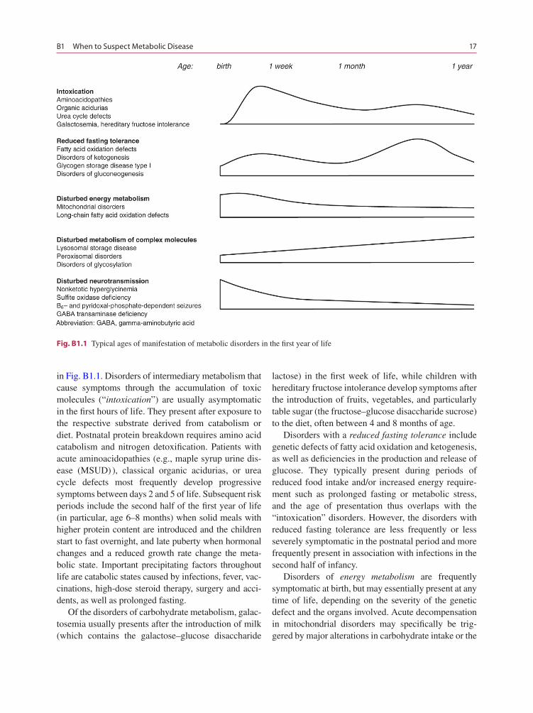
The “typical” ages of manifestation of different groups of metabolic disorders in the fi rst year of life are depicted
B1 When to Suspect Metabolic Disease 17
in Fig. B1.1. Disorders of intermediary metabolism that cause symptoms through the accumulation of toxic molecules (“intoxication”) are usually asymptomatic in the fi rst hours of life. They present after exposure to the respective substrate derived from catabolism or diet. Postnatal protein breakdown requires amino acid catabolism and nitrogen detoxifi cation. Patients with acute aminoacidopathies (e.g., maple syrup urine dis-ease (MSUD) ), classical organic acidurias, or urea cycle defects most frequently develop progressive symptoms between days 2 and 5 of life. Subsequent risk periods include the second half of the fi rst year of life (in particular, age 6–8 months) when solid meals with higher protein content are introduced and the children start to fast overnight, and late puberty when hormonal changes and a reduced growth rate change the meta-bolic state. Important precipitating factors throughout life are catabolic states caused by infections, fever, vac-cinations, high-dose steroid therapy, surgery and acci-dents, as well as prolonged fasting.
Of the disorders of carbohydrate metabolism, galac-tosemia usually presents after the introduction of milk (which contains the galactose–glucose disaccharide
lactose) in the fi rst week of life, while children with hereditary fructose intolerance develop symptoms after the introduction of fruits, vegetables, and particularly table sugar (the fructose–glucose disaccharide sucrose) to the diet, often between 4 and 8 months of age.
Disorders with a reduced fasting tolerance include genetic defects of fatty acid oxidation and ketogenesis, as well as defi ciencies in the production and release of glucose. They typically present during periods of reduced food intake and/or increased energy require-ment such as prolonged fasting or metabolic stress, and the age of presentation thus overlaps with the “intoxication” disorders. However, the disorders with reduced fasting tolerance are less frequently or less severely symptomatic in the postnatal period and more frequently present in association with infections in the second half of infancy.
Disorders of energy metabolism are frequently symptomatic at birth, but may essentially present at any time of life, depending on the severity of the genetic defect and the organs involved. Acute decompensation in mitochondrial disorders may specifi cally be trig-gered by major alterations in carbohydrate intake or the
Fig. B1.1 Typical ages of manifestation of metabolic disorders in the fi rst year of life
18 W. L. Nyhan
ingestion of large amounts of rapidly absorbed carbo-hydrates, while long-chain fatty acids that interfere with energy metabolism in some b-oxidation defects cause clinical features of a mitochondrial disorder dur-ing fasting periods. Another characteristic feature of mitochondrial disorders is a marked and frequently irreversible deterioration of the clinical condition with intercurrent illnesses.
Disorders in the metabolism of complex molecules rarely show acute metabolic crises but present with variable and often progressive organ dysfunction throughout childhood. There are usually no precipitat-ing factors. The clinical presentation of neurotransmit-ter defects and related disorders depends on the ontogenetic expression of neurotransmitter systems and receptors. Affected children are often symptom-atic immediately after birth, and there may even be symptoms of intrauterine epilepsy as evidence of pre-natal disease manifestations. There are usually no pre-cipitating factors.
B1.2 Physical Examination
Every child who is suspected of suffering from an inborn error of metabolism requires a thorough physi-cal examination and a careful evaluation of organ func-tions aided by routine laboratory and imaging investigations. In addition, hearing and vision should be examined at specialist appointments. Depending on the presenting symptoms and the clinical course, a reevaluation, especially a detailed physical examina-tion, should be repeated every 6 months. The detection of additional manifestations is of great importance even if the patient does not complain of them, particu-larly if the fi nal diagnosis is still unknown.
The involvement of multiple organ systems is one of the strongest arguments in favor of an inherited metabolic disease. This is especially true for defects of organelle metabolism such as mitochondrial or per-oxisomal disorders or the quickly enlarging group of glycosylation defects or CDG syndromes. Structural abnormalities such as dysmorphic features or malfor-mations may be caused by disorders in the metabolism of complex molecules as well as disorders affecting mitochondrial energy metabolism, but are not usually observed in other disorders of intermediary metabolism.
Generalized organomegaly is often indicative of a (lys-osomal) storage disorder, while isolated hepatomegaly is observed in a great variety of enzyme defects. Urine color and body odor can provide diagnostic clues, as discussed later. A list of differential diagnoses of char-acteristic symptoms and signs is given in the appendix.
B1.2.1 Unusual Odor
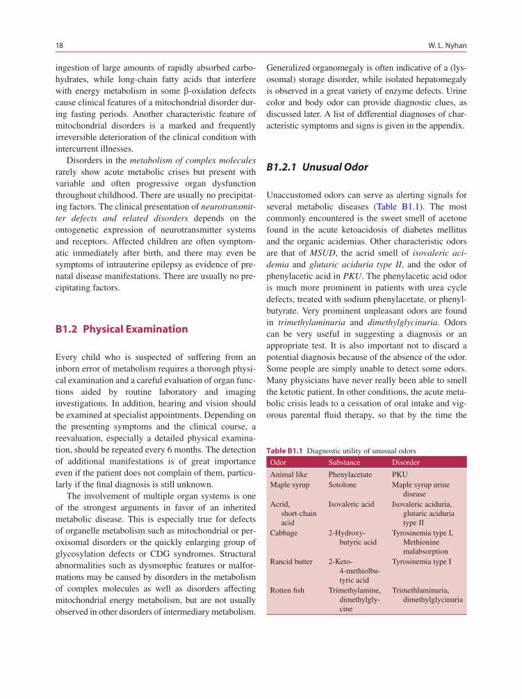
Unaccustomed odors can serve as alerting signals for several metabolic diseases (Table B1.1). The most commonly encountered is the sweet smell of acetone found in the acute ketoacidosis of diabetes mellitus and the organic acidemias. Other characteristic odors are that of MSUD, the acrid smell of isovaleric aci-demia and glutaric aciduria type II, and the odor of phenylacetic acid in PKU. The phenylacetic acid odor is much more prominent in patients with urea cycle defects, treated with sodium phenylacetate, or phenyl-butyrate. Very prominent unpleasant odors are found in trimethylaminuria and dimethylglycinuria. Odors can be very useful in suggesting a diagnosis or an appropriate test. It is also important not to discard a potential diagnosis because of the absence of the odor. Some people are simply unable to detect some odors. Many physicians have never really been able to smell the ketotic patient. In other conditions, the acute meta-bolic crisis leads to a cessation of oral intake and vig-orous parental fl uid therapy, so that by the time the
Odor Substance Disorder
Animal like Phenylacetate PKUMaple syrup Sotolone Maple syrup urine
diseaseAcrid,
short-chain acid
Isovaleric acid Isovaleric aciduria, glutaric aciduria type II
Cabbage 2-Hydroxy-butyric acid
Tyrosinemia type I, Methionine malabsorption
Rancid butter 2-Keto-4-methiolbu-tyric acid
Tyrosinemia type I
Rotten fi sh Trimethylamine, dimethylgly-cine
Trimethlaminuria, dimethylglycinuria
Table B1.1 Diagnostic utility of unusual odors
B1 When to Suspect Metabolic Disease 19
patient reaches the referral hospital the odor has long since disappeared.
The odor of maple syrup led to the recognition and original description of MSUD, before it was known that this was a disorder in the metabolism of the branched-chain amino acids. A keen sense of smell can still be useful in the detection of this disease, but the seriousness of the presentation of metabolic imbal-ance and a readiness to carry out an analysis of amino acids in plasma are such that most patients diagnosed today do not trigger the smell test. This is also true of acute exacerbation in established patients. Testing for urinary ketones with DNPH and organic acid analysis of the urine are also useful in the diagnosis of this dis-ease. The odor of the patient with isovaleric acidemia has been described as like that of sweaty feet, but it does not smell anything like a locker room. The smell is penetrating, pervasive, and readily recognized. It is the odor of a short-chain volatile acid, and the same smell may be appreciated in patients with multiple acyl-CoA dehydrogenase defi ciency (glutaric aciduria type II) during times of acute illness.
Now that screening of newborns for PKU is univer-sal in developed countries, patients with this disease are not likely to be diagnosed because of the character-istic odor, but some of us have made the diagnosis in this way in patients born prior to the development of screening. The odor has variously been described as musty, barny, animal-like or wolf-like. It is actually the odor of phenylacetic acid. Now that patients with defects in the urea cycle are treated with phenylacetic acid or its precursor phenylbutyric acid, specialists in inherited metabolic disease are quite as accustomed to this odor.
Patients with hepatorenal tyrosinemia and other nonmetabolic patients with hepatic cirrhosis may have a very unpleasant odor that results from the accumula-tion of methionine.
The classic unpleasant odor is that of patients with trimethylaminuria. Trimethylamine is the odor of fi sh that is not fresh. The compound is a major end product of nitrogen metabolism of teleost fi shes, which convert it to the oxide and employ the resulting compound to balance their osmotic pressure with surrounding sea water. In man, trimethylamine is formed from dietary trimethylamine oxide in fi sh and from choline absorbed from the intestine and transported to the liver, where the trimethylamine oxide is formed and ultimately excreted in the urine. Patients with trimethylaminuria have an inborn error in the metabolism of the oxide, defective activity of hepatic trimethlyamine N-oxide synthetase. The metabolic abnormality does not appear to produce a disease as we usually know one; its con-sequences are nevertheless terrible. An odor so unpleas-ant leads to social ostracism, poor performance in school, depression, and loss of employment. Suicide is a possibility. Diagnosis is important because a diet low in fi sh, liver, and egg yolks is usually suffi cient to elim-inate the odor. The diagnosis is made by identifying the compound by gas chromatography, gas chromatog-raphy–mass spectroscopy (GC–MS), FAB-MS, or nuclear magnetic resonance (NMR) spectroscopy. Its excretion is increased by loading with choline, and this may be necessary for the diagnosis in patients who have found dietary ways of minimizing their odor. Following a morning specimen of urine, a 5 g oral sup-ply of choline bitartrate in 3 doses over 24 h led to a 44-fold increase in trimethylamine excretion to 1.098 mmol/mg creatinine. Normal individuals excreted 0.0042–0.405 mmol/mg creatinine. The activity of the enzyme has been measured in biopsied liver. It is a fl avin-containing monooxidase, designated FMO
3.
Several mutations in the gene have been identifi ed.Patients have been described in whom the odor of
trimethylamine is mild or intermittent. Mutations have been identifi ed in the FMO
3 gene on chromosome
1q23–25. For instance, the P153L mutation has been identifi ed in patients with severe trimethylaminuria and no enzyme activity in vitro. The patients with the mild phenotype have had an allele with two common polymorphisms, E158K in which a 472G→A mutation coded for a lysine instead of a glutamate, and E3086 in which a 923A→G mutation coded for glycine instead of glutamate. Patients have generally been heterozygous for this allele and a disease-producing mutation, but one patient has been homozygous for the variant allele. The
Remember
Diagnosis by smell is underutilized, but many are not good at it. Too, a characteristic odor maybe absent in a severely ill infant partaking nothing by mouth and receiving parenteral fl uids.
20 W. L. Nyhan
variant allele is common in Caucasian populations; allele frequency was found to be 20% in Germans.
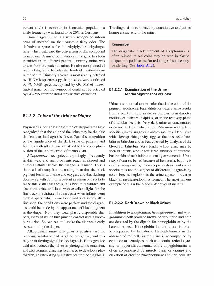
Dimethylglycinuria is a newly recognised inborn error of metabolism that causes a fi shy odor. The defective enzyme is the dimethylglycine dehydroge-nase, which catalyzes the conversion of this compound to sarcosine. A missense mutation in the gene has been identifi ed in an affected patient. Trimethylamine was absent from the patient’s urine. He also complained of muscle fatigue and had elevated levels of creatine kinase in the serum. Dimethylglycine is most readily detected by 1H-NMR spectroscopy. Its presence was confi rmed by 13C-NMR spectroscopy and by GC–MS of nonex-tracted urine, but the compound could not be detected by GC–MS after the usual ethylacetate extraction.
B1.2.2 Color of the Urine or Diaper
Physicians since at least the time of Hippocrates have recognized that the color of the urine may be the clue that leads to the diagnosis. It was Garrod’s recognition of the signifi cance of the dark urine of patients and families with alkaptonuria that led to the conceptual-ization of the inborn errors of metabolism.
Alkaptonuria is recognized surprisingly infrequently in this way, and many patients reach adulthood and clinical arthritis before the diagnosis is made. This is the result of many factors, among them that the black pigment forms with time and oxygen, and that fl ushing does away with both. In a patient in whom one seeks to make this visual diagnosis, it is best to alkalinize and shake the urine and look with excellent light for the fi ne black precipitate. In times past when infants wore cloth diapers, which were laundered with strong alka-line soap, the conditions were perfect, and the diagno-sis could be made by the appearance of black pigment in the diaper. Now they wear plastic disposable dia-pers, many of which turn pink on contact with alkapto-nuric urine. So, we can still make the diagnosis early by examining the diaper.
Alkaptonuric urine also gives a positive test for reducing substance and is glucose-negative, and this may be an alerting signal for the diagnosis. Homogentisic acid also reduces the silver in photographic emulsion, and alkaptonuric urine has been used to develop a pho-tograph, an interesting qualitative test for the diagnosis.
The diagnosis is confi rmed by quantitative analysis of homogentisic acid in the urine.
B1.2.2.1 Examination of the Urine for the Signifi cance of Color
Urine has a normal amber color that is the color of the pigment urochrome. Pale, dilute, or watery urine results from a plentiful fl uid intake or diuresis as in diabetes mellitus or diabetes insipidus, or in the recovery phase of a tubular necrosis. Very dark urine or concentrated urine results from dehydration. Pale urine with a high specifi c gravity suggests diabetes mellitus. Dark urine with a low specifi c gravity suggests the presence of uro-bilin or bilirubin and is best checked by analysis of the blood for bilirubin. Very bright yellow urine may be seen in infants who ingest large amounts of carotene, but the skin of such infants is usually carotenemic. Urine may, of course, be red because of hematuria, but this is readily recognized by microscopic analysis, and such a specimen is not the subject of differential diagnosis by color. Free hemoglobin in the urine appears brown or black as methemoglobin is formed. The most famous example of this is the black water fever of malaria.
B1.2.2.2 Dark Brown or Black Urines
In addition to alkaptonuria, hemoglobinuria and myo-globinuria both produce brown or dark urine and both are detected by the dipstix for hemoglobin or by the benzidine test. Hemoglobin in the urine is often accompanied by hematuria. Hemoglobinuria in the absence of red cells in the urine is accompanied by evidence of hemolysis, such as anemia, reticulocyto-sis, or hyperbilirubinemia, while myoglobinuria is often accompanied by muscle pains or cramps and elevation of creatine phosphokinase and uric acid. An
Remember
The diagnostic black pigment of alkaptonuria is often missed. A red color may be seen in plastic diaper, or a positive test for reducing substance may be alerting (See Table B1.2).
B1 When to Suspect Metabolic Disease 21
attack of myoglobinuria should signal a work-up for a disorder of fatty acid oxidation (Chap. C6). It is also seen in enzyme defects localized to muscle, such as
myophosphorylase defi ciency (McArdle disease) and myodenylate deaminase defi ciency. Melaninuria is seen in disseminated melanotic sarcoma.
B1.2.2.3 Red Urine
Porphyrias are the major metabolic cause of red urine. Congenital erythropoietic porphyria is an autosomal recessive disease caused by mutations in the gene for uroporphyrinogen synthase. Uroporphyria and copropor-phyria are found in the urine. It manifests a variable phenotype from nonimmune hydrops fetalis to a mild adult-onset form with only photosensitive cutaneous lesions. The disease is often fi rst recognized because of a pink, red, or brown stain in the diapers. These patients also develop erythrodontia in which a red fl uorescence of the teeth is visible with ultraviolet illumination.
Red urine may also be seen following the ingestion of large quantities of colored foods. The anthrocyani-nuria of beet ingestion is quite common. Blackberries have also been associated with red urine. Red dyes, such as rhodamine B, used to color foods and cold drinks have led to red urine of so many children after a weekend party that the condition was termed the Monday morning disorder of children. Phenolphthalein in laxatives may also cause red urine. In the neonatal period, distinct red spots in the diaper were seen where crystals of ammonium urate dried out. In previous days when cloth diapers were used and accumulated for a while before laundering, a red diaper syndrome was recognized in which the color developed after 24 h of incubation and came from the growth of the chro-mobacterium, Seratia marcescens, which does not produce pigment in the infant’s intestine, but only after aerobic growth at 25–30°C. Red stools may also be seen after the ingestion of red crayons, and in some patients receiving cefdinir, in most but not all of whom receive oral iron.
B1.2.2.4 Green or Blue Urines
Blue pigment in urine containing urochrome usually leads to a green color. Blue color was seen in the blue diaper syndrome. This disorder of the intestinal absorption of tryptophan was described in two siblings who also had hypercalcemia and nephrocalcinosis.
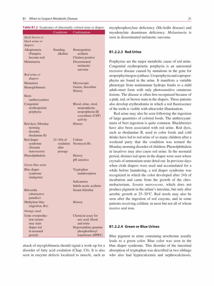
Conditions Confi rmation
Dark brown or black urine or diapers
Alkaptonuria (Pampers become red)
Standing, alkaline
Homogentisic aciduria
Clinitest positiveMelaninuria Disseminated
melanotic sarcoma
Red urine or diapers
Hematuria MicroscopicGuaiac, benzidineHistory
Hemoglobinuria
Beets (anthrocyanins)
Congenital erythropoietic porphyria
Blood, urine, stool, uroporphyrin, uroporphyrin-III cosynthase (CEP) activity
Red dyes (Monday morning disorder, rhodamine B)
History
Red diaper syndrome (Serratia marcescens)
24–36 h of oxidation after passage
CultureNeomycin Rx
Phenolphthalein HistorypH sensitive
Green-blue urine
Blue diaper syndrome (indigotin)
Tryptophan malabsorption
IndicanuriaIndole-acetic aciduria
Biliverdin (obstructive jaundice)
Serum bilirubin
Methylene blue (ingestion, Rx)
History
Orange sand
Urate overproduc-tion (urates may stain diaper red in neonatal period)
Chemical assay for uric acid, blood and urine
Hypoxanthine-guanine phosphoribosyl transferase (HPRT)
Table B1.2 Syndromes of abnormally colored urine or diapers
22 W. L. Nyhan
When tryptophan is not effi ciently absorbed, intesti-nal bacteria convert it to indole metabolites that are absorbed and excreted in the urine. The blue color comes from the oxidative conjugation of two molecules of indican to indigotin, or indigo blue, a water insolu-ble dye. The excretion of indole products is increased by an oral tryptophan load. The condition must be very rare because further patients have not been reported since the initial report in 1964. Indoles including indi-can are also found in the urine of patients with Hartnup disease, in which there is defective renal tubular reab-sorption, as well as intestinal absorption of a number of amino acids including tryptophan, but blue diapers or urine have not been observed.
Biliverdin, the oxidation product of bilirubin, is excreted in the urine, and so green urine may be seen in jaundiced patients, particularly those with chronic obstructive jaundice.
Benign pigments such as methylene blue, found in some tablets, are excreted in urine, and if a suffi cient quantity is taken, will color the urine. Indigo-carmine is another blue dye that may fi nd its way into food stuffs.
B1.3 Routine Laboratory Investigations
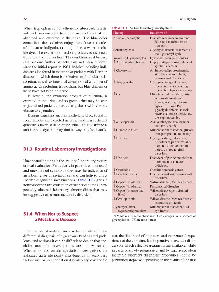
Unexpected fi ndings in the “routine” laboratory require critical evaluation. Particularly in patients with unusual and unexplained symptoms they may be indicative of an inborn error of metabolism and can help to direct specifi c diagnostic investigations. Table B1.3 gives a noncomprehensive collection of such sometimes unex-pectedly obtained laboratory abnormalities that may be suggestive of certain metabolic disorders.
B1.4 When Not to Suspect a Metabolic Disease
Inborn errors of metabolism may be considered in the differential diagnosis of a great variety of clinical prob-lems, and at times it can be diffi cult to decide that spe-cialist metabolic investigations are not warranted. Whether or not certain specialist investigations are indicated quite obviously also depends on secondary factors such as local or national availability, costs of the
test, the likelihood of litigation, and the personal expe-rience of the clinician. It is imperative to exclude disor-ders for which effective treatments are available, while in cases of slowly progressive, and by experience often incurable disorders diagnostic procedures should be performed stepwise depending on the results of the fi rst
Finding Indicative of
Anemia (macrocytic) Disturbances in cobalamin or folic acid metabolism or transport
Reticulocytosis Glycolysis defects, disorders of the g-glutamyl cycle
Vacuolized lymphocytes Lysosomal storage disorders↑ Alkaline phosphatase Hypoparathyreoidism, bile acid
synthesis defects↓ Cholesterol A-, hypobetalipoproteinemia,
sterol synthesis defects, peroxisomal disorders
↑ Triglycerides Glycogen storage disorders, lipoprotein disorders, e.g., lipoprotein lipase defi ciency
↑ CK Mitochondrial disorders, fatty acid oxidation defects, glycogen storage disease types II, III, and IV, glycolysis defects, muscle-AMP-deaminase defi ciency, dystrophinopathies
↑ a-Fetoprotein Ataxia telangiectasia, hepator-enal tyrosinemia
↓ Glucose in CSF Mitochondrial disorders, glucose transport protein defi ciency
↑ Uric acid Glycogen storage disorders, disorders of purine metabo-lism, fatty acid oxidation defects, mitochondrial disorders
↓ Uric acid Disorders of purine metabolism, molybdenum cofactor defi ciency
↓ Creatinine Creatine synthesis defect↑ Iron, transferrin Hemochromatosis, peroxisomal
disorders↓ Copper (in plasma) Wilson disease, Menkes disease↑ Copper (in plasma) Peroxisomal disorders↑ Copper (in urine and
liver)Wilson disease, peroxisomal
disorders↓ Ceruloplasmin Wilson disease, Menkes disease,
aceruloplasminemiaHypothyroidism,
hypoparathyreoidismMitochondrial disorders, CDG
syndromes
Table B1.3 Routine laboratory investigations
AMP adenosine monophosphate; CDG congenital disorders of glycosylation; CK creatine kinase
B1 When to Suspect Metabolic Disease 23
investigations and the appearance and development of signs and symptoms with time. The diagnosis of some metabolic disorders involves procedures that are stress-ful, such as sedation or lumbar puncture, or potentially dangerous for the child (e.g., fasting or loading stud-ies), and that are often also stressful for the parents. Psychosocial factors should be taken into consideration when the diagnostic work-up is planned. The families need to be guided and supported. In the worst case, a specifi c diagnosis with a doomed prognosis that shat-ters the expectations of the parents can even damage the parent–child relationship. On the other hand, in almost all families a specifi c diagnosis no matter how negative will be one of the most important supports for coping, and of course is critical for timely genetic diag-nosis in young families and appropriate counseling.
Specialist metabolic investigations are not usually indicated in children with moderate developmental delay, isolated delay in speech development in early childhood, moderate failure to thrive, frequent infec-tions, occasional seizures, e.g., during fever, or defi ned epileptic syndromes. An inborn error of metabolism is also unlikely in the healthy sibling of an infant who died of SIDS, provided that this child had been
previously asymptomatic. Key factors in the evalua-tion of symptoms are their isolated appearance vs. the presence of additional pathology, however subtle, i.e., the lack or presence of additional neurological and/or systemic abnor malities, and a static vs. a progressive clinical course. Multisystem or progressive disorders are much more likely to be caused by inborn errors of metabolism.
Key References
Moolenaar SH, Poggi-Bach J, Engelke UFH, Corstaensen JMB, Heerschap A, De Jong JGN, Binzak BA, Vockley J, Wevers RA (1999) Defect in dimethylglycine dehydrogenase, a new inborn error of metabolism: NMR spectroscopy study. Clin Chem 45:459–464
Nyhan WL, Barshop BA, Ozand PT (2005) Atlas of metabolic diseases, 2nd edn. Hodder Arnold, London
Saudubray JM, Charpentier C (2001) Clinical phenotypes: diagno-sis/algorithms. In: Scriver CR, Beaudet AL, Valle D, Sly WL (eds) The metabolic and molecular bases of inherited disease, 7th edn, Vol 1 . McGraw-Hill, New York, pp 1327–1403
Zschocke J, Kohlmueller D, Quak E, Meissner T, Hoffmann GF, Mayatepek E (1999) Mild trimethylaminuria caused by common variants in FMO3 gene. Lancet 354:834–835