Embed Size (px)
Citation preview
Washington University School of Medicine Washington University School of Medicine
Digital Commons@Becker Digital Commons@Becker
Open Access Publications
2018
Inherited p40phox deficiency differs from classic chronic Inherited p40phox deficiency differs from classic chronic
granulomatous disease granulomatous disease
Mary C. Dinauer Washington University School of Medicine in St. Louis
et al
Follow this and additional works at: https://digitalcommons.wustl.edu/open_access_pubs
Recommended Citation Recommended Citation Dinauer, Mary C. and et al, ,"Inherited p40phox deficiency differs from classic chronic granulomatous disease." The Journal of Clinical Investigation. 128,9. 3957-3975. (2018). https://digitalcommons.wustl.edu/open_access_pubs/7097
This Open Access Publication is brought to you for free and open access by Digital Commons@Becker. It has been accepted for inclusion in Open Access Publications by an authorized administrator of Digital Commons@Becker. For more information, please contact [email protected].
Inherited p40phox deficiency differs from classicchronic granulomatous disease
Annemarie van de Geer, … , Taco W. Kuijpers, JacintaBustamante
J Clin Invest. 2018;128(9):3957-3975. https://doi.org/10.1172/JCI97116.
Graphical abstract
Research Article Genetics Immunology
Find the latest version:
http://jci.me/97116/pdf
The Journal of Clinical Investigation R E S E A R C H A R T I C L E
3 9 5 7jci.org Volume 128 Number 9 September 2018
Biallelic loss-of-function (LOF) mutations of the NCF4 gene, encoding the p40phox subunit of the phagocyte NADPH oxidase, have been described in only 1 patient. We report on 24 p40phox-deficient patients from 12 additional families in 8 countries. These patients display 8 different in-frame or out-of-frame mutations of NCF4 that are homozygous in 11 of the families and compound heterozygous in another. When overexpressed in NB4 neutrophil-like cells and EBV-transformed B cells in vitro, the mutant alleles were found to be LOF, with the exception of the p.R58C and c.120_134del alleles, which were hypomorphic. Particle-induced NADPH oxidase activity was severely impaired in the patients’ neutrophils, whereas PMA-induced dihydrorhodamine-1,2,3 (DHR) oxidation, which is widely used as a diagnostic test for chronic granulomatous disease (CGD), was normal or mildly impaired in the patients. Moreover, the NADPH oxidase activity of EBV-transformed B cells was also severely impaired, whereas that of mononuclear phagocytes was normal. Finally, the killing of Candida albicans and Aspergillus fumigatus hyphae by neutrophils was conserved in these patients, unlike in patients with CGD. The patients suffer from hyperinflammation and peripheral infections, but they do not have any of the invasive bacterial or fungal infections seen in CGD. Inherited p40phox deficiency underlies a distinctive condition, resembling a mild, atypical form of CGD.
Inherited p40phox deficiency differs from classic chronic granulomatous diseaseAnnemarie van de Geer,1 Alejandro Nieto-Patlán,2,3,4 Douglas B. Kuhns,5 Anton T.J. Tool,1 Andrés A. Arias,6,7 Matthieu Bouaziz,2,3 Martin de Boer,1 José Luis Franco,6 Roel P. Gazendam,1 John L. van Hamme,1 Michel van Houdt,1 Karin van Leeuwen,1 Paul J.H. Verkuijlen,1 Timo K. van den Berg,1,8 Juan F. Alzate,9 Carlos A. Arango-Franco,6,7 Vritika Batura,10 Andrea R. Bernasconi,11 Barbara Boardman,12 Claire Booth,13 Siobhan O. Burns,14,15 Felipe Cabarcas,9,16 Nadine Cerf Bensussan,17,18,19 Fabienne Charbit-Henrion,17,18,19,20 Anniek Corveleyn,21 Caroline Deswarte,2,3 María Esnaola Azcoiti,2,22 Dirk Foell,23 John I. Gallin,24 Carlos Garcés,6 Margarida Guedes,25 Claas H. Hinze,23 Steven M. Holland,26 Stephen M. Hughes,12 Patricio Ibañez,27 Harry L. Malech,24 Isabelle Meyts,28,29 Marcela Moncada-Velez,6 Kunihiko Moriya,2,3 Esmeralda Neves,30 Matias Oleastro,11 Laura Perez,11 Vimel Rattina,2,3 Carmen Oleaga-Quintas,2,3 Neil Warner,31 Aleixo M. Muise,10,31,32 Jeanet Serafín López,4 Eunice Trindade,33 Julia Vasconcelos,30 Séverine Vermeire,34,35 Helmut Wittkowski,23 Austen Worth,13 Laurent Abel,2,3,36 Mary C. Dinauer,37 Peter D. Arkwright,12 Dirk Roos,1 Jean-Laurent Casanova,2,3,38,39 Taco W. Kuijpers,1,40,41 and Jacinta Bustamante2,3,42
1Department of Blood Cell Research, Sanquin Research, Academic Medical Center, University of Amsterdam, Amsterdam, Netherlands. 2Laboratory of Human Genetics of Infectious Diseases, Necker Branch,
INSERM U1163, Necker Hospital for Sick Children, Paris, France. 3Paris Descartes University, Imagine Institute, Paris, France. 4Department of Immunology, National School of Biological Science, National
Polytechnic Institute, ENCB – IPN, Mexico. 5Neutrophil Monitoring Laboratory, Clinical Services Program, Leidos Biomedical Research Inc., Frederick National Laboratory for Cancer Research, Frederick,
Maryland, USA. 6Primary Immunodeficiencies Group, Department of Microbiology and Parasitology, School of Medicine, and 7School of Microbiology, University of Antioquia, Medellin, Colombia. 8Department
of Molecular Cell Biology and Immunology, VU Medical Center, VU University, Amsterdam, Netherlands. 9National Center for Genomic Sequencing – CNSG-SIU, School of Medicine, University of Antioquia,
Medellin, Colombia. 10Department of Pediatrics and Biochemistry, University of Toronto, Hospital for Sick Children, Toronto, Ontario, Canada. 11Service of Immunology and Rheumatology, Garrahan National
Pediatric Hospital, Buenos Aires, Argentina. 12Department of Pediatric Allergy and Immunology, Royal Manchester Children’s Hospital, University of Manchester, Manchester, United Kingdom. 13Department of
Immunology, Great Ormond Street Hospital, NHS Foundation Trust, London, United Kingdom. 14Institute of Immunity and Transplantation, University College London, London, United Kingdom. 15Department
of Clinical Immunology, Royal Free London, NHS Foundation Trust, London, United Kingdom. 16SISTEMIC Group, Electronic Engineering Department, University of Antioquia, Medellin, Colombia. 17Laboratory of
Intestinal Immunity, INSERM U1163, Imagine Institute, Paris, France. 18GENIUS group (GENetically ImmUne-mediated enteropathieS) of the European Society for Pediatric Gastroenterology, Hepatology and
Nutrition (ESPGHAN). 19Paris Descartes University, Paris, France. 20Pediatric Gastroenterology, Hepatology and Nutrition Unit, AP-HP, Necker Hospital for Sick Children, Paris, France. 21Department of Human
Genetics, University Hospitals Leuven, Leuven, Belgium. 22Department of Immunology, Ricardo Gutierrez Children’s Hospital, Buenos Aires, Argentina. 23Department of Pediatric Rheumatology and Immunology,
Munster University Hospital, Munster, Germany. 24Laboratory of Clinical Immunology and Microbiology, National Institute of Allergy and Infectious Diseases (NIAID), NIH, Bethesda, Maryland, USA. 25Department of Pediatrics, Santo Antonio Hospital, Porto, Portugal. 26Laboratory of Clinical Infectious Diseases, NIAID, NIH, Bethesda, Maryland, USA. 27Inflammatory Bowel Disease Program, Gastroenterology
Department, Clinic Las Condes Medical Center, University of Chile, Santiago de Chile, Chile. 28Department of Pediatric Hematology and Oncology and 29Department of Microbiology and Immunology, University
Hospitals Leuven, KU Leuven, Leuven, Belgium. 30Department of Immunology, Santo Antonio Hospital, Porto, Portugal. 31SickKids Inflammatory Bowel Disease Center and Cell Biology Program, Research
Institute, and 32Division of Gastroenterology, Hepatology, and Nutrition, Department of Pediatrics and Biochemistry, University of Toronto, Hospital for Sick Children, Toronto, Ontario, Canada. 33Pediatric
Gastroenterology Unit, Sao Joao Hospital, Porto, Portugal. 34Division of Gastroenterology and Hepatology, University Hospitals Leuven, Leuven, Belgium. 35Department of Experimental Medicine, KU Leuven,
Leuven, Belgium. 36St. Giles Laboratory of Human Genetics of Infectious Diseases, Rockefeller Branch, The Rockefeller University, New York, New York, USA. 37Department of Pediatrics, Washington University
School of Medicine, Saint Louis, Missouri, USA. 38Howard Hughes Medical Institute, New York, New York, USA. 39Pediatric Hematology and Immunology Unit, AP-HP, Necker Hospital for Sick Children, Paris,
France. 40Department of Pediatric Hematology, Immunology and Infectious Diseases, Emma Children’s Hospital, Amsterdam, Netherlands. 41Academic Medical Center, University of Amsterdam, Amsterdam,
Netherlands. 42Center for the Study of Primary Immunodeficiencies, Necker Hospital for Sick Children, Paris, France.
The Journal of Clinical Investigation R E S E A R C H A R T I C L E
3 9 5 8 jci.org Volume 128 Number 9 September 2018
to underlie variant CGD, in which residual activity does not dif-fer between cell types (24). Hypomorphic CYBB mutations, which are more deleterious in monocyte-derived macrophages (MDMs) than in peripheral phagocytes, have been shown to underlie Men-delian susceptibility to mycobacterial disease (MSMD) (25).
Much less is known about the fifth component, p40phox, as biallelic mutations of NCF4 have only been described once before in a young boy with severe colitis (26). p40phox has 3 domains: PX, PB1, and SH3 (27, 28). During phagocytosis, upregulation of the membrane phospholipid phosphatidylinositol 3-phosphate [PI(3)P] and subsequent high-affinity binding of the p40phox PX domain enhance NADPH oxidase activity (21, 29). The p40phox-deficient patient is compound heterozygous for a premature stop codon and a missense mutation in the PX domain that compromises binding to PI(3)P, which results in the impairment of neutrophil phagocytosis–induced oxidase activity (26). As in classic CGD neutrophils, intracellular oxidant production after stimulation with serum-opsonized zymosan (SOZ), IgG beads, or serum- opsonized Staphylococcus aureus is impaired in the patient’s neu-trophils, and S. aureus killing is also defective (20, 26, 30). How-ever, in this patient, unlike in classic CGD patients, the produc-tion of O2
– by neutrophils in response to stimulation with PMA or formyl-methionyl-leucyl-phenylalanine (fMLF) is normal (26). The killing of S. aureus by neutrophils is also impaired in p40phox–deficient and CGD mice (31). However, p40phox-deficient murine neutrophils display highly impaired ROS production upon stimulation with PMA or heat-killed S. aureus, but not SOZ (32). Moreover, gp91phox- and p47phox-KO mice (33–35) seem to have a less severe form of inflammatory colitis than do p40phox-KO mice (36, 37). The cellular and clinical overlap and differences between human p40phox deficiency and classic CGD are largely unknown. Here, we describe the characteristics of 24 patients from 12 fami-lies in 8 countries with biallelic mutations of NCF4.
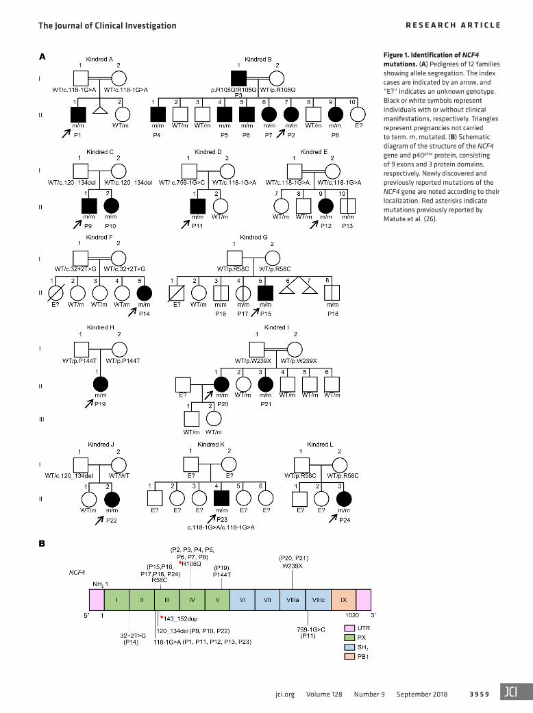
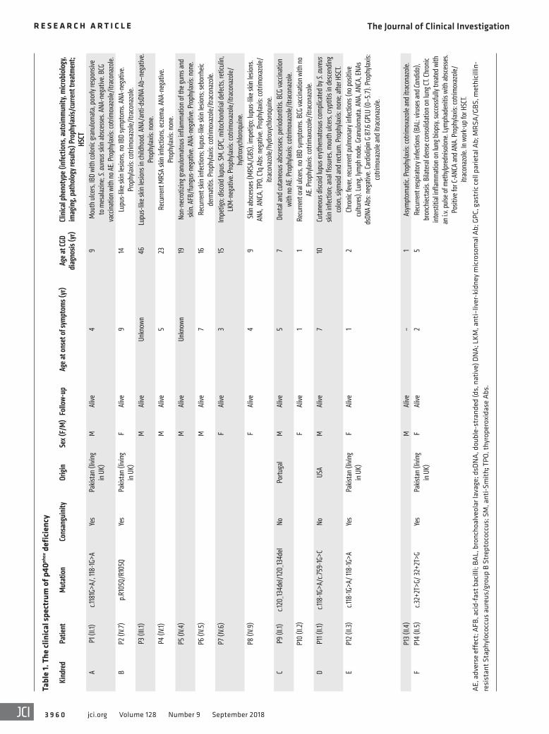
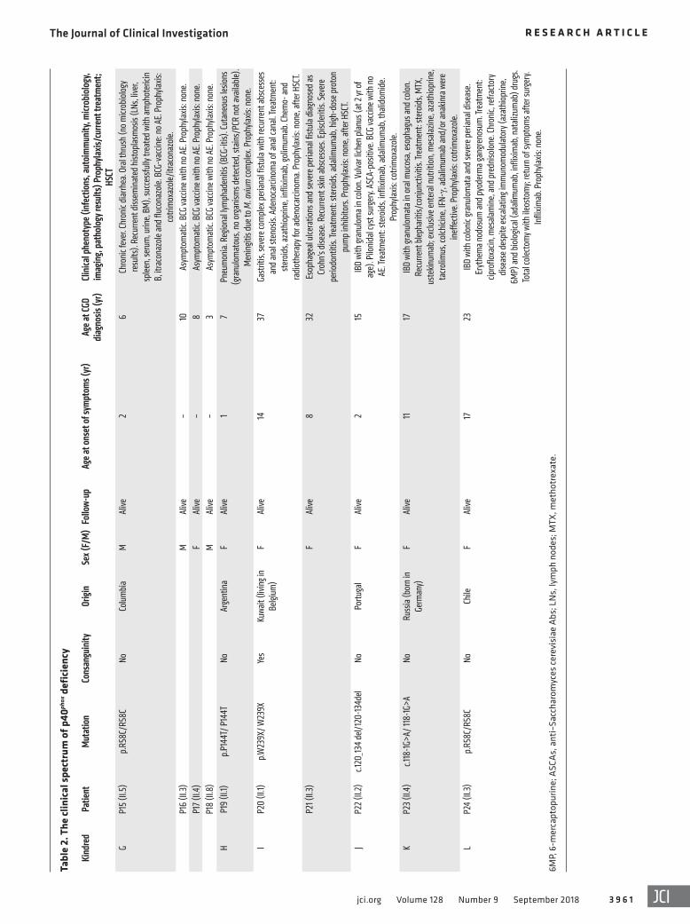
ResultsBiallelic NCF4 mutations in 24 individuals from 12 families in 8 coun-tries. We studied 20 patients from 12 families with clinical features suggestive of CGD (kindreds A–L) (Tables 1 and 2 and Figure 1A). The families originated from Pakistan (kindreds A, B, E, and F), Portugal (kindreds C and J), Australia (kindred D), Colombia (kindred G), Argentina (kindred H), Kuwait (kindred I), Russia (kindred K), and Chile (kindred L). Whole-exome sequencing (WES) or targeted next-generation sequencing (NGS) with a panel of PID genes identified homozygous variants of the NCF4 gene in patients from 11 families: 2 variants affecting essential splice sites (c.118-1G>A in kindreds A, E, and K and c.32+2T>G in kin-dred F); 3 missense variants (c.314G>A in kindred B, resulting in p.R105Q; c.172C>T in kindreds G and L, resulting in p.R58C; and c.430C>A in kindred H, resulting in p.P144T); 1 nonsense variant (c.716G>A in kindred I, resulting in p.W239X); and 1 in-frame deletion (c.120_134del in kindreds C and J). Finally, we detected compound heterozygous splice-site variants in 1 family (c.118-1G>A and c.759-1G>C in kindred D). All variants were verified by Sanger sequencing. In 11 kindreds, the familial segregation of the alleles was consistent with an AR trait. Kindred J displayed unipa-rental isodisomy (UPD), as both copies of chromosome 22 were inherited from the father (Figure 1A and ref. 38). Four asymptom-
IntroductionChronic granulomatous disease (CGD) is a recessive primary immunodeficiency (PID) caused by loss-of-function (LOF) muta-tions of autosomal or X-linked genes encoding 5 components of the phagocyte-reduced NADPH oxidase complex (1). This enzyme catalyzes the production of ROS in phagocytes. CGD was first described in 1954 (2), the first functional disorder of phagocytes was identified in 1966 (3), and the first genetic etiology of CGD, which is X-linked recessive (XR), was discovered in 1986 in the first positional identification of a PID gene (4, 5). The other genetic etiologies proved to be autosomal recessive (AR) (6). Patients with CGD suffer from recurrent, life-threatening, invasive infections with specific bacteria and fungi (7–10). Severe parasitic infections have rarely been reported in CGD patients, who also seem to have no particular predisposition to severe viral illnesses (11). Systemic hyperinflammation, including granulomatous lesions of the respi-ratory and gastrointestinal (GI) tracts in particular, is also common (12–14). Patients with CGD may initially present with unexplained granulomatosis, which is associated with a poor prognosis, (13, 15). Autoimmune conditions, such as systemic lupus erythemato-sus, are rare in patients with CGD (16). The XR (gp91phox [phago-cyte oxidase]) form of CGD is clinically more severe than the AR p47phox-deficient form (7, 9, 17). AR p22phox and p67phox deficiencies appear to be as severe as XR CGD (18, 19). Patients are routinely placed on prophylactic antimicrobial therapy, and the only cura-tive treatment widely available is allogeneic hematopoietic stem cell transplantation (HSCT) (20).
The NADPH oxidase in human phagocytes is a complex of at least 5 subunits. The membrane-bound component is a hetero-dimer consisting of gp91phox and p22phox, encoded by the X-linked CYBB gene and the autosomal CYBA gene, respectively. This heterodimer is known as flavocytochrome b558 and is the catalytic core of the NADPH oxidase complex (21). The second compo-nent is a cytosolic heterotrimer composed of p40phox, p47phox, and p67phox, which form the regulatory part of the NADPH oxidase and are encoded by NCF4, NCF1, and NCF2, respectively (22). The membrane-bound and cytosolic components do not interact in steady-state conditions. Upon phagocyte activation, after the phagocytosis of a bacterium or fungus, for example, the 2 compo-nents and a small GTPase (Rac1 and/or Rac2) are activated and assemble on the phagosomal membrane to produce superoxide (O2
–) in the phagosome (23). This O2– is subsequently converted
into hydrogen peroxide (H2O2) and other ROS, which together contribute to the destruction of the phagocytized microorganisms. LOF mutations of CYBB underlie the XR form of CGD, whereas LOF mutations of CYBA, NCF1, and NCF2 underlie the AR forms. Hypomorphic mutations of some of these genes have been shown
Authorship note: AVDG and ANP contributed equally to this work. AAA, MB, MDB, JLF, RPG, JLVH, MVH, KVL, PJHV, TKVDB, JFA, CAAF, VB, ARB, BB, CB, SOB, FC, NCB, FCH, AC, CD, MEA, DF, JIG, CG, MG, CHH, SMH, SMH, PI, HLM, IM, MMV, KM, EN, MO, LP, VR, COQ, NW, AMM, JSL, ET, JV, SV, HW, AW, LA, MCD, and PDA contributed equally to this work. DR, JLC, TWK, and JB contributed equally to this work.Conflict of interest: The authors have declared that no conflict of interest exists.Submitted: August 30, 2017; Accepted: June 14, 2018.Reference information: J Clin Invest. 2018;128(9):3957–3975. https://doi.org/10.1172/JCI97116.
The Journal of Clinical Investigation R E S E A R C H A R T I C L E
3 9 5 9jci.org Volume 128 Number 9 September 2018
Figure 1. Identification of NCF4 mutations. (A) Pedigrees of 12 families showing allele segregation. The index cases are indicated by an arrow, and “E?” indicates an unknown genotype. Black or white symbols represent individuals with or without clinical manifestations, respectively. Triangles represent pregnancies not carried to term. m, mutated. (B) Schematic diagram of the structure of the NCF4 gene and p40phox protein, consisting of 9 exons and 3 protein domains, respectively. Newly discovered and previously reported mutations of the NCF4 gene are noted according to their localization. Red asterisks indicate mutations previously reported by Matute et al. (26).
The Journal of Clinical Investigation R E S E A R C H A R T I C L E
3 9 6 0 jci.org Volume 128 Number 9 September 2018
Tabl
e 1.
The
clin
ical
spec
trum
of p
40phox
def
icie
ncy
Kind
red
Patie
ntMu
tatio
nCo
nsan
guini
tyOr
igin
Sex (
F/M)
Follo
w-up
Age a
t ons
et of
symp
toms
(yr)
Age a
t CGD
dia
gnos
is (y
r)Cli
nical
phen
otyp
e (inf
ectio
ns, a
utoim
munit
y, mi
crobio
logy,
imag
ing, p
atho
logy r
esult
s) Pr
ophy
laxis/
curre
nt tr
eatm
ent;
HSCT
AP1
(II.1
)c.1
181G
>A/,
118-1G
>AYe
sPa
kista
n (li
ving
in U
K)M
Alive
49
Mou
th u
lcers,
IBD
with
colo
nic g
ranu
lom
ata,
poor
ly re
spon
sive
to m
esala
zine;
S. au
reus s
kin ab
sces
ses.
ANA-
nega
tive.
BCG
vacc
inat
ion
with
no A
E. Pr
ophy
laxis:
cotri
mox
azol
e/itr
acon
azol
e.B
P2 (I
V.7)
p.R1
05Q/
R105
QYe
sPa
kista
n (li
ving
in U
K)F
Alive
914
Lupu
s-lik
e skin
lesio
ns, n
o IBD
sym
ptom
s. AN
A-ne
gativ
e. Pr
ophy
laxis:
cotri
mox
azol
e/itr
acon
azol
e. P3
(III.1
)M
Alive
Unkn
own
46Lu
pus-
like s
kin le
sions
in ch
ildho
od. A
NA/a
nti-d
sDNA
Ab–
nega
tive.
Prop
hylax
is: n
one.
P4 (I
V.1)
MAl
ive5
23Re
curre
nt M
RSA
skin
infe
ction
s, ec
zem
a. AN
A-ne
gativ
e. Pr
ophy
laxis:
non
e.P5
(IV.4
)M
Alive
Unkn
own
19No
n-ne
crotiz
ing g
ranu
lom
atou
s inf
lamm
atio
n of
the g
ums a
nd
skin
. AFB
/fun
gus-
nega
tive.
ANA-
nega
tive.
Prop
hylax
is: n
one.
P6 (I
V.5)
MAl
ive7
16Re
curre
nt sk
in in
fecti
ons;
lupu
s-lik
e skin
lesio
ns; s
ebor
rheic
de
rmat
itis.
Prop
hylax
is: co
trim
oxaz
ole/
itrac
onaz
ole.
P7 (I
V.6)
FAl
ive3
15Im
petig
o; d
iscoi
d lu
pus.
SM, G
PC, m
itoch
ondr
ial d
efec
ts, r
eticu
lin,
LKM
-neg
ative
. Pro
phyla
xis: c
otrim
oxaz
ole/
itrac
onaz
ole/
hydr
oxyc
hlor
oqui
ne.
P8 (I
V.9)
FAl
ive4
9Sk
in ab
sces
ses (
MRS
A/GB
S), im
petig
o; lu
pus-
like s
kin le
sions
. AN
A, A
NCA,
TPO,
C1q A
bs: n
egat
ive. P
roph
ylaxis
: cot
rimox
azol
e/itr
acon
azol
e/hy
drox
ychl
oroq
uine
.C
P9 (I
I.1)
c.120
_134d
el/12
0_13
4del
NoPo
rtuga
lM
Alive
57
Dent
al an
d cu
tane
ous a
bsce
sses
; per
iodo
ntiti
s. BC
G va
ccin
atio
n wi
th n
o AE.
Prop
hylax
is: co
trim
oxaz
ole/
itrac
onaz
ole.
P10
(II.2)
FAl
ive1
1Re
curre
nt or
al ul
cers,
no I
BD sy
mpt
oms.
BCG
vacc
inat
ion
with
no
AE. P
roph
ylaxis
: cot
rimax
ozol
e/itr
acon
azol
e.D
P11 (
II.1)
c.118
-1G>A
/c.75
9-1G
>CNo
USA
MAl
ive7
10Cu
tane
ous d
iscoi
d lu
pus e
ryth
emat
osus
com
plica
ted
by S.
aureu
s sk
in in
fecti
on; a
nal f
issur
es, m
outh
ulce
rs, cr
yptit
is in
des
cend
ing
colo
n, si
gmoi
d an
d re
ctum
. Pro
phyla
xis: n
one;
afte
r HSC
T.E
P12 (
II.3)
c.118
-1G>A
/ 118
-1G>A
Yes
Pakis
tan
(livin
g in
UK)
FAl
ive1
2Ch
roni
c fev
er, re
curre
nt p
ulm
onar
y inf
ectio
ns (n
o pos
itive
cu
lture
s). L
ung,
lymph
nod
e. Gr
anul
omat
a. AN
A, A
NCA,
ENAs
ds
DNA
Abs:
nega
tive.
Card
iolip
in G
87.6
GPL
U (0
–5.7
). Pr
ophy
laxis:
co
trim
oxaz
ole a
nd it
raco
nazo
le.
P13 (
II.4)
MAl
ive–
1As
ympt
omat
ic. P
roph
ylaxis
: cot
rimox
azol
e and
itra
cona
zole.
FP1
4 (II
.5)
c.32+
2T>G
/ 32+
2T>G
Yes
Pakis
tan
(livin
g in
UK)
FAl
ive2
5 Re
curre
nt re
spira
tory
infe
ction
s (BA
L: vir
uses
and
Cand
ida)
, br
onch
iecta
sis. B
ilate
ral d
ense
cons
olid
atio
n on
lung
CT. C
hron
ic in
terst
itial
infla
mm
atio
n on
lung
bio
psy,
succ
essfu
lly tr
eate
d wi
th
an i.v
. pul
se of
met
hylp
redn
isolo
ne. L
ymph
aden
itis w
ith ab
sces
ses.
Posit
ive fo
r C-A
NCA
and A
NA. P
roph
ylaxis
: cot
rimox
azol
e/itr
acon
azol
e. In
wor
k-up
for H
SCT.
AE, a
dver
se e
ffec
t; A
FB, a
cid-
fast
bac
illi;
BAL,
bro
ncho
alve
olar
lava
ge; d
sDN
A, d
oubl
e-st
rand
ed (d
s, n
ativ
e) D
NA
; LKM
, ant
i–liv
er-k
idne
y m
icro
som
al A
b; G
PC, g
astr
ic ce
ll pa
rieta
l Ab;
MR
SA/G
BS, m
ethi
cilli
n-re
sist
ant S
taph
yloc
occu
s au
reus
/gro
up B
Str
epto
cocc
us; S
M, a
nti-S
mith
; TPO
, thy
rope
roxi
dase
Abs
.
The Journal of Clinical Investigation R E S E A R C H A R T I C L E
3 9 6 1jci.org Volume 128 Number 9 September 2018
Tabl
e 2.
The
clin
ical
spec
trum
of p
40phox
def
icie
ncy
Kind
red
Patie
ntMu
tatio
nCo
nsan
guini
tyOr
igin
Sex (
F/M)
Follo
w-up
Age a
t ons
et of
symp
toms
(yr)
Age a
t CGD
dia
gnos
is (y
r)Cli
nical
phen
otyp
e (inf
ectio
ns, a
utoim
munit
y, mi
crobio
logy,
imag
ing, p
atho
logy r
esult
s) Pr
ophy
laxis/
curre
nt tr
eatm
ent;
HSCT
GP1
5 (II
.5)
p.R5
8C/R
58C
NoCo
lum
bia
MAl
ive2
6Ch
roni
c fev
er. Ch
roni
c diar
rhea
. Ora
l thr
ush
(no m
icrob
iolo
gy
resu
lts).
Recu
rrent
diss
emin
ated
hist
oplas
mos
is (L
Ns, li
ver,
splee
n, se
rum
, urin
e, BM
), su
cces
sfully
trea
ted
with
amph
oter
icin
B, it
raco
nazo
le an
d flu
cona
zole.
BCG
-vac
cine:
no A
E. Pr
ophy
laxis:
co
trim
oxaz
ole/
itrac
onaz
ole.
P16
(II.3
)M
Alive
–10
Asym
ptom
atic.
BCG
vacc
ine w
ith n
o AE.
Prop
hylax
is: n
one.
P17 (
II.4)
FAl
ive–
8As
ympt
omat
ic. B
CG va
ccin
e with
no A
E. Pr
ophy
laxis:
non
e.P1
8 (II
.8)
MAl
ive–
3As
ympt
omat
ic. B
CG va
ccin
e with
no A
E. Pr
ophy
laxis:
non
e.H
P19
(II.1)
p.P1
44T/
P14
4TNo
Arge
ntin
a F
Alive
1 7
Pneu
mon
ia. R
egio
nal ly
mph
aden
itis (
BCG-
itis)
. Cut
aneo
us le
sions
(g
ranu
lom
atou
s, no
orga
nism
s det
ecte
d, st
ains/
PCR
not a
vaila
ble)
. M
enin
gitis
due t
o M. a
vium
com
plex
. Pro
phyla
xis: n
one.
IP2
0 (II
.1)
p.W23
9X/ W
239X
Yes
Kuwa
it (li
ving i
n Be
lgium
)F
Alive
1437
Gast
ritis,
seve
re co
mpl
ex p
erian
al fis
tula
with
recu
rrent
absc
esse
s an
d an
al st
enos
is. A
deno
carci
nom
a of a
nal c
anal.
Trea
tmen
t: st
eroi
ds, a
zath
iopr
ine,
infli
ximab
, gol
imum
ab. C
hem
o- an
d ra
diot
hera
py fo
r ade
noca
rcino
ma.
Prop
hylax
is: n
one,
afte
r HSC
T.P2
1 (II.
3)F
Alive
832
Esop
hage
al ul
cera
tions
and
seve
re p
erian
al fis
tula
diag
nose
d as
Cr
ohn’s
dise
ase.
Recu
rrent
skin
absc
esse
s. Ep
iscler
itis.
Seve
re
perio
dont
itis.
Treat
men
t: st
eroi
ds, a
dalim
umab
, high
-dos
e pro
ton
pum
p in
hibi
tors.
Pro
phyla
xis: n
one,
afte
r HSC
T.J
P22 (
II.2)
c.120
_134
del/1
20-13
4del
NoPo
rtuga
lF
Alive
215
IBD
with
gran
ulom
a in
colo
n. Vu
lvar l
ichen
plan
us (a
t 2 yr
of
age)
. Pilo
nida
l cys
t sur
gery.
ASC
A-po
sitive
. BCG
vacc
ine w
ith n
o AE
. Tre
atm
ent:
ster
oids
, infli
ximab
, ada
limum
ab, t
halid
omid
e. Pr
ophy
laxis:
cotri
mox
azol
e.K
P23 (
II.4)
c.118
-1G>A
/ 118
-1G>A
NoRu
ssia
(bor
n in
Ge
rman
y)F
Alive
1117
IBD
with
gran
ulom
ata i
n or
al m
ucos
a, es
opha
gus a
nd co
lon.
Re
curre
nt b
lepha
ritis/
conj
uncti
vitis.
Trea
tmen
t: st
eroi
ds, M
TX,
uste
kinum
ab; e
xclu
sive e
nter
al nu
tritio
n, m
esala
zine,
azat
hiop
rine,
tacro
limus
, col
chici
ne, IF
N-γ,
adali
mum
ab an
d/or
anak
inra
wer
e in
effe
ctive
. Pro
phyla
xis: c
otrim
oxaz
ole.
LP2
4 (II
.3)
p.R5
8C/R
58C
NoCh
ileF
Alive
1723
IBD
with
colo
nic g
ranu
lom
ata a
nd se
vere
per
ianal
dise
ase.
Eryt
hem
a nod
osum
and
pyod
erm
a gan
gren
osum
. Tre
atm
ent:
cipro
floxa
cin, m
esala
min
e, an
d pr
edni
solo
ne. C
hron
ic, re
fracto
ry
dise
ase d
espi
te es
calat
ing i
mm
unom
odul
ator
y (az
athi
oprin
e, 6M
P) an
d bi
olog
ical (
adali
mum
ab, in
flixim
ab, n
atali
zum
ab) d
rugs
. To
tal c
olec
tom
y with
ileos
tom
y; re
turn
of sy
mpt
oms a
fter s
urge
ry.
Infli
ximab
. Pro
phyla
xis: n
one.
6MP,
6-m
erca
ptop
urin
e; A
SCAs
, ant
i–Sa
ccha
rom
yces
cere
visi
ae A
bs; L
Ns,
lym
ph n
odes
; MTX
, met
hotr
exat
e.
The Journal of Clinical Investigation R E S E A R C H A R T I C L E
3 9 6 2 jci.org Volume 128 Number 9 September 2018
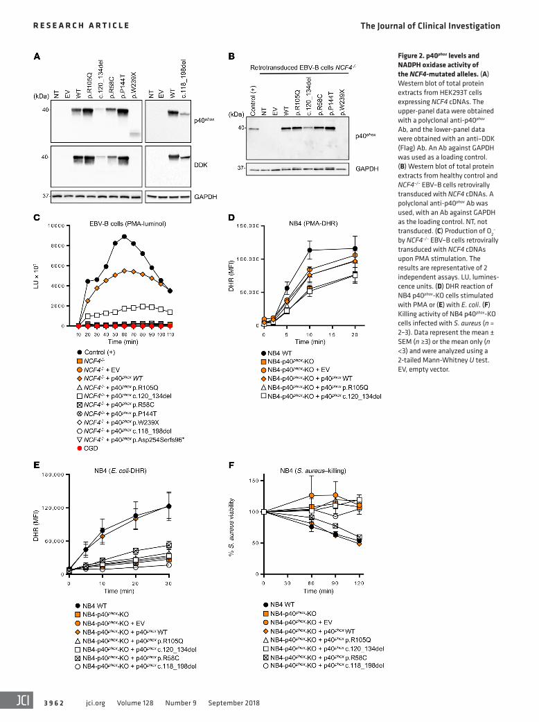
Figure 2. p40phox levels and NADPH oxidase activity of the NCF4-mutated alleles. (A) Western blot of total protein extracts from HEK293T cells expressing NCF4 cDNAs. The upper-panel data were obtained with a polyclonal anti-p40phox Ab, and the lower-panel data were obtained with an anti–DDK (Flag) Ab. An Ab against GAPDH was used as a loading control. (B) Western blot of total protein extracts from healthy control and NCF4–/– EBV–B cells retrovirally transduced with NCF4 cDNAs. A polyclonal anti-p40phox Ab was used, with an Ab against GAPDH as the loading control. NT, not transduced. (C) Production of O2
– by NCF4–/– EBV–B cells retrovirally transduced with NCF4 cDNAs upon PMA stimulation. The results are representative of 2 independent assays. LU, lumines-cence units. (D) DHR reaction of NB4 p40phox-KO cells stimulated with PMA or (E) with E. coli. (F) Killing activity of NB4 p40phox-KO cells infected with S. aureus (n = 2–3). Data represent the mean ± SEM (n ≥3) or the mean only (n <3) and were analyzed using a 2-tailed Mann-Whitney U test. EV, empty vector.
The Journal of Clinical Investigation R E S E A R C H A R T I C L E
3 9 6 3jci.org Volume 128 Number 9 September 2018
atic individuals were shown to carry biallelic mutations (kindreds E and G) (Figure 1A). None of the variants, other than p.R58C (minor allele frequency [MAF] = 0.001), was found in public data-bases (Exome Aggregation Consortium [ExAC], Human Gene Mutation Database [HGMD], gnomAD, and Biomarker Recog-nition and Validation Online [BRAVO]) or in our in-house WES database (~4,500 WES). Furthermore, combined annotation- dependent depletion (CADD) scores predicted all mutations to be deleterious, as these scores were above or close to the mutation significance cutoff (MSC) (ref. 24 and Supplemental Figure 1A; supplemental material available online with this article; https://doi.org/10.1172/JCI97116DS1). The 3 missense mutations affect residues located in the PX domain of the p40phox protein (Figure 1B). These residues are highly conserved in other species, sug-gesting that they have an important function in the p40phox pro-tein (Supplemental Figure 1B). We searched for a founder effect accounting for the presence of the c.120_134del, c.118-1G>A, and p.R58C mutations in different families. The patients homozygous for these mutations were genotyped with the genome-wide array 6.0. The c.120_134del mutation in 2 Portuguese families (kindreds C and J) is within a common haplotype of 884 kb and 1,926 SNPs, demonstrating a founder effect. We dated the most recent com-mon ancestor (MRCA) to 175 years ago (CI: 50–650 years). The c.118-1G>A variant in patients from Pakistan (kindreds A and E) is within a common haplotype of 337 kb and 136 SNPs. The MRCA of these kindreds was dated to 1,950 years ago (CI: 625–8,075 years). The patient from Russia (kindred K) also shared a common haplotype of 152 kb and 41 SNPs in common with the 2 Pakistani patients, suggesting a common ancestor for these 3 patients, and the MRCA was estimated to have lived 6,275 years ago (CI: 2,475–19,000 years) (39). Finally, the c.172C>T (p.R58C) mutation, pres-ent in 2 Latin American kindreds, was within a common haplotype of 122 kb and 47 SNPs. The MRCA of these kindreds was dated to 4,425 years ago (CI: 1,375–16,025 years). Collectively, these data suggest that 24 individuals from 12 kindreds in 8 countries suffer from inherited p40phox deficiency, including 20 symptomatic and 4 asymptomatic individuals (Figure 1A).
Impaired NCF4 mRNA expression in the patients’ cells. We measured NCF4 transcript levels in cells from the patients and healthy controls by quantitative real-time RT-PCR (qPCR). EBV- transformed B cell lines (EBV–B cells) and neutrophils homozy-gous for c.32+2T>G (patient 14 [P14]) produced no NCF4 tran-scripts detectable by qPCR (Supplemental Figure 2A) or cDNA PCR (data not shown). These findings suggest that transcripts car-rying this mutation are subjected to nonsense-mediated mRNA decay. For P12 (c.118-1G>A), P15 (p.R58C), P20 (p.W239X), and P22 (c.120_134del), we found that NCF4 transcript levels in EBV–B cells and neutrophils were lower than those in 10 unrelated con-trols (Supplemental Figure 2A). Cells homozygous for c.118-1G>A or c.120_134del produced transcripts of lower molecular weight (MW), suggesting the occurrence of alternative splicing. Using TOPO-TA cloning, we detected 3 aberrantly spliced variants for c.118-1G>A, all of which were predicted to encode truncated pro-teins, and we identified a fourth minor transcript (6%) with a large in-frame deletion, c.118_198del (Supplemental Figure 2B). For c.120_134del, most of the transcripts carried a 5-codon deletion (p.F41_V45del) at the beginning of exon 3, and another 3 aberrant-
ly spliced variants were predicted to encode truncated proteins (Supplemental Figure 2B). Finally, for the c.759-1G>C allele, we performed exon trapping. We obtained 3 spliced variants: 1 with the retention of 37 bp of intron 8a (63%), 1 in which all of intron 8a was retained (26%), and 1 with the retention of 16 bp of intron 8a (11%) (Supplemental Figure 2C).
The mutant NCF4 alleles are deleterious in EBV–B cells. We assessed the potential impact of all individual NCF4 alleles. We used HEK293T cells for analyses of p40phox protein expression, whereas, for analyses of p40phox function, we used EBV–B cells from P14 (homozygous for c.32+2T>G), designated NCF4–/–, as they produced no detectable NCF4 mRNA. We first evaluated protein expression by transfecting HEK293T cells with expression vectors carrying C terminally Flag-tagged WT or mutant NCF4 cDNAs (R58C, R105Q, P144T, W239X, c.118_198del, and c.120_134del). Immunoblot analyses revealed that the WT, R58C, R105Q, and P144T cDNAs encoded a single, full-length 40-kDa protein that was produced in large amounts. By contrast, cells transfected with the in-frame deletions c.118_198del and c.120_134del produced proteins with a lower MW, at much lower abundance. Finally, the protein encoded by W239X cDNA was not detected with an anti-Flag Ab, whereas a truncated protein of approximately 25 kDa gave a low-intensity signal with an anti-p40phox Ab (Figure 2A). We investigated the mode of functioning of the mutant alleles (LOF or hypomorphic) in EBV–B cells, which express all components of the NADPH oxidase complex and produce ROS (25, 26, 40). We transduced NCF4–/– EBV–B cells from P14 with retroviruses for the overexpression of missense (R58C, R105Q, and P144T), in-frame deletion (c.120_134del), and nonsense (W239X) cDNAs. Western blotting confirmed that the missense proteins were expressed nor-mally, whereas c.120_134del was produced in small amounts, and the W239X protein was undetectable (Figure 2B). We then eval-uated ROS production in NCF4–/– EBV–B cells by measuring O2
– levels after PMA stimulation. EBV–B cells transduced with the R58C, R105Q, P144T, c.118_198del, p.Asp254Serfs96*, and W239X alleles produced no detectable O2
–. By contrast, retrotrans-duction with the c.120_134del variant led to a small, reproducible increase in O2
– production (Figure 2C). No H2O2 was generated following the retrovirus-mediated transduction of NCF4–/– EBV–B cells with any of the mutant alleles in these conditions (Supple-mental Figure 3A). These data suggest that 6 of the 7 alleles tested were LOF, with the remaining allele (c.120_134del) being severely hypomorphic, at least under these experimental conditions.
The mutant NCF4 alleles are deleterious in NB4 cells. We then generated CRISPR-Cas9–KO p40phox NB4 cells, which we allowed to differentiate into neutrophil-like cells. NADPH oxidase activity and S. aureus killing capacity were assessed in p40phox WT cells, NB4-p40phox–KO cells, and in NB4-p40phox–KO cells reconstituted with alleles R105Q, R58C, c.120_134del, or c.118_198del or WT p40phox. We observed no decrease in PMA-induced extracellular H2O2 release or intracellular H2O2 generation in NB4-p40phox–KO cells, as was the case with cells reconstituted with any of the mutant alleles, consistent with the results obtained with neutro-phils from the previously reported p40phox-deficient patient (Fig-ure 2D, Supplemental Figure 3B, and ref. 26). Assessments of extracellular H2O2 release in response to serum-opsonized E. coli and A. fumigatus hyphae showed that NADPH oxidase activity was
The Journal of Clinical Investigation R E S E A R C H A R T I C L E
3 9 6 4 jci.org Volume 128 Number 9 September 2018
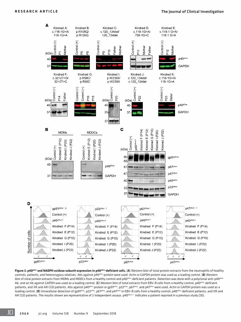
Figure 3. p40phox and NADPH oxidase subunit expression in p40phox-deficient cells. (A) Western blot of total protein extracts from the neutrophils of healthy controls, patients, and heterozygous relatives. Abs against p40phox protein were used. Actin or GAPDH protein was used as a loading control. (B) Western blot of total protein extracts from MDMs and MDDCs from a healthy control and p40phox-deficient patients. Detection was done with a polyclonal anti-p40phox Ab, and an Ab against GAPDH was used as a loading control. (C) Western blot of total extracts from EBV–B cells from a healthy control, p40phox-deficient patients, and XR and AR CGD patients. Abs against p40phox protein or gp91phox, p22phox, p67phox, and p47phox were used. Actin or GAPDH protein was used as a loading control. (D) Intracellular detection of gp91phox, p22phox, p67phox, and p47phox in EBV–B cells from a healthy control, p40phox-deficient patients, and XR and AR CGD patients. The results shown are representative of 2 independent assays. p40phox–/– indicates a patient reported in a previous study (26).
The Journal of Clinical Investigation R E S E A R C H A R T I C L E
3 9 6 5jci.org Volume 128 Number 9 September 2018
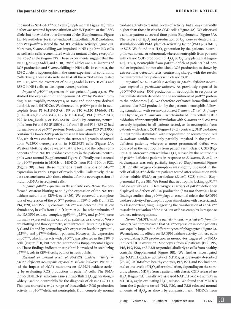
oxidase activity to residual levels of activity, but always markedly higher than those in classic CGD cells (Figure 4A). We observed a similar pattern at several time points (Supplemental Figure 5A). The release of H2O2 and production of O2
– were evaluated after stimulation with PMA, platelet-activating factor (PAF) plus fMLF, or SOZ. We found that H2O2 generation by the patients’ neutro-phils was normal or subnormal, whereas neutrophils from patients with classic CGD produced no H2O2 or O2
– (Supplemental Figure 4C). Thus, neutrophils from p40phox-deficient patients had nor-mal or impaired, but not abolished, ROS production as shown by extracellular detection tests, contrasting sharply with the results for neutrophils from patients with classic CGD.
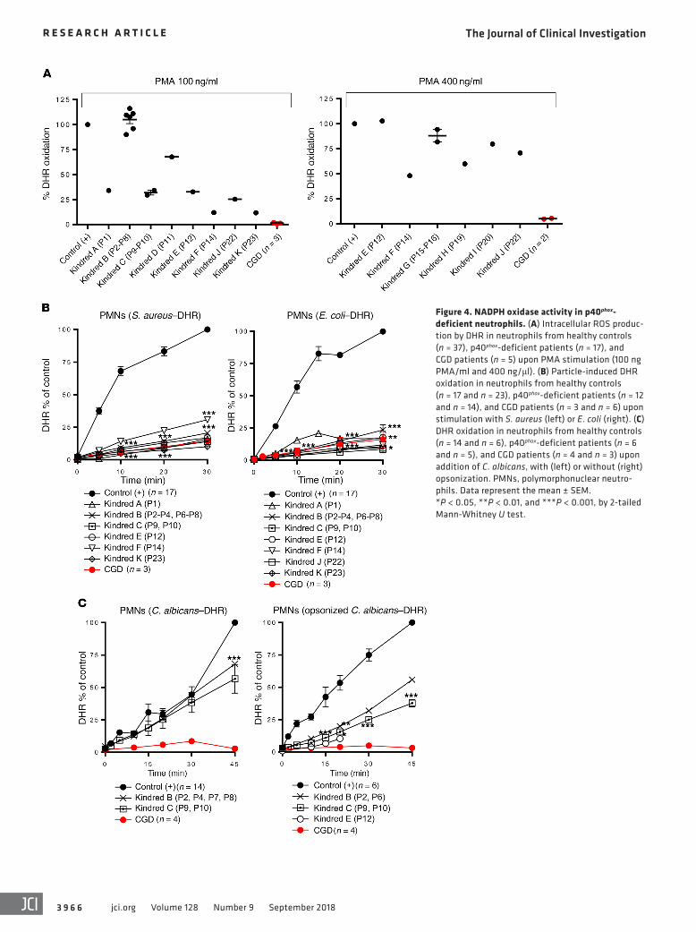
Impaired NADPH oxidase activity in p40phox-deficient neutro-phils exposed to particulate inducers. As previously reported in p40phox-KO mice, ROS production in neutrophils in response to particulate stimuli depends on the recruitment of p40phox protein to the endosomes (31). We therefore evaluated intracellular and extracellular ROS production by the patients’ neutrophils follow-ing stimulation with serum-opsonized S. aureus, E. coli, A. fumig-atus hyphae, or C. albicans. Particle-induced intracellular DHR oxidation after neutrophil stimulation with S. aureus or E. coli was severely impaired in all patients tested, as in neutrophils from patients with classic CGD (Figure 4B). By contrast, DHR oxidation in neutrophils stimulated with unopsonized or serum-opsonized C. albicans conidia was only partially impaired in the p40phox- deficient patients, whereas a more pronounced defect was observed in the neutrophils from patients with classic CGD (Fig-ure 4C). Moreover, extracellular H2O2 release by the neutrophils of p40phox-deficient patients in response to S. aureus, E. coli, or A. fumigatus was only partially impaired (Supplemental Figure 5D). Finally, oxygen consumption was slightly impaired in the cells of all p40phox-deficient patients tested after stimulation with either soluble (PMA) or particulate (E. coli, SOZ) stimuli (Sup-plemental Figure 5E). We found that neutrophils lacking gp91phox had no activity at all. Heterozygous carriers of p40phox deficiency displayed no defects of ROS production (data not shown). These findings confirm that p40phox plays an essential role in the NADPH oxidase activity of neutrophils upon stimulation with bacteria and, to a lesser extent, fungi, suggesting the translocation of at p40phox to assist in activation of the NADPH oxidase complex in response to these microorganisms.
Normal NADPH oxidase activity in other myeloid cells from the patients. We demonstrated that p40phox expression in some patients was equally impaired in different types of phagocytes (Figure 3). We analyzed the effects on NADPH oxidase activity in these cells by evaluating ROS production in monocytes triggered by PMA- induced DHR oxidation. Monocytes from 6 patients (P12, P15, P16, P19, P20, and P22) responded similarly to cells from healthy controls (Supplemental Figure 5B). We further investigated the NADPH oxidase activity of MDMs, as previously described (25, 41). MDMs from healthy controls, P12, P20, and P22 had nor-mal or low levels of H2O2 after stimulation, depending on the stim-ulus, whereas MDMs from a patient with classic CGD released no H2O2 (Figure 5A). Finally, we assessed NADPH oxidase activity in MDDCs, again evaluating H2O2 release. We found that MDDCs from the 3 patients tested (P12, P20, and P22) released normal amounts of H2O2, as shown by comparison with MDDCs from
impaired in NB4-p40phox–KO cells (Supplemental Figure 3B). This defect was restored by reconstitution with WT p40phox or the R58C allele, but not with the other 3 mutant alleles (Supplemental Figure 3B). Nevertheless, for E. coli–induced intracellular DHR oxidation, only WT p40phox restored the NADPH oxidase activity (Figure 2E). Moreover, S. aureus killing was impaired in NB4-p40phox–KO cells as well as in cells reconstituted with the mutant alleles, except for the R58C allele (Figure 2F). These experiments suggest that the R105Q, c.120_134del, and c.118_198del alleles are LOF in terms of ROS production and S. aureus killing in NB4 cells. In contrast, the R58C allele is hypomorphic in the same experimental conditions. Collectively, these data indicate that all the NCF4 alleles tested are LOF, with the exception of c.120_134del in EBV–B cells and R58C in NB4 cells, at least upon overexpression.
Impaired p40phox expression in the patients’ phagocytes. We studied the expression of endogenous p40phox by Western blot-ting in neutrophils, monocytes, MDMs, and monocyte-derived dendritic cells (MDDCs). We detected no p40phox protein in neu-trophils from P1 (c.118-1G>A), P9 or P10 (c.120_134del), P11 (c.118-1G>A/c.759-1G>C), P12 (c.118-1G>A), P14 (c.32+2T>G), P22 (c.120_134del), or P23 (c.118-1G>A). By contrast, neutro-phils from P6 and P8 (R105Q) and from P15 and P16 (R58C) had normal levels of p40phox protein. Neutrophils from P20 (W239X) contained a lower-MW protein present at low abundance (Figure 3A), which was consistent with the truncated protein observed upon W239X overexpression in HEK293T cells (Figure 2A). Western blotting also revealed that the levels of the other com-ponents of the NADPH oxidase complex in the patients’ neutro-phils were normal (Supplemental Figure 4). Finally, we detected no p40phox protein in MDMs or MDDCs from P12, P20, or P22, (Figure 3B). Thus, these mutations result in a loss of p40phox expression in various types of myeloid cells. Collectively, these data are consistent with those obtained for the overexpression of mutant cDNAs in recipient cells.
Impaired p40phox expression in the patients’ EBV–B cells. We per-formed Western blotting to study the expression of the NADPH oxidase subunits in EBV–B cells (26). We observed a complete loss of expression of the p40phox protein in EBV–B cells from P12, P14, P20, and P22. By contrast, p40phox was detected, but at low abundance, in cells from P15 (Figure 3C). The other subunits of the NADPH oxidase complex, gp91phox, p22phox, and p47phox, were normally expressed in the cells of all patients, as shown by West-ern blotting and flow cytometry with intracellular staining (Figure 3, C and D) and by comparing with expression levels in gp91phox-, p22phox-, and p47phox-deficient patients. However, the expression of p67phox, which interacts with p40phox, was affected in the EBV–B cells (Figure 3D), but not the neutrophils (Supplemental Figure 4). These findings indicate that p40phox is involved in stabilizing p67phox levels in EBV–B cells, but not in neutrophils.
Residual to normal levels of NADPH oxidase activity in p40phox-deficient neutrophils exposed to soluble inducers. We stud-ied the impact of NCF4 mutations on NAPDH oxidase activi-ty by evaluating ROS production in patients’ cells. The PMA- induced DHR test, which measures intracellular H2O2 generation, is widely used on neutrophils for the diagnosis of classic CGD (1). This test showed a wide range of intracellular ROS production activity in p40phox-deficient neutrophils, from completely normal
The Journal of Clinical Investigation R E S E A R C H A R T I C L E
3 9 6 6 jci.org Volume 128 Number 9 September 2018
Figure 4. NADPH oxidase activity in p40phox- deficient neutrophils. (A) Intracellular ROS produc-tion by DHR in neutrophils from healthy controls (n = 37), p40phox-deficient patients (n = 17), and CGD patients (n = 5) upon PMA stimulation (100 ng PMA/ml and 400 ng/μl). (B) Particle-induced DHR oxidation in neutrophils from healthy controls (n = 17 and n = 23), p40phox-deficient patients (n = 12 and n = 14), and CGD patients (n = 3 and n = 6) upon stimulation with S. aureus (left) or E. coli (right). (C) DHR oxidation in neutrophils from healthy controls (n = 14 and n = 6), p40phox-deficient patients (n = 6 and n = 5), and CGD patients (n = 4 and n = 3) upon addition of C. albicans, with (left) or without (right) opsonization. PMNs, polymorphonuclear neutro-phils. Data represent the mean ± SEM. *P < 0.05, **P < 0.01, and ***P < 0.001, by 2-tailed Mann-Whitney U test.
The Journal of Clinical Investigation R E S E A R C H A R T I C L E
3 9 6 7jci.org Volume 128 Number 9 September 2018
the patients and neutrophils from healthy controls (Supplemental Figure 7). Finally, we evaluated the formation of neutrophil extra-cellular traps (NETs), a process for which ROS have been shown to be essential (46). We assessed NET formation in response to PMA in 3 patients from kindreds A, B, and C. Only neutrophils from kin-dred B (expressing p40phox protein) produced NETs. We detected no NET production in the other p40phox-deficient families tested (without p40phox protein expression) or for gp91phox-deficient CGD neutrophils (Supplemental Figure 8). These findings suggest that NET formation is dependent on p40phox expression. Collectively, these data indicate that p40phox-deficient neutrophils have normal fungus-killing capacities, unlike classic CGD neutrophils.
Inflammatory lesions in patients with p40phox deficiency. Defi-ciency of p40phox is not characterized by the invasive bacterial and fungal infections seen in patients with classic CGD, but mostly by inflammatory manifestations as well as autoimmunity and periph-eral infections (Tables 1 and 2 and Figure 7A). We identified 20 symptomatic patients, of whom 10 (50%) presented with skin inflammation (P1, P2, P3, P5, P6, P7, P8, P11, P19, and P24), with lupus-like cutaneous lesions (P1, P6, and P8) or discoid lupus (P3, P7, and P11). We found inflammatory, granulomatous GI manifes-tations in 10 patients (50%), and these included oral ulcers (P1 and P10), abscesses/granulomas (P1, P2, P3, P4, P5, P6, P7, P8, P9, P10, P11, P23, and P24), periodontitis (P9 and P21), gingivitis or gum inflammation (P9, P10, P20, P21), esophagitis (P21 and P23), gastritis (P20), Crohn-like inflammatory bowel disease (IBD) (P1, P11, P21, P22, P23, and P24), and perianal abscesses/fistula (P11, P20, P21, and P24). P24 suffered from chronic and severe IBD requiring immunosuppressive treatment and a total colectomy. Some lesions were probably both infectious and inflammatory, as is often seen in CGD. Three patients (15.7%) (P12, P14, and P19) reportedly suffered from pulmonary infections, including P12, who was later diagnosed with interstitial lung disease that responded well to systemic steroids rather than antibiotics, sug-gesting an autoinflammatory component. Ab evaluations were performed in some patients (n = 7) suffering from exacerbated inflammation. P1, P4, P5, P7, P8, and P12 were negative for anti-nuclear Abs (ANAs). By contrast, P14 was positive for ANAs and antineutrophil cytoplasmic Abs (ANCAs), suggesting autoimmune activity in this patient.
Peripheral but not invasive infections in patients with p40phox deficiency. We investigated the possible infectious causes of pulmonary manifestations in the cohort. No pathogen was iden-tified in P19. Cutaneous infections were reported in 8 patients (42%) (P1, P4, P6, P7, P9, P11, P21, and P23), 3 of whom had at least 1 proven episode of S. aureus infection (P1, P11, and P23). One patient (P15) had two episodes of disseminated histoplas-mosis, which was successfully treated with antifungal drugs and steroids. Ten patients from six different families were vaccinated against tuberculosis with live bacillus Calmette-Guérin (BCG) (Tables 1 and 2). No adverse effects were reported in 9 patients, whereas P19 developed local lymphadenitis (BCG-itis). This same patient subsequently suffered from meningitis caused by Mycobacterium avium. Interestingly, this patient was also hetero-zygous for a dominant-negative LOF mutation of STAT1, which is responsible for MSMD (47). Four asymptomatic children, aged 1, 3, 8, and 10 years at diagnosis of p40phox deficiency, were
healthy controls, and unlike MDDCs from classic CGD patients (Figure 5B). Overall, these data suggest that p40phox is redundant in monocytes, MDMs, and MDDCs in terms of the NAPDH oxidase activity triggered by PMA. Further experiments assessing particle- induced ROS production by monocytes, MDMs, and MDDCs are required to determine the extent of this redundancy.
Impaired NADPH oxidase activity in EBV–B cells from patients. We tested EBV–B cells from the p40phox-deficient patients studied here, patients with classic CGD, and the first reported patient with p40phox deficiency (42). In luminol bioluminescence assays, EBV–B cells from P12, P14, P15, and P20 displayed severely impaired NADPH oxidase activity (Figure 5C). By contrast, EBV–B cells from P22 (with c.120_134del) displayed residual luminescence, consistent with the results obtained following the retrovirus- mediated transduction of NCF4–/– EBV–B cells with the c.120_134del allele (Figure 2C). This allele is, therefore, also con-stitutively hypomorphic. We then evaluated the release of H2O2 in EBV–B cells from P12, P14, and P20. H2O2 detection was com-pletely negative, as in cells from classic CGD patients. By contrast, EBV–B cells from P22 and P15 displayed a residual release of H2O2 (Figure 5C). Finally, EBV–B cells from P12, P14, P15, and P20 were transduced with a p40phox WT cDNA. The rescue of p40phox protein expression was confirmed by immunoblotting before functional assays (Supplemental Figure 6A). Production of O2
– in response to PMA stimulation was restored in the transduced EBV–B cells (Supplemental Figure 6B). These experiments with EBV–B cells demonstrate that p40phox is not redundant for NADPH oxidase complex activity in these cells. They also confirm that 1 of the 8 NCF4 alleles is severely hypomorphic.
Impaired killing of Staphylococcus, but not of fungi, by the patients’ cells. We then assessed the ability of the patients’ neu-trophils to kill various microorganisms that commonly cause dis-ease in patients with classic CGD. We found that the phagocyto-sis of serum-opsonized E. coli, S. aureus, C. albicans, and SOZ by the patients’ neutrophils was similar to that by neutrophils from healthy controls and patients with classic CGD (data not shown). Nonoxidative phagocyte killing involves the release of the proteo-lytic content of granules into the phagosome or at the neutrophil plasma membrane. In the p40phox-deficient neutrophils tested, protease release from the azurophilic granules (elastase, cathep-sin G) in the dye-quenched–BSA (DQ-BSA) assay did not differ from that of neutrophils from healthy controls (data not shown) or patients with classic CGD (43). We therefore analyzed the phagocyte-mediated killing of different species of bacteria and fungi. The killing of S. aureus by the patients’ neutrophils was mark-edly impaired for all the patients tested, similar to that observed for the classic CGD neutrophils (Figure 6A and ref. 44). E. coli kill-ing was unaffected in the patients’ neutrophils, as in those from patients with classic CGD (Figure 6B and ref. 45). By contrast, the killing of A. fumigatus was normal or subnormal, whereas neutro-phils from patients with classic CGD were completely incapable of killing these hyphae (Figure 6C). C. albicans killing by the patients’ neutrophils was similar to that by the neutrophils of healthy control cells when C. albicans was opsonized, whereas neutrophils from patients with classic CGD were unable to kill this fungus (Figure 6D). Moreover, we observed that inhibition of the germination and hyphenation of Candida conidia were similar for neutrophils from
The Journal of Clinical Investigation R E S E A R C H A R T I C L E
3 9 6 8 jci.org Volume 128 Number 9 September 2018
eral infections, mostly of the skin and lungs, and inflammation, mostly of the GI tract (Tables 1 and 2).
Favorable clinical outcome of p40phox-deficient individuals. Inher-ited p40phox deficiency differs from classic CGD in terms of cellular
found to have homozygous NCF4 mutations (P13, P16, P17, and P18), demonstrating incomplete clinical penetrance of the dis-ease, at least until adolescence. In summary, p40phox deficiency conferred a variable phenotype marked by noninvasive periph-
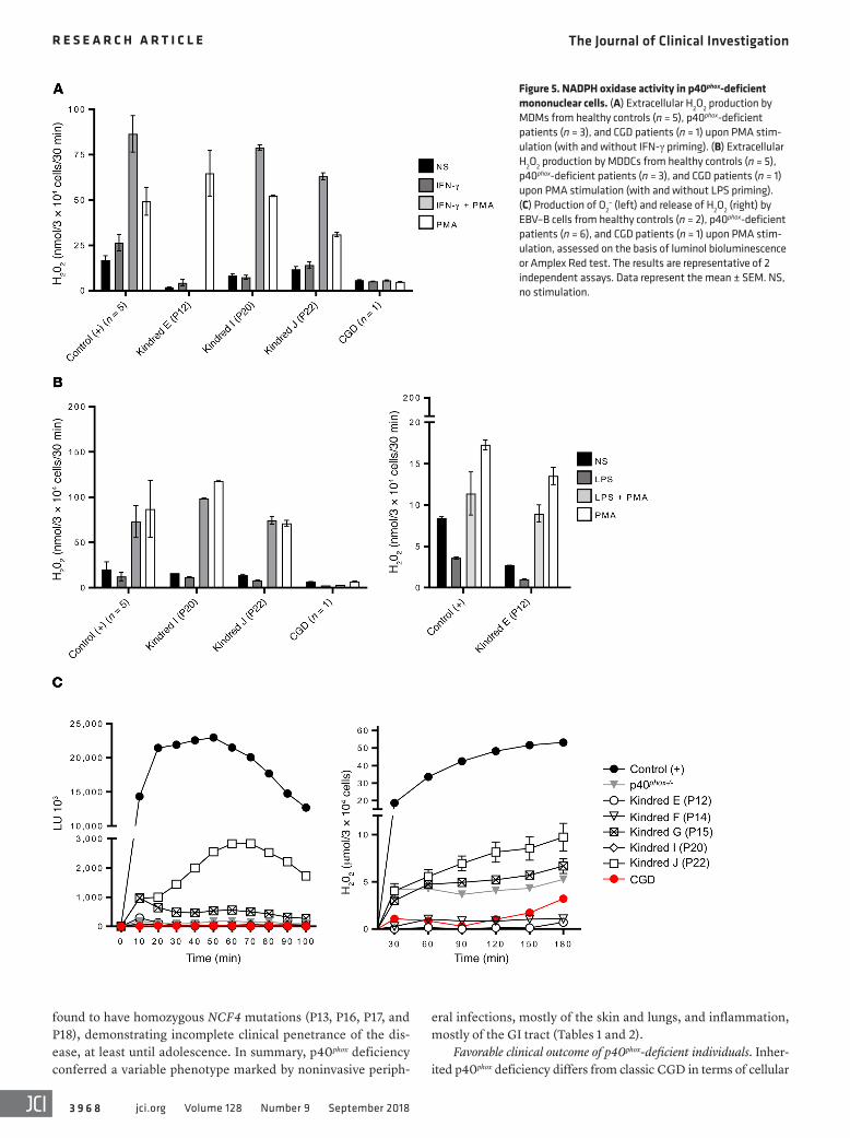
Figure 5. NADPH oxidase activity in p40phox-deficient mononuclear cells. (A) Extracellular H2O2 production by MDMs from healthy controls (n = 5), p40phox-deficient patients (n = 3), and CGD patients (n = 1) upon PMA stim-ulation (with and without IFN-γ priming). (B) Extracellular H2O2 production by MDDCs from healthy controls (n = 5), p40phox-deficient patients (n = 3), and CGD patients (n = 1) upon PMA stimulation (with and without LPS priming). (C) Production of O2
– (left) and release of H2O2 (right) by EBV–B cells from healthy controls (n = 2), p40phox-deficient patients (n = 6), and CGD patients (n = 1) upon PMA stim-ulation, assessed on the basis of luminol bioluminescence or Amplex Red test. The results are representative of 2 independent assays. Data represent the mean ± SEM. NS, no stimulation.
The Journal of Clinical Investigation R E S E A R C H A R T I C L E
3 9 6 9jci.org Volume 128 Number 9 September 2018
are between the ages of 1 and 47 years (mean: 15.3 years), whereas a mortality of close to 4% per year has been reported for classic CGD (6, 17), with the highest mortality rates during the first 2 decades of life. The Kaplan-Meier curves for the survival of the largest classic CGD cohort and this p40phox-deficient cohort differed significant-ly when comparing XR-CGD and p40phox deficiency (P = 0.0164, Mantel-Cox analysis) (Figure 7C and Supplemental Table 2). Fourteen patients were prescribed antibiotic prophylaxis. The
phenotype and clinical manifestations, but also in terms of clinical outcome (Supplemental Table 1). The mean age at symptom onset was 6 years (range: 1–17 years), later than what was reported for symptom onset in most patients with classic CGD (Supplemental Table 2). The mean age at diagnosis (for patients with symptoms) was 15.3 years (range: 1–46 years), whereas classic CGD is typically diagnosed before the age of 5 years (Figure 7B and Supplemental Table 2). All patients included in this study are currently alive and
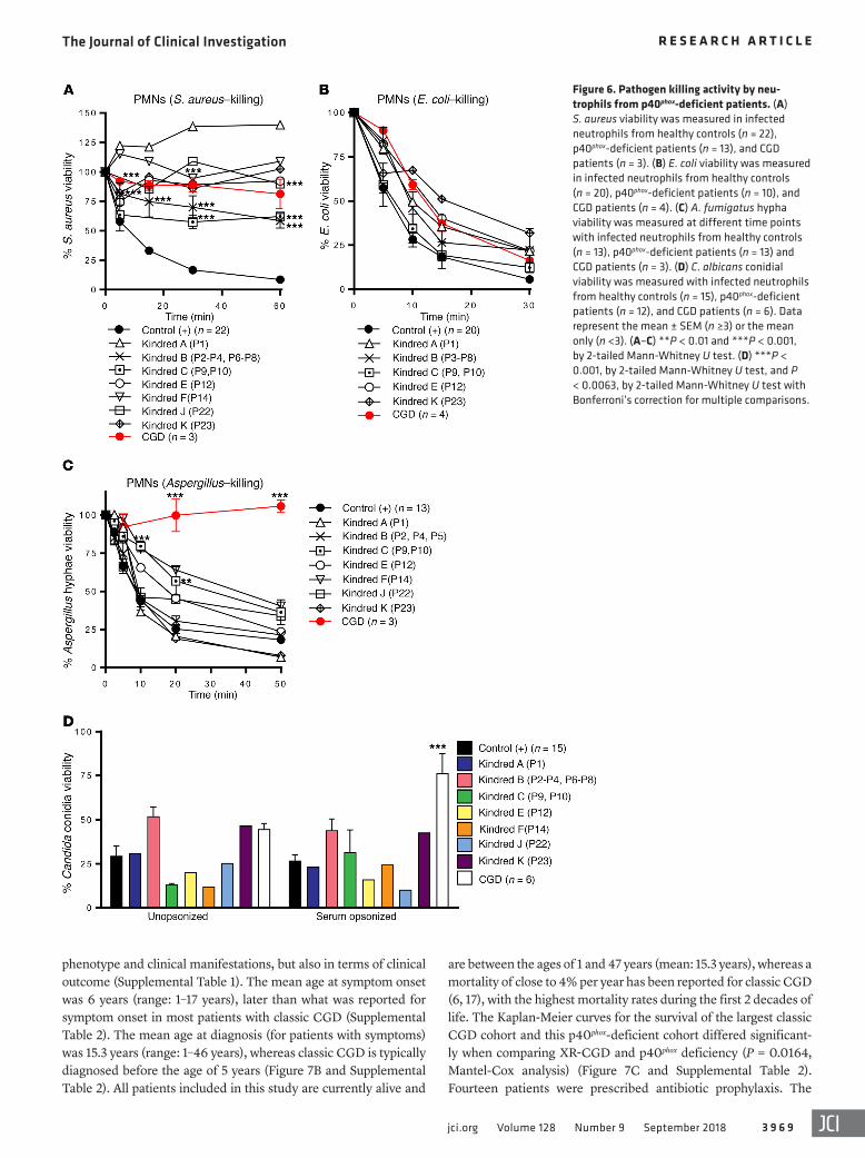
Figure 6. Pathogen killing activity by neu-trophils from p40phox-deficient patients. (A) S. aureus viability was measured in infected neutrophils from healthy controls (n = 22), p40phox-deficient patients (n = 13), and CGD patients (n = 3). (B) E. coli viability was measured in infected neutrophils from healthy controls (n = 20), p40phox-deficient patients (n = 10), and CGD patients (n = 4). (C) A. fumigatus hypha viability was measured at different time points with infected neutrophils from healthy controls (n = 13), p40phox-deficient patients (n = 13) and CGD patients (n = 3). (D) C. albicans conidial viability was measured with infected neutrophils from healthy controls (n = 15), p40phox-deficient patients (n = 12), and CGD patients (n = 6). Data represent the mean ± SEM (n ≥3) or the mean only (n <3). (A–C) **P < 0.01 and ***P < 0.001, by 2-tailed Mann-Whitney U test. (D) ***P < 0.001, by 2-tailed Mann-Whitney U test, and P < 0.0063, by 2-tailed Mann-Whitney U test with Bonferroni’s correction for multiple comparisons.
The Journal of Clinical Investigation R E S E A R C H A R T I C L E
3 9 7 0 jci.org Volume 128 Number 9 September 2018
ciency of p40phox is caused by homozygous (23 patients, 11 fami-lies) or compound heterozygous (1 patient) mutations in families from 8 countries. We identified 8 previously undescribed alleles of the NCF4 gene, 6 of which are completely LOF and caused complete p40phox deficiency in the corresponding patients. Upon overexpression, the allele c.120_134del is hypomorphic in EBV–B cells, and the allele R58C is hypomorphic in NB4 cells. However, we detected no clinical difference between the patient bearing these variants and the other patients. We further characterized phagocytic cells from the p40phox–deficient patients and com-pared them with those of patients with other molecular causes of CGD to define more clearly the contribution of human p40phox to NADPH oxidase activity. Interestingly, NCF4 biallelic mutations
treatments administered for hyperinflammation included corti-costeroids, azathioprine, methotrexate, infliximab, adalimumab, golimumab, ustekinumab, and hydroxychloroquine. Given the severity of clinical manifestations, 4 patients underwent allogeneic HSCT (P11, P14, P20, and P21), resulting in the resolution of symp-toms in all these patients. Overall, p40phox deficiency is a severe condition, but its clinical outcome is nevertheless better than that of classic CGD.
DiscussionWe report the molecular, cellular, and clinical characteristics of 24 patients from 12 families with biallelic mutations of the NCF4 gene, which encodes the NADPH oxidase p40phox protein. Defi-
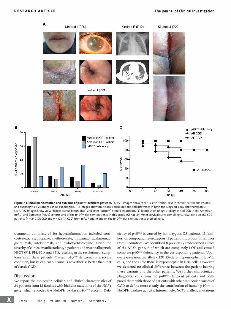
Figure 7. Clinical manifestation and outcome of p40phox-deficient patients. (A) P20 images show cheilitis, episcleritis, severe chronic cutaneous lesions, and esophagitis. P21 images show esophagitis. P12 images show multifocal consolidations and infiltrates in both the lungs on x-ray and thorax on CT scan. P22 images show vulvar lichen planus before (top) and after (bottom) steroid treatment. (B) Distribution of age at diagnosis of CGD in the American (ref. 7) and European (ref. 9) cohorts and of the p40phox-deficient patients in this study. (C) Kaplan-Meier survival curve compiling survival data on 363 CGD patients (n = 240 XR CGD and n = 123 AR CGD) from refs. 7 and 19 and on the p40phox-deficient patients studied here.
The Journal of Clinical Investigation R E S E A R C H A R T I C L E
3 9 7 1jci.org Volume 128 Number 9 September 2018
the patients in our cohort (P20) had adenocarcinoma, possibly related to chronic Crohn’s colitis, although other factors may also be involved.
Other clinical features of the patients can be understood in light of their cellular phenotypes. In vitro S. aureus killing by neu-trophils from patients with p40phox deficiency or classic CGD was severely impaired, whereas E. coli killing by neutrophils from these patients was similar to that by neutrophils from healthy controls. S. aureus killing is a ROS-dependent process, whereas E. coli killing is relatively ROS independent (45, 51). The absence of invasive S. aureus infections in the p40phox-deficient patients was unexpected and remains unexplained, as both in vitro S. aureus–induced oxi-dase activity and S. aureus killing are as severely impaired in these patients as in those with classic CGD, who do suffer from invasive S. aureus infections. The almost normal killing of fungi by neu-trophils from p40phox-deficient patients, even though the killing of Candida and Aspergillus is ROS dependent (52, 53), is of direct clinical relevance and contrasts with the situation in patients with classic CGD (54). The lack of invasive fungal infections is highly consistent with our in vitro biochemical findings for phagocytic cells from p40phox-deficient patients. Residual NADPH oxidase activity was generally higher in p40phox-deficient cells than in classic CGD cells, and in vitro fungal killing was largely unaffect-ed. This situation contrasts with that for classic CGD, in which invasive fungal infections are among the most feared and life- threatening complications, despite antimicrobial prophylaxis (10), and for which fungus killing is impaired in vitro. Given the impor-tance of p40phox-PX interactions with PI(3)P in the phagosome for the NADPH oxidase activity elicited by bacteria, the lack of effect on C. albicans–induced ROS production and killing is surprising. Normal fungal killing in these patients may also result from the residual fungus-induced NADPH oxidase activity observed, sug-gesting a p40phox-independent mechanism for ROS production that is preferentially triggered by fungi. This implies that ROS pro-duction would have to exceed a certain threshold for effective fun-gal killing. Further studies of late phagosome formation in relation to p40phox localization and ROS formation might clarify the differ-ences in S. aureus and C. albicans killing, which diverge in patients with p40phox deficiency, but not in those with classic CGD.
Interestingly, our studies of monocytes, MDMs, and MDDCs suggest that these cell types are not dependent on p40phox for PMA-induced NADPH oxidase activity. This situation contrasts with that for MSMD-causing gp91phox mutations, which disrupt NADPH oxidase activity in MDMs but not in other phagocytes (neutrophils, monocytes, and MDDCs) (25, 41). The conserva-tion of NADPH oxidase activity may explain the milder infectious phenotype of p40phox-deficient patients than of patients with clas-sic CGD, including the absence of invasive bacterial and fungal infections. This feature may be specific to human cells, as p40phox- mutant mouse macrophages (either KO or R58A mice) have impaired NADPH oxidase activity upon stimulation with a phys-iological stimulus (36). The PX domain of p40phox is thought to have a high affinity for and specificity of binding to PI(3)P, a lipid on phagosomal membranes, leading to enhanced NADPH oxi-dase activity. Further studies are required to evaluate NADPH oxidase activity in phagocytic cells other than neutrophils in patients with p40phox deficiency or classic CGD. In this respect,
were found to result in a cellular phenotype related to but clearly different from that of other forms of CGD. In particular, the PMA-induced DHR oxidation test widely used for the diagnosis of CGD gave normal or just below normal values for patients with inherited p40phox deficiency. Diagnosis of p40phox deficiency can be achieved using serum-opsonized E. coli as a stimulus in routine DHR- or Amplex Red–based tests, which must be performed in a diagnostic laboratory with expertise in analysis of NADPH oxidase activity. A genetic approach based on Sanger sequencing or NGS of NCF4 might also facilitate the diagnosis of p40phox deficiency.
The clinical phenotype of patients with p40phox deficiency is also related to but different from that of patients with clas-sic CGD. The patients in this cohort had peripheral infections (mostly staphylococcal) and hyperinflammation (mostly gran-ulomatous lesions of the GI tract and skin). Nevertheless, the frequency of inflammatory lesions is lower than in patients with classic CGD (48). A few of the patients showed autoimmunity, but, strikingly, none had the invasive bacterial and fungal infec-tions commonly seen in patients with classic CGD (7–9, 18). Consistent with these findings, the clinical outcome of p40phox deficiency is better than that of classic CGD. No deaths were recorded in this cohort of patients aged 1 to 46 years, whereas classic CGD–related mortality may reach 35% in its XR and 15% in its AR forms in the first 3 decades of life (10, 17). Moreover, 4 patients remained asymptomatic at 1 to 10 years of age, and the age at clinical onset ranged from 1 to 17 years in patients with symptoms. The incomplete clinical penetrance observed at 1 to 10 years of age also contrasts strongly with observations for clas-sic CGD (7–9, 18). Thus, the healthy siblings of probands should be tested immunologically and genetically. Curative and pre-ventive treatments for CGD are based on prolonged treatment with regimens of antibiotics, antifungal drugs, exogenous IFN-γ, and, increasingly, allogeneic HSCT. Decisions concerning allo-geneic HSCT for patients with p40phox deficiency should be made on a case-by-case basis, given the milder overall phenotype and greater variability between patients. Likewise, the immediate initiation of antifungal prophylaxis may not be required, whereas treatment with immunosuppressive agents may be indicated.
One patient with disseminated histoplasmosis (P15) was included in this cohort. However, to our knowledge, no patient with CGD has ever been reported to suffer from this fungal dis-ease. This situation is reminiscent of that of tuberculosis, which is actually much more common than initially thought in CGD patients living in areas of endemic disease (49). Its pathogen-esis has been attributed to defects of NADPH oxidase activity in mononuclear phagocytes (25). The normal NADPH oxidase activity of the p40phox-deficient mononuclear phagocytes tested suggests that histoplasmosis may actually be due to the disrup-tion of NADPH oxidase activity in tissue-resident macrophages and DCs, which were not tested in patients with classic CGD or p40phox deficiency (50). Another patient in this cohort (P19) had BCG and M. avium diseases. BCG disease has been reported in CGD patients, but cases of M. avium complex infection have rarely been reported in this population. Moreover, these 2 myco-bacterial infections are probably not related to p40phox deficien-cy in P19, because this patient also carries a dominant-nega-tive STAT1 mutation responsible for MSMD (47). Finally, 1 of

The Journal of Clinical Investigation R E S E A R C H A R T I C L E
3 9 7 2 jci.org Volume 128 Number 9 September 2018
Scientific). Cells were stimulated with PMA (100 ng/ml), PAF (1 μM) followed by fMLF (1 μM) (all from Sigma-Aldrich), E. coli (OD600 0.1, strain ML-35 in the presence of 2 mM azide), and S. aureus (OD600 0.1, strain Oxford in the presence of 2 mM azide) or A. fumigatus hyphae (clinical isolate). We used 0.25 × 10⁶ cells/ml, and the experiments were performed as previously described (65). The same method was used for the MDMs, MDDCs, and EBV–B cells, except that 400 ng/ml PMA was also added to the cells. The above-mentioned reagents and E. coli, S. aureus, and A. fumigatus strains were used in all experiments. Particles were opsonized with 10% (v/v) pooled human serum. MDMs and MDDCs were obtained by differentiation as previously described (25, 41). MDMs were cultured for 16 to 18 hours with 1 mg/ml puri-fied protein derivatives (PPDs) (tuberculin PPD Batch RT49, Statens Serum Institute) or 5 × 10³ IFN-γ IU/ml (Imukin, Boehringer Ingel-heim) and then washed and activated by incubation for 30 minutes with PMA, or left unactivated. MDDCs were treated with LPS (1 μg/ml Salmonella minnesota, Sigma-Aldrich), alone or immediately before treatment with PMA.
The production of intracellular ROS by neutrophils, monocytes, and NB4 cells was analyzed with DHR (Thermo Fisher Scientif-ic), as previously described (66). We added 0.5 μM DHR in DMSO to 2.5 × 10⁶ neutrophils. PMA (100 ng/ml), serum-opsonized E. coli or S. aureus (OD600 0.1 in the presence of 2 mM azide), C. albicans (OD625 0.1, strain SC5314), or SOZ (0.1 mg/ml) was then added. At the desired time points, samples were added to STOP buffer (0.5% [w/v] PFA, 1% [w/v] BSA, and 20 mM NaF in PBS), and the amount of rhodamine-1,2,3 (mean MFI [mean fluorescence intensity]) was assessed by flow cytometry using a FACSCanto II/LSR (BD Bioscienc-es) and expressed as the percentage of rhodamine-1,2,3 produced by healthy control cells.
ROS production by monocytes was analyzed by assessing DHR after stimulation with PMA (400 ng/ml) in kindreds E, F, G, H, and J, using a Galios FACS Analyzer (Beckman Coulter) and an LSR Fortessa Cell Analyzer (BD). DHR is expressed as a percentage of the control, and MFI values were normalized against the healthy control.
Generation of NB4 p40phox-KO cells. The Optimized CRISP Design Tool (http://crispr.mit.edu/) was used (Zhang laboratory) to deter-mine the Cas9 target sites present in the coding sequence of p40phox. An oligomer was cloned into the BsmBI site of pLentiCRISPR v2. The constructs were grown in E. coli Stbl3. Lentiviral particles were gen-erated by the transient cotransfection of HEK293T cells with pLenti-CRISPRv2, p40phox-KO, psPAX2, and pCMV-VSVg using TransIT-LT1 Transfection Reagent (Mirusbio). The day after transfection, the cells were plated on NB4 medium. The virus-containing supernatant was harvested on days 2 and 3 after transfection and filtered through 0.45-μm pores, and 1 ml was used with 5 × 10⁵ NB4 cells on 2 successive days. Transduced NB4 cells were selected with 1 μg/ml puromycin (InvivoGen). Surviving cells were plated on limiting dilution medium in a 96-well plate at a density of 0.5 cells/150 μl, and growing clones were routinely maintained. The p40phox levels of these clones were assessed by Western blotting. A clone with no p40phox expression was obtained when the 5′-GTCAGCAATGTTGGCCGAGA-3′ target sequence was used.
Statistics. Statistical analysis was performed with GraphPad Prism, version 7.02 (GraphPad Software). All data sets were considered non-normally distributed, as the numbers were too low for normality tests. A 2-tailed Mann Whitney U test was used for statistical analy-
approaches involving the derivation of different types of phago-cytes from induced pluripotent stem cells may be particularly effective (55).
Excessive granulomatous inflammation of the skin and GI tract was the most common clinical phenotype observed in this cohort of patients. Systemic lupus–like symptoms are rare in patients with CGD (7, 12, 20) and in the p40phox-deficient patients reported here. Enhanced inflammation has also been reported in mice with p40phox deficiency (36, 37, 56). Mouse p40phox has been reported to be required for the expression of glycan structure–modifying enzymes, which may underlie the recruitment of neu-trophils to resolve intestinal inflammation and facilitate wound healing (37). Mouse macrophages with p40phox p.R58A/R58A or KO mutations also produce smaller than normal amounts of ROS (36). The excessive inflammation seen in classic CGD and p40phox deficiency indicates that ROS play a role in immune regulation (12, 57). Patients with p40phox deficiency display at least as much inflammation as those with classic CGD, but have higher levels of ROS production. The ROS produced by neutrophils may con-tribute to the clearance of apoptotic cells or cell debris, and their deficiency may contribute to inflammation in various ways, including aberrant efferocytosis, for example (58–62). Alterna-tively, the inflammation observed in classic CGD and p40phox deficiency may be due to abnormal ROS production by circulating B cells. This possibility was already suggested in the first report describing a p40phox-deficient patient (42). Our results also suggest that NADPH activity is defective in all p40phox-deficient EBV–B cells tested, confirming that p40phox is not redundant in human B cells. An understanding of the excessive inflammation in patients with classic CGD or p40phox deficiency will require further stud-ies of the respiratory burst in various other cell types, including tissue-resident DCs and macrophages, and in different types of circulating B cells.
MethodsCell lines. NB4 cells were obtained from DSMZ (catalog ACC 207). HEK293T cells were obtained from the American Tissue Culture Col-lection (ATCC) (catalog CRL-3216).
Ion torrent sequencing, WES, copy number variation, and Sanger sequenc-ing. We extracted genomic DNA from blood samples with the iPrep Pure-Link gDNA Blood Kit and iPrep Instruments (Life Technologies, Thermo Fisher Scientific). An AmpliSeq Custom Panel (Thermo Fisher Scientific) was used in families A–E, J, and K. A targeted NGS panel for PID was used in kindred I (63) and another for IBD was used in kindred J (64).
WES was performed with 3 μg DNA from kindreds F, G, H, and L. Exome capture was performed with the SureSelect Human All Exon 72 Mb Kit (Agilent Technologies). SNP arrays for CNV or ancestral founder effect were used with the Genome-Wide Human SNP Array 6.0 (Affymetrix) in kindreds A, C, E, G, J, K, and L.
For confirmation of the mutations detected and the analysis of familial segregation for kindred members, we amplified the flanking regions of the variants obtained, including part of the intronic regions for all NCF4 exons studied. Primers and conditions are described in detail in Supplemental Table 3.
NADPH oxidase functional assays. NADPH oxidase activity on neu-trophils and NB4 cells was assessed by measuring extracellular H2O2 release with an Amplex Red Kit (Life Technologies, Thermo Fisher
The Journal of Clinical Investigation R E S E A R C H A R T I C L E
3 9 7 3jci.org Volume 128 Number 9 September 2018
cess Development Cellular Products Fund (PPOC 1957). ANP was supported by the CONACYT National PhD Fellowship Pro-gram. The HGMI Laboratory was funded in part by the NIAID (5R01AI089970 and 5R37AI095983), the National Center for Research Resources, and the National Center for Advancing Sci-ences of the NIH (8UL1TR000043), The Rockefeller University, the St. Giles Foundation, INSERM, Paris Descartes University, the Integrative Biology of Emerging Infectious Diseases Labo-ratory of Excellence (ANR-10-LABX-62-IBEID), and the French National Research Agency (ANR) under the “Investments for the Future” program (ANR-10IAHU-01, to the Imagine Institute), ANR-IFNPHOX/ANR-13-ISV3-0001-01, and ANR-GENMS-MD/ANR-16-CE17-0005-01 (to JB), ECOSNord-C14S01 (to JB and JLF), and COLCIENCIAS (576-2013:111556934990, to JLF). KM was supported by a Japan Foundation for Pediatric Research fellowship grant. AMM was funded by the Canadian Institutes of Health Research (CIHR) and The Leona M. and Harry B. Helms-ley Charitable Trust. This work was supported in part by the Division of Intramural Research, NIAID, NIH and was funded in part with federal funds from the National Cancer Institute, NIH (HHSN261200800001E). The content of this publication does not necessarily reflect the views or policies of the Department of Health and Human Services, nor does mention of trade names, commercial products, or organizations imply endorsement by the US Government.
Address correspondence to: Dirk Roos, Sanquin Research, Ples-manlaan 125, 1066 CX Amsterdam, The Netherlands. Phone: 00.31.20.5123317; Email: [email protected]. Or to: Jean-Laurent Casanova, The Rockefeller University, 1230 York Avenue, New York, New York 10065, USA. Phone: 212.327.7331; Email [email protected]. Or to: Taco W. Kuijpers, Sanquin Research, P.O. Box 9190, 1006 AD Amsterdam, The Netherlands. Phone: 00.31.20.5123224; Email [email protected]. Or to: Jacinta Bustamante, Paris Descartes University, Imagine Institute, Necker Hospital for Sick Children, INSERM U-1163, Laboratory of Human Genetics of Infectious Diseases, 24 Boulevard du Mont-parnasse, 75015 Paris, France. Phone: 33.1.42.75.43.20; Email [email protected].
sis of 2-group comparisons: e.g., controls vs. kindred A, controls vs. kindred B, etc. A P value of less than 0.05 was considered statistically significant. For data sets containing multiple independent variables, the Bonferroni correction for multiple testing was applied, adjusting the P value depending on the number of variables. When the sample number was 3 or more, error bars were added to the graph. Where no error bars are visible, the SEM was smaller than the symbol (for n ≥3).
Study approval. The study was approved by the IRBs of the INSERM, The Rockefeller University, and Necker Hospital for Sick Children; the Medical Ethics Committee of the Academic Medical Center/Emma Children’s Hospital; and by the bioethics committee for research in humans (CBE-SIU) of the UdeA. All procedures were conducted in accordance with the 1975 Declaration of Helsinki, as revised in 2013.
Author contributionsTWK and JB are the principal investigators and conceived the study. AVDG, ANP, DBK, ATJT, PJHV, AAA, LP, MVH, MG, COQ, CAAF, MEA, NW, CD, EN, RPG, JLVH, and KM performed the experiments. DR, DBK, JIG, JLC, MCD, and TKVDB contributed to the design of the study. ARB, SOB, JV, SM Holland, ET, BB, MO, MMV, SV, IM, JLF, JSL, AC, FCH, PI, AMM, NCB, PDA, AW, CB, DF, CHH, HW, SM Hughes, and HLM contributed to the collec-tion of samples from the patients and provided a clinical overview. CG, MDB, KVL, JFA, FC, VR, VB, MB, and LA were responsible for genetic analysis. All authors revised the manuscript and approved the final manuscript. AVDG, ANP, DR, JLC, JB, and TWK ana-lyzed the results and wrote the manuscript.
AcknowledgmentsThe authors thank all the patients and their relatives as well as the treatment teams for their participation in this study. We also thank Céline Desvallées, Dominick Papandrea, Cécile Patissier, and Yelena Nemirovskaya at the HGMI laboratory (Rockefeller University branch) and the Institute Imagine branch for admin-istrative assistance. We thank all the members of the Blood Cell Research Department and the HGMI Laboratory for helpful dis-cussions. We acknowledge the use of the biological resources of the Imagine Institute DNA-Biobank (BB-33-00065). AVDG was supported by the Sanquin Blood Supply Product and Pro-
1. Roos D, Tool ATJ, van Leeuwen K, de Boer M. Biochemical and genetic diagnosis of chronic granulomatous disease. In: Seger RA, Roos D, Segal BH, Kuijpers TW, eds. Chronic Granulo-matous Disease. Geneticcs, Biology And Clinical Management. New York, New York, USA: Nova Biomedical Press; 2017:231–300.
2. Janeway CA, Craig J, Davidson M, Downey W, Gitlin D, Sullivan JC. Hypergammaglobulin-emia associated with severe, recurrent and chronic non-specific infection. Am J Dis Child. 1954;88:388–392.
3. Holmes B, Quie PG, Windhorst DB, Good RA. Fatal granulomatous disease of childhood. An inborn abnormality of phagocytic function. Lancet. 1966;1(7449):1225–1228.
4. Royer-Pokora B, et al. Cloning the gene for an inherited human disorder — chronic granuloma-tous disease — on the basis of its chromosomal
location. Nature. 1986;322(6074):32–38. 5. Dinauer MC, Orkin SH, Brown R, Jesaitis AJ,
Parkos CA. The glycoprotein encoded by the X-linked chronic granulomatous disease locus is a component of the neutrophil cytochrome b complex. Nature. 1987;327(6124):717–720.
6. Segal AW, et al. Absence of cytochrome b-245 in chronic granulomatous disease. A multicenter European evaluation of its incidence and rele-vance. N Engl J Med. 1983;308(5):245–251.
7. Winkelstein JA, et al. Chronic granulomatous disease. Report on a national registry of 368 patients. Medicine (Baltimore). 2000;79(3):155–169.
8. de Oliveira-Junior EB, et al. Clinical and Geno-typic Spectrum of Chronic Granulomatous Dis-ease in 71 Latin American Patients: First Report from the LASID Registry. Pediatr Blood Cancer. 2015;62(12):2101–2107.
9. van den Berg JM, et al. Chronic granulomatous
disease: the European experience. PLoS One. 2009;4(4):e5234.
10. Marciano BE, et al. Common severe infections in chronic granulomatous disease. Clin Infect Dis. 2015;60(8):1176–1183.
11. Parvaneh N, et al. Visceral leishmaniasis in two patients with IL-12p40 and IL-12Rβ1 deficien-cies. Pediatr Blood Cancer. 2017;64(6):e26362.
12. Magnani A, et al. Inflammatory manifestations in a single-center cohort of patients with chronic granulomatous disease. J Allergy Clin Immunol. 2014;134(3):655–662.e8.
13. Marciano BE, Segal BH, Holland SM. Inflamma-tory manifestations in chronic granulomatous disease. In: Seger RA, Roos D, Segal H, Kuijpers TW, eds. Chronic Granulomatous Disease. Geneticcs, Biology And Clinical Management. New York, New York, USA: Nova Biomedical Press; 2017:163–201.
14. Huang C, et al. Genetic Risk for Inflammatory
The Journal of Clinical Investigation R E S E A R C H A R T I C L E
3 9 7 4 jci.org Volume 128 Number 9 September 2018
Bowel Disease Is a Determinant of Crohn’s Dis-ease Development in Chronic Granulomatous Dis-ease. Inflamm Bowel Dis. 2016;22(12):2794–2801.
15. van de Veerdonk FL, Dinauer MC. Dysfunctional processes regulating IL-1B in chronic granuloma-tous disease. In: Seger RA, Roos D, Segal H, Kui-jpers TW, eds. Chronic Granulomatous Disease. Geneticcs, Biology And Clinical Management. New York, New York, USA: Nova Biomedical Press; 2017:69–87.
16. Ben Abdallah Chabchoub R, Turki H, Mah-foudh A. [Systemic lupus erythematosus in a boy with chronic granulomatous disease: case report and review of the literature]. Arch Pediatr. 2014;21(12):1364–1366.
17. Kuhns DB, et al. Residual NADPH oxidase and survival in chronic granulomatous disease. N Engl J Med. 2010;363(27):2600–2610.
18. Wolach B, et al. Chronic granulomatous disease: Clinical, functional, molecular, and genetic studies. The Israeli experience with 84 patients. Am J Hematol. 2017;92(1):28–36.
19. Köker MY, et al. Clinical, functional, and genetic characterization of chronic granulomatous disease in 89 Turkish patients. J Allergy Clin Immunol. 2013;132(5):1156–1163.e5.
20. Kang EM, Marciano BE, DeRavin S, Zarember KA, Holland SM, Malech HL. Chronic granulo-matous disease: overview and hematopoietic stem cell transplantation. J Allergy Clin Immunol. 2011;127(6):1319–26;quiz1327.
21. Segal BH, Leto TL, Gallin JI, Malech HL, Holland SM. Genetic, biochemical, and clinical features of chronic granulomatous disease. Medicine (Baltimore). 2000;79(3):170–200.
22. Roos D. Chronic granulomatous disease. Br Med Bull. 2016;118(1):50–63.
23. Roos D, de Boer M. Molecular diagnosis of chronic granulomatous disease. Clin Exp Immu-nol. 2014;175(2):139–149.
24. Roos D, et al. Two CGD families with a hypomorphic mutation in the activation domain of p67phox. J Clin Cell Immunol. 2014;5(3):1000231.
25. Bustamante J, et al. Germline CYBB mutations that selectively affect macrophages in kindreds with X-linked predisposition to tuberculous mycobacterial disease. Nat Immunol. 2011;12(3):213–221.
26. Matute JD, et al. A new genetic subgroup of chronic granulomatous disease with autosomal recessive mutations in p40 phox and selective defects in neutrophil NADPH oxidase activity. Blood. 2009;114(15):3309–3315.
27. Zhan S, et al. Genomic structure, chromosomal localization, start of transcription, and tissue expression of the human p40-phox, a new component of the nicotinamide adenine dinu-cleotide phosphate-oxidase complex. Blood. 1996;88(7):2714–2721.
28. Matute JD, Arias AA, Dinauer MC, Patiño PJ. p40phox: the last NADPH oxidase subunit. Blood Cells Mol Dis. 2005;35(2):291–302.
29. Nunes P, Demaurex N, Dinauer MC. Regulation of the NADPH oxidase and associated ion fluxes during phagocytosis. Traffic. 2013;14(11):1118–1131.
30. Odell EW, Segal AW. Killing of pathogens asso-ciated with chronic granulomatous disease
by the non-oxidative microbicidal mecha-nisms of human neutrophils. J Med Microbiol. 1991;34(3):129–135.
31. Ellson CD, Davidson K, Ferguson GJ, O’Connor R, Stephens LR, Hawkins PT. Neutrophils from p40phox–/– mice exhibit severe defects in NADPH oxidase regulation and oxidant-dependent bac-terial killing. J Exp Med. 2006;203(8):1927–1937.
32. Bagaitkar J, Matute JD, Austin A, Arias AA, Dinauer MC. Activation of neutrophil respi-ratory burst by fungal particles requires phosphatidylinositol 3-phosphate binding to p40phox in humans but not in mice. Blood. 2012;120(16):3385–3387.
33. Deffert C, et al. Hyperinflammation of chronic granulomatous disease is abolished by NOX2 reconstitution in macrophages and dendritic cells. J Pathol. 2012;228(3):341–350.
34. Rodrigues-Sousa T, et al. Deficient production of reactive oxygen species leads to severe chronic DSS-induced colitis in Ncf1/p47phox-mutant mice. PLoS One. 2014;9(5):e97532.
35. Bao S, Carr ED, Xu YH, Hunt NH. Gp91(phox) contributes to the development of experimental inflammatory bowel disease. Immunol Cell Biol. 2011;89(8):853–860.
36. Bagaitkar J, et al. PI(3)P-p40phox binding regu-lates NADPH oxidase activation in mouse macro-phages and magnitude of inflammatory respons-es in vivo. J Leukoc Biol. 2017;101(2):449–457.
37. Conway KL, et al. p40phox expression regulates neutrophil recruitment and function during the resolution phase of intestinal inflammation. J Immunol. 2012;189(7):3631–3640.
38. Prando C, et al. Paternal uniparental isodisomy of chromosome 6 causing a complex syndrome including complete IFN-gamma receptor 1 defi-ciency. Am J Med Genet A. 2010;152A(3):622–629.
39. Genin E, Tullio-Pelet A, Begeot F, Lyonnet S, Abel L. Estimating the age of rare disease mutations: the example of Triple-A syndrome. J Med Genet. 2004;41(6):445–449.
40. Dusi S, et al. Nicotinamide-adenine dinucleotide phosphate oxidase assembly and activation in EBV-transformed B lymphoblastoid cell lines of normal and chronic granulomatous disease patients. J Immunol. 1998;161(9):4968–4974.
41. Conti F, et al. Phagocyte nicotinamide adenine dinucleotide phosphate oxidase activity in patients with inherited IFN-γR1 or IFN-γR2 deficiency. J Allergy Clin Immunol. 2015;135(5):1393–1395.e1.
42. Crotzer VL, et al. Cutting edge: NADPH oxidase modulates MHC class II antigen presentation by B cells. J Immunol. 2012;189(8):3800–3804.
43. Voetman AA, Weening RS, Hamers MN, Meerhof LJ, Bot AA, Roos D. Phagocytosing human neu-trophils inactivate their own granular enzymes. J Clin Invest. 1981;67(5):1541–1549.
44. Repine JE, Clawson CC. Quantitative measure-ment of the bactericidal capability of neutrophils from patients and carriers of chronic granuloma-tous disease. J Lab Clin Med. 1977;90(3):522–528.
45. Rosen H, Michel BR. Redundant contribution of myeloperoxidase-dependent systems to neutrophil-mediated killing of Escherichia coli. Infect Immun. 1997;65(10):4173–4178.
46. Fuchs TA, et al. Novel cell death program leads to neutrophil extracellular traps. J Cell Biol.
2007;176(2):231–241. 47. Kagawa R, et al. Alanine-scanning mutagenesis of
human signal transducer and activator of transcrip-tion 1 to estimate loss- or gain-of-function variants. J Allergy Clin Immunol. 2017;140(1):232–241.
48. Khangura SK, et al. Gastrointestinal features of chronic granulomatous disease found during endoscopy. Clin Gastroenterol Hepatol. 2016;14(3):395–402.e5.
49. Conti F, et al. Mycobacterial disease in patients with chronic granulomatous disease: a retrospec-tive analysis of 71 cases. J Allergy Clin Immunol. 2016;138(1):241–248.e3.
50. Subramanian Vignesh K, Landero Figueroa JA, Porollo A, Caruso JA, Deepe GS. Granulocyte macrophage-colony stimulating factor induced Zn sequestration enhances macrophage super-oxide and limits intracellular pathogen survival. Immunity. 2013;39(4):697–710.
51. Zhao XW, et al. Defects in neutrophil granule mobilization and bactericidal activity in familial hemophagocytic lymphohistiocytosis type 5 (FHL-5) syndrome caused by STXBP2/Munc18-2 mutations. Blood. 2013;122(1):109–111.
52. Gazendam RP, et al. Human neutrophils use different mechanisms to kill Aspergillus fumigatus Conidia and Hyphae: evidence from phagocyte defects. J Immunol. 2016;196(3):1272–1283.
53. Boyle KB, Stephens LR, Hawkins PT. Activation of the neutrophil NADPH oxidase by Aspergillus fumigatus. Ann N Y Acad Sci. 2012;1273:68–73.
54. Lehrer RI, Cline MJ. Interaction of Candida albicans with human leukocytes and serum. J Bacteriol. 1969;98(3):996–1004.
55. Hetzel M, et al. Hematopoietic stem cell gene therapy for IFNγR1 deficiency protects mice from mycobacterial infections. Blood. 2018;131(5):533–545.
56. Winter S, Hultqvist Hopkins M, Laulund F, Holm-dahl R. A reduction in intracellular reactive oxy-gen species due to a mutation in NCF4 promotes autoimmune arthritis in mice. Antioxid Redox Signal. 2016;25(18):983–996.
57. De Ravin SS, et al. Chronic granulomatous dis-ease as a risk factor for autoimmune disease. J Allergy Clin Immunol. 2008;122(6):1097–1103.
58. Fernandez-Boyanapalli RF, et al. Impaired apoptotic cell clearance in CGD due to altered macrophage programming is reversed by phos-phatidylserine-dependent production of IL-4. Blood. 2009;113(9):2047–2055.
59. Greenlee-Wacker MC, Rigby KM, Kobayashi SD, Porter AR, DeLeo FR, Nauseef WM. Phago-cytosis of Staphylococcus aureus by human neutrophils prevents macrophage efferocytosis and induces programmed necrosis. J Immunol. 2014;192(10):4709–4717.
60. Greenlee-Wacker MC, Nauseef WM. IFN-γ targets macrophage-mediated immune responses toward Staphylococcus aureus. J Leukoc Biol. 2017;101(3):751–758.
61. Fernandez-Boyanapalli R, et al. Impaired phago-cytosis of apoptotic cells by macrophages in chronic granulomatous disease is reversed by IFN-γ in a nitric oxide-dependent manner. J Immunol. 2010;185(7):4030–4041.
62. Marks DJ, Miyagi K, Rahman FZ, Novelli M, Bloom SL, Segal AW. Inflammatory bowel dis-
The Journal of Clinical Investigation R E S E A R C H A R T I C L E
3 9 7 5jci.org Volume 128 Number 9 September 2018
ease in CGD reproduces the clinicopathological features of Crohn’s disease. Am J Gastroenterol. 2009;104(1):117–124.
63. Frans G, et al. PID in disguise: molecular diagnosis of IRAK-4 deficiency in an adult previously misdiagnosed with autosomal dominant hyper IgE syndrome. J Clin Immunol. 2015;35(8):739–744.
64. Charbit-Henrion F, et al. Diagnostic yield of next-generation sequencing in very early-onset inflammatory bowel diseases: a multicenter study [published online ahead of print May 18, 2018]. J Crohns Colitis. https://doi.org/10.1093/ ecco-jcc/jjy068.
65. Kuijpers TW, Alders M, Tool AT, Mellink C, Roos D, Hennekam RC. Hematologic abnormalities
in Shwachman Diamond syndrome: lack of genotype-phenotype relationship. Blood. 2005;106(1):356–361.
66. van Pelt LJ, van Zwieten R, Weening RS, Roos D, Verhoeven AJ, Bolscher BG. Limitations on the use of dihydrorhodamine 123 for flow cytometric analysis of the neutrophil respiratory burst. J Immunol Methods. 1996;191(2):187–196.