Embed Size (px)
Citation preview

Inhibition of cervical cancer cell growth in vitro and in vivowith dual shRNAs
Author
Gu, W, Payne, E, Sun, S, Burgess, M, McMillan, NAJ
Published
2011
Journal Title
Cancer Gene Therapy: the journal of cancer gene and cellular therapies
DOI
https://doi.org/10.1038/cgt.2010.72
Copyright Statement
© 2011 Nature Publishing Group. This is the author-manuscript version of this paper.Reproduced in accordance with the copyright policy of the publisher. Please refer to the journalwebsite for access to the definitive, published version.
Downloaded from
http://hdl.handle.net/10072/44374
Griffith Research Online
https://research-repository.griffith.edu.au

Effective gene silencing and inhibition of cervical cancer cell
growth in vitro and in vivo with dual shRNAs
Wenyi Gu1*, Elizabeth Payne1, Surong Sun2, Melinda Burgess1, Kong-Nan Zhao3, and Nigel
A.J.McMillan1
1. UQ Diamantina Institute for Cancer, Immunology and Metabolic Medicine, University of
Queensland, Brisbane, Australia. 2. College of Life Science and Technology, Xinjiang University,
Urumqi, P.R. China. 3. Queensland Institute for Medical Research, Brisbane, Australia.
*: Corresponding author:
Address: UQ Diamantina Institute, R-Wing, Princess Alexandra Hospital, Ipswich Rd, Brisbane QLD
4102, Australia
Tel: 61-7-32405387
Fax: 61-7-32405946
Email: [email protected]
Running title: Inhibiting cervical cancer cell growth with dual shRNAs
Abbreviations: RNAi: RNA interference; shRNA: short hairpin RNA; HPV: human papillomavirus;
siRNA: short interfering RNA; VEGF: vascular endothelial growth factor; LV: lentiviral vector.

Abstract:
RNA interference holds great promise for the treatment of disease but delivery remains a key issue.
Lentivirus vectors are widely used for stable transfer of short-hairpin RNA (shRNA) in to cells and are
expected to deliver a stable and durable interference. However this does not appear to be the case.
Here we show that lentiviral-delivered shRNAs directed against HPV E6/E7 oncogenes are effect for
less than three weeks. This short-lived RNAi was not due to the loss of the vector in the host cells but
was more likely to be related to shRNA expression or RNAi machinery itself. Using this vector to carry
two copies of the same shRNA or two shRNAs targeting at different genes (HPV E6 and VEGF) was
more effectiveness at silencing the gene targets and inhibiting cell or tumor growth than their single
shRNA counterparts. These results indicate that a multi-shRNA strategy is a more attractive approach
than single-shRNA for developing a RNAi treatment for cancer.
Key words: Lentiviral vector, dual shRNA, RNAi; HPV E6/E7, cervical cancer, VEGF.

Introduction:
RNAi has been proved to be a powerful tool for gene function studies and shows great potential for the
treatment of viral diseases, genetic disorders, and cancer (1-5). To date, at least 5 siRNA treatments
have been tested in clinical trials (6-7) and many more are expected soon. For cancer treatment,
cervical cancer represents an ideal model for testing RNAi treatment due to its association with human
papillomaviruses (HPV) infection. It is also known that the viral early genes, E6 and E7, of high-risk
HPV types are primarily responsible for the transformation of the epithelial cells and moreover their
continuous expression is essential for cancer cell survival (8-10). Therefore, these two oncogenes are
regarded as ideal targets for developing and testing RNAi-based therapeutic treatments. Previously,
studies, including our own, have demonstrated that silencing HPV E6/E7, with siRNA or shRNA, leads
to cervical cancer cells undergoing apoptosis or senescence (11-14) and inhibition of tumor growth in
vivo (14-17).
These results suggest that RNAi therapy could be developed for cervical cancer treatment. However,
for this treatment to become a reality a number of issues need to be addressed including achieving
stable and durable RNAi. Previously, we showed that lentiviral-delivered shRNA triggers effective
silencing of E6 and E7 and leads to cervical cancer cell growth inhibition in vitro and in vivo (14).
However, while lentiviral-mediated silencing is thought to be permanent we had observed reduced
suppression of E7 protein two weeks after treatment. Others have found similarity, albeit using a
slightly different vector, that lentiviral delivery did not result in a lasting and stable shRNA suppression
(18).
In addition to this problem, cervical cancer cell lines CaSki and SiHa have been reported to be
able to develop resistance to RNAi via the expression of a previously unknown 50kDa cytoplasmic
protein that unwound the antisense strand of E7 siRNA from the RISC (21). The resistance was not

only seen in siRNA treatment but may also be seen in shRNA (22). Such resistance in cervical cancer
cells could be an obstacle to the development of RNAi-based treatment. One potential solution to
overcome these issues is developing a multiple shRNA strategy or combining RNAi treatment with
chemical-based cancer drugs (23-24). The multiple shRNA strategy has already been tried in the
context of HIV-1 treatment and shown to successfully overcome viral escape and mutation (25-26). For
example, Brake et al showed that HIV-1 could escape from a single shRNA attack but not from four
shRNAs that were simultaneously expressed in a cell (27). A multiple shRNA strategy was also used to
silence hepatitis C or B virus and superior inhibition of viral replication was observed (28-29).
However, using shRNAs to target multiple genes has not been previously investigated for cervical
cancer and is all the more important as this may overcome RNAi resistance.
Here we investigated the durability of lentiviral-delivered RNAi treatment as well as a multiple shRNA
strategy in cervical cancer HeLa cells, targeting HPV 18E6E7 and VEGF, a well-known angiogenesis
factor important for tumor formation and growth. We show that both shRNAs worked well at slicing
their target genes in the dual shRNA construct and dual shRNA therapy was more effective than single
shRNA in gene silencing and inhibition of cell and tumor growth.
Materials and Methods:
shRNA design and cloning:
The shRNA targeting at HPV 18E6 (18E6-1) was as previously described (14). This shRNA targets at a
common site of all three alternative spliced mRNA classes of HPV 18 and was shown to be effective at
simultaneously knockdown of both E6 and E7 expression as they are bi-cistronically transcribed (14).
The shRNA targeting human VEGF-A (shVEGF-2) was adopted from previous publication and was
shown to be effective at silencing VEGF (30). For dual shRNA design, we used two simple methods:

direct synthesis and linking method. In the first case, the two shRNA expression cassette and their
complementary strands were synthesized as one DNA fragment (PROLIGO, Lismore, Australia) and
annealed in the annealing buffer (100mM K-acetate, 30mM HEPES-KOH pH7.4, 2mM Mg-acetate) by
heating to 95°C for 5 min followed by cooling to room temperature. The two cassettes were spaced by
Bam H I site (for further cloning or restriction digesting check) and the first shRNA Xho1 site was
replaced by the Bam H I site. For the linking method, each shRNA expression cassette and its
complementary strand were synthesized and annealed as above. The first shRNA expression cassette
ended with Bam H I site rather than Xho1 site and the second one started with Bam H I site rather than
Hpa I site. The annealed two shRNA expression cassettes were linked together using the method
previously described (31). Above annealed oligo DNA was cloned into plasmid pLentiLox3.7 (pLL3.7,
transfer vector) as described previously (14). The insert was confirmed by both restriction enzyme
digestion and DNA sequencing. Plasmid pLL3.7 has a self inactivating LRT (32) for the bio-safety
consideration and also contains an eGFP gene under CMV promoter, this enables monitoring the
infection of lentiviruses by eGFP expression. Other lentiviral packaging plasmids were as described in
detail by Dull et al (33) and were used for third generation lentiviral vector production.
LV-shRNA production
Lentiviral production and titration were as previously described (14). Briefly, the packing plasmids
and pLL3.7 were amplified in E. coli and purified using W/Endo-free Qiagen Maxi-Prep Kits
(Promega, Sydney Australia) according to the manufacturer’s instructions. Packing cell line 293T cells
were transfected with 6.6µg pLL3.7 (-/+ insert) and 3.3µg of each packaging plasmid in 133µl 1.25M
CaCl2, 0.5ml H2O, and 0.66ml 2x HBS (140mM NaCl, 1.5mM Na2HPO4, 50mM HEPES, pH 7.05) in a
T75 flask. The viral supernatant was harvested and concentrated using Vivaspin 20ml Concentrators
(100 MW, VivaScience Sartorius Group, Sydney Australia). The lentiviral stocks were stored in small
aliquot at -80oC for titration and cell infection.

Cell transduction and flow cytometry
HeLa cells were maintained in DMEM supplemented with 10% heat-inactivated foetal calf serum,
100 units/ml penicillin G, 100ug/ml streptomycin sulphate and 0.29mg/ml of L-Glutamine (Gibco-
Invitrogen). For transduction, cells were plated in 6-well plates (1x10⁵ cells /well) or in T25 flasks (2x
105 /flask) and were cultured overnight. LV-shRNAs diluted in medium containing 8µg/ml polybrene
were added to the cells in 0.5ml (6-well plate) or 1ml (flask) polybrene-medium for incubation for 1
hour at 37oC. Fresh polybrene DMEM medium was added and incubation continued for 48 hours.
Transduced HeLa cells were continuously cultured and harvested by trypsin and washed with PBS
before resuspending in 0.5ml 1% paraformaldehyde/PBS for flow cytometry analysis using FACS
Calibur or FACS Canto.
Colony forming assay
Transduced HeLa cells were harvested and counted. One hundred cells were seeded in each well of 12-
well plates with 1 ml complete DMEM, each group has 3 or 4 repeats. The cells were cultured for 7
days and then fixed with 95% ethanol for 30 mins at room temperature and stained with 0.1% crystal
violet for 5 mins. The colony was counted and the data was presented in mean of the 3 repeats.
Western blotting
Western blotting was conducted as previously described (14). HeLa cell lysates were collected after
lentiviral transduction or at intervals of two weeks during continuous culture. Anti-HPV 18E7
polyclonal antibody was purchased from Santa Cruz Biotechnology. Anti-human Bax polyclonal
antibody was from Cell Signaling Technology. Anti-human β-tubulin antibody was from Sigma.

PCR and Real-time PCR
Freshly transduced HeLa cells were harvested for total RNA and DNA extraction using TRIzol®
reagent (Invitrogen) as instructed by the manufacturer. Reverse transcription reactions were performed
with oligo-dT primer. The primers for RT-PCR were as following: 18E7; Forward 5’-
AAAATGAAATTCCGGTTGA-3’; Reverse 5’-GGCTGGTAAATGTTGATGAT-3’; VEGF: Forward
5’-TCTACCTCCACCATGCCAAGT-3’; Reverse 5’-GATGATTCTGCCCTCCTCCTT-3’; Human β-
actin: Forward 5’-AGCCTCGCCTTTGCCGA-3’; Reverse: 5’-CTGGTGCCTGGGGCG-3’. Real-time
PCR was carried out with SYBgreen master mixture (Promega) using a Rotor-Gene RG-3000 (Corbett
Research, Australia) with the following program: pre-heating 95oC 10min; then 40 cycles of 94 oC
15sec; 58 oC 30sec; and 72 oC 30sec. The results were analyzed using Rotor-Gene6 software and
presented as mean ± SEM of relative gene expression, which were normalized by actin expression as
previously described (34).
PCR was performed for pLL3.7 vector and 18s rDNA in transduced HeLa cells. The primers for
pLL3.7 were (Forward 5’-CAGTGCAGGGGAAAGAATAGT-3’; Reverse 5’-ATGGGCTATGAA
CTAATGAC-3’) and human 18S rDNA (Forward 5’-CCA TCG AAC GTC TGC CCT A-3’; Reverse
5’-TCA CCC GTG GTC ACC ATG-3’) were as described previously (35). 18s rDNA was a single
copy gene and was used as DNA loading control. The PCR was performed in 20 µl volume with 2.5 µl
of reverse transcription product. PCR program was pre-heating 95oC for 5 min; the cycle of 94 oC for
45sec; 56 oC for 1min; and 72 oC for 2 min. Twenty seven cycles were used for detecting E6/E7 mRNA
and 40 cycles for short form mRNA detection.
Cell Titer-blue assay for cell viability:
Transduced HeLa cells were cultured and counted, 3 x 10⁴ cells were seed in each well of 96-well
plates. Each transduced cell line has 4-6 repeats. DMEM medium were used as blanks. The cells were
cultured overnight and then 15µl CellTiter-BlueTM reagent (Promega) was added to each well and the

cells were incubated for 30-40 minutes at 37oC. The reaction was stopped by transferring the
supernatant to another plate and the plates were read by Taqman 7700 (AB Applied Biosystems) for
fluorescence.
Animal experiment
Female Rag-/- mice of 6 weeks were used in this study. Transduced HeLa cells were harvested and
resuspended in PBS at 1x 10⁷cells /ml. For tumor transplant model, the mice were subcutaneously
injected (5 mice/group) with 50 µl cell suspension to the neck scruff and the tumors was monitored by
size and were collected and weighted at day 35 after injection. All animal experiments were approved
by the University of Queensland animal ethics committee.
Data analysis
Data was collected and expressed as mean ± S.D for each group and unpaired t-test was used to analyze
the differences and discriminate the significant differences (two-tails, P<0.05) between two groups.
Results:
1. RNAi is Short-lived in HeLa cells
We had previously observed that shRNA-mediated suppression of E7 protein appeared to become
reduced over a two week time period (14) and wished to explore this over a longer period of time
asking if a stable and lasting gene silencing can be achieved in cervical cancer cells with the lentiviral
vector. Therefore we analyzed E7 protein levels over a 16 week period using HeLa cells transduced
with a lentivirally delivery shRNA to HPV 18 E6 (18E6-1). We have previously shown this shRNA
to be effective at reducing E7 protein expression as E7 is expressed from the same mRNA as E6 (14).

HPV 18E7 protein level expression was low at week 1 but back to normal expression levels by week 3
(Fig 1A). This loss of suppression continued for 4 months. A control lentiviral shRNA against another
HPV type, HPV 16E7 (16E7-2) was used as a negative control (36), and we observed no changes of E7
protein levels during the same period. To further confirm the loss of shRNA suppression we carried out
colony forming assays on shRNA-treated cells. It can be seen that by week three, colony forming
ability was restored to control levels (Fig 1B), a result consistent with E7 protein level (Fig 1A). These
results show that gene silencing mediated by the shRNA 18E6-1 lasted less than 3 weeks, after which
the E7 protein level and cell growth ability were no different from non-transduced HeLa cells.
One explanation for these results is that the loss of gene silencing is due to the loss of the vector in host
genomic DNA or the outgrowth of non-transfected cells. To address this we examined transduced cells
for the presence of our lentiviral transfer plasmid, pLL3.7, using PCR. The results show that in the
detection period of 14 weeks, the plasmid was persistently present in cells without any obvious loss or
decrease (Fig 1C). The vector, pLL3.7, encodes not only the shRNA but also eGFP driven from a CMV
promoter which is used as the basis for selection of transfected cells. Moreover, we had previously
shown to eGFP expression correlated with gene copy number and silencing efficacy (14). Therefore, to
further confirm cells contained the vector we examined eGFP expression using FACS. We observed
that eGFP expression did not decline for at least 5 weeks (Fig 1D). From the above data we concluded
that the vector was present in the host genome and that the loss of suppression was not due to a
population of non-transfected cells.
2. Construction of twin and dual-target shRNAs in a lentiviral vector
Cervical cancer cells can develop resistance to RNAi triggered by synthetic siRNA (21) and our data
above demonstrates that shRNa delivered by lentiviral is also short-lived. One strategy to overcome
this is the multi-copy or multi-target approach which allows additive or synergistic effects to induce
rapid cell death before resistance occurs. To investigate this possibility in cervical cancer we designed
two, dual shRNA constructs; one contained two copies of the 18E6-1 shRNA (twin 18E6-1) and the
other contained shRNAs targeting HPV 18E6/E7 and human VEGF-A (dual-target shRNA). Fig 2A
shows the predicted RNA fold structure. Figure 2B shows the confirmation of inserts by restriction
enzyme digestion. The inserts were further confirmed by DNA sequencing.
3. Dual- shRNA constructs silence their target genes more effectively than single shRNA
To test if dual shRNA constructs can effectively silence their target genes, we produced lentiviruses
expressing each of them. HeLa cells were transduced with lentiviruses at a 2x dose, that is twice the
dose required to give 100% transduction efficiency, and RNA and protein samples were collected for
real-time RT-PCR and Western blot analysis at various times following incubation. The single copy
vectors expressing shRNA against just 18E6-1 or VEGF were also produced and used to compare with
the dual shRNA constructs. A 16E7-2 shRNA was used as a negative control. Both mRNA and protein
levels of HPV 18E7 (Fig 3A and C) and VEGF (Fig 3B) expression indicated that the dual target
shRNA construct could effectively silence their target genes. As expected, Twin 18E6-1 was more
effective than single copy 18E6-1 shRNA at silencing E6/E7 expression (Fig 3A). Interestingly, the
dual-target shRNA was more effective at silencing E7 and VEGF than the single shRNA constructs
even though one would predict they expressed the same level shRNA. This result suggests an
interaction between HPV E6/E7 and VEGF. A few studies have shown that HPV E6/E7 could regulate
VEGF expression (37-39). However, the current result indicates that VEGF may also modulate HPV
E7 expression.
4. Dual shRNA constructs are more effective at inhibiting cancer cell growth in vitro
To further confirm that twin 18E6-1 and dual-target shRNA have more effect on cervical cancer cell
growth, we tested cell viability and colony forming ability of the transduced HeLa cells. Consistent

with E7 protein level, HeLa cells transduced with twin 18E6-1 shRNA and dual-target shRNA were
much less viable and formed fewer colonies compared with the negative controls in both assays (Fig 4).
Whereas VEGF shRNA alone did no significantly affect HeLa cells, cells transduced with the single
18E6-1 were significantly less viable (Fig 4 A) and formed fewer colonies (Fig 4B, C) than the control.
These data further have proved that the dual-target shRNA has an additive or synergistic effect on
target gene silencing.
5. Dual-target shRNA is more effective at inhibiting tumor growth in vivo and caused more
apoptosis
To verify our in vitro results, we carried out tumor transplant studies using RAG knockout mice.
Transduced HeLa cells (2x dose) were subcutaneously injected to mice and tumors were collected 35
days after injection and weighted. Compared with untreated control (HeLa) and LV-shRNA control
16E7-2, the average tumor weights of 18E6-1, twin 18E6-1, VEGF, and dual-target shRNA groups
were all reduced (Fig5 A). However, only the dual-target shRNA group was significantly reduced
(P<0.05) compared with the controls. This result is consistent with in vitro data, and further confirms
that the dual-target shRNA is more effective at not only silencing the target genes but also inhibiting
tumor growth in vivo. To investigate if the dual target shRNA was more effective at inhibiting cell
growth was due to apoptosis we detected Bax protein level in the transduced HeLa cells, a downstream
component of the p53-related apoptosis pathway. The dual-target shRNA triggered more Bax
expression (apoptosis) in the cancer cells than other single or even twin shRNAs (Fig 5B) perhaps
explaining why the dual-target shRNA was more effective than other shRNAs in inhibiting tumor
growth.
6. Short-lived RNAi for dual shRNA constructs

To investigate if RNAi triggered by our dual shRNA constructs would last longer than single shRNAs,
we examined the expression of VEGF and E7 following transduction (Fig 6). We observed that the
suppression of both VEGF (Fig 6A) and E7 (Fig6 B) were lost at the same rate as our single 18E6-1
shRNA vectors, confirming that lentiviral delivered shRNA (single or dual) is short-lived and only lasts
about 2-3 weeks.
Discussion:
The lentiviral vector has been explored as a vehicle for stable and durable gene therapy in preclinical
treatment and clinical trials (19; 20) therefore we hoped it could do so for RNAi in cervical cancer
cells. However, our data indicate the gene silence delivered by this vector can last only about two
weeks. This result is consistent with a previous observation though in different cells (18). The reason
for this seems not due to the loss of the shRNA expressing vector as the pLL3.7 plasmid had been
present in the cells for 3.5 months after transduction. In addition, the high percentage of eGFP positive
cell population lasted at least 5 weeks, suggesting that the plasmid can still actively expressed the
carried genes after 3 weeks and that the overgrowth of the negative cell population did not take place.
One previously reported explanation is that the U6 promoter was less stable than H1 promoter in
lentiviral transduced, in this case in human primary lymphocytes (40). Our vectors contain the U6
promoter and indeed one could replace it with an H1 promoter but this is not without problems as H1 is
less effective than U6 at gene silencing (40). Another potential explanation is that the transduced HeLa
cells have developed resistance to RNAi, as previously described in cervical cancer cells (21).
Whatever the reason is, the use of current lentiviral vectors for RNAi treatment does not result in long
lasting suppression and therefore introduces the possibility of cancer cell escape.

A alternative strategy using multiple shRNA was tested to overcome this issue, an approach that has
the added benefit of targeting multiple genes or pathways central to cancer cell maintenance. Other
advantages of this strategy include being able to simultaneously target isotypes of a gene and using one
vector to carry multiple copies of shRNA targeting at different sites of a gene. In the current study, we
explored both a two-copy and two-target shRNA strategy. Although the two dual shRNA constructs did
not offer longer RNAi, the data showed that targeting HPV 18E6 and VEGF-A together gave enhanced
gene silencing of each target gene compared to single targeted shRNA suggesting an additive or
synergistic gene silencing was occurring. In vitro, the dual-target shRNA seemed almost equally
effective to twin 18E6-1. However, in vivo, only the 18E6-1vegf group had significantly smaller
tumors than the controls, suggesting that silencing 18E6 together with VEGF in vivo is more efficient
at inhibiting tumor growth in vivo than the twin 18E6-1which inhibited only E6/E7 genes. Several
studies have shown that silencing VEGF can slow or stop blood vessels formation (angiogenesis) in
few different cancer types (30; 41; 42). A recent study also showed that silencing VEGF, hTERT, and
Bcl-xl simultaneously was more effective at blocking human laryngeal squamous carcinoma (Hep-2)
cell growth and suggested this was a more attractive approach for treating human cancer (43). In
addition, it has been shown that silencing both VEGF isotypes A and C was beneficial for inhibiting
lymph node and lung metastases (42). These data demonstrate that a multiple shRNA strategy combing
E7 and VEGF is a good treatment approach. To extend this concept, the multiple-target strategy will be
a better option for other viral vectors that would stimulate immune response in the host and the repeat
treatments are not possible (eg. Adenovirus).
An interesting observation in this study was the relationship between HPV viral oncogene E6/E7 and
angiogenesis factor VEGF. Few studies have shown that HPV 16 E6 onco-protein can increase VEGF
level in cervical cancer cells by activating its promoter (44) or expression of HPV 16 E6/E7 in human
primary keratinocytes increased the VEGF level in these cells (39). These reports suggest that there is a
relationship between oncogene E6/E7 and angiogenesis factor VEGF by the former modulates the later

expression. However, in this study, we showed that silencing VEGF could also affect E7 expression.
As far as us aware, this observation has not been reported. This observation may explain why silencing
both genes had significant effect on E7 expression (mRNA and protein levels) and cell growth in vitro.
As how VEGF affect E7 expression or if this was through the similar pathway as E6/E7 modulate
VEGF is not clear and worthy further investigation. However, this result suggests that using this
strategy as a tool we can investigate the biological function relationship for the two genes or more
genes.
Our data have proved that our dual shRNA constructs work well at silencing their target genes,
indicating that the simple way we constructed the dual shRNA expression cassettes in lentiviral vectors
is useful. These constructs can be easily modified to add another one or two shRNAs (two synthesized
or linked shRNA with BamH I site) using the BamH I cloning site to make a multiple shRNA
construct. However, care must be taken to space each shRNA expression cassette because the simple
combination of the expression cassettes and the repeat sequence may result in reduced activity of each
shRNA or even deletion of some shRNA (45). In addition, from our data it is obvious that although the
multi-shRNA strategy has some advantages over single shRNA, the short-lived gene silencing is not
avoidable with dual-shRNA constructs. Therefore, the multiple shRNA strategy should emphasis more
effective and simultaneous hits to cancer cells to trigger apoptosis in the cells before they develop
resistance to RNAi.
Acknowledgement:
The authors have no conflicting financial interests. The author’s would like to thank Dr. Ibtissam
Abumdo for her technical support of cell sorting. Funding sources are NHMRC project grant (NM, GL)

and NHMRC Peter Doherty Fellowship (WG) and Early Career Researcher Grant (WG) of the
University of Queensland, Brisbane, Australia.
Conflict of interest:
Authors declare that there is no competing financial interest in relation to the work described.
References:
1. Bitko V, Musiyenko A, Shulyayeva O, Barik S. Inhibition of respiratory viruses by nasally
administered siRNA. Nat Med 2005; 11: 50-55.
2. Dasgupta R, Perrimon N. Using RNAi to catch Drosophila genes in a web of interactions:
insights into cancer research. Oncogene 2004; 23: 8359-8365.
3. Hannon GJ, Rossi JJ. Unlocking the potential of the human genome with RNA interference.
Nature 2004; 431: 371-378.
4. Capodici J, Kariko K, Weissman D. Inhibition of HIV-1 infection by small interfering RNA-
mediated RNA interference. J Immunol 2002; 169: 5196-5201.
5. Jacque JM, Triques K, Stevenson M. Modulation of HIV-1 replication by RNA interference.
Nature 2002; 418: 435-438.
6. Novobrantseva TI, Akinc A, Borodovsky A, de Fougerolles A. Delivering silence:
advancements in developing siRNA therapeutics. Curr Opin Drug Discov Devel 2008; 11: 217-
224.
7. de Fougerolles AR. Delivery vehicles for small interfering RNA in vivo. Hum Gene Ther 2008;
19: 125-132.
8. Goodwin EC, DiMaio D. Repression of human papillomavirus oncogenes in HeLa cervical
carcinoma cells causes the orderly reactivation of dormant tumor suppressor pathways. Proc
Natl Acad Sci U S A 2000; 97: 12513-12518.
9. Munger K, Werness BA, Dyson N, Phelps WC, Harlow E, Howley PM. Complex formation of
human papillomavirus E7 proteins with the retinoblastoma tumor suppressor gene product.
Embo J 1989; 8: 4099-4105.
10. von Knebel Doeberitz M, Oltersdorf T, Schwarz E, Gissmann L. Correlation of modified

human papilloma virus early gene expression with altered growth properties in C4-1 cervical
carcinoma cells. Cancer Res 1988; 48: 3780-3786.
11. Jiang M, Milner J. Selective silencing of viral gene expression in HPV-positive human cervical
carcinoma cells treated with siRNA, a primer of RNA interference. Oncogene 2002; 21: 6041-
6048.
12. Butz K, Ristriani T, Hengstermann A, Denk C, Scheffner M, Hoppe-Seyler F. siRNA targeting
of the viral E6 oncogene efficiently kills human papillomavirus-positive cancer cells. Oncogene
2003; 22: 5938-5945.
13. Putral LN, Bywater MJ, Gu W, Saunders NA, Gabrielli BG, Leggatt GR et al. RNA
interference against human papillomavirus oncogenes in cervical cancer cells results in
increased sensitivity to cisplatin. Mol Pharmacol 2005; 68: 1311-1319.
14. Gu W, Putral L, Hengst K, Minto K, Saunders NA, Leggatt G et al. Inhibition of cervical
cancer cell growth in vitro and in vivo with lentiviral-vector delivered short hairpin RNA
targeting human papillomavirus E6 and E7 oncogenes. Cancer Gene Ther 2006; 13: 1023-1032.
15. Yoshinouchi M, Yamada T, Kizaki M, Fen J, Koseki T, Ikeda Y et al. In vitro and in vivo
growth suppression of human papillomavirus 16-positive cervical cancer cells by E6 siRNA.
Mol Ther 2003; 8: 762-768.
16. Fujii T, Saito M, Iwasaki E, Ochiya T, Takei Y, Hayashi S et al. Intratumor injection of small
interfering RNA-targeting human papillomavirus 18 E6 and E7 successfully inhibits the growth
of cervical cancer. Int J Oncol 2006; 29: 541-548.
17. Jonson AL, Rogers LM, Ramakrishnan S, Downs LS, Jr. Gene silencing with siRNA targeting
E6/E7 as a therapeutic intervention in a mouse model of cervical cancer. Gynecol Oncol 2008;
111: 356-364.
18. Fish RJ, Kruithof EK. Short-term cytotoxic effects and long-term instability of RNAi delivered
using lentiviral vectors. BMC Mol Biol 2004; 5: 9.
19. Levine BL, Humeau LM, Boyer J, MacGregor RR, Rebello T, Lu X et al. Gene transfer in
humans using a conditionally replicating lentiviral vector. Proc Natl Acad Sci U S A 2006; 103:
17372-17377.
20. Manilla P, Rebello T, Afable C, Lu X, Slepushkin V, Humeau LM et al. Regulatory
considerations for novel gene therapy products: a review of the process leading to the first
clinical lentiviral vector. Hum Gene Ther 2005; 16: 17-25.
21. Tang S, Tao M, McCoy JP, Jr., Zheng ZM. Short-term induction and long-term suppression of
HPV16 oncogene silencing by RNA interference in cervical cancer cells. Oncogene 2006; 25:

2094-2104.
22. Zheng ZM, Tang S, Tao M. Development of resistance to RNAi in mammalian cells. Ann N Y
Acad Sci 2005; 1058: 105-118.
23. Gu W, Putral LN, Irving A, McMillan NA. The development and future of oligonucleotide-
based therapies for cervical cancer. Curr Opin Mol Ther 2007; 9: 126-131.
24. Chen J, A. Irving, N. McMillan and W. Gu. Future of RNAi-based therapies for human
papillomavirus-aasociated cervical cancer. Future Virology 2007; 2: 9.
25. ter Brake O, Konstantinova P, Ceylan M, Berkhout B. Silencing of HIV-1 with RNA
interference: a multiple shRNA approach. Mol Ther 2006; 14: 883-892.
26. Anderson J, Akkina R. CXCR4 and CCR5 shRNA transgenic CD34+ cell derived macrophages
are functionally normal and resist HIV-1 infection. Retrovirology 2005; 2: 53.
27. Brake OT, Hooft K, Liu YP, Centlivre M, Jasmijn von Eije K, Berkhout B. Lentiviral vector
design for multiple shRNA expression and durable HIV-1 inhibition. Mol Ther 2008; 16: 557-
564.
28. Henry SD, van der Wegen P, Metselaar HJ, Tilanus HW, Scholte BJ, van der Laan LJ.
Simultaneous targeting of HCV replication and viral binding with a single lentiviral vector
containing multiple RNA interference expression cassettes. Mol Ther 2006; 14: 485-493.
29. Wu KL, Zhang X, Zhang J, Yang Y, Mu YX, Liu M et al. Inhibition of Hepatitis B virus gene
expression by single and dual small interfering RNA treatment. Virus Res 2005; 112: 100-107.
30. Yoo JY, Kim JH, Kwon YG, Kim EC, Kim NK, Choi HJ et al. VEGF-specific short hairpin
RNA-expressing oncolytic adenovirus elicits potent inhibition of angiogenesis and tumor
growth. Mol Ther 2007; 15: 295-302.
31. Sambrook, Russell. Molecular Cloning, 3rd edn, vol. 2. Cold Spring Harbor Laboratory Press:
Cold Spring Harbor New York, 1989.
32. Miyoshi, H., U. Blomer, M. Takahashi, F. H. Gage, and I. M. Verma. Development of a self-
inactivating lentivirus vector. J Virol 1998; 72:8150-7.
33. Dull, T., R. Zufferey, Kelly M., Mandel R.J., Nguyen M. Trono D., et al. A third-generation
lentivirus vector with a conditional packaging system. J Virol. 1998; 72(11): 8463-71.
34. Zhao KN, Gu W, Fang NX, Saunders NA, Frazer IH. Gene codon composition determines
differentiation-dependent expression of a viral capsid gene in keratinocytes in vitro and in vivo.
Mol Cell Biol 2005; 25: 8643-8655.
35. Klein D., Bugl B., Gunzburg W H., and Salmons B. Accurate estimation of transduction
efficiency necessitates a multiplex real-time PCR. Gene Therapy 2000; 7:458-63.

36. Gu W, Cochrane M, Leggatt GR, Payne E, Choyce A, Zhou F et al. Both treated and untreated
tumors are eliminated by short hairpin RNA-based induction of target-specific immune
responses. Proc Natl Acad Sci U S A 2009; 106: 8314-8319.
37. Tang X, Zhang Q, Nishitani J, Brown J, Shi S, Le AD. Overexpression of human
papillomavirus type 16 oncoproteins enhances hypoxia-inducible factor 1 alpha protein
accumulation and vascular endothelial growth factor expression in human cervical carcinoma
cells. Clin Cancer Res 2007; 13: 2568-2576.
38. Le Buanec H, D'Anna R, Lachgar A, Zagury JF, Bernard J, Ittele D et al. HPV-16 E7 but not
E6 oncogenic protein triggers both cellular immunosuppression and angiogenic processes.
Biomed Pharmacother 1999; 53: 424-431.
39. Toussaint-Smith E, Donner DB, Roman A. Expression of human papillomavirus type 16 E6 and
E7 oncoproteins in primary foreskin keratinocytes is sufficient to alter the expression of
angiogenic factors. Oncogene 2004; 23: 2988-2995.
40. An DS, Qin FX, Auyeung VC, Mao SH, Kung SK, Baltimore D et al. Optimization and
functional effects of stable short hairpin RNA expression in primary human lymphocytes via
lentiviral vectors. Mol Ther 2006; 14: 494-504.
41. Wang S, Liu H, Ren L, Pan Y, Zhang Y. Inhibiting colorectal carcinoma growth and metastasis
by blocking the expression of VEGF using RNA interference. Neoplasia 2008; 10: 399-407.
42. Shibata MA, Morimoto J, Shibata E, Otsuki Y. Combination therapy with short interfering
RNA vectors against VEGF-C and VEGF-A suppresses lymph node and lung metastasis in a
mouse immunocompetent mammary cancer model. Cancer Gene Ther 2008; 15: 776-786.
43. Chen SM, Wang Y, Xiao BK, Tao ZZ. Effect of blocking VEGF, hTERT and Bcl-xl by
multiple shRNA expression vectors on the human laryngeal squamous carcinoma xenograft in
nude mice. Cancer Biol Ther 2008; 7: 734-739.
44. Lopez-Ocejo O, Viloria-Petit A, Bequet-Romero M, Mukhopadhyay D, Rak J, Kerbel RS.
Oncogenes and tumor angiogenesis: the HPV-16 E6 oncoprotein activates the vascular
endothelial growth factor (VEGF) gene promoter in a p53 independent manner. Oncogene
2000; 19: 4611-4620.
45. ter Brake O, t Hooft K, Liu YP, Centlivre M, von Eije KJ, Berkhout B. Lentiviral vector design
for multiple shRNA expression and durable HIV-1 inhibition. Mol Ther 2008; 16: 557-564.
Titles and legends to figures:

Fig 1. Short lived RNAi in HeLa cells transduced with lentiviral 18E6-1 shRNA. HeLa cells
transduced with lentiviral shRNA 18E6-1 against HPV 18 E6 had low level of E7 protein by Western
blot assay at week 1 (1W) after transduction (A). However, the E7 protein level came back to the level
of HeLa cells before transduction (H) and lasted for 16 weeks (A). As a negative control, HeLa cells
transduced with ineffective lentiviral shRNA 16E7-2 were not observed any changes (A). In both cases,
human beta-tubulin (Tub) was also assayed as loading controls. B represents the result of colony
forming assay from HeLa cells transduced with 18E6-1 and the control 16E7-2 during a period of 9
weeks. Each time point has at least three repeats. C. the PCR results of the expression of pLL3.7 (PLL)
in the LV-18E6-1 (18E6-1) and lentiviral vector carrying only pLL3.7 plasmid (PLL) transduced HeLa
cells during the period of 14 weeks. Human 18s DNA was similarly assayed as loading control. D.
FACS result of eGFP expression of 18E6-1 and PLL transduced HeLa cells during the period of 5
weeks. Negative control was the HeLa cells with mock transductions. *: P<0.05.
Figure 2. Dual shRNA secondary structures and the cloning result. A and B show the
computational RNA folding images of twin 18E6-1 shRNA (A) and dual-target shRNA (B). C, the
agarose gel image showing the DNA fragment sizes of inserts in pLL3.7 after restriction enzyme
digestion reaction (with Xba I and Not 1). 1, DNA ladders; 2, pLL3.7 plasmid alone; 3, 18E6-1 plasmid
; 4, twin 18E6-1 plasmid; 5, dual-target shRNA plasmid.
Figure 3. Gene silencing efficacy of dual shRNA constructs. Real-time RT-PCR results of HPV
18E7 (A) and human VEGF-A (B) gene expression in HeLa cells after transduction of lentiviral
shRNA. Each column represents at least 3 repeats. C represents the Western blotting results of 18E7
protein levels knocking down by dual shRNAs. E6-1: 18E6-1; Tw: twin 18E6-1; E6-1VE: dual-target
shRNA of 18E6-1 and VEGF; 16E7: 16E7-2. **: P<0.01; ***: P<0.001.
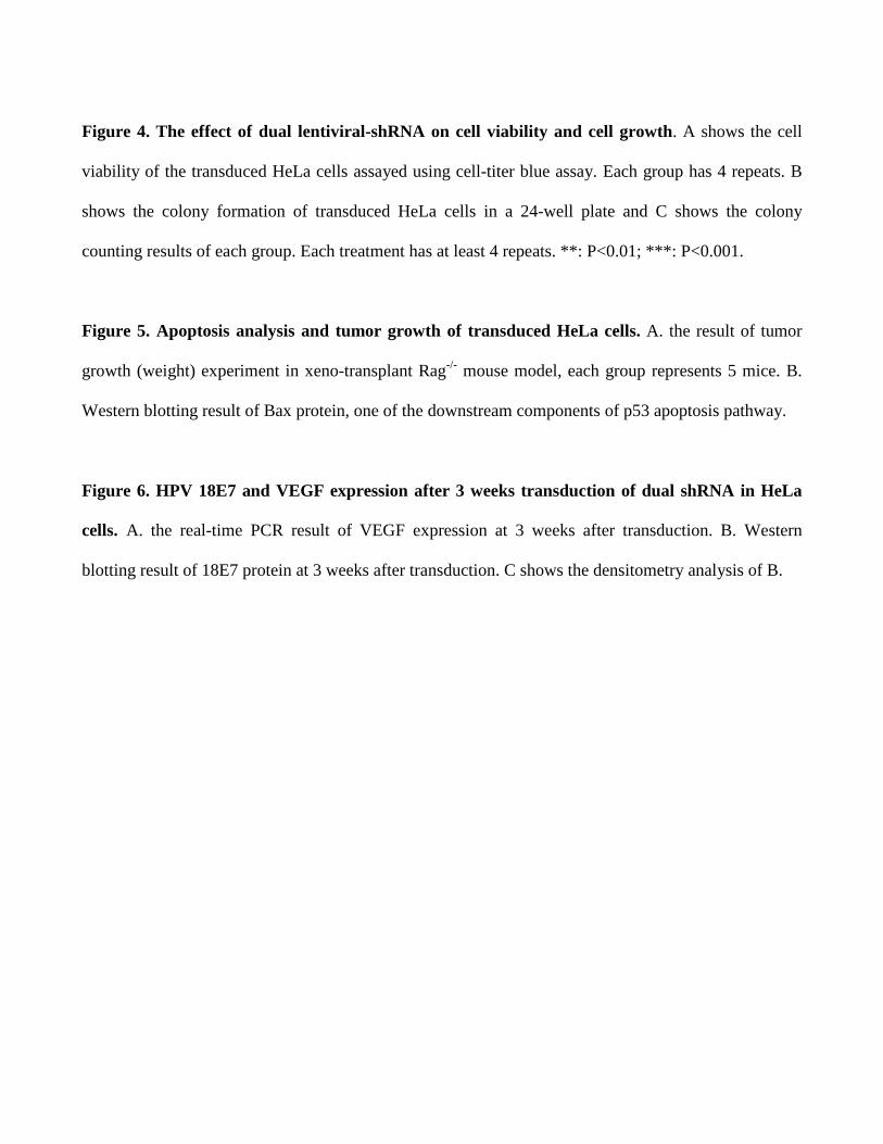
Figure 4. The effect of dual lentiviral-shRNA on cell viability and cell growth. A shows the cell
viability of the transduced HeLa cells assayed using cell-titer blue assay. Each group has 4 repeats. B
shows the colony formation of transduced HeLa cells in a 24-well plate and C shows the colony
counting results of each group. Each treatment has at least 4 repeats. **: P<0.01; ***: P<0.001.
Figure 5. Apoptosis analysis and tumor growth of transduced HeLa cells. A. the result of tumor
growth (weight) experiment in xeno-transplant Rag-/- mouse model, each group represents 5 mice. B.
Western blotting result of Bax protein, one of the downstream components of p53 apoptosis pathway.
Figure 6. HPV 18E7 and VEGF expression after 3 weeks transduction of dual shRNA in HeLa
cells. A. the real-time PCR result of VEGF expression at 3 weeks after transduction. B. Western
blotting result of 18E7 protein at 3 weeks after transduction. C shows the densitometry analysis of B.

Fig 1.
A. B. H 1W 3W 5W 7W 9W
E7
Tub
E7
Tub
18E6-1
16E7-2
W1 W2 W3 W7 W925
50
75
100
18E6-1
16E7-2
* *
Time (weeks)
Co
lon
y N
o.

C.
Negative control
1W 3W 5W
PLL
MFI: 3708 3637 1585
18E6-1
MFI: 3700 1928 1130

Fig 2.
A. B.
C.
500bp →
1 2 3 4 5

Fig 3.
A. B.
C.
Dencitometry:
E7 Tub
HeLa PLL E6-1 Tw E6-1vegf
18E7 Expression
16E7-
2
VEGF
18E6-
1
T-18E
6-1
18E6-
1VEGF
0.0
0.2
0.4
0.6
0.8
1.0
***
***
*** ***Rela
tive E
xp
ressio
n
VEGF Expression
16E7-
2
18E6-
1
T-18E
6-1
VEGF
18E6-
1VEGF
2.0×10⁻⁵
4.0×10⁻⁵
6.0×10⁻⁵
8.0×10⁻⁵
5.0×10⁻³
1.0×10⁻²
1.5×10⁻²
2.0×10⁻² P=0.258
**
**
Rela
tive E
xp
ressio
n
HeL
aPLL
E6-
1Tw
E61
vegf
0
50
100
Perc
en
t %
Fig 4.
A.
B. C.
18E6-1VEGF
16E7-2
18E6-1
16E7-
2
VEG
F
18E6-
1
T-18E
6-1
18E6-
1VEG
F
0
20
40
60
80
100
**
*** ***
P=0.062
Co
lon
y N
o.
16E7-
2
VEGF
18E6-
1
T-18E
6-1
18E6-
1VEGF
0
2000
4000
6000
8000
P=0.309
**
*** ***
CT
B c
ou
nts

Fig. 5.
A
B
Rapamycin
HeL
a
HeL
a+PLL
PLL+
18E6-
1
18E6-
1+
T-18E
6-1
T-18E
6-1+
0
2000
4000
6000
8000
P<0.05
CT
B c
ou
nts
SAHA
HeL
a
HeL
a+PLL
PLL+
18E6-
1
18E6-
1+
TWH
TWH+
18E6-
1veg
f
18E6-
1veg
f+
0
2000
4000
6000
8000
*****
*
CT
B c
ou
nts

Fig 6.
A.
B.
16E7 vegf E6-1 Tw E6-1vegf
Bax
Tub
HeL
a
16E7-
2
18E6-
1
T-18E
6-1
18E6-
1VEGF
VEGF
0.0
0.2
0.4
0.6
0.8
1.0
*
Tu
mo
r W
eig
ht
(g)


![Research Article Synthesis and In Vitro Inhibition Effect of ...Synthesis and In Vitro Inhibition Effect of New Pyrido[2,3-d]pyrimidine Derivatives on Erythrocyte Carbonic Anhydrase](https://img.pdfslide.net/doc/110x75/60b84bfde6a72e7d1963a0c3/research-article-synthesis-and-in-vitro-inhibition-effect-of-synthesis-and-in.jpg)