Embed Size (px)
Citation preview
MQP-BIO-DSA-3196
Inhibition of HIV-1 Tat Induced Inflammation by the NF-κκκκB Subunit RelB
A Major Qualifying Project Report
Submitted to the Faculty of the
WORCESTER POLYTECHNIC INSTITUTE
in partial fulfillment of the requirements for the
Degree of Bachelor of Science
in
Biology and Biotechnology
by
_________________________ Donna C. Davidson
January 10, 2007
APPROVED:
_________________________ _________________________ Sanjay B. Maggirwar, Ph.D. David S. Adams, Ph.D. Dept. Microbiology and Immunology Dept of Biology and Biotechnology University of Rochester WPI Project Advisor Major Advisor
2
ABSTRACT
The NF-kappaB (NF-κB)/Rel family of transcription factors serves an important
regulatory role in the production of immune responses and inflammation. The NF-κB
family member RelB displays unusual properties in comparison to the other family
members, as it exudes inhibitory effects on the pathway along with its traditional function
of transcription initiation. Luciferase assays and TNFα ELISAs were used to determine
if RelB represses transcriptional activity of NF-κB in a subunit-specific manner, and also
to determine if RelB is able to attenuate NF-κB activity in response to HIV-encoded Tat.
This in turn would lead to decreased TNFα synthesis. The outcome may have therapeutic
potential in the context of neuroAIDS.
3
TABLE OF CONTENTS
Signature Page ………………………………………………………………………. 1 Abstract ……………………………………………………………………………… 2 Table of Contents ……………………………………………………………….…… 3 Acknowledgements ………………………………………………………………….. 4 Background ………………………………………………………………………….. 5 Project Purpose ………………………………………………………………………. 17 Methods ……………………………………………………………………………… 18 Results ……………………………………………………………………………….. 21 Discussion …………………………………………………………………………… 28 Bibliography ………………………………………………………………………… 31
4
ACKNOWLEDGEMENTS
I would first like to thank Dr. Sanjay Maggirwar for allowing me to work in his
lab at the University of Rochester, Rochester, NY, and for all his help throughout this
entire project. I would also like to thank Michelle Crandall, Ziye Sui, and Lynn
Sniderhan for all of their help with this project and for the countless things that I learned
from them in the lab. Thanks to Drs. Avi Nath (Johns Hopkins Univ., Maryland, MD),
Bernd Baumann (Ulm University, Ulm, Germany), and Rosario Donato (University of
Perugia, Perugia, Italy) for their kind contributions, providing useful reagents, plasmid
DNA, and the murine microglial BV-2 cell line, respectively. Finally, I would like to
thank Dr. David Adams (Worcester Polytechnic Institute, Worcester, MA) for helping to
initiate this project and for help editing the final report.
5
BACKGROUND
NeuroAIDS
As Acquired Immunodeficiency Syndrome (AIDS) continues to grow as a global
epidemic, now affecting more then 40 million people world wide (Joint United Nations,
2005), there are various associated conditions causing increasing concern. Among these
conditions are neurological disorders connected with the virus; the most common being
HIV-associated dementia (HAD), along with a lesser form of neurological dysfunction
known as minor cognitive/motor disorder (MCMD). The symptoms most commonly
associated with HAD are cognitive, behavioral, and motor dysfunction including memory
loss, slowing of mental processes, and some reading and comprehension difficulties
(McArthur et al., 2003). MCMD causes similar dysfunctions as HAD, albeit to a lesser
degree, and as a result, seems slightly more incidental. MCMD affects about 37% of
HIV-1 infected people, as compared to about 10% for HAD (McArthur et al., 1993;
McArthur, 2004). The advent and subsequent introduction of highly active antiretroviral
therapies (HAART) in the mid nineties offered some hope for the control of such HIV-
associated neurological disorders. When first introduced, the incidence rate fell by about
half, from 20-30% of infected individuals to the 10% previously mentioned (Sacktor et
al., 2001; Kaul et al., 2001; McArthur, 2004). Although the use of HAART has also
severely decreased the number of AIDS related deaths (Jones and Power, 2005), with
these therapies in place the prevalence of these disorders is actually on the rise
(McArthur, 2004).
While it seems that HAART did have an initial positive impact on the incidence
of HAD and other HIV-associated neurological dysfunctions, the increasing prevalence
6
of these disorders suggests that HAART does not provide complete protection (Kaul et
al., 2001; McArthur, 2004; Jones and Power, 2005). As a result, HIV-associated
dementia is now thought to be the number one cause of dementia in people under the age
of 40 (Kaul et al., 2001).
With such vast numbers of HIV infected individuals suffering from some form of
neurological dysfunction, it seems imperative to try to understand where and how
problems are occurring within the central nervous system (CNS). Several theories exist,
and although there is evidence supporting each, a definite cause has yet to be determined
(Kramer-Hammerle et al., 2005). HIV is known to drastically affect the peripheral
nervous system (PNS), however, it is after the virus is able to enter the central nervous
system that neurological problems begin to occur. The CNS is protected by the blood-
brain barrier (BBB), a membrane that serves to control the passage of materials from the
blood into the brain and the central nervous system (Ghafouri et al., 2006). Even with this
elaborate protection the virus is able to surpass the barrier and enter the CNS very early
in infection (Gaul et al., 2001).
HIV Entrance to the CNS
Three main hypotheses remain as possible methods of entry into the CNS for the
virus, including the “Trojan horse” method, the passage of cell-free virus into the brain,
and the release of viral particles into the brain by infected endothelial cells (Albright et
al., 2003; Kramer-Hammerle et al., 2005). The cell-free virions may be able to gain
access to the CNS when aided by infected endothelial cells, and although these two
methods of entry remain controversial (Kramer-Hammerle et al., 2005), there is evidence
7
suggesting that initial entry could be caused by these events (Liu et al., 2002). Brain
microvascular endothelial cells (BMVECs) that are found in the blood brain barrier can
reportedly take up the virus in vacuoles and release them on the other side of the barrier
in a process called transcytosis, giving rise to the cell-free virion hypothesis. In addition,
these cells may also be susceptible to infection by HIV-1 (Kramer-Hammerle et al.,
2005). Once infected, the BMVECs may then be able to infect surrounding cells within
the BBB (Kramer-Hammerle et al., 2005), and in either event, the integrity of the barrier
seems to be somewhat compromised. This destruction compromises an already less
restrictive barrier resulting from the initial immune response, which when mounted
against the virus or other diseases, causes an increase in paracellular transport, allowing
various cytokines and pro-inflammatory molecules into the brain (Liu et al., 2002;
Kramer-Hammerle et al., 2005). This increased movement through the blood brain barrier
provides access to the CNS for both the free virions and infected cells (Liu et al., 2002;
Albright et al., 2003).
The “Trojan horse” model is one of the most widely accepted models of how
HIV-1 enters the brain, and is thought to happen later in the course of HIV infection. In
this model, HIV is carried into the CNS via infected monocytes that later differentiate
into macrophages once inside the brain (Albright et al., 2003). Once macrophage
infiltration has occurred, productive viral particles can be spread to other cell types within
the CNS, such as microglia. As more and more cells of the CNS are infected, infiltration
subsequently causes a wide range of other effects, especially neuronal injury and death
(Albright et al., 2003).
8
HIV-Induced Neuronal Injury
The exact mechanism of neuronal injury after viral localization in the CNS also
remains a controversial issue, with evidence supporting both direct and indirect causes.
The direct injury hypothesis seems slightly less accepted, as this theory relies on the idea
that HIV proteins can cause direct injury to neurons without the aid of other cell types
(Kaul et al., 2001). A strong, and possibly the most convincing, example of direct
neurotoxicity can be demonstrated by the HIV protein gp120. This protein is able to
interact with various chemokine receptors, and it is thought that direct injury may result
from chemokine receptor signaling (Kaul et al., 2001). Other HIV proteins such as Tat
(transactivator of transcription) and Vpr (viral protein R) have also reportedly been able
to cause direct injury of neurons through receptor mediated mechanisms and the
formation of cation-permeable channels respectively (Kaul et al., 2001; Ghafouori et al.,
2006).
Although direct injury remains a possibility, many indirect mechanisms have been
proposed with a wide range of strong supporting data. The basic principle behind this
theory is that macrophages and microglia, the main targets of HIV infection in the CNS,
become activated or infected and, as a result, release a large amount of cytokines, pro-
inflammatory molecules, and HIV particles that accumulate in the CNS and eventually
lead to neuronal death (Rausch and Davis, 2001). Specifically, activated or infected
macrophages/microglia tend to release substances that lead to high levels of activation of
the N-methyl-D-aspartate (NMDA) receptors, triggering an increase in cellular Ca2+
levels, as well as the release of inflammatory molecules, such as tumor necrosis factor – �
(TNF�) and interleukin (IL)-1 (Kaul et al., 2001; Kramer-Hammerle et al., 2005). TNF�
9
has been known to induce neuronal apoptosis alone, and some studies have found that the
HIV-1 Tat protein and TNF� can synergize to promote neuron injury and death (Kaul et
al., 2001). In addition, many signaling pathways are stimulated by HIV infection and
activation of macrophages/microglia, such as the nuclear factor – kappa B (NF-�B)
pathway and the p38 signal transduction pathway. The activation of these pathways can
trigger the release of additional pro-inflammatory molecules or apoptosis (Kaul et al.,
2001; Kramer-Hammerle et al., 2005).
NF-kB Signaling Pathway
The nuclear factor – kappa B (NF-�B) family of transcription factors, also known
as the Rel family, serves an important role in the activation and regulation of genes that
are responsible for the production of many inflammatory molecules. The secretion of
inflammatory molecules acts as a first line of defense against infection, serving to recruit
more immune cells to areas of insult (Minghetti et al, 2004). As a result, this pathway is
very important in both adaptive and innate immunity (Bonizzi and Karin, 2004), and is
therefore vulnerable to the effects of an HIV-1 infection. This pathway has been
connected to neuronal injuries associated with HIV infection, as inflammatory molecules
are the main suspects of the indirect injury theory, and this pathway leads to the
production of these molecules (Peruzzi et al., 2005). Not only do pro-inflammatory
cytokines recruit more microglia to sites of infection, causing the accumulation of various
cells and molecules and leading to the development of large, multinucleated giant cells
(King et al., 2006), they can also cause the secretion of more viral particles and
neurotoxic factors (D’Aversa et al., 2005). Inflammatory molecules can stimulate the
10
release of L-cysteine from macrophages, which in turn, causes increased stimulation of
NMDA receptors. As previously mentioned, high levels of NMDA receptor activation
leads to an excess of Ca2+ within cells and promotes neuronal apoptosis (Kaul et al.,
2001).
The NF-�B family consists of five subunits including p50 (NF�B1), p52
(NF�B2), RelA (p65), cRel, and RelB. Each of these members have a consistent Rel
homology domain that allows them to form homo- or heterodimers, translocate from the
cytoplasm to the nucleus, and bind DNA to initiate transcription (Maier et al., 2003;
Karin et al., 2004). Inhibition of the NF-�B proteins is controlled by a group of proteins
known as the inhibitory �B proteins, or I�Bs. These proteins bind to the subunits of the
NF-�B family at their Rel homology domain and keep them localized in the cytoplasm
(Maier et al., 2003). Upon activation of the NF-�B pathway, the I�Bs undergo
phosphorylation induced ubiquitination, and release the NF-�B dimers (Karin et al.,
2004). The dimers then translocate to the nucleus, bind to DNA at specific sites within
some gene promoters, and activate their target genes (Karin et al., 2004).
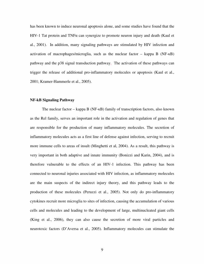
Activation of the NF-�B pathway can occur in response to several different types
of stimuli (Figure 1). When various types of receptors on the cell surface interact with
substrates, these pathways signal specific I�B kinases (IKKs) that phosphorylate the
I�Bs, causing dissociation from the NF-�B dimers (Bonizzi and Karin, 2004). The three
main types of receptors responsible for activating the NF-�B signaling pathway include
pattern-recognition receptors, the most common being Toll-like receptors (TLR) (left side
of diagram), tumor necrosis factor (TNF) receptors (middle of diagram), and T-cell
receptors (TCR) (right side of diagram) (Li and Verma, 2002). The Toll-like receptors
11
are able to recognize specific components of bacterial cell walls and pathogen derived
materials such as lipopolysaccharide (LPS), peptidoglycans, lipoproteins, lipoteichoic
acid, unmethylated bacterial DNA, and double-stranded RNA (Li and Verma, 2002;
Bonizzi and Karin, 2004).
NF-�B activation induced by cyokine expression occurs through the use of TNF
receptors. These receptors, found on many cell types, respond to the presence of
Figure 1. Activation of the NF-�B signaling pathway. Three main types of receptors, (a) pattern-recognition, or Toll-like, (b) tumor necrosis factor, and (c) T-cell receptors, are responsible for the stimulation of I�B kinases (IKKs). These kinases then lead to the phosphorylation and ubiquitination of I�Bs, and eventually NF-�B dimer dissociation and translocation into the nucleus to activate transcription. Figure from Li, Q., Verma, I.M. (2002) NF-kB regulation in the immune system. Nature Rev. Immunol. 2, 725-734.
12
cytokines, including TNF� and IL-1, released from activated macrophages and
monocytes (Li and Verma, 2002). Finally, T-cell receptor activation of NF-�B occurs
with co-stimulation of TCR and CD28 by antigen presenting cells, causing the
subsequent activation of protein kinase C (PKC). This eventually leads to stimulation of
I�Bs, and translocation of NF-�B dimers to the nucleus (Li and Verma, 2002). Once
activation occurs in response to various types of insult, the NF-�B pathway turns on the
genes responsible for producing inflammatory molecules, cytokines, chemokines, and
even various types of adhesion molecules. This in turn leads to further activation of the
innate and adaptive immune systems (Yamamoto and Gaynor, 2004).
RelB
The NF-�B subunit RelB has been found to have some unusual properties in
comparison to the other members of the NF-κB family. As a result, it has been proposed
that RelB serves as a feedback inhibitor of the pathway, providing alternative inhibition
that is especially important during periods of high or constant stimulation, such as during
HIV infection, when I�Bs are already dissociated from the NF-κB dimers (Ruben et al,
1992).
One vast difference found between RelB and the other subunits of the NF-κB
family is that RelB has a much lower DNA binding affinity than the others members
(Ryseck et al, 1992). The amino terminal domain of RelB is thought to be the cause of
this low affinity, as it was found to have inhibitory effects on DNA binding. However, it
was determined that RelB is able to bind DNA when associated with one of the two
subunits with which it can dimerize, p50 or p52 (Ryseck et al, 1992). The RelB subunit
13
can only form heterodimers with these two subunits to activate transcription, and it is not
capable of forming homodimers, as are the other subunits (Marienfeld et al, 2003).
Another unusual characteristic displayed by RelB is that it is thought to be more
loosely controlled by the common NF-κB inhibitors, IκBs, than other NF-κB subunits.
As a result, RelB is more often localized in the nucleus rather than in the cytoplasm with
the other Rel transcription factor family members (Marienfeld et al, 2001). Furthermore,
RelB is thought to display degradation characteristics similar to IκBs, undergoing signal-
dependent phosphorylation, which is not thought to be a characteristic of the other family
members. It has been determined that the processing of RelB takes place in three steps
including phosphorylation at Thr84 and Ser552, cleavage at the N-terminal end, and
finally complete degradation in the proteasomes (Marienfeld et al, 2001).
The mechanism by which RelB is thought to act as an inhibitor of the NF-κB
pathway is through the formation of inactive complexes. The “classic NF-κB” dimer,
consisting of p50 and RelA, is often considered to be the largest contributor to the
activation of transcription. In addition, RelA is a very important subunit necessary for
the anti-apoptotic and pro-inflammatory roles of the NF-κB pathway (Marienfeld et al,
2003). Thus, these two p50 and RelA subunits display desirable characteristics as targets
of RelB during the formation of inactive complexes to carry out its inhibitory role. RelB
is able to form dimers with p50 to transactivate transcription, however, this complex
results in significantly lower amounts of activation as compared to other NF-κB dimers
(Ryseck et al, 1992). To reduce the number of highly active transcription activating
complexes, it was found that RelB competes with RelA for binding p50 (Ruben et al,
1992), and once RelB binds with RelA, the complex is rendered completely inactive and
14
is no longer able to bind DNA (Marienfeld et al, 2003). It has also been noted that TNF�
stimulation promotes the association of RelA with RelB, further indicating the feedback
inhibition of RelB (Jacque et al., 2005).
It is obvious that RelB plays a significant role in the regulation of the NF-κB
pathway, and in addition, its presence is crucial for the proper functioning of immune
responses. The study of relb knockout mice has provided strong evidence in support of
the indispensable role of RelB. Several methods have been employed to observe the
effects of the absence of RelB, and each has arrived at the same conclusion. RelB
deficient mice display many abnormalities upon stimulation with inflammatory triggers,
such as lipopolysaccharide, including defects in immune responses, extremely high
amounts of inflammation, myeloid hyperplasia, splenomegaly, and a reduction in the
amount of thymic dendritic cells (Weih et al, 1995, Weih et al, 1997, Xia et al, 1997).
Additionally, it was found that the absence of RelB could not be compensated for with
the substitution of another NF-κB family member (Weih et al, 1995). Xia et al found that
these harmful effects could be reversed with the transfection of RelB cDNA into the
knockout mice, demonstrating that the deficiency was the probable cause of the defects,
and emphasizing the importance of RelB (1997).
HIV-1 Tat
The HIV-1 protein Tat (transactivator of transcription) plays a major role in the
productive infection of HIV as it is required for HIV replication. This protein is released
from infected cells into the extracellular space, sera, and cerebrospinal fluid, and can be
taken up by uninfected cells to cause a variety of harmful events (King et al, 2006).
15
Although the exact mechanism that Tat employs to activate transcription is still unknown,
it is understood that Tat is a eukaryotic transcription factor that directs transcription from
the HIV long terminal repeat (LTR) to promote replication (Jeang, 1996). In addition,
the uptake of Tat results in a variety of intracellular events including the production and
secretion of cytokines and chemokines, activation of numerous receptors, and the
expression of apoptotic proteins (King et al, 2006).
The intracellular changes caused by the uptake of Tat are thought to begin
through interactions with cell surface receptors. In the CNS, the cells most commonly
affected are the microglia. Once internalized within these cells, Tat activates various
signaling pathways and receptors that in turn lead to the over-production of numerous
pro-inflammatory molecules and recruitment of more immune cells. This is thought to be
one of the major ways in which Tat is able to cause neuronal damage and eventual death
(Minghetti et al, 2004). Pro-inflammatory molecules such as cytokines and chemokines
are produced as part of an immune response in an effort to recruit lymphocytes to areas of
infection or damage, and are the product of the NF-κB signaling pathway. However,
when these molecules are produced in excess amounts, they can actually be harmful to
cells. It has been suggested that Tat is able to bind NF-κB directly and most of the
mechanisms by which Tat exerts its effects are NF-κB dependent (Peruzzi et al, 2005;
Nicolini et al, 2001).
Increased levels of Tat-induced NF-κB activation, and subsequent production of
cytokines such as TNF� and IL-1�, leads to the recruitment of more microglia to sites of
infection. Additionally, it has been shown that Tat is able to stimulate the secretion of the
chemokine CCL2 (monocyte chemoattractant protein-1, MCP-1), which is involved in
16
the recruitment of monocytes and microglia to areas of inflammation, and is also able to
induce the migration of these cells across the blood-brain barrier (King et al, 2006,
D’Aversa et al, 2005). This recruitment then facilitates further infection, and can also
lead to the formation of large, multinucleated, giant cells (D’Aversa et al, 2005).
The HIV-1 Tat protein has the additional ability of stimulating various receptors
to cause further neuronal damage than that induced by over-expression of cytokines and
chemokines. NMDA receptors in particular are stimulated by the presence of Tat,
resulting in an intracellular calcium overload and subsequent neuronal death (King et al,
2006; Kaul et al., 2001).
There are many characteristics of Tat that make it a critical component of
productive HIV infection, especially within the central nervous system. This makes it a
prime target of investigation for the understanding of HIV associated neurological
disorders.
17
PROJECT PURPOSE
The HIV-1 protein Tat is necessary for productive HIV-1 infection. This protein
has been shown to cause excess inflammation and neuronal death in the CNS. The
intracellular mechanisms of Tat are thought to be dependent on its ability to directly
interact with the NF-κB pathway, and as a result, it seems imperative to focus on the
proteins of this transcription factor when considering HIV-1 infection in the CNS.
Based on previous studies proposing an inhibitory role of the NF-κB subunit
RelB, we hypothesized that in microglial cells RelB may be able to render the cells
resistant to the action of the HIV-1 protein Tat. HIV associated changes in RelB could
therefore be related to the incidence of inflammation, and eventually neuronal death, in
neuroAIDS. NF-κB reporter luciferase assays and TNF-α ELISAs were used to
determine if RelB represses transcriptional activity of NF-κB in a subunit-specific
manner, and also to determine if RelB is able to attenuate NF-κB activation in response
to HIV-encoded Tat. This in turn would lead to decreased TNFα synthesis. Immunoblot
analysis was used to observe cellular levels of RelB in the presence of Tat. The outcome
of these experiments together may have therapeutic potential in the context of HIV-
associated neurological disorders and neuroAIDS.
18
MATERIALS AND METHODS
Reagents
HIV-1 Tat was a kind gift of Dr. Avi Nath (Johns Hopkins Univ., Maryland,
MD), and this reagent was used at a concentration of 100nM. The proteasome inhibitor
MG132 (BioMol International, Plymouth Meeting, PA), resuspended in
dimethylsulfoxide (DMSO; Sigma Aldrich, St. Louis, MO), was used at a concentration
of 50µg/mL. The RelB plasmid DNAs were a kind gift from Dr. Bernd Baumann (Ulm
University, Ulm, Germany), and other plasmids were as previously described (Maggirwar
et al., 2000).
Murine Microglial Cell-Line (BV-2)
The BV-2 cell line (kind gift from Dr. Rosario Donato, University of Perugia,
Perugia, Italy) was maintained in DMEM containing 10% fetal bovine serum, 2mM
glutamine, and antibiotics.
Transient Transfections and Luciferase Assay
BV-2 cells were plated at a density of 1.5 x 10⁵ cells/well in 24-well plates and
incubated for 24h prior to use in experiments. The cells were transfected with an NF-κB-
driven luciferase reporter plasmid (κB Luc), together with plasmids encoding various
NF-κB subunits using Lipofectamine LTX with PLUS reagent (Invitrogen, Carlsbad,
CA). Twenty-four hours post transfection the cells were either left untreated or incubated
for 8h with Tat; cell lysates were then prepared using reporter lysis buffer (Promega Life
19
Science, Madison, WI, USA), and upon addition of luciferase substrate to the lysates
luciferase activity was measured with a Lumicount Microplate Luminometer (Packard
Instrument Co., Meriden, CT, USA).
TNF-αααα ELISA
TNFα levels were measured in culture supernatant (pre-cleared by brief
centrifugation) by using a mouse TNFα ELISA Kit (Biosource International, Camarillo,
CA) according to the manufacturer’s instructions. Briefly, 100µL of cell culture
supernatant (at a 1:2 dilution) was incubated in a 96-well plate pre-coated with a TNFα-
specific monoclonal antibody for 90 minutes. After extensive washing, binding of TNFα
was detected by incubation with biotinylated antibodies, followed by Streptavidin-
Peroxidase colorimetric enzyme assays (optical density was recorded at 450nm).
Immunoblot
This assay was performed as previously described (Sui et al., 2006). BV-2 whole
cell lysates were prepared using ELB buffer (50mM HEPES pH7, 250nM NaCl, 0.1% NP
40, 5nM EDTA, 10mM NaF, 0.1mM Na3VO4, 50�M ZnCl2, supplemented with 0.1mM
PMSF, 1mM DTT, and a mixture of protease and phosphatase inhibitors) and
centrifugation was then used to remove cellular debris. Equal amounts of each lysate
were run on a 10% SDS-PAGE gel for separation, and the protein was transferred to a
Hybond ECL nitrocellulose membrane (Amersham, Arlington Heights, IL). A 5% milk
solution was used to block for nonspecific proteins. The membranes were incubated
overnight with primary antibody raised against RelB or �-tubulin (at 1:500 dilution;
20
Santa Cruz Biotechnology, Santa Cruz, CA) and, after incubation with the HRP-
conjugated, species specific secondary antibody (at 1:3000 dilution; Santa Cruz
Biotechnology, Santa Cruz, CA), the blot was exposed and developed. Enhanced
chemiluminescence (ECL) reagent (Pierce Biotechnology, Rockford, IL) was used to
develop the blots, followed by exposure to X-Ray film.
21
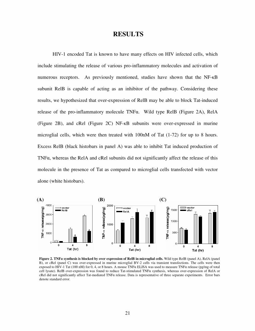
Figure 2. TNF� synthesis is blocked by over expression of RelB in microglial cells. Wild type RelB (panel A), RelA (panel B), or cRel (panel C) was over-expressed in murine microglial BV-2 cells via transient transfections. The cells were then exposed to HIV-1 Tat (100 nM) for 0, 4, or 8 hours. A mouse TNF� ELISA was used to measure TNF� release (pg/mg of total cell lysate). RelB over-expression was found to reduce Tat-stimulated TNF� synthesis, whereas over-expression of RelA or cRel did not significantly affect Tat-mediated TNF� release. Data is representative of three separate experiments. Error bars denote standard error.
(A) (B) (C)
RESULTS
HIV-1 encoded Tat is known to have many effects on HIV infected cells, which
include stimulating the release of various pro-inflammatory molecules and activation of
numerous receptors. As previously mentioned, studies have shown that the NF-�B
subunit RelB is capable of acting as an inhibitor of the pathway. Considering these
results, we hypothesized that over-expression of RelB may be able to block Tat-induced
release of the pro-inflammatory molecule TNF�. Wild type RelB (Figure 2A), RelA
(Figure 2B), and cRel (Figure 2C) NF-�B subunits were over-expressed in murine
microglial cells, which were then treated with 100nM of Tat (1-72) for up to 8 hours.
Excess RelB (black histobars in panel A) was able to inhibit Tat induced production of
TNF�, whereas the RelA and cRel subunits did not significantly affect the release of this
molecule in the presence of Tat as compared to microglial cells transfected with vector
alone (white histobars).
22
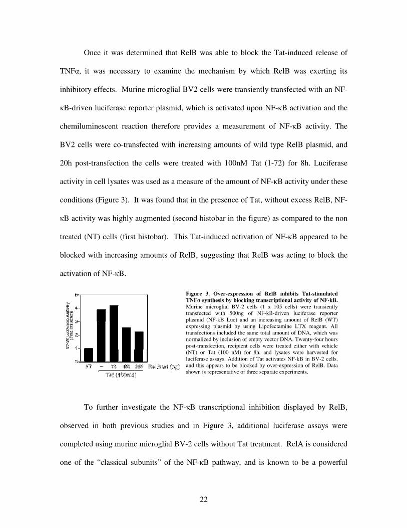
Once it was determined that RelB was able to block the Tat-induced release of
TNF�, it was necessary to examine the mechanism by which RelB was exerting its
inhibitory effects. Murine microglial BV2 cells were transiently transfected with an NF-
�B-driven luciferase reporter plasmid, which is activated upon NF-�B activation and the
chemiluminescent reaction therefore provides a measurement of NF-�B activity. The
BV2 cells were co-transfected with increasing amounts of wild type RelB plasmid, and
20h post-transfection the cells were treated with 100nM Tat (1-72) for 8h. Luciferase
activity in cell lysates was used as a measure of the amount of NF-�B activity under these
conditions (Figure 3). It was found that in the presence of Tat, without excess RelB, NF-
�B activity was highly augmented (second histobar in the figure) as compared to the non
treated (NT) cells (first histobar). This Tat-induced activation of NF-�B appeared to be
blocked with increasing amounts of RelB, suggesting that RelB was acting to block the
activation of NF-�B.
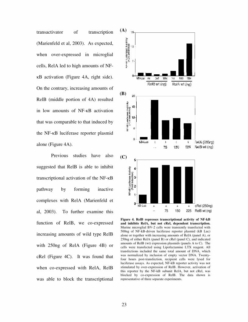
To further investigate the NF-�B transcriptional inhibition displayed by RelB,
observed in both previous studies and in Figure 3, additional luciferase assays were
completed using murine microglial BV-2 cells without Tat treatment. RelA is considered
one of the “classical subunits” of the NF-�B pathway, and is known to be a powerful
Figure 3. Over-expression of RelB inhibits Tat-stimulated TNF� synthesis by blocking transcriptional activity of NF-kB. Murine microglial BV-2 cells (1 x 10⁵ cells) were transiently transfected with 500ng of NF-kB-driven luciferase reporter plasmid (NF-kB Luc) and an increasing amount of RelB (WT) expressing plasmid by using Lipofectamine LTX reagent. All transfections included the same total amount of DNA, which was normalized by inclusion of empty vector DNA. Twenty-four hours post-transfection, recipient cells were treated either with vehicle (NT) or Tat (100 nM) for 8h, and lysates were harvested for luciferase assays. Addition of Tat activates NF-kB in BV-2 cells, and this appears to be blocked by over-expression of RelB. Data shown is representative of three separate experiments.
23
�� ��
�� ��
�� ��
�� ��
�� ��
�� ��
kB-Luc + + + + cRel (250ng) 75 150 225 RelB wt (ng)_
NF-k
B Lu
cife
rase
Act
ivity
(Fol
d In
duct
ion)
Figure 4. RelB represses transcriptional activity of NF-kB and inhibits RelA, but not cRel, dependent transcription. Murine microglial BV-2 cells were transiently transfected with 500ng of NF-kB-driven luciferase reporter plasmid (kB Luc) alone or together with increasing amounts of RelA (panel A), or 250ng of either RelA (panel B) or cRel (panel C), and indicated amounts of RelB (wt) expression plasmids (panels A to C). The cells were transfected using Lipofectamine LTX reagent. All transfections included the same total amount of DNA, which was normalized by inclusion of empty vector DNA. Twenty-four hours post-transfection, recipient cells were lysed for luciferase assays. As expected, NF-kB reporter activity was not stimulated by over-expression of RelB. However, activation of this reporter by the NF-kB subunit RelA, but not cRel, was blocked by co-expression of RelB. The data shown is representative of three separate experiments.
(A)
(B)
(C)
transactivator of transcription
(Marienfeld et al, 2003). As expected,
when over-expressed in microglial
cells, RelA led to high amounts of NF-
�B activation (Figure 4A, right side).
On the contrary, increasing amounts of
RelB (middle portion of 4A) resulted
in low amounts of NF-�B activation
that was comparable to that induced by
the NF-�B luciferase reporter plasmid
alone (Figure 4A).
Previous studies have also
suggested that RelB is able to inhibit
transcriptional activation of the NF-�B
pathway by forming inactive
complexes with RelA (Marienfeld et
al, 2003). To further examine this
function of RelB, we co-expressed
increasing amounts of wild type RelB
with 250ng of RelA (Figure 4B) or
cRel (Figure 4C). It was found that
when co-expressed with RelA, RelB
was able to block the transcriptional
24
0
2
4
6
8
kB-Luc + + + RelA (250ng) + RelB wt (225ng)_ _
+ RelB S368A (225ng)__
NF-k
B Lu
cife
rase
Act
ivity
(Fol
d In
duct
ion)
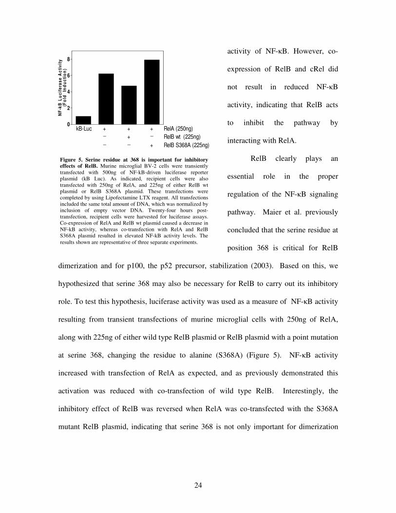
Figure 5. Serine residue at 368 is important for inhibitory effects of RelB. Murine microglial BV-2 cells were transiently transfected with 500ng of NF-kB-driven luciferase reporter plasmid (kB Luc). As indicated, recipient cells were also transfected with 250ng of RelA, and 225ng of either RelB wt plasmid or RelB S368A plasmid. These transfections were completed by using Lipofectamine LTX reagent. All transfections included the same total amount of DNA, which was normalized by inclusion of empty vector DNA. Twenty-four hours post-transfection, recipient cells were harvested for luciferase assays. Co-expression of RelA and RelB wt plasmid caused a decrease in NF-kB activity, whereas co-transfection with RelA and RelB S368A plasmid resulted in elevated NF-kB activity levels. The results shown are representative of three separate experiments.
activity of NF-�B. However, co-
expression of RelB and cRel did
not result in reduced NF-�B
activity, indicating that RelB acts
to inhibit the pathway by
interacting with RelA.
RelB clearly plays an
essential role in the proper
regulation of the NF-�B signaling
pathway. Maier et al. previously
concluded that the serine residue at
position 368 is critical for RelB
dimerization and for p100, the p52 precursor, stabilization (2003). Based on this, we
hypothesized that serine 368 may also be necessary for RelB to carry out its inhibitory
role. To test this hypothesis, luciferase activity was used as a measure of NF-�B activity
resulting from transient transfections of murine microglial cells with 250ng of RelA,
along with 225ng of either wild type RelB plasmid or RelB plasmid with a point mutation
at serine 368, changing the residue to alanine (S368A) (Figure 5). NF-�B activity
increased with transfection of RelA as expected, and as previously demonstrated this
activation was reduced with co-transfection of wild type RelB. Interestingly, the
inhibitory effect of RelB was reversed when RelA was co-transfected with the S368A
mutant RelB plasmid, indicating that serine 368 is not only important for dimerization
25
and p100 stabilization (Maier et al., 2003) but is also critical for the proper regulatory
functioning of RelB.
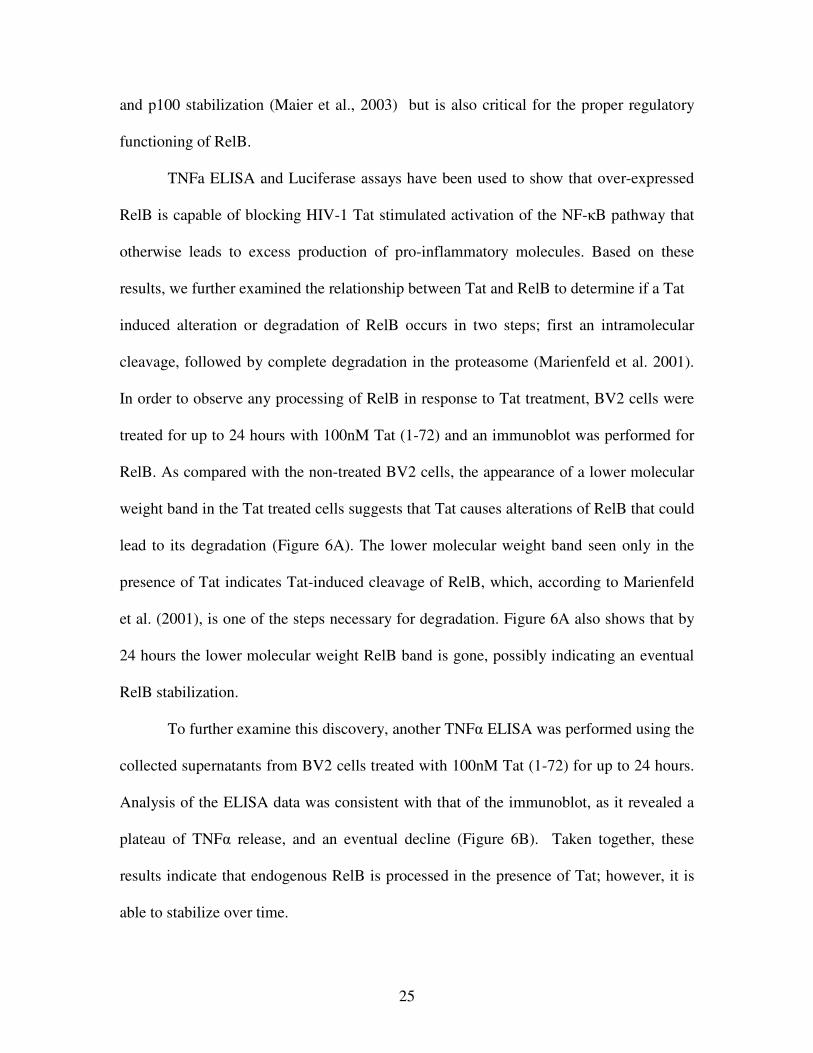
TNFa ELISA and Luciferase assays have been used to show that over-expressed
RelB is capable of blocking HIV-1 Tat stimulated activation of the NF-�B pathway that
otherwise leads to excess production of pro-inflammatory molecules. Based on these
results, we further examined the relationship between Tat and RelB to determine if a Tat
induced alteration or degradation of RelB occurs in two steps; first an intramolecular
cleavage, followed by complete degradation in the proteasome (Marienfeld et al. 2001).
In order to observe any processing of RelB in response to Tat treatment, BV2 cells were
treated for up to 24 hours with 100nM Tat (1-72) and an immunoblot was performed for
RelB. As compared with the non-treated BV2 cells, the appearance of a lower molecular
weight band in the Tat treated cells suggests that Tat causes alterations of RelB that could
lead to its degradation (Figure 6A). The lower molecular weight band seen only in the
presence of Tat indicates Tat-induced cleavage of RelB, which, according to Marienfeld
et al. (2001), is one of the steps necessary for degradation. Figure 6A also shows that by
24 hours the lower molecular weight RelB band is gone, possibly indicating an eventual
RelB stabilization.
To further examine this discovery, another TNF� ELISA was performed using the
collected supernatants from BV2 cells treated with 100nM Tat (1-72) for up to 24 hours.
Analysis of the ELISA data was consistent with that of the immunoblot, as it revealed a
plateau of TNF� release, and an eventual decline (Figure 6B). Taken together, these
results indicate that endogenous RelB is processed in the presence of Tat; however, it is
able to stabilize over time.
26
0h 4h 8h 12h 16h 20h 24h0
5000
10000
15000
20000
25000
30000
35000
Treatment Timecourse
TNF αα αα
Rel
ease
(pg/
mg)75kD
50kD 75kD 50kD
�RelB 68kD
��-Tubulin
NT 4h 8h 12h 24h ________________ 100nM Tat 1-72
(A) (B)
Figure 6. Tat induced TNF� synthesis is associated with processing of endogenous RelB in microglial cells. (A) Murine microglial BV-2 cells were treated with HIV-1 Tat 1-72 (100 nM) for up to 24 hours. Immunoblot analysis revealed a lower molecular weight RelB band that appeared in the presence of Tat and disappeared by 24h. (B) Murine microglial BV-2 cells were once again treated with HIV-1 Tat 1-72 (100 nM) for 24 hours and a mouse TNFα ELISA was used to measure TNFα release (pg/mg of total cell lysate). The stabilization of RelB by 24h in part (A) is consistent with the leveling off of TNF� synthesis displayed in part (B). Each set of data is representative of three separate experiments. The error bars denote standard error.
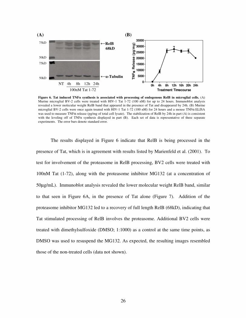
The results displayed in Figure 6 indicate that RelB is being processed in the
presence of Tat, which is in agreement with results listed by Marienfeld et al. (2001). To
test for involvement of the proteasome in RelB processing, BV2 cells were treated with
100nM Tat (1-72), along with the proteasome inhibitor MG132 (at a concentration of
50µg/mL). Immunoblot analysis revealed the lower molecular weight RelB band, similar
to that seen in Figure 6A, in the presence of Tat alone (Figure 7). Addition of the
proteasome inhibitor MG132 led to a recovery of full length RelB (68kD), indicating that
Tat stimulated processing of RelB involves the proteasome. Additional BV2 cells were
treated with dimethylsulfoxide (DMSO; 1:1000) as a control at the same time points, as
DMSO was used to resuspend the MG132. As expected, the resulting images resembled
those of the non-treated cells (data not shown).
27
75kD 50kD
�RelB 68kD
NT 30’ 4h 8h 30’ 4h 8h ___________ ___________ 100nM Tat 1-72 100nM Tat 1-72 + MG132
Figure 7. Inhibition of proteasome blocks Tat stimulated processing of RelB. Murine microglial BV-2 cells were treated with HIV-1 Tat 1-72 (100 nM) +/- the proteasome inhibitor MG132 for up to 8 hours. Immunoblot analysis exposed a lower molecular weight RelB band in the presence of Tat. This band disappeared with the addition of MG132, suggesting that Tat stimulated processing of RelB involves degradation within the proteasome. The lower band indicates equal loading in each lane. Data is representative of three separate experiments.
28
DISCUSSION
The NF-kappaB (NF-κB)/Rel family of transcription factors serve an important
regulatory role in the production of immune responses and inflammation. Previous
studies have shown that the NF-κB subunit RelB displays unusual characteristics as
compared to the other subunits. The most significant difference noted was the ability of
RelB to serve as a feedback inhibitor for the NF-κB pathway (Ruben et al, 1992). The
absence of properly functioning RelB leads to accumulation of pro-inflammatory
molecules and various other defects, as demonstrated in multiple studies of relb knockout
mice (Weih et al, 1995, Weih et al, 1997, Xia et al, 1997). These harmful effects were
reversed upon transfection with wild type RelB, demonstrating that the RelB deficiency
was the probable cause (Xia et al, 1997).
As previously discussed, the HIV-1 protein Tat is known to be necessary for
productive HIV-1 infection, and in addition stimulates the release of many pro-
inflammatory molecules such as TNF� from cells in the CNS. Based on this, we
hypothesized that RelB may be able to render cells resistant to the action of Tat, and
furthermore, HIV associated changes in RelB may be to blame, at least in part, for the
incidence of inflammation, and eventually neuronal death, in neuroAIDS.
The results reported here provide supporting evidence for this hypothesis. We
demonstrate that Tat is able to stimulate the release of TNF�, a feat which can be
overcome with the over-expression of RelB, but is not affected by the presence of excess
RelA or cRel. Additionally, over-expression of RelB was not able to stimulate NF-κB
activity, further indicating the inhibitory role of RelB. Taken together, these results are
29
consistent with the idea that RelB, but not RelA or cRel, is capable of providing
microglial cells with protection from the effects of the HIV-1 protein Tat.
Further analysis of the inhibitory function of RelB revealed that when co-
expressed with RelA, but not cRel, the subunits are not able to stimulate NF-�B activity.
This result was expected, as previous reports conclude that RelB forms transcriptionally
inactive complexes with RelA (Marienfeld et al., 2003). These inactive complexes could
therefore be the mechanism by which RelB exudes its inhibitory effects. This regulatory
role of RelB was reversed with the addition of a point mutation at serine residue 368,
which was previously noted to be a vital residue for the dimerization of RelB with other
subunits (Maier et at., 2003). These results further support our hypothesis that RelB
serves as an inhibitory element within the NF-�B pathway.
Marienfeld et al. noted that RelB degradation occurs in at least two steps,
cleavage at the N-terminal end, and complete degradation in the proteasome (2001). In
the presence of Tat, a lower molecular weight RelB band was observed via immunoblot
analysis. RelB stabilization was also noted at 24 hours, as demonstrated with both
immunoblot and TNF� ELISA analysis. Although the �-tubulin blot in the lane
representing the 24 hour Tat treatment in Figure 6A appears to contain a slightly lower
amount of protein, the band at the full length RelB position remains extremely apparent
and there is obvious disappearance of the lower band. In addition, these results are
representative of three replicates of the same experiment. These results suggest that
endogenous RelB is being processed as a consequence of the Tat treatment. Although
there is recovery of RelB after 24 hours, which may help attenuate the effects of Tat
within the infected cell, the release of pro-inflammatory cytokines and recruitment of
30
more inflammatory cells would occur prior to this recovery. This in turn, would still lead
to the release of excess pro-inflammatory molecules and eventual neuronal injury.
Recovery of full length RelB was also observed with the addition of the
proteasome inhibitor MG132. This data was also in agreement with the criteria listed for
RelB degradation according to Marienfeld et al. (2001) and supported the idea that Tat
treatment of microglial cells leads to the processing of RelB which involves the
proteasome. Based on these results, future investigation involving mutations in RelB at
phosphoacceptor sites may also prove to be beneficial, with potential implications for
therapeutic advances. Marienfeld et al. suggest a signal specific and phosphorylation-
dependent degradation of RelB, adding a third step to their proposed degradation criteria
(2001). If this third step of RelB degradation is necessary, mutagenesis at the two
proposed phosphoacceptor sites, threonine 84 and serine 552 (Marienfeld et al., 2001),
could potentially prevent RelB degradation in the presence of Tat. This in turn would
then lead to a reduction in Tat induced NF-κB activation, and subsequently decreased
TNFα synthesis and neuronal injury.
Taken all together, it seems that RelB is able to act as an inhibitor on the NF-�B
signaling pathway, and as a result, is a possible target for the HIV-1 protein Tat. In
addition, the over-expression of RelB is capable of partially inhibiting Tat induced TNF�
release, which may subsequently reduce neuronal injury and death that occurs as a result
of excess inflammation. These results represent a significant advance in the
understanding of the mechanisms by which the HIV-1 protein Tat contributes to
inflammation and subsequent neuronal injury in the CNS. Further study of this
mechanism may reveal potential therapies in the context of neuroAIDS.
31
BIBLIOGRAPHY Albright, A.V., Soldan, S.S., Gonzalez-Scarano, F. (2003) Pathogenesis of human immunodeficiency virus-induced neurological disease. J. Neurovirol. 9, 222-227. Bonizzi, G., Karin, M. (2004) The two NF-�B activation pathways and their role in innate and adaptive immunity. Trends Immunol. 25, 280-288. D’Aversa. T.G., Eugenin, E.A., Berman, J.W. (2005) NeuroAIDS: Contributions of the Human Immunodeficiency Virus-1 proteins Tat and gp120 as well as CD40 to microglial activation. J. Neurosci. Res. 81, 436-446. Ghafouri, M., Amini, S., Khalili, K., Sawaya, B.E. (2006) HIV-1 associated dementia: symptoms and causes. Retrovirology 3:28. Jacque, E., Tchenio, T., Piton, G., Romeo, PH., Baud, V. (2005) RelA repression of RelB activity induces selective gene activation downstream of TNF receptors. PNAS, 102, 14635-14640. Jeang, KT. (1996) HIV-1 Tat: Structure and Function. National Institute of Health. Joint United Nations Programme on HIV/AIDS, World Health Organization. (2005) AIDS epidemic update: special report on HIV prevention. http://www.unaids.org/epi/2005/doc/EPIupdate2005_pdf_en/epi-update2005_en.pdf Jones, G., Power, C. (2005) Regulation of neuronal cell survival by HIV-1 infection. Neurobiol. Dis. 21, 1-17. Karin, M., Yamamoto, Y., Wang, Q.M. (2004) The IKK NF-kB system: a treasure trove for drug development. Nature Rev. Drug Discov. 3, 17-26. Kaul, M., Garden, G.A., Lipton, S.A. (2001) Pathways to neuronal injury and apoptosis in HIV-associated dementia. Nature 410, 988-994. King, J.E., Eugenun, E.A., Buckner, C.M., Berman, J.W. (2006) HIV Tat and neurotoxicity. Microb. Infect. 8, 1347-1357. Kramer-Hammerle, S., Rothenaigner, I., Wolff, H., Bell, J.E., Brack-Werner, R. (2005) Cells of the central nervous system as targets and reservoirs of the human immunodeficiency virus. Virus Res. 111, 194-213. Li, Q., Verma, I.M. (2002) NF-kB regulation in the immune system. Nature Rev. Immunol. 2, 725-734.
32
Liu, N.Q., Lossinaky, A.S., Popik, W., Li, X., Gujuluva, C., Kriederman, B., Roberts, J., Pushkarsky, T., Bukrinsky, M., Witte, M., Weinand, M., Fiana, M. (2002) Human immunodeficiency virus type 1 enters brain microvascular endothelia by macropinocytosis dependent in lipid rafts and the mitogen-activated protein kinase signaling pathway. J. Virol. 76, 6689-6700. Maggirwar, S.B., Ramirez, S., Tong, N., Gelbard, H.A., Dewhurst, S. (2000) Functional interplay between nuclear factor-kappaB and c-Jun integrated by coactivator p300 determines the survival of nerve growth factor-dependent PC12 cells. J Neurochem. 74, 527-39. Maier, H. J., Marienfeld, R., Wirth, T., Baumann, B. (2003) Critical Role of Rel B Serine 368 for Dimerization and p100 Stabalization. J. Biol. Chem. 278, 39242-39250. Marienfeld, R., Berberich-Siebelt, F., Berberich, I., Denk, A., Serfling, E., and Neumann, M. (2001) Signal-specific and Phosphorylation-dependent RelB Degradation: a Potential Mechanism of NF-kB Control. Oncogene 20, 8142-8147 Marienfeld, R., May, M. J., Berberich, I., Serfling, E., Ghosh, S. & Neumann, M. (2003) RelB Forms Transcriptionally Inactive Complexes with RelA/p65. J. Biol. Chem. 278, 19852-19860. McArthur, J.C., Hoover, D.R., Bacellar, H., Miller, E.N., Cohen, B.A., Becker, J.T. (1993) Dementia in AIDS patients: incidence and risk factors. Neurology 43, 2245–2252. McArthur, J.C., Haughey, N., Gartner, S., Conant, K., Pardo, C., Nath, A., Sacktor, N. (2003) Human immunodeficiency virus-associated dementia: an evolving disease. J. Neurovirol. 9, 205-221. McArthur, J.C. (2004) HIV dementia: an evolving disease. J. Neuroimmunol. 157, 3-10. Minghetti, L., Visentin, S., Patrizio, M., Franchini, L., Ajmone-Cat, M.A., Levi, G. (2004) Multiple Actions of the Human Immunodeficiency Virus Type-1 Tat Protein on Microglial Cell Function. Neurochem. Res. 29, 965-978. Nicolini, A., Ajmone-Cat, M.A., Bernardo, A., Levi, G., Minghetti, L. (2001) Human Immunodeficiency Virus Type-1 Tat Protein induces nuclear factor (NF)-kB activation and oxidative stress in microglial cultures by independent mechanisms. J. Neurochem. 79, 713-716. Peruzzi, F. Bergonzini, V., Aprea, S., Reiss., K., Azuaya, B.E., Rappaport, J., Amini, S., Khalili, K. (2005) Cross talk between growth factors and viral and cellular factors alters neuronal signaling pathways: Implication for HIV-associated dementia. Brain Res. Rev. 50, 114-125.
33
Rausch, D.M., Davis, M.R. (2001) HIV in the CNS: Pathogenic relationships to systemic HIV disease and other CNS diseases. J. Neurovirol. 7, 85-96. Ruben, S.M., Klement, J.F., Coleman, T.A., Maher, M., Chen, C., Rosen, C.A. (1992) I-Rel: a novel rel-related protein that inhibits NF-�B transcriptional activity. Genes Dev. 6, 745-760. Ryseck, R., Bull, P., Takamiya, M., Bours, V., Siebenlist, U., Dobrzanski, P., Bravo, R. (1992) RelB, a new Rel family transcription activator that can interact with p50- NF-κB. Mol. Cell Biol. 12, 674-684. Sacktor N., Lyles R.H., Skolasky R., Kleeberger C., Selnes O.A., Miller E.N., Becker J.T., Cohen B., McArthur J.C. (2001) HIV-associated neurologic disease incidence changes: Multicenter AIDS Cohort Study, 1990-1998. Neurology. 56, 257-60. Sui, Z., Fan, S., Sniderhan, L., Reisinger, E., Litzburg, A., Schifitto, G., Gelbard, H.A., Dewhurst, S., Maggirwar, S.B. (2006) Inhibition of mixed lineage kinase 3 prevents HIV-1 Tat-mediated neurotoxicity and monocyte activation. J. Immunol. 177, 702-711. Weih, F., Carrasco, D., Durham, S.K., Barton, D.S., Rizzo, C.A., Ryseck, R., Lira, S.A, Bravo, R. (1995) Multiorgan Inflammation and Hematopoietic Abnormalities in Mice with a Targeted Disruption of RelB, a Member of the NF-kB/Rel Family. Cell 80, 331-340. Weih, F., Warr, G., Yang, H., Bravo, R. (1997) Multifocal Defects in Immune Responses in RelB-Deficient Mice. J. Immunol. 158, 5211-5218. Xia, Y., Pauza, M.E., Feng, L., Lo, D. (1997) RelB Regulation of Chemokine Expression Modulates Local Inflammation. Am. J. Pathol. 151, 375-387. Yamamoto, Y., Gaynor, R.B. (2004) IkB kinases: key regulators of the NF-kB pathway. Trends Biochem. Sci. 29, 72-79.