Embed Size (px)
Citation preview
8/3/2019 Inhibition of NFkappaB by the Natural Product Withaferin a in Cellular Models of Cystic Fibrosis Inflammation.
http://slidepdf.com/reader/full/inhibition-of-nfkappab-by-the-natural-product-withaferin-a-in-cellular-models 1/5
BioMed Central
Page 1 of 5(page number not for citation purposes)
Journal of Inflammation
Open AccesShort Report
Inhibition of NFκB by the natural product Withaferin A in cellularmodels of Cystic Fibrosis inflammation
Rangan Maitra*1
, Melissa A Porter 1
, Shan Huang 2
and Brian P Gilmour 1
Address: 1Center for Organic and Medicinal Chemistry, The Research Triangle Institute, Research Triangle Park, NC 27709, USA and 2Department of Chemistry, Duke University, Box 90354, Durham, NC 27708-0354, USA
Email: Rangan Maitra* - [email protected]; Melissa A Porter - [email protected]; Shan Huang - [email protected];Brian P Gilmour - [email protected]
* Corresponding author
Abstract
Cystic Fibrosis (CF) is one of the most common autosomal genetic disorders in humans. This
disease is caused by mutations within a single gene, coding for the cystic fibrosis transmembrane
conductance regulator (CFTR) protein. The phenotypic hallmark of CF is chronic lung infection and
associated inflammation from opportunistic microbes such as Pseudomonas aeruginosa (PA),
Haemophilus influenzae, and Staphylococcus aureus. This eventually leads to deterioration of lungfunction and death in most CF patients. Unfortunately, there is no approved therapy for correcting
the genetic defect causal to the disease. Hence, controlling inflammation and infection in CFpatients are critical to disease management. Accordingly, anti-inflammatory agents and antibiotics
are used to manage chronic inflammation and infection in CF patients. However, most of the anti-
inflammatory agents in CF have severe limitations due to adverse side effects, and resistance to
antibiotics is becoming an even more prominent problem. Thus, new agents that can be used tocontrol chronic inflammation in CF are needed in the absence of a cure for the disease. Activation
of the transcription factor NFκB through Toll-like receptors (TLR) following bacterial infection is
principally involved in regulating lung inflammation in CF. NFκB regulates the transcription of
several genes that are involved in inflammation, anti-apoptosis and anti-microbial activity, and
hyper-activation of this transcription factor leads to a potent inflammatory response. Thus, NFκB
is a potential anti-inflammatory drug target in CF. Screening of several compounds from natural
sources in an in vitro model of CF-related inflammation wherein NFκB is activated by filtrates of a
clinically isolated strain of PA (PAF) led us to Withaferin A (WFA), a steroidal lactone from the
plant Withania Somnifera L. Dunal . Our data demonstrate that WFA blocks PAF-induced activationof NFκB as determined using reporter assays, IL-8 measurements and high-content fluorescent
imaging of NFκB subunit p65 translocation. Since the airways of CF patients can be specifically
targeted for delivery of therapeutics, we propose that WFA should be further studied as an anti-
inflammatory agent in models of CF related inflammation mediated by NFκB.
FindingsCystic Fibrosis (CF) is one of the most common lethalautosomal recessive diseases in humans. It is caused by
mutations within a single gene, coding for the cystic fibro-sis transmembrane conductance regulator (CFTR) protein(reviewed in [1]). Loss of lung function causes over 90%
Published: 13 May 2009
Journal of Inflammation 2009, 6:15 doi:10.1186/1476-9255-6-15
Received: 7 November 2008Accepted: 13 May 2009
This article is available from: http://www.journal-inflammation.com/content/6/1/15
© 2009 Maitra et al; licensee BioMed Central Ltd.This is an Open Access article distributed under the terms of the Creative Commons Attribution License (http://creativecommons.org/licenses/by/2.0),which permits unrestricted use, distribution, and reproduction in any medium, provided the original work is properly cited.
8/3/2019 Inhibition of NFkappaB by the Natural Product Withaferin a in Cellular Models of Cystic Fibrosis Inflammation.
http://slidepdf.com/reader/full/inhibition-of-nfkappab-by-the-natural-product-withaferin-a-in-cellular-models 2/5
Journal of Inflammation 2009, 6:15 http://www.journal-inflammation.com/content/6/1/15
Page 2 of 5(page number not for citation purposes)
of all CF deaths [2,3], which is brought about by chronic bacterial infections involving drug-resistant pathogenic strains of Pseudomonas aeruginosa (PA), Haemophilus influ-enzae and Staphylococcus aureus [3-5] among others.Chronic and uncontrolled stimulation of cellular signal-
ing by bacterial products through toll-like receptors(TLRs) lead to hyper-activation of the transcription factor NFκB and over-expression of a number of pro-inflamma-tory cytokines [6-8]. Consequently, an overwhelming number of neutrophils and macrophages are attracted tothe site of infection and these cells release proteases andother agents that cause structural damage to the airways.
Anti-inflammatory agents are used to manage lung inflammation in CF, but have adverse effects [9] that limit their use. Thus, there is a need to identify drugs with lim-ited toxicity to treat lung inflammation in CF [10].
Screening of natural products with purported anti-inflam-
matory activity led us to Withaferin A (WFA), a steroidallactone isolated from the herb Withania somnifera (alsoknown as Indian Ginseng and Ashwagandha), which is
widely used in traditional Indian medicine as an anti-inflammatory agent [11]. Recent reports indicate that thisnatural product is an inhibitor of NFκB activity [12,13].
The overall goal of this study was to characterize the effect of this compound on NFκB in cellular models of CF-related inflammation. In our studies, filtrates of PA iso-lated from a CF patient were used. This is an establishedmethod to experimentally induce inflammation in thefield of CF research and is relevant to airway inflamma-tion noted in patients [14,15]. Inflammation in CF is
caused by a complex mixture of bacterial products includ-ing secreted toxins, lipoproteins, lipopolysaccharides andbacterial DNA [16]. The filtrates used in our studies iso-lated from post log-phase cultures of PA contain many of these harmful agents. These products differentially acti-
vate various TLRs expressed in airway epithelial cells andultimately increase expression of pro-inflammatory genesregulated by NFκB [17].
Unless specified, all reagents were purchased from Sigma Aldrich (St. Louis Missouri). The KKLEB immortalized CFairway cell line homozygous for ΔF508 mutation and theCF 14 clinically isolated mucoid strain of PA (originally
from the laboratory of Dr. M.C. Wolfgang, University of North Carolina) were donated by Dr. S. Randell (Univer-sity of North Carolina) [15]. HEK 293 cells were obtainedfrom ATCC (Manassas, VA). Cells were maintained inDMEM/F12 medium with 10% fetal bovine serum andantibiotics. A NFκB reporter plasmid was constructed asfollows: Complementary oligonucleotides bearing NFκBconsensus DNA-binding sequence (5'-gctagc tgg gga ctt tcc gct ggg gac ttt ccg ctg ggg act ttc cgc tgg gga ctt tcc gct ggg gac ttt ccg c aagctt-3') were synthesized with flanking NheIand HindIII sites (underlined), annealed and introduced
into the pGL4.26 (luc2/minP/Hygro) vector (Promega,Madison, WI). The construct was linearized with Bsu36Iand transfected into HEK293 cells using Fugene HD(Roche Diagnostics, Indianapolis, IN). Clonal cell-linesstably expressing the construct were identified following
selection in Hygromycin-containing media and tested for NFκB-mediated induction of luciferase reporter activity using recombinant TNF-α and filtrates of a clinically iso-lated mucoid strain of PA (PAF) from a CF patient (datanot shown). A stable cell line designated HEK293/NFκB-luc was used for the reported experiments. For transient transfection assays, KKLEB cells were batch transfectedusing Fugene HD reagent in suspension and subsequently plated out into 24-well plates. This approach nullified theneed to use a second reporter gene for data normalization.Unless otherwise noted, cells were allowed to incubateovernight and then induced with PAF for 24 hr in serum-containing media. Typically, cells were pre-incubated with
WFA (Chromadex, Santa Ana, CA) for 2 hr and then stim-ulated with PAF that were prepared essentially asdescribed previously [15]. Briefly, the clinically isolatedmucoid strain of PA was grown for 72 hr in LB media. Thesupernatant from the culture was removed by centrifuga-tion, boiled for 10 min to inactivate proteolytic activity,aliquoted, and stored at -80°C. Luciferase assays wereconducted using a kit obtained from Promega in a TECANplate-reader. Quantification of IL-8 in media was per-formed using a commercially available sandwich ELISA
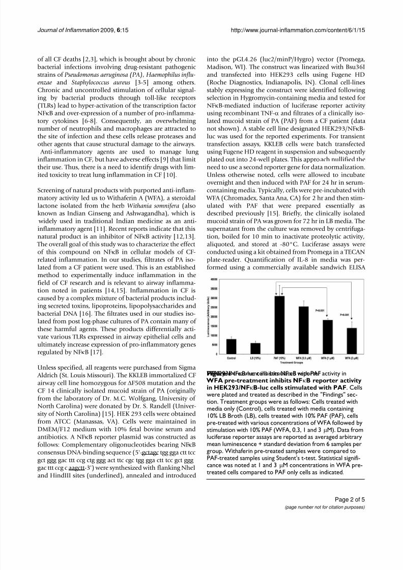
WFA pre-treatment inhibits NFκB reporter activity inHEK293/NFκB-luc cells stimulated with PAFFigure 1 WFA pre-treatment inhibits NFκB reporter activityin HEK293/NFκB-luc cells stimulated with PAF. Cellswere plated and treated as described in the "Findings" sec-tion. Treatment groups were as follows: Cells treated withmedia only (Control), cells treated with media containing10% LB Broth (LB), cells treated with 10% PAF (PAF), cellspre-treated with various concentrations of WFA followed bystimulation with 10% PAF (WFA, 0.3, 1 and 3 μM). Data fromluciferase reporter assays are reported as averaged arbitrarymean luminescence + standard deviation from 6 samples pergroup. Withaferin pre-treated samples were compared toPAF-treated samples using Student's t-test. Statistical signifi-cance was noted at 1 and 3 μM concentrations in WFA pre-treated cells compared to PAF only cells as indicated.
8/3/2019 Inhibition of NFkappaB by the Natural Product Withaferin a in Cellular Models of Cystic Fibrosis Inflammation.
http://slidepdf.com/reader/full/inhibition-of-nfkappab-by-the-natural-product-withaferin-a-in-cellular-models 3/5
Journal of Inflammation 2009, 6:15 http://www.journal-inflammation.com/content/6/1/15
Page 3 of 5(page number not for citation purposes)
kit (Biolegend Inc., San Diego, CA). For the NFκB subunit p65 translocation studies using immunofluorescencemicroscopy, KKLEB cells were seeded in black optical-bot-tom 96-well plates and treated as described above withPAF and WFA. Following treatment, cells were fixed with
3.7% formaldehyde in PBS, and fluorescently labeledusing a commercially available kit (NFκB activation HCSkit, Thermo Scientific, Waltham, MA). Fluorescent images
were acquired at 20× magnification using a Discovery 1automated fluorescent microscope (MDS AnalyticalDevices) with filter sets appropriate for FITC (for p65detection) and DAPI (nuclear stain). Six images were ana-lyzed per group resulting in analysis of roughly 300 cellsper treatment. Nuclear translocation of p65 was measuredusing the enhanced translocation module from the Metax-press image analysis software provided with the instru-ment. Input settings delineating cell "compartment"(nucleus) and "regions for measurement" (cytoplasm)
were entered as follows: Compartment-width = 10 μm,intensity above background = 200 gray levels, minimumarea = 5 μm2, and maximum area = 1000 μm2. Regions for Measurement (RFM) were entered as follows: Inner regiondistance from edge = 1 μm, outer region distance fromedge = 1 μm, outer region width = 6 μm, background cor-rection = none. Cells were scored as positive for nuclear translocation of p65 if the correlation coefficient was 0.75or greater. Cell viability was monitored using the Cell-
Titer Glo Luminescent Cell Viability Assay (Promega Cor-poration) following the manufacturer's suggestion. Allconcentrations used for our studies were non-cytotoxic to
the cells (data not shown) under the experimental condi-tions.
An NFκB-responsive luciferase reporter construct wasused to test the hypothesis that PAF-stimulated NFκBactivity diminished upon treatment with WFA. As demon-strated in Figure 1, WFA pre-treatment significantly inhib-ited NFκB luciferase reporter activity stimulated by PAF ina concentration-dependent fashion by as much as 70% inHEK293/NFκB-luc cells. Past reports indicate that HEK293 cells express certain TLR isoforms that were acti-
vated by bacterial factors present in PAF used for our stud-ies leading to NFκB reporter activity [18,19]. Further
testing of WFA was conducted in a more relevant in vitromodel of CF inflammation. In this model, the immortal-ized CF epithelial cell line KKLEB harboring the most common and severe CFTR mutation (ΔF508, noted in >90% CF patients) was stimulated with PAF and NFκBactivity was subsequently measured using three different methods. First, a transiently transfected luciferase reporter
was used to monitor NFκB activation by PAF with or with-out WFA pre-treatment. As reported in Figure 2, WFA pre-treatment significantly diminished luciferase activity com-pared to control groups in a concentration-dependent
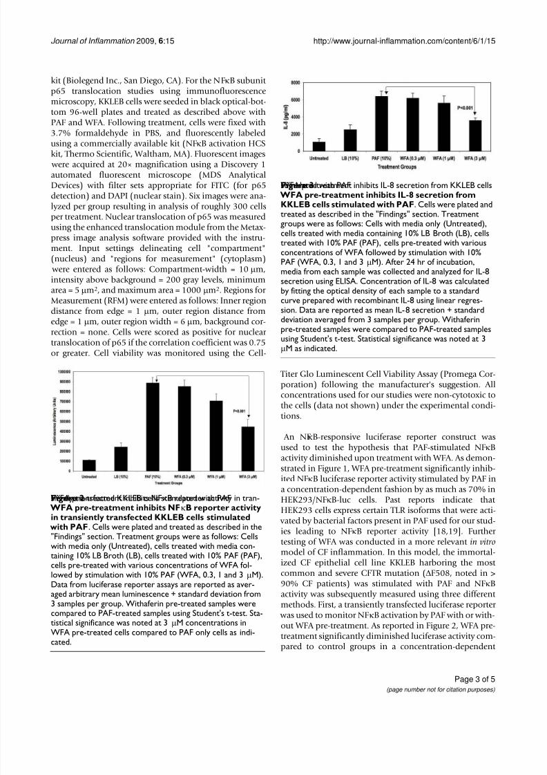
WFA pre-treatment inhibits IL-8 secretion from KKLEB cellsstimulated with PAFFigure 3 WFA pre-treatment inhibits IL-8 secretion fromKKLEB cells stimulated with PAF. Cells were plated andtreated as described in the "Findings" section. Treatmentgroups were as follows: Cells with media only (Untreated),cells treated with media containing 10% LB Broth (LB), cellstreated with 10% PAF (PAF), cells pre-treated with variousconcentrations of WFA followed by stimulation with 10%PAF (WFA, 0.3, 1 and 3 μM). After 24 hr of incubation,
media from each sample was collected and analyzed for IL-8secretion using ELISA. Concentration of IL-8 was calculatedby fitting the optical density of each sample to a standardcurve prepared with recombinant IL-8 using linear regres-sion. Data are reported as mean IL-8 secretion + standarddeviation averaged from 3 samples per group. Withaferinpre-treated samples were compared to PAF-treated samplesusing Student's t-test. Statistical significance was noted at 3μM as indicated.
WFA pre-treatment inhibits NFκB reporter activity in tran-siently transfected KKLEB cells stimulated with PAFFigure 2 WFA pre-treatment inhibits NFκB reporter activityin transiently transfected KKLEB cells stimulated with PAF. Cells were plated and treated as described in the"Findings" section. Treatment groups were as follows: Cellswith media only (Untreated), cells treated with media con-taining 10% LB Broth (LB), cells treated with 10% PAF (PAF),cells pre-treated with various concentrations of WFA fol-lowed by stimulation with 10% PAF (WFA, 0.3, 1 and 3 μM).Data from luciferase reporter assays are reported as aver-aged arbitrary mean luminescence + standard deviation from3 samples per group. Withaferin pre-treated samples werecompared to PAF-treated samples using Student's t-test. Sta-tistical significance was noted at 3 μM concentrations inWFA pre-treated cells compared to PAF only cells as indi-cated.
8/3/2019 Inhibition of NFkappaB by the Natural Product Withaferin a in Cellular Models of Cystic Fibrosis Inflammation.
http://slidepdf.com/reader/full/inhibition-of-nfkappab-by-the-natural-product-withaferin-a-in-cellular-models 4/5
Journal of Inflammation 2009, 6:15 http://www.journal-inflammation.com/content/6/1/15
Page 4 of 5(page number not for citation purposes)
fashion by ~70% in the KKLEB cells. Inflammation in CFis regulated largely by activation of NFκB and transcrip-tion of pro-inflammatory genes regulated by this tran-scription factor [16,20]. Therefore, we examined the effect
of WFA pre-treatment on IL-8 secretion upon challenge with 10% PAF in KKLEB cells using ELISA. Inhibition of PAF-stimulated secretion of IL-8 protein was noted uponpre-treatment with WFA (Figure 3) by ~50% in KKLEBcells in agreement with reporter assays.
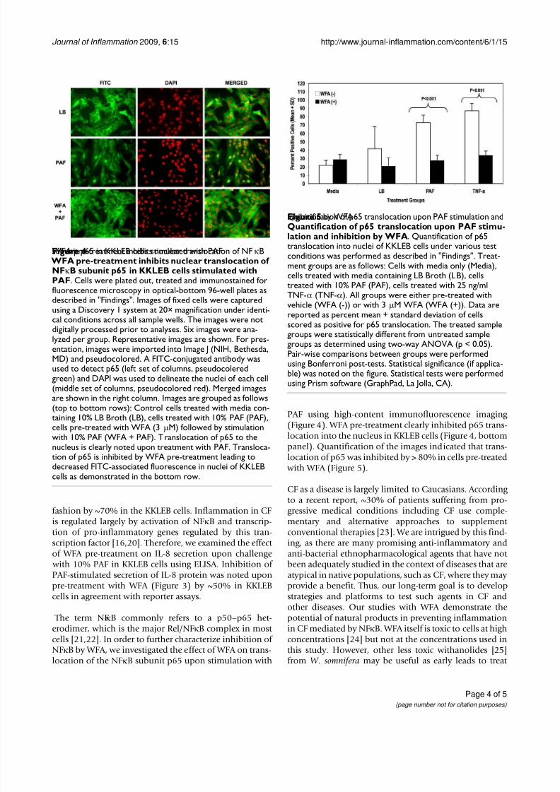
The term NFκB commonly refers to a p50–p65 het-erodimer, which is the major Rel/NFκB complex in most cells [21,22]. In order to further characterize inhibition of NFκB by WFA, we investigated the effect of WFA on trans-location of the NFκB subunit p65 upon stimulation with
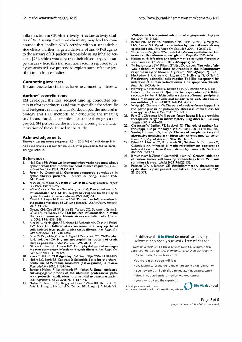
PAF using high-content immunofluorescence imaging (Figure 4). WFA pre-treatment clearly inhibited p65 trans-location into the nucleus in KKLEB cells (Figure 4, bottompanel). Quantification of the images indicated that trans-location of p65 was inhibited by > 80% in cells pre-treated
with WFA (Figure 5).
CF as a disease is largely limited to Caucasians. According to a recent report, ~30% of patients suffering from pro-gressive medical conditions including CF use comple-mentary and alternative approaches to supplement conventional therapies [23]. We are intrigued by this find-ing, as there are many promising anti-inflammatory and
anti-bacterial ethnopharmacological agents that have not been adequately studied in the context of diseases that areatypical in native populations, such as CF, where they may provide a benefit. Thus, our long-term goal is to developstrategies and platforms to test such agents in CF andother diseases. Our studies with WFA demonstrate thepotential of natural products in preventing inflammationin CF mediated by NFκB. WFA itself is toxic to cells at highconcentrations [24] but not at the concentrations used inthis study. However, other less toxic withanolides [25]from W. somnifera may be useful as early leads to treat
Quantification of p65 translocation upon PAF stimulation andinhibition by WFAFigure 5Quantification of p65 translocation upon PAF stimu-lation and inhibition by WFA. Quantification of p65translocation into nuclei of KKLEB cells under various testconditions was performed as described in "Findings". Treat-ment groups are as follows: Cells with media only (Media),
cells treated with media containing LB Broth (LB), cellstreated with 10% PAF (PAF), cells treated with 25 ng/mlTNF-α (TNF-α). All groups were either pre-treated withvehicle (WFA (-)) or with 3 μM WFA (WFA (+)). Data arereported as percent mean + standard deviation of cellsscored as positive for p65 translocation. The treated samplegroups were statistically different from untreated samplegroups as determined using two-way ANOVA (p < 0.05).Pair-wise comparisons between groups were performedusing Bonferroni post-tests. Statistical significance (if applica-ble) was noted on the figure. Statistical tests were performedusing Prism software (GraphPad, La Jolla, CA).
WFA pre-treatment inhibits nuclear translocation of NFκBsubunit p65 in KKLEB cells stimulated with PAFFigure 4 WFA pre-treatment inhibits nuclear translocation of NFκB subunit p65 in KKLEB cells stimulated withPAF. Cells were plated out, treated and immunostained forfluorescence microscopy in optical-bottom 96-well plates asdescribed in "Findings". Images of fixed cells were capturedusing a Discovery 1 system at 20× magnification under identi-cal conditions across all sample wells. The images were notdigitally processed prior to analyses. Six images were ana-lyzed per group. Representative images are shown. For pres-entation, images were imported into Image J (NIH, Bethesda,MD) and pseudocolored. A FITC-conjugated antibody wasused to detect p65 (left set of columns, pseudocoleredgreen) and DAPI was used to delineate the nuclei of each cell(middle set of columns, pseudocolored red). Merged imagesare shown in the right column. Images are grouped as follows(top to bottom rows): Control cells treated with media con-
taining 10% LB Broth (LB), cells treated with 10% PAF (PAF),cells pre-treated with WFA (3 μM) followed by stimulationwith 10% PAF (WFA + PAF). Translocation of p65 to thenucleus is clearly noted upon treatment with PAF. Transloca-tion of p65 is inhibited by WFA pre-treatment leading todecreased FITC-associated fluorescence in nuclei of KKLEBcells as demonstrated in the bottom row.
8/3/2019 Inhibition of NFkappaB by the Natural Product Withaferin a in Cellular Models of Cystic Fibrosis Inflammation.
http://slidepdf.com/reader/full/inhibition-of-nfkappab-by-the-natural-product-withaferin-a-in-cellular-models 5/5
Publish with BioMed Central and everyscientist can read your work free of charge
"BioMed Central will be the most significant development for
disseminating the results of biomedical research in our lifetime."
Sir Paul Nurse, Cancer Research UK
Your research papers will be:
available free of charge to the entire biomedical community
peer reviewed and published immediately upon acceptance
cited in PubMed and archived on PubMed Central
yours — you keep the copyright
Submit your manuscript here:
http://www.biomedcentral.com/info/publishing_adv.asp
BioMedcentral
Journal of Inflammation 2009, 6:15 http://www.journal-inflammation.com/content/6/1/15
Page 5 of 5(page number not for citation purposes)
inflammation in CF. Alternatively, structure activity stud-ies of WFA using medicinal chemistry may lead to com-pounds that inhibit NFκB activity without undesirableside effects. Further, targeted delivery of anti-NFκB agentsto the airways of CF patients is possible using inhaled aer-
osols [26], which would restrict their effects largely to tar-get tissues where this transcription factor is reported to behyper-activated. We propose to explore some of these pos-sibilities in future studies.
Competing interests The authors declare that they have no competing interests.
Authors' contributionsRM developed the idea, secured funding, conducted cer-tain in vitro experiments and was responsible for scientific and budgetary management. BG developed the molecular biology and HCS methods. MP conducted the imaging
studies and provided technical assistance throughout theproject. SH performed the molecular cloning and charac-terization of the cells used in the study.
AcknowledgementsThis work was supported by a grant (1R21NSO61743-01) to RM from NIH.
Additional financial support for the project was provided by the Research
Triangle Institute.
References1. Ma J, Davis PB: What we know and what we do not know about
cystic fibrosis transmembrane conductance regulator. Clinicsin Chest Medicine 1998, 19:459-471.
2. Ferrari M, Cremonesi L: Genotype-phenotype correlation incystic fibrosis patients. Annales de Biologie Clinique 1996,
54:235-241.3. Pilewski JM, Frizzell RA: Role of CFTR in airway disease. Physiol
Rev 1999, 79:S215-255.4. Witko-Sarsat V, Sermet-Gaudelus I, Lenoir G, Descamps-Latscha B:
Inflammation and CFTR: might neutrophils be the key incystic fibrosis? Mediators Inflamm 1999, 8(8):7-11.
5. Chmiel JF, Berger M, Konstan MW: The role of inflammation inthe pathophysiology of CF lung disease. Clin Rev Allergy Immunol 2002, 23:5-27.
6. Greene CM, Carroll TP, Smith SG, Taggart CC, Devaney J, Griffin S,O'Neill SJ, McElvaney NG: TLR-induced inflammation in cysticfibrosis and non-cystic fibrosis airway epithelial cells. J Immu-nol 2005, 174:1638-1646.
7. Aldallal N, McNaughton EE, Manzel LJ, Richards AM, Zabner J, FerkolTW, Look DC: Inflammatory response in airway epithelialcells isolated from patients with cystic fibrosis. Am J Respir CritCare Med 2002, 166:1248-1256.
8. Salva PS, Doyle NA, Graham L, Eigen H, Doerschuk CM: TNF-alpha,
IL-8, soluble ICAM-1, and neutrophils in sputum of cysticfibrosis patients. Pediatr Pulmonol 1996, 21:11-19.
9. Gibson RL, Burns JL, Ramsey BW: Pathophysiology and manage-ment of pulmonary infections in cystic fibrosis. Am J Respir CritCare Med 2003, 168:918-951.
10. Kawai T, Akira S: TLR signaling. Cell Death Differ 2006, 13:816-825.11. Mishra LC, Singh BB, Dagenais S: Scientific basis for the thera-
peutic use of Withania somnifera (ashwagandha): a review. Altern Med Rev 2000, 5:334-346.
12. Bargagna-Mohan P, Ravindranath PP, Mohan R: Small moleculeanti-angiogenic probes of the ubiquitin proteasome path- way: potential application to choroidal neovascularization.Invest Ophthalmol Vis Sci 2006, 47:4138-4145.
13. Mohan R, Hammers HJ, Bargagna-Mohan P, Zhan XH, Herbstritt CJ,Ruiz A, Zhang L, Hanson AD, Conner BP, Rougas J, Pribluda VS:
Withaferin A is a potent inhibitor of angiogenesis. Angiogen-esis 2004, 7:115-122.
14. Becker MN, Sauer MS, Muhlebach MS, Hirsh AJ, Wu Q, VergheseMW, Randell SH: Cytokine secretion by cystic fibrosis airwayepithelial cells. Am J Respir Crit Care Med 2004, 169:645-653.
15. Wu Q, Lu Z, Verghese MW, Randell SH: Airway epithelial cell tol-erance to Pseudomonas aeruginosa. Respir Res 2005, 6:26.
16. Heijerman H: Infection and inflammation in cystic fibrosis: Ashort review. J Cyst Fibros 2005, 4(Suppl 2):3-5.17. Terheggen-Lagro SW, Rijkers GT, Ent CK van der: The role of air-
way epithelium and blood neutrophils in the inflammatoryresponse in cystic fibrosis. J Cyst Fibros 2005, 4(Suppl 2):15-23.
18. MacRedmond R, Greene C, Taggart CC, McElvaney N, O'Neill S:Respiratory epithelial cells require Toll-like receptor 4 forinduction of human beta-defensin 2 by lipopolysaccharide.Respir Res 2005, 6:116.
19. Hornung V, Rothenfusser S, Britsch S, Krug A, Jahrsdorfer B, Giese T,Endres S, Hartmann G: Quantitative expression of toll-likereceptor 1–10 mRNA in cellular subsets of human peripheralblood mononuclear cells and sensitivity to CpG oligodeoxy-nucleotides. J Immunol 2002, 168:4531-4537.
20. Wright JG, Christman JW: The role of nuclear factor kappa B inthe pathogenesis of pulmonary diseases: implications fortherapy. Am J Respir Med 2003, 2:211-219.
21. Park GY, Christman JW: Nuclear factor kappa B is a promising
therapeutic target in inflammatory lung disease. Curr Drug Targets 2006, 7:661-668.22. Christman JW, Sadikot RT, Blackwell TS: The role of nuclear fac-
tor-kappa B in pulmonary diseases. Chest 2000, 117:1482-1487.23. Samdup DZ, Smith RG, Il Song S: The use of complementary and
alternative medicine in children with chronic medical condi-tions. Am J Phys Med Rehabil 2006, 85:842-846.
24. Falsey RR, Marron MT, Gunaherath GM, Shirahatti N, Mahadevan D,Gunatilaka AA, Whitesell L: Actin microfilament aggregationinduced by withaferin A is mediated by annexin II. Nat ChemBiol 2006, 2:33-38.
25. Jayaprakasam B, Zhang Y, Seeram NP, Nair MG: Growth inhibitionof human tumor cell lines by withanolides from Withaniasomnifera leaves. Life Sci 2003, 74:125-132.
26. Prescott WA Jr, Johnson CE: Antiinflammatory therapies forcystic fibrosis: past, present, and future. Pharmacotherapy 2005,25:555-573.