Embed Size (px)
Citation preview
HAL Id: ineris-00961905https://hal-ineris.archives-ouvertes.fr/ineris-00961905
Submitted on 20 Mar 2014
HAL is a multi-disciplinary open accessarchive for the deposit and dissemination of sci-entific research documents, whether they are pub-lished or not. The documents may come fromteaching and research institutions in France orabroad, or from public or private research centers.
L’archive ouverte pluridisciplinaire HAL, estdestinée au dépôt et à la diffusion de documentsscientifiques de niveau recherche, publiés ou non,émanant des établissements d’enseignement et derecherche français ou étrangers, des laboratoirespublics ou privés.
Inhibition of rainbow trout (Oncorhynchus mykiss) P450aromatase activities in brain and ovarian microsomes by
various environmental substancesNathalie Hinfray, Jean-Marc Porcher, François Brion
To cite this version:Nathalie Hinfray, Jean-Marc Porcher, François Brion. Inhibition of rainbow trout (Oncorhynchusmykiss) P450 aromatase activities in brain and ovarian microsomes by various environmental sub-stances. Comparative Biochemistry and Physiology. Part C - Toxicology and Pharmacology, 2006,144 (3), pp.252-262. <10.1016/j.cbpc.2006.09.002>. <ineris-00961905>
1
Inhibition of rainbow trout (Oncorhynchus mykiss) P450 aromatase activities in brain and
ovarian microsomes by various environmental substances.
Nathalie Hinfray, Jean-Marc Porcher and François Brion
Unité d’évaluation des risques écotoxicologiques, Direction des Risques Chroniques, Institut
National de l’Environnement Industriel et des Risques (INERIS), BP 2, F-60550 Verneuil-en-
Halatte, France
To whom correspondence should be addressed:
François Brion
Institut National de l’Environnement Industriel et des Risques (INERIS),
Direction des Risques Chroniques
Unité d’évaluation des risques écotoxicologiques
BP 2, F-60550 Verneuil-en-Halatte, France
Phone : +33 (0)3 44 55 65 12
Fax : +33 (0)3 44 55 67 67
Email : [email protected]
2
ABSTRACT
Aromatase, a key steroidogenic enzyme that catalyses the conversion of androgens to
estrogens, represent a target for endocrine disrupting chemicals. However, little is known
about the effect of pollutants on aromatase enzymes in fish. In this study, we first optimised a
rainbow trout (Oncorhynchus mykiss) microsomal aromatase assay to measure the effects of
43 substances belonging to diverse chemical classes (steroidal and non steroidal aromatase
inhibitors, pesticides, heavy metals, organotin compounds, dioxins, polycyclic aromatic
hydrocarbons) on brain and ovarian aromatase activities in vitro. Our results showed that 12
compounds were able to inhibit brain and ovarian aromatase activities in a dose-dependent
manner with IC50 values ranging from the low nM to the high µM range depending on the
substance: steroidal and non steroidal inhibitors of aromatase (4-hydroxyandrostenedione,
androstatrienedione, aminogluthethimide), imidazole fungicides (clotrimazole, imazalil,
prochloraz), triazole fungicides (difenoconazole, fenbuconazole, propiconazole, triadimenol),
the pyrimidine fungicide fenarimol and methylmercury. Overall, this study demonstrates that
rainbow trout brain and ovarian microsomal aromatase assay is suitable for evaluating
potential aromatase inhibitors in vitro notably with respect to environmental screening. The
results highlight that methylmercury and some pesticides that are currently used throughout
the world, have the potential to interfere with the biosynthesis of endogenous estrogens in
fish.
Key words: aromatase, brain, endocrine disrupting chemicals, ovary, rainbow trout, tritiated
water assay.
3
INTRODUCTION
It has now been well established that many environmental pollutants are able to
disturb the normal physiology and endocrinology of organisms. These substances, termed
Endocrine Disrupting Chemicals (EDCs), have been defined as “exogenous substances that
cause adverse health effects in an intact organism, or its progeny, secondary to changes in
endocrine function” (OECD,1997). Exposure of fish to EDCs has been associated with
reproductive adverse effects at both individual and population level in a variety of fish species
(Jobling et al., 2002; Brion et al., 2004; Nash et al., 2004; Mills and Chichester, 2005). These
substances have multiple modes of action since they can potentially act on the synthesis,
secretion, transport, action and elimination of endogenous hormones (Segner et al., 2003). To
date, research has focused mainly on compounds that interfere with sex steroids receptors,
particularly the estrogen receptor. However, the endocrine system may also be disrupted by
environmental substances through pathways and mechanisms other than those that are ER-
mediated. The knowledge of critical molecular and biochemical targets of EDCs in fish is thus
of a great interest.
The biosynthesis of steroid hormones represents a target for EDC action, particularly
the steps catalysed by cytochrome P450-dependent enzymes (Monod et al., 1993). In
vertebrates, an essential sex-related enzyme is aromatase (P450aro). Aromatase is an
enzymatic complex including a NADPH-dependent cytochrome P450 reductase and a
cytochrome P450 aromatase which catalyzes the final, rate-limiting step in the conversion of
androgens into estrogens (Simpson et al., 1994). In fish, this enzyme has been shown to be
mainly expressed in ovary and brain. In mammals except pig, only one gene encodes the
aromatase while in several teleost fish such as zebrafish (Danio rerio), goldfish (Carassius
auratus) or rainbow trout (Oncorhynchus mykiss), aromatase is encoded by two different
genes : cyp19a (or cyp19A1) and cyp19b (cyp19A2) (Callard and Tchoudakova, 1997;
Kishida and Callard, 2001; Dalla Valle et al., 2002). These two distinct genes generate two
structurally and functionally different aromatase proteins, CYP19A1 or P450 Aro A (AroA),
and CYP19A2 or P450 Aro B (AroB) (Tchoudakova and Callard, 1998; Chiang et al., 2001a;
Blazquez and Piferrer, 2004). These genes have distinct expression patterns : the brain
aromatase activity is mainly due to the expression of the cyp19b gene while the ovarian
aromatase activity is mainly due to the expression of the cyp19a gene (Tchoudakova and
Callard, 1998; Chiang et al., 2001b; Forlano et al., 2001; Kishida and Callard, 2001; Trant et
al., 2001; Dalla Valle et al., 2002; Menuet et al., 2005).
4
In teleost fish, brain aromatase activity is much higher than in mammals (Pasmanik
and Callard, 1985; Pasmanik and Callard, 1988). This very high expression of brain
aromatase could be linked to the capability of fish brain to grow during adulthood (Gelinas et
al., 1998; Forlano et al., 2001). In the gonads, transcription of the aromatase gene has been
proposed as a key step in the process of ovarian differentiation. For instance, in the rainbow
trout (Oncorhynchus mykiss), inhibition of ovarian aromatase in undifferentiated female
resulted in a complete masculinization of an all-female population (Guiguen et al., 1999).
Moreover, during the female reproductive cycle, ovarian secretion of 17-estradiol controls
the hepatic synthesis of vitellogenin, a phospho-lipoprotein corresponding to the major
precursor of embryonic trophics reserves (Flouriot et al., 1997).
Recent studies have reported alterations of brain and/or ovarian P450 aromatase
activities in wild fish collected from contaminated sites (Noaksson et al., 2001; Orlando et al.,
2002; Noaksson et al., 2003; Lavado et al., 2004) suggesting that fish populations are exposed
to substances that perturb the biosynthesis of estrogens. However the nature (and the levels)
of substances involved in these biological responses remains to be determined. In fish, a few
studies have shown that environmental chemicals can interfere with aromatase activity. In
these studies, the range of substances was limited to some pesticides (Monod et al., 1993,
Noakson et al., 2003, Ankley et al., 2005 ) and polycyclic aromatic hydrocarbons (PAHs)
(Monteiro et al., 2000). Thus, there is a need to provide a broader, more systematic
knowledge on aromatase inhibiting potencies of environmental chemicals. Further, most of
the available studies have analysed only gonadal aromatase and it remains to be determined
whether the brain form is likewise affected.
Considering the critical role of aromatase in development and reproduction in fish as
well as the large number of chemical substances that can potentially affect this enzyme, the
aim of this study was to investigate whether several substances belonging to diverse chemical
classes (pesticides, polycyclic aromatic hydrocarbon, heavy metals) affect brain and ovarian
aromatase activities in rainbow trout (Oncorhynchus mykiss) in vitro. For this purpose, we
first optimized a microsomal aromatase assay to measure the effect of xenobiotics on
aromatase activity in fish.
5
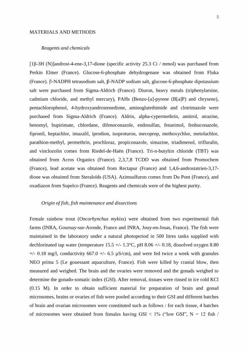
MATERIALS AND METHODS
Reagents and chemicals
[1β-3H (N)]androst-4-ene-3,17-dione (specific activity 25.3 Ci / mmol) was purchased from
Perkin Elmer (France). Glucose-6-phosphate dehydrogenase was obtained from Fluka
(France). -NADPH tetrasodium salt, -NADP sodium salt, glucose-6-phosphate dipotassium
salt were purchased from Sigma-Aldrich (France). Diuron, heavy metals (triphenylarsine,
cadmium chloride, and methyl mercury), PAHs (Benzo-[a]-pyrene (B[a]P) and chrysene),
pentachlorophenol, 4-hydroxyandrostenedione, aminoglutethimide and clotrimazole were
purchased from Sigma-Aldrich (France). Aldrin, alpha-cypermethrin, amitrol, atrazine,
benomyl, bupirimate, chlordane, difenoconazole, endosulfan, fenarimol, fenbuconazole,
fipronil, heptachlor, imazalil, iprodion, isoproturon, mecoprop, methoxychlor, metolachlor,
parathion-methyl, permethrin, prochloraz, propiconazole, simazine, triadimenol, trifluralin,
and vinclozolin comes from Riedel-de-Haën (France). Tri-n-butyltin chloride (TBT) was
obtained from Acros Organics (France). 2,3,7,8 TCDD was obtained from Promochem
(France), lead acetate was obtained from Rectapur (France) and 1,4,6-androstatrien-3,17-
dione was obtained from Steraloïds (USA). Azimsulfuron comes from Du Pont (France), and
oxadiazon from Supelco (France). Reagents and chemicals were of the highest purity.
Origin of fish, fish maintenance and dissections
Female rainbow trout (Oncorhynchus mykiss) were obtained from two experimental fish
farms (INRA, Gournay-sur-Aronde, France and INRA, Jouy-en-Josas, France). The fish were
maintained in the laboratory under a natural photoperiod in 500 litres tanks supplied with
dechlorinated tap water (temperature 15.5 +/- 1.3°C, pH 8.06 +/- 0.18, dissolved oxygen 8.80
+/- 0.18 mg/l, conductivity 667.0 +/- 6.5 µS/cm), and were fed twice a week with granules
NEO prima 5 (Le gouessant aquaculture, France). Fish were killed by cranial blow, then
measured and weighed. The brain and the ovaries were removed and the gonads weighed to
determine the gonado-somatic index (GSI). After removal, tissues were rinsed in ice cold KCl
(0.15 M). In order to obtain sufficient material for preparation of brain and gonad
microsomes, brains or ovaries of fish were pooled according to their GSI and different batches
of brain and ovarian microsomes were constituted such as follows : for each tissue, 4 batches
of microsomes were obtained from females having GSI < 1% (“low GSI”, N = 12 fish /
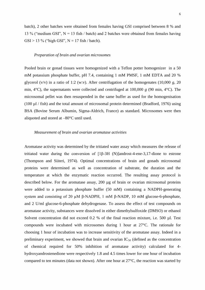
6
batch), 2 other batches were obtained from females having GSI comprised between 8 % and
13 % (“medium GSI”, N = 13 fish / batch) and 2 batches were obtained from females having
GSI > 13 % (“high GSI”, N = 17 fish / batch).
Preparation of brain and ovarian microsomes
Pooled brain or gonad tissues were homogenized with a Teflon potter homogenizer in a 50
mM potassium phosphate buffer, pH 7.4, containing 1 mM PMSF, 1 mM EDTA and 20 %
glycerol (v/v) in a ratio of 1:2 (w:v). After centrifugation of the homogenates (10,000 g, 20
min, 4°C), the supernatants were collected and centrifuged at 100,000 g (90 min, 4°C). The
microsomal pellet was then resuspended in the same buffer as used for the homogenisation
(100 µl / fish) and the total amount of microsomal protein determined (Bradford, 1976) using
BSA (Bovine Serum Albumin, Sigma-Aldrich, France) as standard. Microsomes were then
aliquoted and stored at –80°C until used.
Measurement of brain and ovarian aromatase activities
Aromatase activity was determined by the tritiated water assay which measures the release of
tritiated water during the conversion of [1β-3H (N)]androst-4-ene-3,17-dione to estrone
(Thompson and Siiteri, 1974). Optimal concentrations of brain and gonads microsomal
proteins were determined as well as concentration of substrate, the duration and the
temperature at which the enzymatic reaction occurred. The resulting assay protocol is
described below. For the aromatase assay, 200 µg of brain or ovarian microsomal proteins
were added to a potassium phosphate buffer (50 mM) containing a NADPH-generating
system and consisting of 20 µM β-NADPH, 1 mM β-NADP, 10 mM glucose-6-phosphate,
and 2 U/ml glucose-6-phosphate dehydrogenase. To assess the effect of test compounds on
aromatase activity, substances were dissolved in either dimethylsulfoxide (DMSO) or ethanol
Solvent concentration did not exceed 0.2 % of the final reaction mixture, i.e. 500 µl. Test
compounds were incubated with microsomes during 1 hour at 27°C. The rationale for
choosing 1 hour of incubation was to increase sensitivity of the aromatase assay. Indeed in a
preliminary experiment, we showed that brain and ovarian IC50 (defined as the concentration
of chemical required for 50% inhibition of aromatase activity) calculated for 4-
hydroxyandrostenedione were respectively 1.8 and 4.5 times lower for one hour of incubation
compared to ten minutes (data not shown). After one hour at 27°C, the reaction was started by
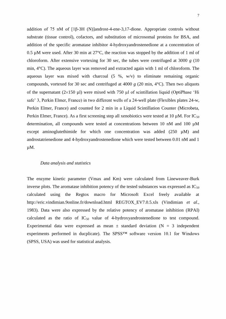
7
addition of 75 nM of [1β-3H (N)]androst-4-ene-3,17-dione. Appropriate controls without
substrate (tissue control), cofactors, and substitution of microsomal proteins for BSA, and
addition of the specific aromatase inhibitor 4-hydroxyandrostenedione at a concentration of
0.5 µM were used. After 30 min at 27°C, the reaction was stopped by the addition of 1 ml of
chloroform. After extensive vortexing for 30 sec, the tubes were centrifuged at 3000 g (10
min, 4°C). The aqueous layer was removed and extracted again with 1 ml of chloroform. The
aqueous layer was mixed with charcoal (5 %, w/v) to eliminate remaining organic
compounds, vortexed for 30 sec and centrifuged at 4000 g (20 min, 4°C). Then two aliquots
of the supernatant (2150 µl) were mixed with 750 µl of scintillation liquid (OptiPhase ‘Hi
safe’ 3, Perkin Elmer, France) in two different wells of a 24-well plate (Flexibles plates 24-w,
Perkin Elmer, France) and counted for 2 min in a Liquid Scintillation Counter (Microbeta,
Perkin Elmer, France). As a first screening step all xenobiotics were tested at 10 µM. For IC50
determination, all compounds were tested at concentrations between 10 nM and 100 µM
except aminoglutethimide for which one concentration was added (250 µM) and
androstatrienedione and 4-hydroxyandrostenedione which were tested between 0.01 nM and 1
µM.
Data analysis and statistics
The enzyme kinetic parameter (Vmax and Km) were calculated from Lineweaver-Burk
inverse plots. The aromatase inhibition potency of the tested substances was expressed as IC50
calculated using the Regtox macro for Microsoft Excel freely available at
http://eric.vindimian.9online.fr/download.html REGTOX_EV7.0.5.xls (Vindimian et al.,
1983). Data were also expressed by the relative potency of aromatase inhibition (RPAI)
calculated as the ratio of IC50 value of 4-hydroxyandrostenedione to test compound.
Experimental data were expressed as mean ± standard deviation (N = 3 independent
experiments performed in ducplicate). The SPSS™ software version 10.1 for Windows
(SPSS, USA) was used for statistical analysis.
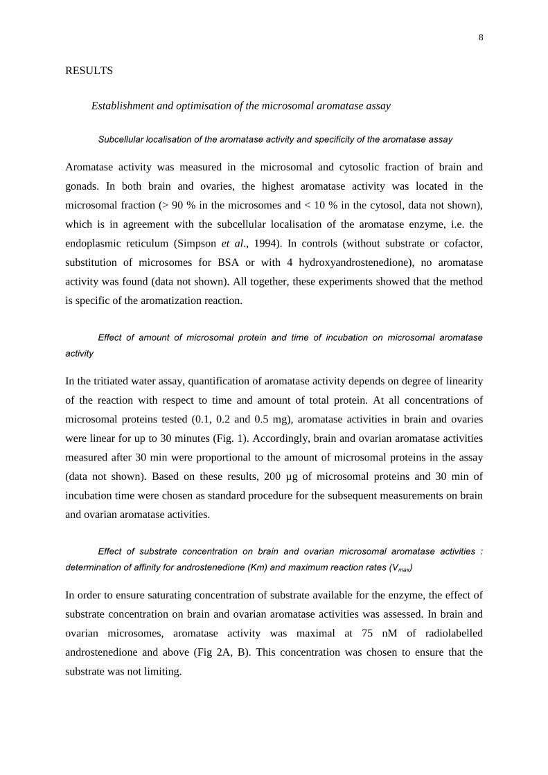
8
RESULTS
Establishment and optimisation of the microsomal aromatase assay
Subcellular localisation of the aromatase activity and specificity of the aromatase assay
Aromatase activity was measured in the microsomal and cytosolic fraction of brain and
gonads. In both brain and ovaries, the highest aromatase activity was located in the
microsomal fraction (> 90 % in the microsomes and < 10 % in the cytosol, data not shown),
which is in agreement with the subcellular localisation of the aromatase enzyme, i.e. the
endoplasmic reticulum (Simpson et al., 1994). In controls (without substrate or cofactor,
substitution of microsomes for BSA or with 4 hydroxyandrostenedione), no aromatase
activity was found (data not shown). All together, these experiments showed that the method
is specific of the aromatization reaction.
Effect of amount of microsomal protein and time of incubation on microsomal aromatase
activity
In the tritiated water assay, quantification of aromatase activity depends on degree of linearity
of the reaction with respect to time and amount of total protein. At all concentrations of
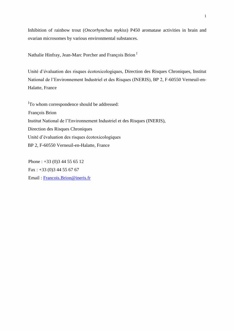
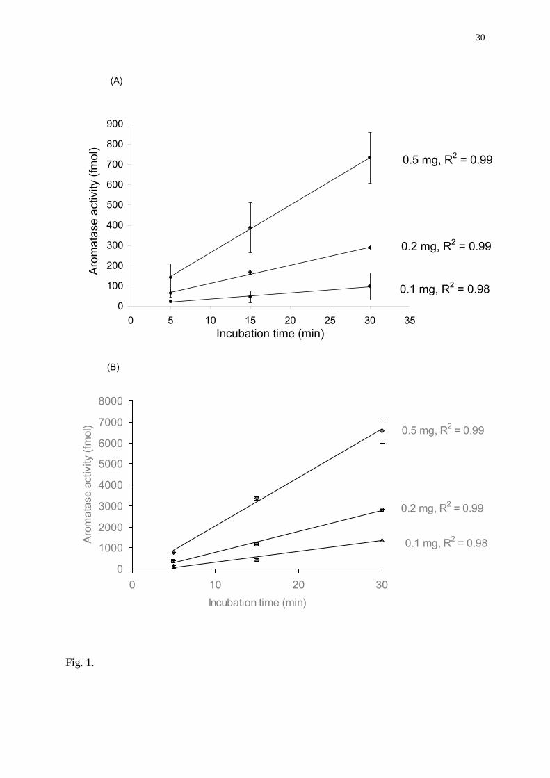
microsomal proteins tested (0.1, 0.2 and 0.5 mg), aromatase activities in brain and ovaries
were linear for up to 30 minutes (Fig. 1). Accordingly, brain and ovarian aromatase activities
measured after 30 min were proportional to the amount of microsomal proteins in the assay
(data not shown). Based on these results, 200 µg of microsomal proteins and 30 min of
incubation time were chosen as standard procedure for the subsequent measurements on brain
and ovarian aromatase activities.
Effect of substrate concentration on brain and ovarian microsomal aromatase activities :
determination of affinity for androstenedione (Km) and maximum reaction rates (Vmax)
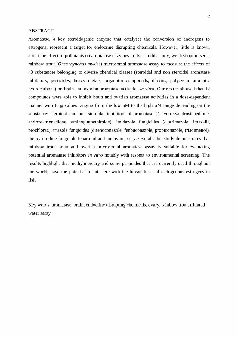
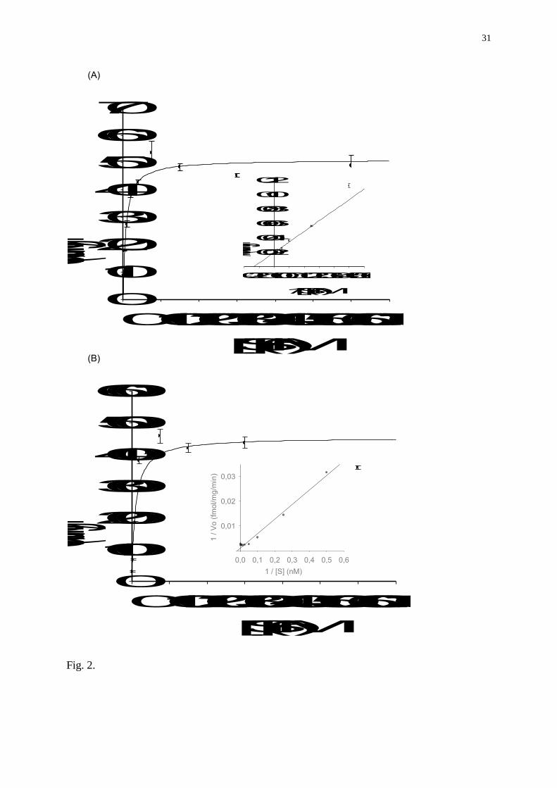
In order to ensure saturating concentration of substrate available for the enzyme, the effect of
substrate concentration on brain and ovarian aromatase activities was assessed. In brain and
ovarian microsomes, aromatase activity was maximal at 75 nM of radiolabelled
androstenedione and above (Fig 2A, B). This concentration was chosen to ensure that the
substrate was not limiting.
9
By using the established aromatase assay, the rainbow trout aromatase affinity for
androstenedione (Km) and its maximum reaction rate (Vmax) were determined in both brain
and ovarian microsomes (Fig 2A, B). The Km values for androstenedione in brain and ovary
were 9.9 +/- 2.6 nM and 7.48 +/- 1.14 respectively without significant difference. In contrast,
Vmax were much higher in brain (Vmax = 453.7 +/- 24.5) than in ovary (51.0 +/- 1.5
fmol/mg/min) (Fig 2A, B).
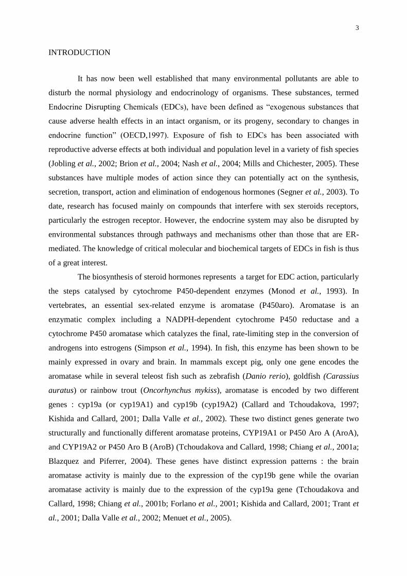
Effect of incubation temperature of microsomes on aromatase activity
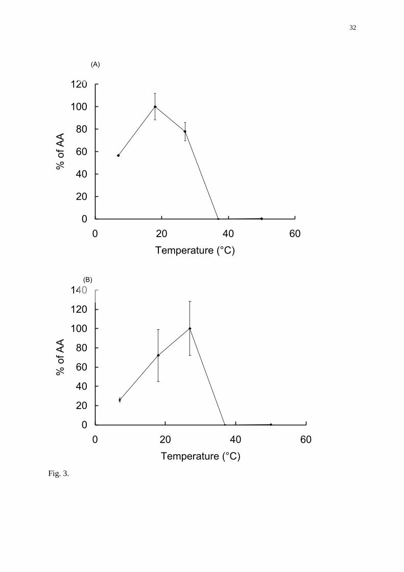
Variations of the incubation temperature resulted in significant changes in aromatase activity
(Fig. 3A, B). In brain and in ovarian microsomes, aromatase activity was maximal at 18°C
and 27°C respectively. At 37°C, aromatase activities in both brain and gonad were completely
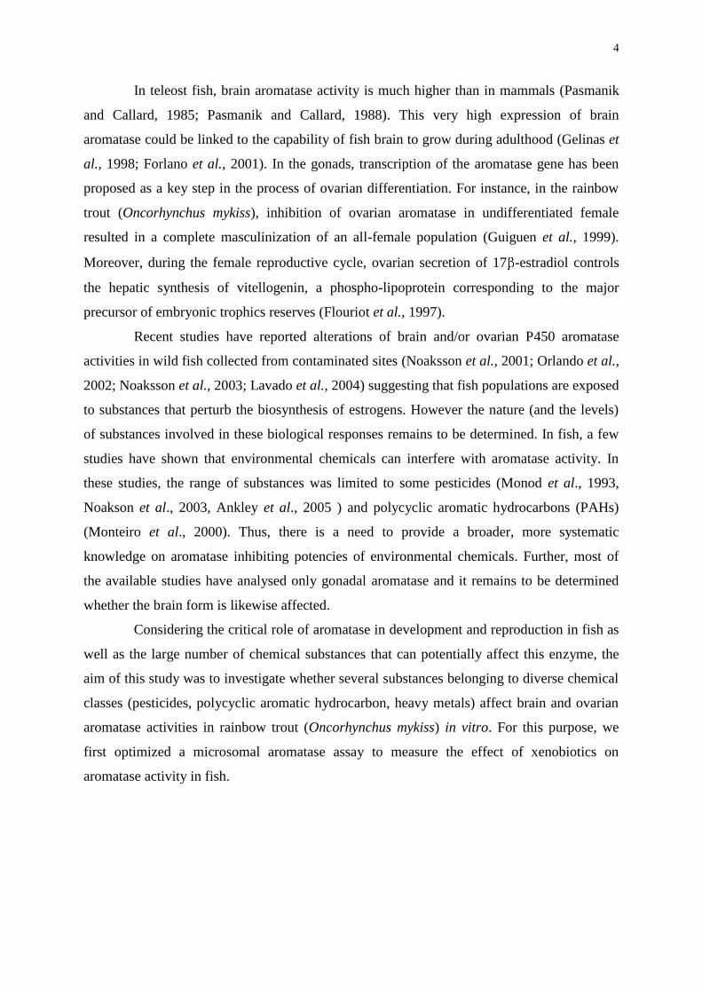
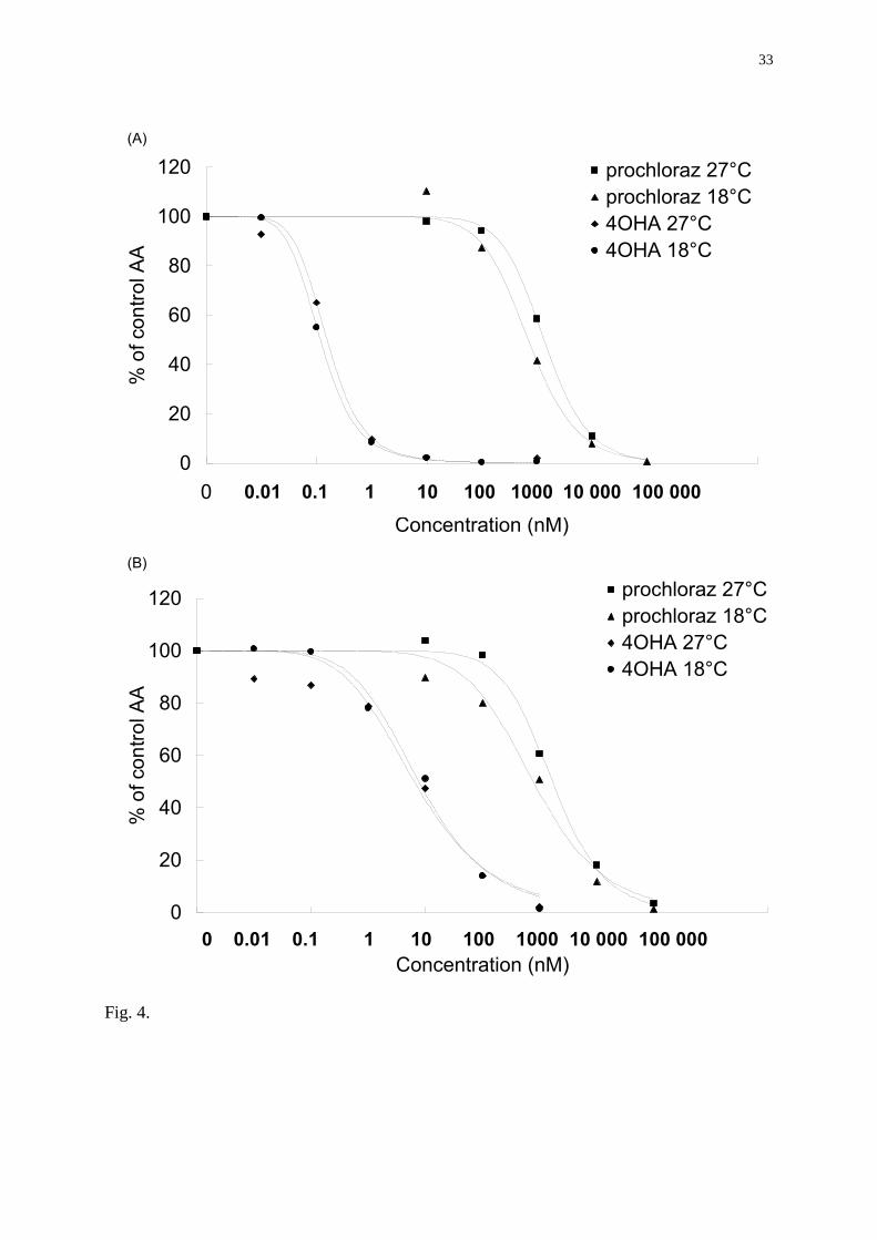
inhibited. In order to verify that temperature did not influence the effects of xenobiotics in the
aromatase assay, microsomes were exposed to known aromatase inhibitors and incubated at
18 or 27°C. As shown by the Figure 4, the calculated brain and ovarian IC50 for 4-
hydroxyandrostenedione and prochloraz were the same at both temperatures. Based on this
data, the incubation temperature used for the microsomal assay was set at 27°C.
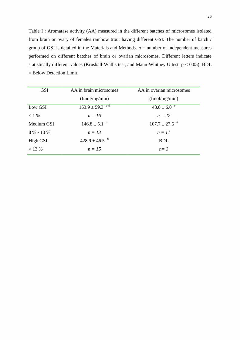
Brain and ovarian aromatase activities measured in the different batches of microsomes
Whatever the batch of microsomes, brain aromatase activity was systematically higher than
ovarian activity (Table I). Moreover, brain and ovarian aromatase activities were significantly
different among batches : in microsomes isolated from the “high GSI” group, brain aromatase
activity was significantly higher than in microsomes isolated from fish of the “low” and
“medium” GSI groups. In contrast, no aromatase activity was measured in ovarian
microsomes isolated from females of the “high GSI” group. From a practical point of view,
the effect of environmental pollutants on brain and ovarian aromatase activities was tested on
microsomes of the “medium GSI group”, where both brain and ovarian aromatase activities
were high.
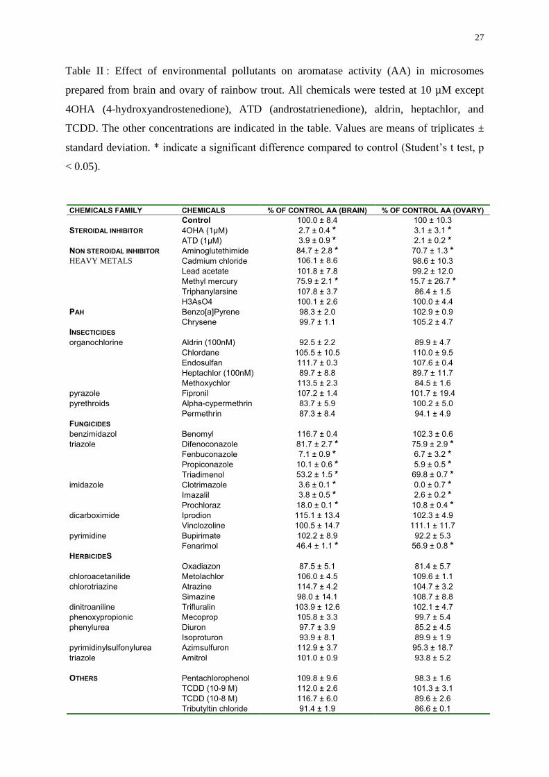
Effect of environmental pollutants on brain and ovarian aromatase activities
Forty three environmental chemicals from different chemical classes were tested for their
ability to interfere with brain and ovarian aromatase activities. As a first screening step,
experiments were conducted by testing all the compounds at 10µM except for few of them
10
which where tested at lower concentrations due to solubility limitations. The results from
these experiments are summarized in Table II. Twelve out of 43 substances were potent
inhibitors of aromatase activity in vitro at 10 µM both in the brain and ovaries:
androstatrienedione, 4-hydroxyandrostenedione, aminoglutethimide, clotrimazole, fenarimol,
difenoconazole, fenbuconazole, imazalil, prochloraz, propiconazole, triadimenol, and
methylmercury (Table II). None of the other tested chemicals exhibited any significant effect
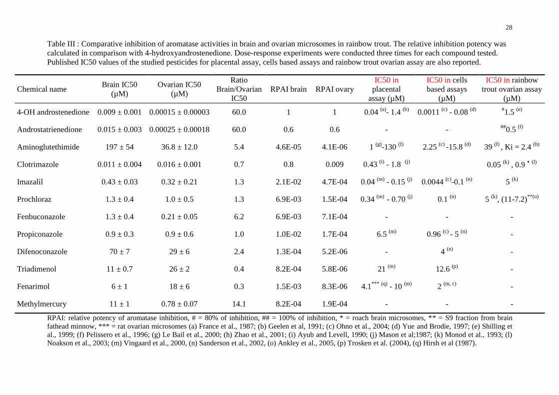
on aromatase activity at the concentrations tested. For the active substances, dose-response
experiments were then conducted to determine their IC50 values. The results are summarised
in Table III. Steroidal compounds (4-hydroxyandrostenedione and androstatrienedione) were
found to be the most potent aromatase inhibitors with IC50 values in the low nM range. The
non steroidal compound aminoglutethimide was found to be the least potent aromatase
inhibitor among all the inhibiting substances tested in this study with a relative potency of
aromatase inhibition (RPAI) for brain and ovary equal to 4.6 10-5 and 4.1 10-6 respectively
compared to 4-hydroxyandrostenedione. In contrast, the imidazole fungicide clotrimazole
inhibited the brain and ovarian aromatase activities with a potency of inhibition in brain close
to that of 4-hydroxyandrostenedione (RPAIbrain = 0.8 and RPAIovary = 0.009). With the
exception of methylmercury, all the aromatase inhibiting xenobiotics were fungicides
belonging to the triazole, imidazole and pyrimidine families and were characterized by IC50
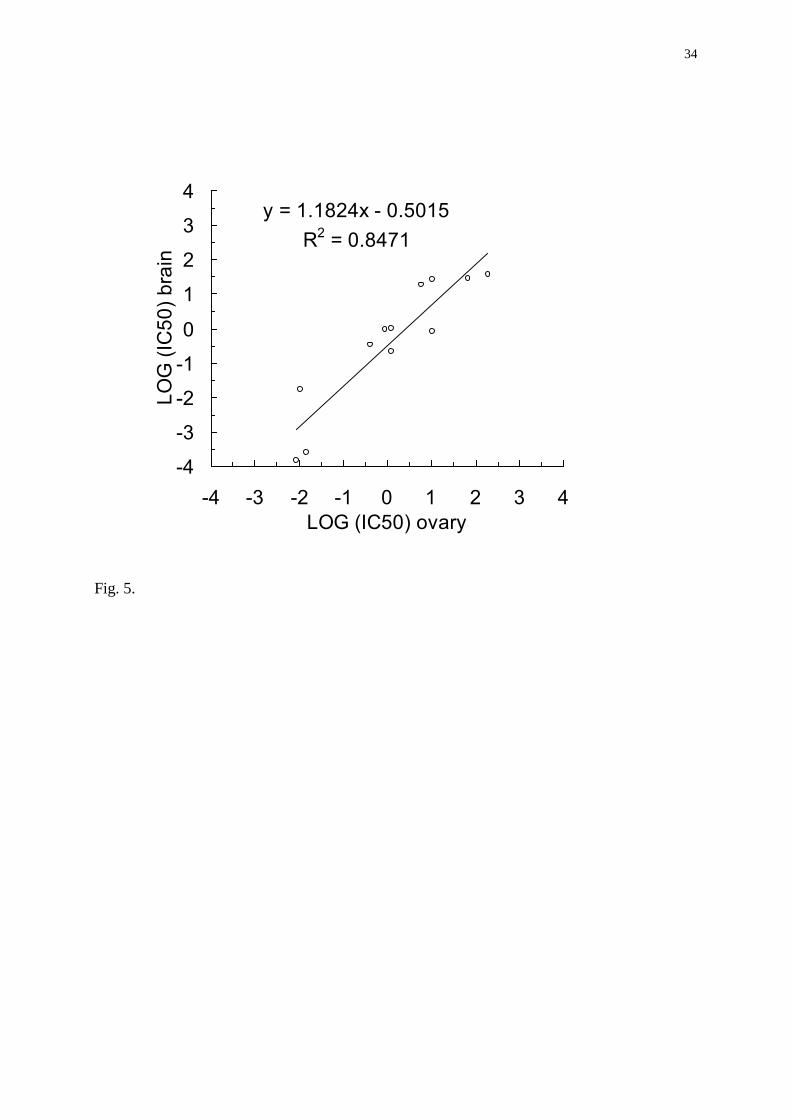
values ranging from the low to the high µM range (Table III). Linear regression analysis
showed that there exists a significant and positive correlation between the inhibitory action of
a substance on the ovarian aromatase and on the brain aromatase (R2 = 0.85; Pearson’s test,
p< 0.01, Fig. 5). Interestingly, despite this overall correlation, tissue specific effects were
highlighted for certain chemicals. For the four fungicides (clotrimazole, imazalil, prochloraz
and propiconazole), no significant differences of their brain and ovarian IC50 values were
noted. In contrast, 4 out of 12 compounds (4-hydroxyandrostenedione, androstatrienedione,
difenoconazole, and methylmercury) showed ovarian IC50 values significantly lower than
those measured in the brain (student’s t test, p<0.05). IC50 of aminoglutethimide and
fenbuconazole were also lower than brain IC50 but the difference was not statistically
significant (Student’s –t test, p = 0.06 and 0.07 respectively). Finally, triadimenol had
stronger inhibitory effect on the brain than on the ovarian aromatase activity (Student’s t test,
p<0.05). Fenarimol had the same pattern of inhibitory effect as triadimenol but the difference
was not statistically significant.
11
DISCUSSION
This paper describes the potential endocrine disrupting activity of xenobiotics by
assessing their capacities to inhibit in vitro brain and ovarian aromatase activities of rainbow
trout. We first optimized the tritiated water assay, characterized aromatase activity in the two
target tissues and determined the kinetic parameters of the aromatase in brain and ovarian
microsomes. Then we showed that several environmental substances are able to inhibit both
brain and ovarian aromatase activities in a dose-dependent manner, indicating that they can
potentially interfere with biosynthesis of endogenous estrogens and alter the androgen :
estrogen ratio. Among them, methylmercury and the triazole fungicide fenbuconazole were
newly identified as in vitro inhibitors of aromatase activity in a vertebrate model.
Rainbow trout brain and ovarian microsomal aromatase assay
The tritiated water assay to measure aromatase activity has been already applied to
several fish species, including goldfish (Carassius auratus) (Pasmanik and Callard, 1985),
medaka (Oryzias latipes) (Melo and Ramsdell, 2001), perch (Perca fluviatilis) and roach
(Rutilus rutilus) (Noaksson et al., 2001), sea bass (Dicentrarchus labrax) (Gonzalez and
Piferrer, 2002; Gonzalez and Piferrer, 2003) and rainbow trout (Oncorhynchus mykiss)
(Monod et al., 1993; Shilling et al., 1999). We aimed to optimize the assay in such a way that
it fits to both ovarian and brain aromatase activities, and to determine whether brain and
ovarian aromatase differ in their key kinetic parameters, Vmax and Km. Amount of protein,
length of incubation time, and substrate concentration were found to influence aromatase
activity, a finding which is in accordance with the observations of Gonzalez and Piferrer
(2002) on aromatase activity in sea bass. Also temperature influenced aromatase activity. The
temperature effect on aromatase activity can be associated with decreasing cyp19 mRNA
expression in tilapia with increasing water temperature (D'Cotta et al., 2001; Tsai et al.,
2003). Aromatase activity of rainbow trout showed lower temperature maxima than aromatase
activity of sea bass (30°C and 40°C for brain and ovary, respectively, in sea bass (Gonzalez
and Piferrer, 2002)), a difference that might be related to the fact that rainbow trout is a cold
water teleost fish species while sea bass live at higher temperatures.
Comparison of the catalytic properties
12
To our knowledge, this study is the first to report Vmax and Km values for both the
brain and ovarian P450 aromatase in rainbow trout. The Vmax value of the brain aromatase is
nine-fold higher than that of ovarian aromatase (453.72 24.50 and 51.04 1.52
fmol/mg/min respectively). These results are in accordance with the ten-fold higher Vmax of
goldfish brain aromatase isoform compared to ovary isoform (Zhao et al., 2001) and are in
line with the four-fold higher Vmax of sea bass brain aromatase compared to ovary (Gonzalez
and Piferrer, 2002). Moreover, the Vmax calculated for the ovaries is similar to the Vmax
reported by Shilling et al. (1999) for rainbow trout (71.1 fmol/mg/min). While brain exhibited
higher catalytic activity compared to the ovary, brain and ovarian Km values were not
significantly different, demonstrating similar and very high affinities of both aromatases for
androstenedione. Our data are well in accordance with Km values reported in literature for
other teleost fish species : 5 nM in goldfish brain homogenates (Pasmanik and Callard, 1988),
8.2 nM in goldfish brain microsomes (Zhao et al., 2001), 4.1 and 3.4 nM in brain and ovarian
sea bass microsomes respectively (Gonzalez and Piferrer, 2002). However, it should be noted
that our ovarian Km value is in disagreement with that reported by Shilling et al. (1999) for
rainbow trout who found a 40-fold higher value.
Effect of test chemicals on rainbow trout brain and ovarian aromatase activities
Aromatase activity inhibition by known aromatase inhibitors
In this study three aromatase inhibitors were used, namely two steroidal inhibitors, 4-
hydroxyandrostenedione and androstatrienedione, and a non steroidal inhibitor,
aminoglutethimide. Steroidal inhibitors are steroid analogues of androstenedione. They bind
irreversibly to the active site of the enzyme while non steroidal inhibitor act reversibly on
P450 aromatase by interacting with the heme prosthetic group of the enzyme (Brodie et al.,
1986; Yue and Brodie, 1997). The capacities of these pharmaceuticals to inhibit aromatase
have been extensively studied in mammals notably within the context of estrogen-dependant
cancer therapy (see Geisler and Lonning, 2005 for a review). By using different in vitro
human systems (i.e., placental microsomes, human adrenocortical cells H295R, human
choriocarcinoma-derived JEG3 cells), the human IC50 values reported for these molecules
ranged from 0.0015 µM to 1.5 µM for 4-hydroxyandrostenedione and from 0.0146 µM to 55
µM for aminoglutethimide (see Table III). Our results clearly show that these compounds act
as aromatase inhibitors in rainbow trout as well. That rainbow trout aromatase activity
13
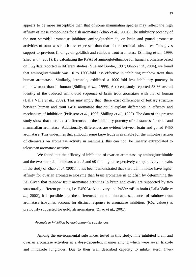
appears to be more susceptible than that of some mammalian species may reflect the high
affinity of these compounds for fish aromatase (Zhao et al., 2001). The inhibitory potency of
the non steroidal aromatase inhibitor, aminoglutethimide, on brain and gonad aromatase
activities of trout was much less expressed than that of the steroidal substances. This gives
support to previous findings on goldfish and rainbow trout aromatase (Shilling et al., 1999;
Zhao et al., 2001). By calculating the RPAI of aminoglutethimide for human aromatase based
on IC50 data reported in different studies (Yue and Brodie, 1997; Ohno et al., 2004), we found
that aminoglutethimide was 10 to 1200-fold less effective in inhibiting rainbow trout than
human aromatase. Similarly, letrozole, exhibited a 1000-fold less inhibitory potency in
rainbow trout than in human (Shilling et al., 1999). A recent study reported 53 % overall
identity of the deduced amino-acid sequence of brain trout aromatase with that of human
(Dalla Valle et al., 2002). This may imply that there exist differences of tertiary structure
between human and trout P450 aromatase that could explain differences in efficacy and
mechanism of inhibition (Pelissero et al., 1996; Shilling et al., 1999). The data of the present
study show that there exist differences in the inhibitory potency of substances for trout and
mammalian aromatase. Additionally, differences are evident between brain and gonad P450
aromatase. This underlines that although some knowledge is available for the inhibitory action
of chemicals on aromatase activity in mammals, this can not be linearly extrapolated to
teleostean aromatase activity.
We found that the efficacy of inhibition of ovarian aromatase by aminoglutethimide
and the two steroidal inhibitors were 5 and 60 fold higher respectively comparatively to brain.
In the study of Zhao et al. (2001) it has been demonstrated that steroidal inhibitor have higher
affinity for ovarian aromatase isozyme than brain aromatase in goldfish by determining the
Ki. Given that rainbow trout aromatase activities in brain and ovary are supported by two
structurally different proteins, i.e. P450AroA in ovary and P450AroB in brain (Dalla Valle et
al., 2002), it is possible that the differences in the amino-acid sequences of rainbow trout
aromatase isozymes account for distinct response to aromatase inhibitors (IC50 values) as
previously suggested for goldfish aromatases (Zhao et al., 2001).
Aromatase Inhibition by environmental substances
Among the environmental substances tested in this study, nine inhibited brain and
ovarian aromatase activities in a dose-dependent manner among which were seven triazole
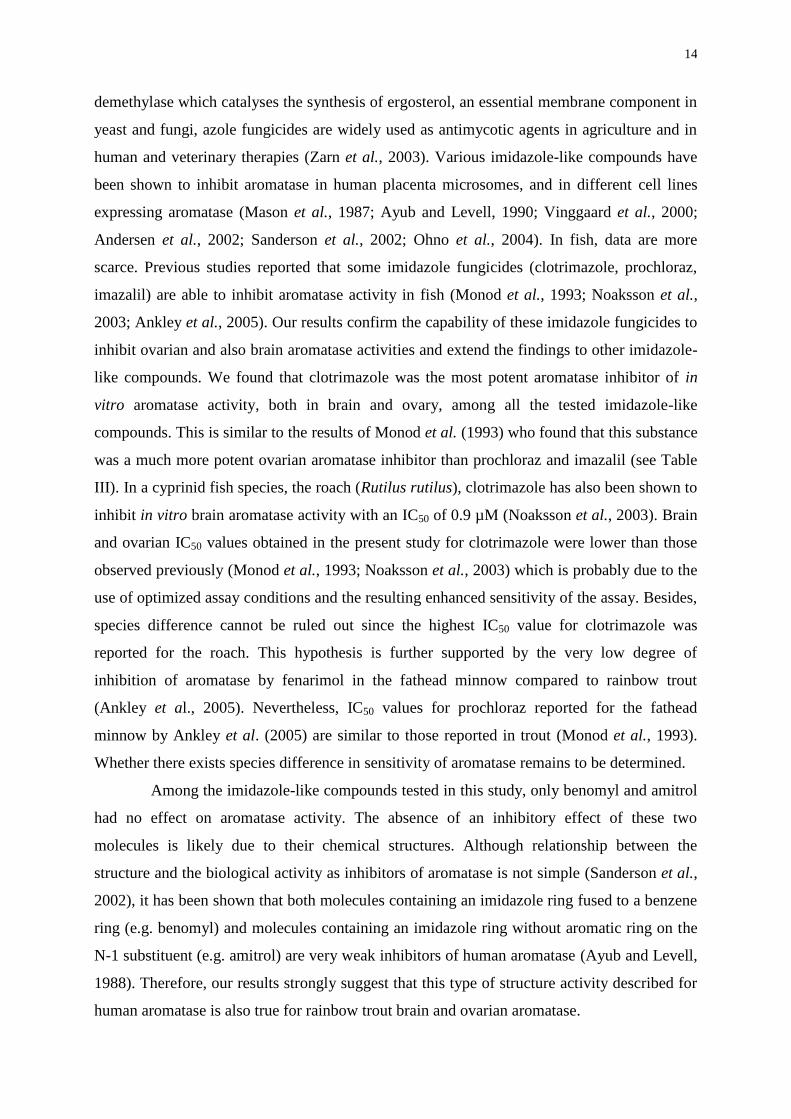
and imidazole fungicides. Due to their well described capacity to inhibit sterol 14--
14
demethylase which catalyses the synthesis of ergosterol, an essential membrane component in
yeast and fungi, azole fungicides are widely used as antimycotic agents in agriculture and in
human and veterinary therapies (Zarn et al., 2003). Various imidazole-like compounds have
been shown to inhibit aromatase in human placenta microsomes, and in different cell lines
expressing aromatase (Mason et al., 1987; Ayub and Levell, 1990; Vinggaard et al., 2000;
Andersen et al., 2002; Sanderson et al., 2002; Ohno et al., 2004). In fish, data are more
scarce. Previous studies reported that some imidazole fungicides (clotrimazole, prochloraz,
imazalil) are able to inhibit aromatase activity in fish (Monod et al., 1993; Noaksson et al.,
2003; Ankley et al., 2005). Our results confirm the capability of these imidazole fungicides to
inhibit ovarian and also brain aromatase activities and extend the findings to other imidazole-
like compounds. We found that clotrimazole was the most potent aromatase inhibitor of in
vitro aromatase activity, both in brain and ovary, among all the tested imidazole-like
compounds. This is similar to the results of Monod et al. (1993) who found that this substance
was a much more potent ovarian aromatase inhibitor than prochloraz and imazalil (see Table
III). In a cyprinid fish species, the roach (Rutilus rutilus), clotrimazole has also been shown to
inhibit in vitro brain aromatase activity with an IC50 of 0.9 µM (Noaksson et al., 2003). Brain
and ovarian IC50 values obtained in the present study for clotrimazole were lower than those
observed previously (Monod et al., 1993; Noaksson et al., 2003) which is probably due to the
use of optimized assay conditions and the resulting enhanced sensitivity of the assay. Besides,
species difference cannot be ruled out since the highest IC50 value for clotrimazole was
reported for the roach. This hypothesis is further supported by the very low degree of
inhibition of aromatase by fenarimol in the fathead minnow compared to rainbow trout
(Ankley et al., 2005). Nevertheless, IC50 values for prochloraz reported for the fathead
minnow by Ankley et al. (2005) are similar to those reported in trout (Monod et al., 1993).
Whether there exists species difference in sensitivity of aromatase remains to be determined.
Among the imidazole-like compounds tested in this study, only benomyl and amitrol
had no effect on aromatase activity. The absence of an inhibitory effect of these two
molecules is likely due to their chemical structures. Although relationship between the
structure and the biological activity as inhibitors of aromatase is not simple (Sanderson et al.,
2002), it has been shown that both molecules containing an imidazole ring fused to a benzene
ring (e.g. benomyl) and molecules containing an imidazole ring without aromatic ring on the
N-1 substituent (e.g. amitrol) are very weak inhibitors of human aromatase (Ayub and Levell,
1988). Therefore, our results strongly suggest that this type of structure activity described for
human aromatase is also true for rainbow trout brain and ovarian aromatase.
15
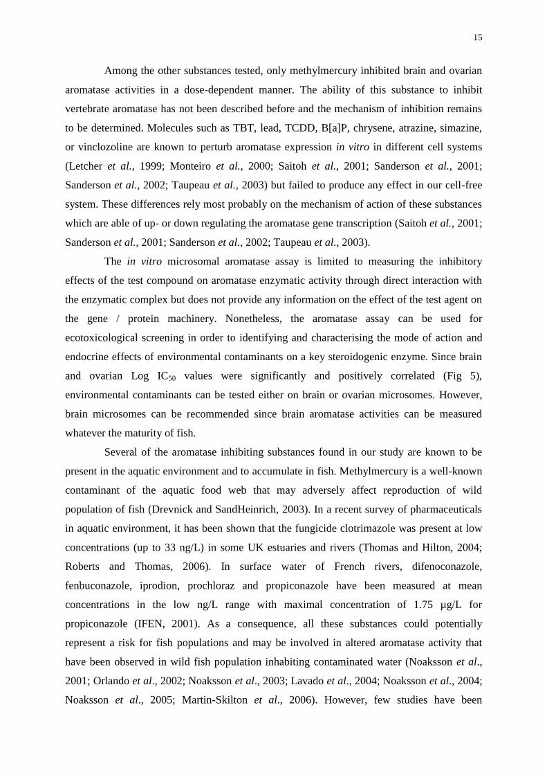
Among the other substances tested, only methylmercury inhibited brain and ovarian
aromatase activities in a dose-dependent manner. The ability of this substance to inhibit
vertebrate aromatase has not been described before and the mechanism of inhibition remains
to be determined. Molecules such as TBT, lead, TCDD, B[a]P, chrysene, atrazine, simazine,
or vinclozoline are known to perturb aromatase expression in vitro in different cell systems
(Letcher et al., 1999; Monteiro et al., 2000; Saitoh et al., 2001; Sanderson et al., 2001;
Sanderson et al., 2002; Taupeau et al., 2003) but failed to produce any effect in our cell-free
system. These differences rely most probably on the mechanism of action of these substances
which are able of up- or down regulating the aromatase gene transcription (Saitoh et al., 2001;
Sanderson et al., 2001; Sanderson et al., 2002; Taupeau et al., 2003).
The in vitro microsomal aromatase assay is limited to measuring the inhibitory
effects of the test compound on aromatase enzymatic activity through direct interaction with
the enzymatic complex but does not provide any information on the effect of the test agent on
the gene / protein machinery. Nonetheless, the aromatase assay can be used for
ecotoxicological screening in order to identifying and characterising the mode of action and
endocrine effects of environmental contaminants on a key steroidogenic enzyme. Since brain
and ovarian Log IC50 values were significantly and positively correlated (Fig 5),
environmental contaminants can be tested either on brain or ovarian microsomes. However,
brain microsomes can be recommended since brain aromatase activities can be measured
whatever the maturity of fish.
Several of the aromatase inhibiting substances found in our study are known to be
present in the aquatic environment and to accumulate in fish. Methylmercury is a well-known
contaminant of the aquatic food web that may adversely affect reproduction of wild
population of fish (Drevnick and SandHeinrich, 2003). In a recent survey of pharmaceuticals
in aquatic environment, it has been shown that the fungicide clotrimazole was present at low
concentrations (up to 33 ng/L) in some UK estuaries and rivers (Thomas and Hilton, 2004;
Roberts and Thomas, 2006). In surface water of French rivers, difenoconazole,
fenbuconazole, iprodion, prochloraz and propiconazole have been measured at mean
concentrations in the low ng/L range with maximal concentration of 1.75 µg/L for
propiconazole (IFEN, 2001). As a consequence, all these substances could potentially
represent a risk for fish populations and may be involved in altered aromatase activity that
have been observed in wild fish population inhabiting contaminated water (Noaksson et al.,
2001; Orlando et al., 2002; Noaksson et al., 2003; Lavado et al., 2004; Noaksson et al., 2004;
Noaksson et al., 2005; Martin-Skilton et al., 2006). However, few studies have been
16
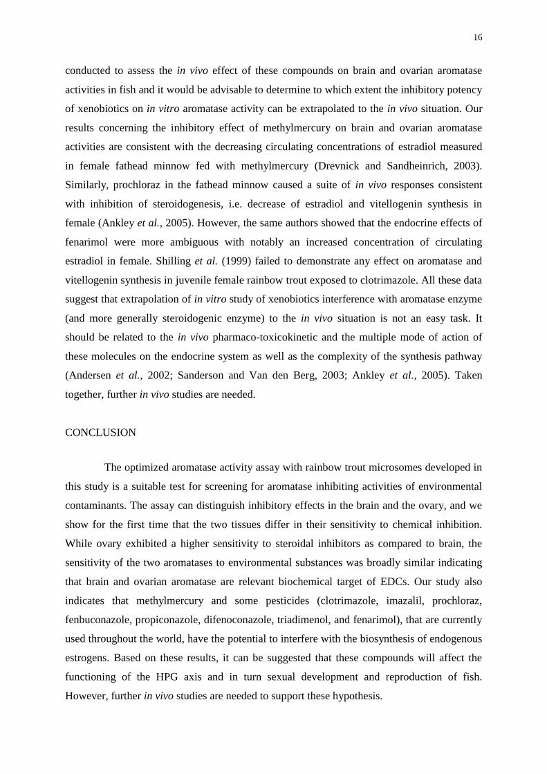
conducted to assess the in vivo effect of these compounds on brain and ovarian aromatase
activities in fish and it would be advisable to determine to which extent the inhibitory potency
of xenobiotics on in vitro aromatase activity can be extrapolated to the in vivo situation. Our
results concerning the inhibitory effect of methylmercury on brain and ovarian aromatase
activities are consistent with the decreasing circulating concentrations of estradiol measured
in female fathead minnow fed with methylmercury (Drevnick and Sandheinrich, 2003).
Similarly, prochloraz in the fathead minnow caused a suite of in vivo responses consistent
with inhibition of steroidogenesis, i.e. decrease of estradiol and vitellogenin synthesis in
female (Ankley et al., 2005). However, the same authors showed that the endocrine effects of
fenarimol were more ambiguous with notably an increased concentration of circulating
estradiol in female. Shilling et al. (1999) failed to demonstrate any effect on aromatase and
vitellogenin synthesis in juvenile female rainbow trout exposed to clotrimazole. All these data
suggest that extrapolation of in vitro study of xenobiotics interference with aromatase enzyme
(and more generally steroidogenic enzyme) to the in vivo situation is not an easy task. It
should be related to the in vivo pharmaco-toxicokinetic and the multiple mode of action of
these molecules on the endocrine system as well as the complexity of the synthesis pathway
(Andersen et al., 2002; Sanderson and Van den Berg, 2003; Ankley et al., 2005). Taken
together, further in vivo studies are needed.
CONCLUSION
The optimized aromatase activity assay with rainbow trout microsomes developed in
this study is a suitable test for screening for aromatase inhibiting activities of environmental
contaminants. The assay can distinguish inhibitory effects in the brain and the ovary, and we
show for the first time that the two tissues differ in their sensitivity to chemical inhibition.
While ovary exhibited a higher sensitivity to steroidal inhibitors as compared to brain, the
sensitivity of the two aromatases to environmental substances was broadly similar indicating
that brain and ovarian aromatase are relevant biochemical target of EDCs. Our study also
indicates that methylmercury and some pesticides (clotrimazole, imazalil, prochloraz,
fenbuconazole, propiconazole, difenoconazole, triadimenol, and fenarimol), that are currently
used throughout the world, have the potential to interfere with the biosynthesis of endogenous
estrogens. Based on these results, it can be suggested that these compounds will affect the
functioning of the HPG axis and in turn sexual development and reproduction of fish.
However, further in vivo studies are needed to support these hypothesis.
17
ACKNOWLEDGEMENTS
This study was supported by the French Ministry of Ecology and Sustainable Development
(Budget Civil de Recherche et Développement, BCRD Grant AP17-02). N. Hinfray is funded
by INERIS and ARNT (National association for technical research). The authors would like
to express their thanks to Dr Sélim Aït-Aissa (Unité d’évaluation des risques
écotoxicologiques, INERIS), Professor Helmut Segner (University of Bern, Centre for Fish
and Wildlife Health, Switzerland) and Professor Roger A. Coulombe (Utah State University,
Department of veterinary sciences, USA) for their critical reading and comments of the
manuscript and for English corrections.
18
REFERENCES
Andersen, H.R., Vinggaard, A.M., Rasmussen, T.H., Gjermandsen, I.M., Bonefeld-Jorgensen,
E.C., 2002. Effects of currently used pesticides in assays for estrogenicity, androgenicity, and
aromatase activity in vitro. Toxicol. Appl. Pharmacol., 179 (1), 1-12.
Ankley, G.T., Jensen, K.M., Durhan, E.J., Makynen, E.A., Butterworth, B.C., Kahl, M.D.,
Villeneuve, D.L., Linnum, A., Gray, L.E., Cardon, M., Wilson, V.S., 2005. Effects of two
fungicides with multiple modes of action on reproductive endocrine function in the fathead
minnow (Pimephales promelas). Toxicol. Sci., 86 (2), 300-308.
Ayub, M., Levell, M.J., 1988. Structure-activity relationships of the inhibition of human
placental aromatase by imidazole drugs including ketoconazole. J. Steroid. Biochem., 31 (1),
65-72.
Ayub, M., Levell, M.J., 1990. The inhibition of human prostatic aromatase activity by
imidazole drugs including ketoconazole and 4-hydroxyandrostenedione. Biochem.
Pharmacol., 40 (7), 1569-1575.
Blazquez, M., Piferrer, F., 2004. Cloning, sequence analysis, tissue distribution, and sex-
specific expression of the neural form of P450 aromatase in juvenile sea bass (Dicentrarchus
labrax). Mol. Cell. Endocrinol., 219 (1-2), 83-94.
Bradford, M., 1976. A rapid and sensitive method for the quantitation of microgram quantities
of protein utilizing the principle of protein-dye binding. Anal. Biochem., 72 248-254.
Brion, F., Tyler, C.R., Palazzi, X., Laillet, B., Porcher, J.M., Garric, J., Flammarion, P., 2004.
Impacts of 17-estradiol, including environmentally relevant concentrations, on reproduction
after exposure during embryo-larval-, juvenile- and adult-life stages in zebrafish (Danio
rerio). Aquat. Toxicol., 68 (3), 193-217.
Brodie, A.M., Wing, L.Y., Goss, P., Dowsett, M., Coombes, R.C., 1986. Aromatase inhibitors
and their potential clinical significance. J. Steroid Biochem., 25 (5B), 859-865.
19
Callard, G.V., Tchoudakova, A., 1997. Evolutionary and functional significance of two
CYP19 genes differentially expressed in brain and ovary of goldfish. J. Steroid Biochem.
Mol. Biol., 61 (3-6), 387-392.
Chiang, E.F., Yan, Y.L., Guiguen, Y., Postlethwait, J., Chung, B., 2001a. Two Cyp19 (P450
aromatase) genes on duplicated zebrafish chromosomes are expressed in ovary or brain. Mol.
Biol. Evol., 18 (4), 542-550.
Chiang, E.F., Yan, Y.L., Tong, S.K., Hsiao, P.H., Guiguen, Y., Postlethwait, J., Chung, B.C.,
2001b. Characterization of duplicated zebrafish cyp19 genes. J. Exp. Zool., 290 (7), 709-714.
Dalla Valle, L., Ramina, A., Vianello, S., Belvedere, P., Colombo, L., 2002. Cloning of two
mRNA variants of brain aromatase cytochrome P450 in rainbow trout (Oncorhynchus mykiss
Walbaum). J. Steroid Biochem. Mol. Biol., 82 (1), 19-32.
D'Cotta, H., Fostier, A., Guiguen, Y., Govoroun, M., Baroiller, J.-F., 2001. Aromatase plays a
key role during normal and temperature-induced sex differentiation of tilapia Oreochromis
niloticus. Mol. Reprod. Dev., 59 (3), 265-276.
Drevnick, P.E., Sandheinrich, M.B., 2003. Effects of dietary methylmercury on reproductive
endocrinology of fathead minnows. Environ. Sci. Technol., 37 (19), 4390-4396.
Flouriot, G., Pakdel, F., Ducouret, B., Ledrean, Y., Valotaire, Y., 1997. Differential regulation
of two genes implicated in fish reproduction: vitellogenin and estrogen receptor genes. Mol.
Reprod. Dev., 48 (3), 317-323.
Forlano, P.M., Deitcher, D.L., Myers, D.A., Bass, A.H., 2001. Anatomical distribution and
cellular basis for high levels of aromatase activity in the brain of teleost fish: aromatase
enzyme and mRNA expression identify glia as source. J. Neurosci., 21 (22), 8943-8955.
France, J. T., Mason, J. I., Magness, R. R., Murry, B. A., Rosenfeld, C. R., 1987. Ovine
placental aromatase: Studies of activity levels, kinetic characteristics and effects of aromatase
inhibitors. J. Steroid Biochem., 28, 155–160.

20
Geelen, J.A., Deckers, G.H., van der Wardt, J.T., Loozen, H.J., Tax, L.J., Kloosterboer, H.J.,
1991. Selection of 19-(ethyldithio)-androst-4-ene-3,17-dione (ORG 30958): a potent
aromatase inhibitor in vivo. J. Steroid Biochem. Mol. Biol., 38 (2), 181-188.
Geisler, J., Lonning, P.E., 2005. Aromatase inhibition: translation into a successful
therapeutic approach. Clin. Cancer Res., 11 (8), 2809-2821.
Gelinas, D., Pitoc, G., Callard, G.V., 1998. Isolation of a goldfish brain cytochrome P450
aromatase cDNA : mRNA expression during the seasonal cycle and after steroid treatment.
Mol. Cell. Endocrinol., 138 (1-2), 81-93.
Gonzalez, A., Piferrer, F., 2002. Characterization of aromatase activity in the sea bass: effects
of temperature and different catalytic properties of brain and ovarian homogenates and
microsomes. J. Exp. Zool., 293 (5), 500-510.
Gonzalez, A., Piferrer, F., 2003. Aromatase activity in the European sea bass (Dicentrarchus
labrax L.) brain. Distribution and changes in relation to age, sex, and the annual reproductive
cycle. Gen. Comp. Endocrinol. , 132 (2), 223-230.
Guiguen, Y., Baroiller, J.-F., Ricordel, M.-J., Iseki, K., McMeel, O.M., Martin, S.A.M.,
Fostier, A., 1999. Involvement of estrogens in the process of sex differentiation in two fish
species: The rainbow trout (Oncorhynchus mykiss) and a tilapia (Oreochromis niloticus). Mol.
Reprod. Dev., 54 (2), 154-162.
Hirsch K. S., Weaver D. E., Black L. J., Falcone J. F., MacLusky N. J., 1987. Inhibition of
central nervous system aromatase activity: A mechanism for fenarimol-induced infertility in
the male rat. Toxicol. Appl. Pharmacol., 91, 235-245.
Institut Français de l'Environnement (IFEN), 2001. Pesticides in water, Fifth annual report. 1-
67.
Jobling, S., Beresford, N., Nolan, M., Rodgers-Gray, T., Brighty, G.C., Sumpter, J.P., Tyler,
C.R., 2002. Altered sexual maturation and gamete production in wild roach (Rutilus rutilus)
living in rivers that receive treated sewage effluents. Biol. Reprod., 66 (2), 272-281.
21
Kishida, M., Callard, G.V., 2001. Distinct cytochrome P450 aromatase isoforms in zebrafish
(Danio rerio) brain and ovary are differentially programmed and estrogen regulated during
early development. Endocrinology, 142 (2), 740-750.
Lavado, R., Thibaut, R., Raldua, D., Martin, R., Porte, C., 2004. First evidence of endocrine
disruption in feral carp from the Ebro River. Toxicol. Appl. Pharmacol., 196 (2), 247-257.
Le Bail, J.C., Champavier, Y., Chulia, A.J., Habrioux, G., 2000. Effects of phytoestrogens on
aromatase, 3beta and 17beta-hydroxysteroid dehydrogenase activities and human breast
cancer cells. Life Sci., 66 (14), 1281-1291.
Letcher, R.J., van Holsteijn, I., Drenth, H.-J., Norstrom, R.J., Bergman, A., Safe, S., Pieters,
R., van den Berg, M., 1999. Cytotoxicity and Aromatase (CYP19) Activity Modulation by
Organochlorines in Human Placental JEG-3 and JAR Choriocarcinoma Cells. Toxicol. Appl.
Pharmacol., 160 (1), 10-20.
Martin-Skilton, R., Lavado, R., Thibaut, R., Minier, C., Porte, C., 2006. Evidence of
endocrine alteration in the red mullet, Mullus barbatus from the NW Mediterranean. Environ.
Pollut., 141 (1), 60-68.
Mason, J.I., Carr, B.R., Murry, B.A., 1987. Imidazole antimycotics: selective inhibitors of
steroid aromatization and progesterone hydroxylation. Steroids, 50 (1-3), 179-189.
Melo, A.C., Ramsdell, J.S., 2001. Sexual dimorphism of brain aromatase activity in medaka:
induction of a female phenotype by estradiol. Environ. Health Perspect., 109 (3), 257-264.
Menuet A., Pellegrini E., Brion F., Gueguen M-M, Dujardin T, Anglade I, Marmignon M,
Pakdel F, Kah O. (2005). Expression and estrogen-dependent regulation of the zebrafish brain
aromatase gene. J. Comp. Neurol. 16;485(4):304-20.
Mills, L.J., Chichester, C., 2005. Review of evidence: are endocrine-disrupting chemicals in
the aquatic environment impacting fish populations? Sci. Total Environ., 343 (1-3), 1-34.
22
Monod, G., De Mones, A., Fostier, A., 1993. Inhibition of ovarian microsomal aromatase and
follicular estradiol suppression by imidazole fungicides in rainbow trout. Mar. Environ. Res.,
35 153-157.
Monteiro, P.R.R., Reis-Henriques, M.A., Coimbra, J., 2000. Polycyclic aromatic
hydrocarbons inhibit in vitro ovarian steroidogenesis in the flounder (Platichthys flesus L.).
Aquat. Toxicol., 48 (4), 549-559.
Nash, J.P., Kime, D.E., Van der Ven, L.T., Wester, P.W., Brion, F., Maack, G., Stahlschmidt-
Allner, P., Tyler, C.R., 2004. Long-term exposure to environmental concentrations of the
pharmaceutical ethynylestradiol causes reproductive failure in fish. Environ. Health Perspect.,
112 (17), 1725-1733.
Noaksson, E., Tjarnlund, U., Bosveld, A.T.C., Balk, L., 2001. Evidence for Endocrine
Disruption in Perch (Perca fluviatilis) and Roach (Rutilus rutilus) in a Remote Swedish Lake
in the Vicinity of a Public Refuse Dump. Toxicol. Appl. Pharmacol., 174 (2), 160-176.
Noaksson, E., Linderoth, M., Bosveld, A.T., Norrgren, L., Zebuhr, Y., Balk, L., 2003.
Endocrine disruption in brook trout (Salvelinus fontinalis) exposed to leachate from a public
refuse dump. Sci. Total Environ., 305 (1-3), 87-103.
Noaksson, E., Gustavsson, B., Linderoth, M., Zebuhr, Y., Broman, D., Balk, L., 2004. Gonad
development and plasma steroid profiles by HRGC/HRMS during one reproductive cycle in
reference and leachate-exposed female perch (Perca fluviatilis). Toxicol. Appl. Pharmacol.,
195 (2), 247-261.
Noaksson, E., Linderoth, M., Tjarnlund, U., Balk, L., 2005. Toxicological effects and
reproductive impairments in female perch (Perca fluviatilis) exposed to leachate from
Swedish refuse dumps. Aquat. Toxicol., 75 (2), 162-177.
OECD (1997). Draft detailed review paper : appraisal of test methods for sex-hormone
disrupting chemicals. Paris, OECD: 290.
23
Ohno, K., Araki, N., Yanase, T., Nawata, H., Iida, M., 2004. A novel nonradioactive method
for measuring aromatase activity using a human ovarian granulosa-like tumor cell line and an
estrone ELISA. Toxicol. Sci., 82 (2), 443-450.
Orlando, E.F., Davis, W.P., Guillette, L.J., Jr., 2002. Aromatase activity in the ovary and
brain of the eastern mosquitofish (Gambusia holbrooki) exposed to paper mill effluent.
Environ. Health Perspect., 110 Suppl 3 429-433.
Pasmanik, M., Callard, G.V., 1985. Aromatase and 5 alpha-reductase in the teleost brain,
spinal cord, and pituitary gland. Gen. Comp. Endocrinol. , 60 (2), 244-251.
Pasmanik, M., Callard, G.V., 1988. Changes in brain aromatase and 5 alpha-reductase
activities correlate significantly with seasonal reproductive cycles in goldfish (Carassius
auratus). Endocrinology, 122 1349-1356.
Pelissero, C., Lenczowski, M.J.P., Chinzi, D., Davail-Cuisset, B., Sumpter, J.P., Fostier, A.,
1996. Effects of flavonoids on aromatase activity, an in vitro study. J. Steroid Biochem. Mol.
Biol., 57 (3-4), 215-223.
Roberts, P.H., Thomas, K.V., 2006. The occurrence of selected pharmaceuticals in
wastewater effluent and surface waters of the lower Tyne catchment. Sci. Total Environ., 356
(1-3), 143-153.
Saitoh, M., Yanase, T., Morinaga, H., Tanabe, M., Mu, Y.-M., Nishi, Y., Nomura, M., Okabe,
T., Goto, K., Takayanagi, R., Nawata, H., 2001. Tributyltin or Triphenyltin Inhibits
Aromatase Activity in the Human Granulosa-like Tumor Cell Line KGN*1. Biochem.
Biophys. Res. Commun., 289 (1), 198-204.
Sanderson, J.T., Letcher, R.J., Heneweer, M., Giesy, J.P., van den Berg, M., 2001. Effects of
chloro-s-triazine herbicides and metabolites on aromatase activity in various human cell lines
and on vitellogenin production in male carp hepatocytes. Environ. Health Perspect., 109 (10),
1027-1031.
24
Sanderson, J.T., Boerma, J., Lansbergen, G.W.A., van den Berg, M., 2002. Induction and
Inhibition of Aromatase (CYP19) Activity by Various Classes of Pesticides in H295R Human
Adrenocortical Carcinoma Cells. Toxicol. Appl. Pharmacol., 182 (1), 44-54.
Sanderson, T., van den Berg, M., 2003. Interactions of xenobiotics with the steroid hormone
biosynthesis pathway. Pure Appl. Chem., 75 (11-12), 1957-1971.
Segner, H., Caroll, K., Fenske, M., Janssen, C.R., Maack, G., Pascoe, D., Schafers, C.,
Vandenbergh, G.F., Watts, M., Wenzel, A., 2003. Identification of endocrine-disrupting
effects in aquatic vertebrates and invertebrates: report from the European IDEA project.
Ecotoxicol. Environ. Saf., 54 (3), 302-314.
Shilling, A.D., Carlson, D.B., Williams, D.E., 1999. Rainbow trout, Oncorhynchus mykiss, as
a model for aromatase inhibition. J. Steroid Biochem. Mol. Biol., 70 (1-3), 89-95.
Simpson, E.R., Mahendroo, M.S., Means, G.D., Kilgore, M.W., Hinshelwood, M.M.,
Graham-Lorence, S., Amarneh, B., Ito, Y., Fisher, C.R., Michael, M.D., 1994. Aromatase
cytochrome P450, the enzyme responsible for estrogen biosynthesis. Endocr. Rev., 15 (3),
342-355.
Taupeau, C., Poupon, J., Treton, D., Brosse, A., Richard, Y., Machelon, V., 2003. Lead
reduces messenger RNA and protein levels of cytochrome p450 aromatase and estrogen
receptor beta in human ovarian granulosa cells. Biol. Reprod., 68 (6), 1982-1988.
Tchoudakova, A., Callard, G.V., 1998. Identification of multiple CYP19 genes encoding
different cytochrome P450 aromatase isozymes in brain and ovary. Endocrinology, 139 (4),
2179-2189.
Thomas, K.V., Hilton, M.J., 2004. The occurrence of selected human pharmaceutical
compounds in UK estuaries. Mar. Pollut. Bull., 49 (5-6), 436-444.
Thompson, E.A., Jr.,Siiteri, P.K., 1974. Utilization of oxygen and reduced nicotinamide
adenine dinucleotide phosphate by human placental microsomes during aromatization of
androstenedione. J. Biol. Chem., 249 (17), 5364-5372.
25
Trant, J.M., Gavasso, S., Ackers, J., Chung, B.C., Place, A.R., 2001. Developmental
expression of cytochrome P450 aromatase genes (CYP19a and CYP19b) in zebrafish fry
(Danio rerio). J. Exp. Zool., 290 (5), 475-483.
Trosken, E.R., Scholz, K., Lutz, R.W., Volkel, W., Zarn, J.A., Lutz, W.K., 2004. Comparative
assessment of the inhibition of recombinant human CYP19 (aromatase) by azoles used in
agriculture and as drugs for humans. Endocr. Res., 30 (3), 387-394.
Tsai, C.L., Chang, S.L., Wang, L.H., Chao, T.Y., 2003. Temperature influences the
ontogenetic expression of aromatase and oestrogen receptor mRNA in the developing tilapia
(Oreochromis mossambicus) brain. J. Neuroendocrinol., 15 (1), 97-102.
Vindimian, E., Robaut, C., Fillion, G., 1983. A method for cooperative or noncooperative
binding studies using nonlinear regression analysis on a microcomputer. J. Appl. Biochem., 5
(4-5), 261-268.
Vinggaard, A.M., Hnida, C., Breinholt, V., Larsen, J.C., 2000. Screening of selected
pesticides for inhibition of CYP19 aromatase activity in vitro. Toxicol. in Vitro, 14 (3), 227-
234.
Yue, W., Brodie, A.M.H., 1997. Mechanisms of the actions of aromatase inhibitors 4-
hydroxyandrostenedione, fadrozole, and aminoglutethimide on aromatase in JEG-3 cell
culture. J. Steroid Biochem. Mol. Biol., 63 (4-6), 317-328.
Zarn, J.A., Bruschweiler, B.J., Schlatter, J.R., 2003. Azole fungicides affect mammalian
steroidogenesis by inhibiting sterol 14 alpha-demethylase and aromatase. Environ. Health
Perspect., 111 (3), 255-261.
Zhao, J., Mak, P., Tchoudakova, A., Callard, G., Chen, S., 2001. Different catalytic properties
and inhibitor responses of the Goldfish brain and ovary aromatase isozymes. Gen. Comp.
Endocrinol. , 123 (2), 180-191.
26
Table I : Aromatase activity (AA) measured in the different batches of microsomes isolated
from brain or ovary of females rainbow trout having different GSI. The number of batch /
group of GSI is detailed in the Materials and Methods. n = number of independent measures
performed on different batches of brain or ovarian microsomes. Different letters indicate
statistically different values (Kruskall-Wallis test, and Mann-Whitney U test, p < 0.05). BDL
= Below Detection Limit.
GSI AA in brain microsomes
(fmol/mg/min)
AA in ovarian microsomes
(fmol/mg/min)
Low GSI
< 1 %
153.9 ± 59.3 a,d
n = 16
43.8 ± 6.0 c
n = 27
Medium GSI
8 % - 13 %
146.8 ± 5.1 a
n = 13
107.7 ± 27.6 d
n = 11
High GSI
> 13 %
428.9 ± 46.5 b
n = 15
BDL
n= 3
27
Table II : Effect of environmental pollutants on aromatase activity (AA) in microsomes
prepared from brain and ovary of rainbow trout. All chemicals were tested at 10 µM except
4OHA (4-hydroxyandrostenedione), ATD (androstatrienedione), aldrin, heptachlor, and
TCDD. The other concentrations are indicated in the table. Values are means of triplicates ±
standard deviation. * indicate a significant difference compared to control (Student’s t test, p
< 0.05).
CHEMICALS FAMILY CHEMICALS % OF CONTROL AA (BRAIN) % OF CONTROL AA (OVARY)
Control 100.0 ± 8.4 100 ± 10.3
STEROIDAL INHIBITOR 4OHA (1µM) 2.7 ± 0.4 * 3.1 ± 3.1 *ATD (1µM) 3.9 ± 0.9 * 2.1 ± 0.2 *
NON STEROIDAL INHIBITOR Aminoglutethimide 84.7 ± 2.8 * 70.7 ± 1.3 *HEAVY METALS Cadmium chloride 106.1 ± 8.6 98.6 ± 10.3
Lead acetate 101.8 ± 7.8 99.2 ± 12.0
Methyl mercury 75.9 ± 2.1 * 15.7 ± 26.7 *Triphanylarsine 107.8 ± 3.7 86.4 ± 1.5
H3AsO4 100.1 ± 2.6 100.0 ± 4.4
PAH Benzo[a]Pyrene 98.3 ± 2.0 102.9 ± 0.9
Chrysene 99.7 ± 1.1 105.2 ± 4.7
INSECTICIDES
organochlorine Aldrin (100nM) 92.5 ± 2.2 89.9 ± 4.7
Chlordane 105.5 ± 10.5 110.0 ± 9.5
Endosulfan 111.7 ± 0.3 107.6 ± 0.4
Heptachlor (100nM) 89.7 ± 8.8 89.7 ± 11.7
Methoxychlor 113.5 ± 2.3 84.5 ± 1.6
pyrazole Fipronil 107.2 ± 1.4 101.7 ± 19.4
pyrethroids Alpha-cypermethrin 83.7 ± 5.9 100.2 ± 5.0
Permethrin 87.3 ± 8.4 94.1 ± 4.9
FUNGICIDES
benzimidazol Benomyl 116.7 ± 0.4 102.3 ± 0.6
triazole Difenoconazole 81.7 ± 2.7 * 75.9 ± 2.9 *Fenbuconazole 7.1 ± 0.9 * 6.7 ± 3.2 *Propiconazole 10.1 ± 0.6 * 5.9 ± 0.5 *Triadimenol 53.2 ± 1.5 * 69.8 ± 0.7 *
imidazole Clotrimazole 3.6 ± 0.1 * 0.0 ± 0.7 *Imazalil 3.8 ± 0.5 * 2.6 ± 0.2 *Prochloraz 18.0 ± 0.1 * 10.8 ± 0.4 *
dicarboximide Iprodion 115.1 ± 13.4 102.3 ± 4.9
Vinclozoline 100.5 ± 14.7 111.1 ± 11.7
pyrimidine Bupirimate 102.2 ± 8.9 92.2 ± 5.3
Fenarimol 46.4 ± 1.1 * 56.9 ± 0.8 *HERBICIDES
Oxadiazon 87.5 ± 5.1 81.4 ± 5.7
chloroacetanilide Metolachlor 106.0 ± 4.5 109.6 ± 1.1
chlorotriazine Atrazine 114.7 ± 4.2 104.7 ± 3.2
Simazine 98.0 ± 14.1 108.7 ± 8.8
dinitroaniline Trifluralin 103.9 ± 12.6 102.1 ± 4.7
phenoxypropionic Mecoprop 105.8 ± 3.3 99.7 ± 5.4
phenylurea Diuron 97.7 ± 3.9 85.2 ± 4.5
Isoproturon 93.9 ± 8.1 89.9 ± 1.9
pyrimidinylsulfonylurea Azimsulfuron 112.9 ± 3.7 95.3 ± 18.7
triazole Amitrol 101.0 ± 0.9 93.8 ± 5.2
OTHERS Pentachlorophenol 109.8 ± 9.6 98.3 ± 1.6
TCDD (10-9 M) 112.0 ± 2.6 101.3 ± 3.1
TCDD (10-8 M) 116.7 ± 6.0 89.6 ± 2.6
Tributyltin chloride 91.4 ± 1.9 86.6 ± 0.1
28
Table III : Comparative inhibition of aromatase activities in brain and ovarian microsomes in rainbow trout. The relative inhibition potency was calculated in comparison with 4-hydroxyandrostenedione. Dose-response experiments were conducted three times for each compound tested. Published IC50 values of the studied pesticides for placental assay, cells based assays and rainbow trout ovarian assay are also reported.
Chemical name Brain IC50
(µM) Ovarian IC50
(µM)
Ratio Brain/Ovarian
IC50 RPAI brain RPAI ovary
IC50 in placental
assay (µM)
IC50 in cells based assays
(µM)
IC50 in rainbow trout ovarian assay
(µM)
4-OH androstenedione 0.009 ± 0.001 0.00015 ± 0.00003 60.0 1 1 0.04 (a)- 1.4 (b) 0.0011 (c) - 0.08 (d) #1.5 (e)
Androstatrienedione 0.015 ± 0.003 0.00025 ± 0.00018 60.0 0.6 0.6 - - ##0.5 (f)
Aminoglutethimide 197 ± 54 36.8 ± 12.0 5.4 4.6E-05 4.1E-06 1 (g)-130 (f) 2.25 (c) -15.8 (d) 39 (f) , Ki = 2.4 (h)
Clotrimazole 0.011 ± 0.004 0.016 ± 0.001 0.7 0.8 0.009 0.43 (i) - 1.8 (j) 0.05 (k) , 0.9 (l)
Imazalil 0.43 ± 0.03 0.32 ± 0.21 1.3 2.1E-02 4.7E-04 0.04 (m) - 0.15 (j) 0.0044 (c)-0.1 (n) 5 (k)
Prochloraz 1.3 ± 0.4 1.0 ± 0.5 1.3 6.9E-03 1.5E-04 0.34 (m) - 0.70 (j) 0.1 (n) 5 (k), (11-7.2)**(o)
Fenbuconazole 1.3 ± 0.4 0.21 ± 0.05 6.2 6.9E-03 7.1E-04 - - -
Propiconazole 0.9 ± 0.3 0.9 ± 0.6 1.0 1.0E-02 1.7E-04 6.5 (m) 0.96 (c) - 5 (n) -
Difenoconazole 70 ± 7 29 ± 6 2.4 1.3E-04 5.2E-06 - 4 (n) -
Triadimenol 11 ± 0.7 26 ± 2 0.4 8.2E-04 5.8E-06 21 (m) 12.6 (p) -
Fenarimol 6 ± 1 18 ± 6 0.3 1.5E-03 8.3E-06 4.1*** (q) - 10 (m) 2 (m, c) -
Methylmercury 11 ± 1 0.78 ± 0.07 14.1 8.2E-04 1.9E-04 - - -
RPAI: relative potency of aromatase inhibition, # = 80% of inhibition, ## = 100% of inhibition, * = roach brain microsomes, ** = S9 fraction from brain fathead minnow, *** = rat ovarian microsomes (a) France et al., 1987; (b) Geelen et al, 1991; (c) Ohno et al., 2004; (d) Yue and Brodie, 1997; (e) Shilling et al., 1999; (f) Pelissero et al., 1996; (g) Le Bail et al., 2000; (h) Zhao et al., 2001; (i) Ayub and Levell, 1990; (j) Mason et al;1987; (k) Monod et al., 1993; (l) Noakson et al., 2003; (m) Vingaard et al., 2000, (n) Sanderson et al., 2002, (o) Ankley et al., 2005, (p) Trosken et al. (2004), (q) Hirsh et al (1987).
29
FIGURE LEGENDS
Fig. 1. Effect of the amount of microsomal proteins and incubation time on the aromatase
activities (AA) in rainbow trout ovaries (A) and brain (B). In all experiments, the
concentration of substrate was 75 nM and the reaction was carried out at a temperature of
27°C. Each value represents the mean of two independent experiments ± SD.
Fig. 2. Saturation analysis of aromatase activity (AA) in : (A) ovarian microsomes, and (B)
brain microsomes of rainbow trout. Brain and ovaries were incubated 30 min at 27°C with
increasing concentrations of 1-[3H]-androstenedione from 2 to 600 nM. Inserts :
Lineweaver-Burk plots to determine the Vmax and the Km.
Fig. 3. Effect of temperature on the rainbow trout aromatase activities (AA) in brain (A) and
ovary (B).The concentration of substrate was 75 nM and the amount of protein 200 µg. AA
were expressed as percent of maximal AA measured for each tissue.
Fig. 4. Effect of temperature on brain and ovarian IC50 values for two aromatase inhibiting
substances (prochloraz and 4-hydroxyandrostenedione (4-OHA)). IC50 values calculated in
ovary (A) and brain (B) for each substance at 18°C and 27°C were not significantly different
(Student’s t test, p > 0.05).
Fig. 5. Linear regression analysis between brain and ovarian LOG (IC50) values. Brain and
ovarian IC50 data were positively and significantly correlated (Pearson’s test, p<0.01).
30
Fig. 1.
0.5 mg, R2 = 0.99
0.2 mg, R2 = 0.99
0.1 mg, R2 = 0.98
0
100
200
300
400
500
600
700
800
900
0 5 10 15 20 25 30 35
Incubation time (min)
Aro
ma
tase
activity (
fmo
l)(A)
(B)
0.5 mg, R2 = 0.99
0.2 mg, R2 = 0.99
0.1 mg, R2 = 0.98
0
1000
2000
3000
4000
5000
6000
7000
8000
0 10 20 30
Incubation time (min)
Aro
ma
tase a
ctivity (
fmo
l)
31
Fig. 2.
[S] (nM)
0100200300400500600700
Vo (fmol/min/mg)
0
10
20
30
40
50
60
70
1 / [S] (nM)
-0.2-0.10.00.10.20.30.40.50.6
1 / Vo (fmol/min/mg) 0.020.040.06
0.08
0.10
0.12
[S] (nM)
0100200300400500600700
Vo (fmol/min/mg)
0
100
200
300
400
500
600
(A)
(B)
1 / [S] (nM)
-0,1 0,0 0,1 0,2 0,3 0,4 0,5 0,6
1 /
Vo (
fmol/m
g/m
in)
0,00
0,01
0,02
0,03
32
Fig. 3.
0
20
40
60
80
100
120
140
0 20 40 60
Temperature (°C)
% o
f A
A
0
20
40
60
80
100
120
0 20 40 60
Temperature (°C)
% o
f A
A(A)
(B)
33
Fig. 4.
0
20
40
60
80
100
120
0 2 4 6 8 10Concentration (nM)
% o
f co
ntr
ol A
A
prochloraz 27°C
prochloraz 18°C
4OHA 27°C
4OHA 18°C
0
20
40
60
80
100
120
0 2 4 6 8 10
Concentration (nM)
% o
f co
ntr
ol A
A
prochloraz 27°C
prochloraz 18°C
4OHA 27°C
4OHA 18°C
0.01 0.1 1 10 100 1000 10 000 100 000
0 0.01 0.1 1 10 100 1000 10 000 100 000
(A)
(B)
34
Fig. 5.
y = 1.1824x - 0.5015
R2 = 0.8471
-4
-3
-2
-1
0
1
2
3
4
-4 -3 -2 -1 0 1 2 3 4
LOG (IC50) ovary
LO
G (
IC5
0)
bra
in