Embed Size (px)
Citation preview
462 SHORT COMMUNICATIONS
BBA 4 3 2 0 2
Inhibition of spinach chloroplast fructose-i,6-diphosphatase by MgATP z-, MgADP', and magnesium pyrophosphate (MgPzO7 2-)
Fructose-I,6-diphosphatase catalyzes the hydrolysis of fructose 1,6-diphosphate to fructose 6-phosphate and Pi. In photosynthetic cells it appears to be an important control point in the fixation of carbon dioxide by the reductive pentose phosphate cycle 1. The mechanism of its regulation is not clear, and the present study is part of an at tempt to clarify the problem.
Chloroplasts were extracted from store-bought spinach leaves by the method of JENSEN AND ]3ASSHAM 2. The chloroplast pellet was resuspended in distilled water and broken by sonication. After centrifugation at 2 ° for 30 rain at 40000 x g the clear green supernatant was used as the crude extract of soluble protein. The crude extract was partially purified by ammonium sulphate fractionation and the fraction precipitating between 58 and 9 ° % saturation was used as the enzyme preparation in these studies. The assay nfixture contained in I.o ml: 5 ° ~moles Tris-HC1 (pH 8.7), 5.0/~moles MgC12, 2.0 ~moles EDTA (dipotassium salt), I.O/,mole fructose 1,6-di- phosphate, 0.2/,mole NADP +, 5.0/~g phospboglucose isomerase (D-glucose-6-phos- phate ketol-isomerase, EC 5.3.1.9) and 1.5/zg glucose-6-phosphate dehydrogenase (D-glueose-6-phosphate:NADP oxidoreductase, EC 1.1.1.49 ). The reaction was started by adding 0.05 ml of an enzyme preparation containing I.O mg protein per ml. Reduction of the pyridine nucleotide was measured by following absorption at 34 ° m/~ with a Cary (Model I4) spectrophotometer. After a delay of about 3 ° sec (possibly due to thermal equilibration) the reaction was linear at maximal rate for 2--3 rain.
The activity of fructose-I,6-diphosphatase from animal and microbial systems is regulated by the allosteric inhibitor 5'-adenylate (AMP) 3-6. However, AMP at a concentration of 20 mM has no effect on the activity of the enzyme from spinach chloroplasts. Other compounds which have no effect (at concentrations of 20 raM) include glucose 6-phosphate, fructose 6-phosphate, dihydroxyacetone phosphate, 3-phosphoglyceraldehyde, 3-phosphoglyceric acid, ribose 5-phosphate, ribulose 1,5- diphosphate, 6-phosphogiuconate, phosphoenolpyruvate, UTP, CTP, NADPH.
Inhibition of activity was observed with MgATP 2-, MgADP- and magnesium pyrophosphate (MgP20~-). (The stability constants for MgATP s- (ref. 7) and MgP20 ~- (ref. 8) are sufficiently high that one can assume that in the presence of excess Mg s+ the concentration of the free deficient ion is negligible; tile stability constant for MgADP- (ref. 7) is lower, and at the excess Mg 2+ concentrations used in this investigation 93-95 % of the ADP is present as the complex and the remainder as the free ion.)
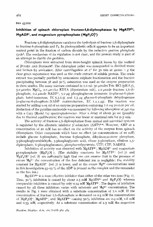
MgATP 2- is a more effective inhibitor than either of the other two ions (Fig. I). Thus, 50 % inhibition is caused by about 2.5 mM MgADP- and MgP20 ~- whereas comparable inhibition is caused by only 0.74 mM MgATP 2-. The degree of inhibition caused by all three inhibitors varies with substrate and Mg ~+ concentration. The results in Fig. I were obtained with a substrate concentration of I.O raM. If the concentration of fructose 1,6-diphosphate is decreased to o.5 mM the concentrations of MgP20~-, MgADP-, and MgATP 2- causing 50 % inhibition are 0. 9 raM, 1.6 mM and 0.53 raM, respectively. At a substrate concentration of 2.5 mM the respective
Biochim. Biophys. Acta, 162 (1068) 462-464
SHORT COMMUNICATIONS 463
concentrations are 4-4 raM, 5.8 mM and 1.2 raM. Increasing or decreasing the free Mg =+ concentration affects the degree of inhibition in a similar way.
,oo 80
60 x
._~
< 40
¢,
20
0 I I I I I I
0 2.0 4.0 6.0 Inhibitor (mM)
Fig. I. The effect of MgATP 2- ( A - - A ) , M g A D P - ( X - - X ) and MgP~O~- ( Q - - O ) on fructose- 1 ,6-diphosphatase ac t iv i ty . The s u b s t r a t e concen t ra t ion is i .o mM and free Mg 2+ concen t ra t ion is 3.o mM. The inh ib i to r concen t ra t ions are 1. 5 mM. Act iv i t i e s are expressed as re la t ive va lues wi th the a c t i v i t y in the absence of i nh ib i to r t a k e n to be ioo.
PRIESS, WYMAN BIGGS AND GREENBERG 9 have observed a sigmoidal relationship between fructose-I,6-diphosphatase activity and substrate concentration, particularly at low Mg ~+ concentration. This observation has been confirmed, and it has also been observed that the sigmoidal nature becomes more pronounced in the presence of the three inhibitors MgATP 2-, MgADP- and MgP~O~-. The relationship between activity and Mg 2+ concentration is also sigmoidal and the three inhibitors enhance this feature. Hill plots of the data show that the interaction coefficients (n*) with respect to sub- strate and with respect to Mg 2+ concentration increase with increasing concentration of inhibitors (up to 2.5 mM).
Although certain characteristics of the inhibition described here therefore resemble those of several known 'regulatory enzymes'4,1°-13, the role of the three inhibitors in the control of fructose-I,6-diphosphatase activity in vivo is not clear. Thus, their combined concentrations decrease when chloroplasts or Chlorella cells are transferred from the light to darkness1,14. This change is the reverse of that which would be expected if the inhibitors were responsible for the decrease in activity of the diphosphatase thought to occur when photosynthetic cells are transferred from illuminated conditions to darkness.
The original observations of PEDERSEN, KIRK AND BASSHAM 1 also suggested that the activity of ribulose-I,5-diphosphate carboxylase (3-phospho-D-glycerate carboxy-lyase (dimerizing), EC 4.1.1.39) also decreased when Chlorella cells are trans- ferred from light to darkness. Of possible interest is the fact that preliminary obser- vations indicate that the same three inhibitors (MgATP 2-, MgADP-, MgP20~- ) also inhibit the activity of the carboxylating enzyme extracted from spinach chloroplasts. (The inhibitors do not have a general inhibitory effect on chloroplast enzymes since they fail to inhibit the activity of aldolase (ketose-I-phosphate aldehyde-lyase, EC4.I.2.7) and triose phosphate dehydrogenase (n-glyceraldehyde-3-phosphate: NADP oxidoreductase (phosphorylating), EC 1.2.1.13) from spinach chloroplasts.)
BiocMm. Biophys. Acta, 162 (1968) 462-464
464 SHORT COMMUNICATIONS
The work was done while on leave of absence in The Biodynamics Labora to ry , Lawrence Rad ia t ion Labora to ry , Univers i ty of California, Berkeley, Calif. and was suppor t ed b y the U.S. Atomic Energy Commission.
Botany Department, University College London, London (Great Britain)
IAN MORRIS
1 T. A. PEDERSEN, M. KIRK AND J. A. ]~ASSHAM, Physiol. Plantarum, 19 (1966) 219. 2 1~. G. JENSEN AND J. A. BASSHAM, Proc. Natl. Acad. Sci. U.S., 56 (1966) lO95. 3 ]3. L. HORECKER, S. PONTREMOLY, O. ROSEN AND S. ROSEN, Federation Proc., 25 (1965) 1521. 4 K. TAKETA AND 13. M. POGELL, J. Biol. Chem., 24 ° (1965) 651. 5 C. GANCEDO, M.C. SALAS, A. GINER AND A. SOLS, Biochem. Biophys. Res. Commun., 20 (1965) 15. 6 A. M. UNDERWOOD AND E. A. ~NEWSHOLME, Biochem. J., 95 (1965) 767 . 7 W. J. O'SULLIVAN AND D. D. PERRIN, Biochemistry, 3 (1964) 18. 8 L. G. SILLEN AND A. E, MARTELL, Chem. Soc. London Spec. Publ., 17 (1964). 9 J- PRIESS, M. L. WYMAN BIGGS AND E. GREENBERG, J. Biol. Chem., 242 (1967) 2292.
IO D. E. ATKINSON, J. A. HATHAWAY AND E. C. SMITH, J. Biol. Chem., 240 (1965) 2682. II M. P. GHOSH AND J. PRIESS, J, Biol. Chem., 241 (1966) 4491. 12 N. GENTNER AND J. PRIESS, Biochem. Biophys. Res. Commun., 27 (1967) ¢17, 13 B. KEECH AND G. J. BARRITT, J. Biol. Chem., 242 (1967) 1983. 14 J. A. BASSHAM, M. KIRK AND R. G. JENSEN, Biochim. Biophys. Acta, 153 (1968) 211.
Received May 2oth, 1968
Biochim. Biophys. Acta, 162 (I968) 462-464
BBA 43204
Phytochrome spectrum of Pisum leaves and stems
Repor t s in this journaP and elsewhere 2,3 indicate t ha t the phy toch rome differ- ence spec t rum ob ta ined on e t io la ted Pisum (pea) leaves, or ex t rac t s of them, differs subs tan t i a l ly from corresponding spec t ra ob ta ined from other sources, including P isum stems. These in teres t ing observat ions seemed to accord with suggestions made b y the wr i te r (ref. 4, P- 318), and a t t e m p t s were immed ia t e ly made to confirm them in this l abora tory . This communica t ion summarizes the failure of these a t t empts .
At first, i t seemed unnecessary to repea t the earl ier procedures precisely, par- t i cu la r ly in view of the l ikel ihood (see later) t ha t t hey might in t roduce errors. A differ- ence of the magni tude repor ted z in phy tochrome spect ra should be easi ly de tec ted by more s t ra igh t - forward methods , so the following were adopted . Seedlings of Pisum sativum cv. Alaska were grown for 7 days in to ta l darkness at abou t 26 ° (ref. 5). Samples of s tem tissue consisted of 20-50 5-mm segments cut jus t below the apical hook, while leaf samples comprised 15-5o apical buds cut jus t above the hook and excluding as much s tem tissue as possible. I m m e d i a t e l y af ter cut t ing, the samples were packed in cyl indr ical a luminum cells 14 m m in d iamete r (cf. ref. 5), held on ice for abou t i o min, then exposed on ice to lO-3O rain of white incandescent l ight , abou t 45ooo lux, to sa tu ra te p ro toch lorophyl l conversion. Spec t ra were then t aken a t ice t empera tu re using the Biospect Model 61 (Agricul tural Special t ies Co., Beltsvil le, Md.), a s ingle-beam recording spec t ropho tomete r capable of measur ing at high ab-
Biochim. Biophys. Acta, 162 (1968) 464-466