Embed Size (px)
Citation preview
Inhibitors of the cell-surface proteinases of the periodontal pathogen Porphyromonas gingivalis
N.L. Huq, C.A. Seers, S.G. Dashper, K.J. Cross and E.C. Reynolds*
Oral Health Cooperative Research Centre, Melbourne Dental School, Bio21 Institute of Molecular Science and Biotechnology, The University of Melbourne, Victoria, Australia. *Corresponding author.
Porphyromonas gingivalis is a major pathogen associated with chronic periodontitis. Major virulence factors of this organism are its cell-surface located cysteine proteinases known as gingipains that include the Arg-specific proteinases RgpA, RgpB and the Lys-specific proteinase Kgp. The gingipains have been demonstrated to be essential for the virulence of the organism in animal models of disease. Hence there is considerable interest in the development of specific inhibitors of these proteinases that would be safe for human use. Review of the reported peptide-derived and non-peptide inhibitors of the gingipains reveals a surprising diversity of affinity, specificity and structural features. The ability of the gingipain active site to accommodate various substrates makes structure-based drug design of high specificity inhibitors a challenge. Here, we summarize the structural diversity of known gingipain substrates and inhibitors, including those derived from bovine milk and the gingipain propeptides. Inhibition of the gingipains using these peptides has been shown to reduce bacterial growth, disrupt biofilm development and significantly reduce lesion development in an animal model of infection. Hence these inhibitors may have potential to develop as therapeutics for P. gingivalis-associated periodontitis.
Keywords gingipain; Porphyromonas gingivalis; propeptide; casein
1. Introduction
Periodontal diseases are chronic inflammatory diseases of the supporting tissues of the teeth associated with bacteria in a biofilm (dental plaque). The diseases range from the relatively mild form, gingivitis, to the more aggressive forms, such as chronic periodontitis, which are characterized by the destruction of the tooth's supporting structures that can lead to tooth loss. The bacterial aetiology of chronic periodontitis is acknowledged to be polymicrobial in nature and the consensus is that anaerobic, proteolytic, amino acid fermenting species including Porphromonas gingivalis, Treponema denticola, and Tannerella forsythia play crucial roles in disease [1-4]. Based on animal model data P. gingivalis has recently been proposed to be a “keystone pathogen” that manipulates the host response in a manner that favours proliferation of other oral bacterial species which then results in disease progression [5]. P. gingivalis’ main virulence factors are its cysteine proteinases; the Arg- and Lys-specific gingipains which are located on the cell surface and on vesicles [6,7]. The membrane vesicles are specifically protein-loaded and are released into the environment where they can elicit immune responses and penetrate into host tissues and cells [8-12]. The gingipains are essential for the virulence of the organism in animal models of disease [13,14]. P. gingivalis is asaccharolytic and derives its carbon, nitrogen, and energy from amino acids and peptides in its local environment, thus gingipains are also important for the production of peptides and amino acids from host protein for bacterial nutrition [15,16]. In addition, the gingipains play an important role in acquisition and storage of the essential micronutrient iron, especially in the form of haem [16-20] as well as in the processing of other outer membrane proteins such as fimbriae [21]. The well-conserved gingipains found in all strains of P. gingivalis, are encoded by three genes, rgpA, rgpB and kgp [22-25]. All three gene products contain a signal peptide of ~22 amino acids in length, an unusually long propeptide of around 200 amino acids in length, and a catalytic domain of ~480 amino acids. In addition, rgpA and kgp encode haemagglutinin-adhesin (HA) domains in the polypeptide region C-terminal to the catalytic domains. The RgpA and Kgp polypeptides are cleaved into multiple domains that remain non-covalently associated forming large outer membrane protein complexes [25]. RgpB lacks haemagglutinin-adhesin domains and is located in a monomeric form on the outer membrane. Furthermore, the P. gingivalis genome contains hagA encoding the large polyprotein HagA that has multiple repeating HA domains [26] which can associate with other cell surface proteins including the gingipains [27]. A series of processing events occur for the RgpA and Kgp polyproteins involving N-terminal and C-terminal processing and modifications which culminate in the surface attachment and assembly of non-covalent proteinase-adhesin complexes [25,27-31] that are large enough to be visualized by electron microscopy [32,33]. Loss of gingipain function in spontaneous mutants resulted in avirulence in an animal subcutaneous lesion model of disease [34]. Isogenic rgpA, rgpB and kgp knock-out mutants also have reduced virulence in a murine periodontitis model [35]. The RgpA and Kgp proteinases work in concert to process the domains of the proteinase-adhesin complexes thus production of the wild-type form of this virulence factor requires each proteinase to be functional [28,29]. The RgpA and Kgp proteinase-adhesin complexes have binding constants for host proteins fibrinogen, fibronectin, haemoglobin, collagen type V and laminin in the nanomolar range and are essential for tissue invasion in
Microbial pathogens and strategies for combating them: science, technology and education (A. Méndez-Vilas, Ed.)
© FORMATEX 2013
____________________________________________________________________________________________
1726

animal models of disease [13,20,28]. Hence, as the gingipains are essential for virulence of P. gingivalis reducing the efficacy of the gingipains by application of inhibitors should reduce the pathogenicity of the bacterium. In this review we summarize the structural diversity of known gingipain substrates and inhibitors including those derived from milk and the gingipain propeptides which may have potential for the development of therapeutics for P. gingivalis-associated periodontitis.
2. Gingipain subsite specificity
2.1. Analysis of the S, S' subsites and P, P' preferences of the gingipains
To describe the specificity of proteases, a model of an active site composed of contiguous pockets termed subsites S1, S2 … etc is used with P1, P2… representing the amino acyl residues of the substrate occupying the corresponding subsites [36]. The residues in the substrate sequence are numbered consecutively outward from the cleavage sites: -P4-P3-P2-P1+P1'-P2'-P3'-P4'- -S4-S3-S2-S1*S1'-S2'-S3'-S4'- The scissile bond represented by the symbol + is located between the P1 and P1' positions, while the catalytic site is represented by the symbol *. The substrate P, P' preferences reflect the protease subsite specificities and enable the identification of the subsites in the absence of the X-ray structure of substrate bound protease. To define the P, P' preferences of the gingipains, the reported data of gingipain cleaved proteins and synthetic substrates were extracted from the literature and databases, collated and analysed. The specificity matrix of combined RgpA and RgpB, and that of Kgp were determined by calculating the frequency of each residue in positions P4 to P4', obtained from over eighty known cleavage sites derived from more than 20 natural and synthetic substrates: apolipoprotein B precursor [37]; Bz-Arg-NHPhNO2 (BAPNA) [37]; consensus arginyl bond [37]; cystatin C precursor [37]; coagulation factor X preproprotein [37]; coagulation factor IX preproprotein [37]; fimbrilin type 1 long precursor isoform [37]; H-kininogen [37]; interleukin (IL) 8 precursor; insulin B chain (oxidized) [38]; IL-6; mellitin [38]; Pg-II fimbriae B precursor [37]; progingipain R [37]; tumor necrosis factor alpha [37]; hemoglobin [16] ;synthetic histatin 5 analogs [39]; mucin 2 [40]; adrenocorticotrophin [37]; beta-endorphin [37]; HagA protein [37]; and neurotensin porcine [37]. These specificity matrices indicated an absolute requirement for Arg or Lys in the P1 position. However the size, charge or shape preferences for the P2, P3, P4 or P1', P2', P3' or P4' positions are not clear cut. Several groups have experimentally investigated the P, P` preferences of the gingipains. Ally and co-workers (2003) examined the P2' preferences of RgpA and RgpB, using fluorescence-quenched substrates with 18 different amino acids at this position, excluding substrates with an Arg or Cys residue [41]. Similarly the P3' preference was investigated using substrates with 16 substitutions at this position, with Cys, Arg, Phe, and Glu excluded [41]. Overall these studies also indicated that there are no distinctive P1-P4' preferences. Bialas and co-workers (2006) probed the subsites of the gingipains, by synthesizing a series of chloromethylketone inhibitors with differing substituents at P2 and P3 positions, and testing for inhibition against Kgp and RgpB [42]. Molecular modelling of the active site of Kgp and RgpB with the series of inhibitors, in conjunction with the evidence of the differing potencies, offered a description of the S2 and S3 subsites with P2 preference being hydrophobic in Kgp but a hydrogen-bond-donor group in RgpB [42]. The recent X-ray structure of RgpB inactivated by exogenous application of its propeptide produced as a recombinant protein in Escherichia coli revealed that the S1 pocket was lined by Gln282, Val242, Met288, His166 and Thr209, with Asp163 contributing a salt bridge with the P1 Arg102 side chain [43]. The S2 subsite accommodated the hydrophilic Ser101 residue, while the S3 subsite accommodated the hydrophobic Ile100 [43]. In summary, the gingipain active site is able to accommodate various substrates with a strong specificity only for an Arg or Lys residue in the P1 position.
3. Synthetic inhibitors of the gingipains
A variety of synthetic inhibitors of the gingipains have been reported in the literature. The molecular structures, inhibition types and affinities were analysed for evidence of size, charge and shape pre-requisites. Table 1 shows examples of synthetic inhibitors of gingipains with the inhibition constants reported as Ki/Ka/IC(50)/MIC or rate constant kass. Iodoacetamide and chloromethylketone with Lys or Arg residues in their P1 positions, are effective inhibitors of the gingipains [44]. A synthetic reversible inhibitor, 1-(3-phenylpropionyl) piperidine-3(R,S)-carboxylic acid-[4-amino-1(S)-(benzothiazole-2-carbonyl)butyl]amide (A71561) was found to be specific against the Lys-specific gingipain (Kgp) [45]. A synthetic inhibitor DX-9065a, that was designed and developed as an anticoagulant and selective inhibitor of Factor Xa has also shown RgpA and RgpB inhibitory activity [46,47]. Most of the known inhibitors of the gingipains display competitive kinetics and are substrate-like or partially substrate-like, forming non-covalent interactions with the active site. In contrast, chloromethylketones are irreversible inhibitors of the gingipains with potencies depending on the peptidyl components of the inhibitor [44]. Carbobenzoxy-Phe-Lys-CH2OCO-2,4,6-Me3-Ph is a specific inhibitor of Kgp D-Phe-Pro-Arg-CH2Cl and D-Phe-Phe-Arg-CH2Cl are
Microbial pathogens and strategies for combating them: science, technology and education (A. Méndez-Vilas, Ed.)
© FORMATEX 2013
____________________________________________________________________________________________
1727

specific inhibitors of Rgps [44]. The small synthetic peptides carbobenzoxy-Lys-Arg-CO-Lys-N-(CH3)2 (KYT-1) and carbobenzoxy-Glu[NHN(CH3)Ph]-Lys-CO-NHCH2Ph (KYT-36) were found to be the most potent inhibitors of Rgp and Kgp respectively [48]. Table 1 Examples of synthetic inhibitors of gingipains with the inhibition constants reported as Ki/Ka/IC50/MIC or rate constant kass.
Inhibitors Proteinase Inhibited Inhibition / Rate Constant
Ki/Ka/IC50/ kass
Inhibition Potency‡
A71561 Kgp [45] Ki = 9 x 10⁻¹⁰ M strong DX-9065a Rgp [46,47] Ki (RgpA) = 1.45 x 10⁻⁵ M
Ki (RgpB) = 1.3 x 10⁻⁵ M moderate
Cbz-Phe-Lys-CH2OCO-2,4,6-Me3-Ph Kgp [44] kass = 4.2 x 10⁶ M-1s-1 moderate D-Phe-Pro-Arg-CH2Cl Rgp [44] kass = 1.0-2.2 x 10⁷ M-1s-1 moderate D-Phe-Phe-Arg-CH2Cl Rgp [44] kass = 2.1-4.7 x 10⁷ M-1s-1 moderate KYT-1
Rgp [48] Ki = 1.9 x 10⁻¹⁰ M
strong
KYT-36 Kgp [48] Ki = 1.3 x 10⁻¹⁰ M strong Chlorhexidine Rgp and Kgp [49] Ki (RgpA) = 3.87 x10⁻⁴ M
Ki (RgpB) = 2.62 x10⁻⁴ M Ki (Kgp) = 1.65 x10⁻⁴ M
weak weak weak
E-64 Rgp [50] Ki = 1.9-2.3 x 10⁻⁶ M
moderate
‡(Strong: Ki ≤ 10-9 M, Ka ≤ 109 M; moderate: 10-8≤ Ki ≤ 10-5 M; 109≤ Ka ≤ 106 M; and weak: Ki ≥ 10-4 M, Ka ≥ 103 M).
Chlorhexidine which is used in a mouthwash to help prevent periodontal disease is a weak inhibitor of both Kgp and RgpB [49], with Arg-gingipain inhibition enhanced in the presence of Zn(II) [51]. Tetracycline and doxycycline currently used as antibiotics for periodontitis, and their analogues have been reported to inhibit RgpB by a mechanism that is uncompetitive and by an interaction that is likely to be outside the substrate binding site [52]. Although a common structure in the synthetic gingipain inhibitors is the presence of polyphenol groups, these do not determine specificity. Most of the potent synthetic gingipain inhibitors (Table 1) including Nα-Tosyl-Lys-chloromethylketone (TLCK) also demonstrate inhibitory activity against other proteases and hence are non-specific and are not compatible with human use.
4. Natural inhibitors of gingipains
A variety of natural non-peptide and peptide-based gingipain inhibitors have been reported. Although some natural inhibitors contain polyphenol groups (cranberry extract, green tea catechins), overall naturally derived non-peptide gingipain inhibitors share little in common (Table 2). Furthermore, for virulence attenuation, it is desirable that the inhibitor is potent against Kgp as animal model studies have shown that Kgp is the dominant virulence factor [7]. Table 2 Examples of natural non-peptide based inhibitors of gingipains with the inhibition constants reported as Ki/Ka/IC(50)/MIC.
Natural Non-Peptide Based Inhibitors Proteinase Inhibited
Inhibition Constant
Ki/Ka/IC50/MIC
Inhibition Potency‡
Green tea catechins
Rgp and Kgp Rgp: IC50 = 3->300 μM Kgp: IC50 = 100 μM [53]
Weak to moderate
Garlic (Allicin/ thio-2-propene-1-sulfinic acid S-allyl ester)
Rgp MIC = 55 μg/ml [54]
weak
Cranberry polyphenols Rgp and Kgp [55] Rgp: IC50 = 76 mg/ml Kgp: IC50 = 25 mg/ml
weak
‡(Strong: Ki ≤ 10-9 M, Ka ≤ 109 M; moderate: 10-8≤ Ki ≤ 10-5 M; 109≤ Ka ≤ 106 M; and weak: Ki ≥ 10-4 M, Ka ≥ 103 M). In the case of naturally occurring peptide-based inhibitors, a few do share in common the presence of an Arg or Lys residue assumed to be in the P1 position of the peptide sequence (Table 3). Leupeptin is a tripeptide (acetyl-Leu-Leu-Arg-aldehyde) known as a reversible thiol and trypsin-like serine protease inhibitor as well as a potent Arg-gingipain inhibitor, with Lys-gingipain being much less sensitive [56,57].
Microbial pathogens and strategies for combating them: science, technology and education (A. Méndez-Vilas, Ed.)
© FORMATEX 2013
____________________________________________________________________________________________
1728

Table 3 Examples of natural peptide based inhibitors of gingipains with inhibition constants reported as Ki/Ka/IC50/MIC.
Peptide-based Inhibitors Proteinase Inhibited Inhibition Constant
Ki/Ka/IC50/MIC
Inhibition
Potency‡
Leupeptin Rgp [56] MIC = 1 x10⁻⁶ M moderate Porcine pancreatic secretory trypsin inhibitor (PSTI)
Kgp [58]
Ka = 5.1 x 10⁴ M-1 moderate
Bovine PSTI Rgp [59] Ka=1.6 x 10⁶ M-1 Phenylalanyl-ureido-citrullinyl-valinyl-cycloarginal(FA-70C1) isolated from Streptomyces spp. strain FA-70
Rgp [59] Ki = 4.5 x 10⁻⁹ M strong
Cowpox virus cytokine-response modifier A (CrmA) (Asp-Lys mutation)
Kgp [60] Ki < 0.2 x 10⁻⁹ M strong
Histatin 5 Rgp and Kgp [61] IC50(Rgp) = 22 x 10-6 M IC50(Kgp) = 13.8 x 10⁻⁶ M weak
Protamines: clupeine Rgp[58] Ki = 0.2 x 10⁻⁶ M
moderate
Protamines: salmine Rgp[58] Ki = 0.4 x 10⁻⁶ M
moderate ‡(Strong: Ki ≤ 10-9 M, Ka ≤ 109 M; moderate: 10-8≤ Ki ≤ 10-5 M; 109≤ Ka ≤ 106 M; and weak: Ki ≥ 10-4 M, Ka ≥ 103 M).
These peptide based inhibitors (Table 3) may show moderate to strong potencies; however they have low specificity and hence are also not suitable for human use.
4.1. Inhibitory peptides derived from milk proteins
Protease inhibitory peptides have recently been discovered in a variety of mammalian milks. For example, milk proteins from the brushtail possum (Trichosurus vulpecula) and tammar wallaby (Macropus eugenii) show significant identity with a few Kunitz-type (serine) protease inhibitors [62]. A cysteine protease inhibitor, cystatin has also been isolated from the milk of the red kangaroo (Macropus rufus) and the tammar wallaby [63]. From bovine milk, recently four casein-derived peptides have been reported to have some inhibitory activity against the gingipains [64] (Table 4): β-casein(193-209) YQEPVLGPVRGPFPIIV, β-casein(193-205) YQEPVLGPVRGPF, αS1-casein(11-23) LPQEVLNENLLRF, and κ-casein(109-137) PPKKNQDKTEIPTINTIASGEPTSTPTTE. The β-casein peptide had been shown in previous studies to inhibit angiotensin-converting enzyme (ACE) [65]. These ACE inhibitors have the sequence 193YQEPVL198 and 193YQEPVLQPVR202. The peptide αS1-casein(11-23) is part of a larger antimicrobial peptide, Isracidin, 1RPKHPIKHQGLPQEVLNENLLRF23 that is effective against a variety of Gram-negative and Gram-positive bacteria and has strong antibacterial activity in vivo at concentrations similar to known antibiotics [66]. The κ-casein(109-137) peptide is part of the previously discovered Kappacin peptide which is a known growth inhibitor of P. gingivalis and Streptococcus mutans [67]. Table 4 The effect of casein-derived bioactive peptides and lactoferrin on proteolytic activity, growth, and biofilm formation. The values in brackets represent the inhibitor concentrations.
Peptides % Inhibition of P. gingivalis
Lys-activity
% Inhibition of P. gingivalis
Arg-activity
% Inhibition of T. denticola proteolytic activity
% Inhibition of T. forsythia proteolytic activity
% Inhibition of P. gingivalis biofilm inhibition
% Inhibition of P. gingivalis growth
αS1-casein(11-23) 60 (100 μM) 36 (100 μM) 15 (500 μM) 20 (1.5 mM) β-casein(193-209) 35 (200 μM) 35 (200 μM) 20 (200 μM) κ-casein(109-137) 82 (100μM) 62 (100 μM) 40 (200 μM) κ-casein(106-169) 45 (200 μM) 20 (200 μM) κ-casein(117-123) 8 (1 mM) κ-casein(127-137) 18 (1 mM) lactoferrin 70 (125 μM) 40 (12.5 μM) 84 (0.125μM)
Microbial pathogens and strategies for combating them: science, technology and education (A. Méndez-Vilas, Ed.)
© FORMATEX 2013
____________________________________________________________________________________________
1729

Using P. gingivalis W50 whole cells κ-casein(109-137) exhibited the most significant inhibition of Arg- and Lys-gingipains with 62% inhibition against the Arg-gingipains and 82% inhibition against Lys-gingipain at 100 μM of the synthetic peptide. The other peptides exhibited weak to moderate inhibitory activity. Despite little sequence similarity between the surface-located proteases of the three bacteria P. gingivalis, T. forsythia, and T. denticola, β-casein(193-209) and κ-casein (106-169) inhibited the major proteolytic activity of all three species, κ-casein(109-137) inhibited T. denticola dentilisin activity while αS1-casein(11-23) only inhibited gingipain activity(Table 4). β-casein(193-209) and κ-casein(106-169) exhibited synergism with Zn2+ in the inhibition of P. gingivalis gingipains [68]. Using short synthetic peptides, the active region for inhibition of κ-casein(109-137) was identified as κ-casein(117-137). Kinetic studies revealed that κ-casein(109-137) inhibits in an uncompetitive manner indicating that the peptide binds to the substrate-bound protease [64]. A model of the inhibition was proposed combining the synergism with zinc and the uncompetitive mode [64]. The gingipains penetrate periodontal connective tissue causing direct tissue damage as well as dysregulating the host immune response [20]. Host-tissue destruction potential can be assessed by measuring subcutaneous lesion development or alveolar bone loss in murine models of disease. A mouse subcutaneous lesion model used extensively to determine the invasive potential of P. gingivalis and other oral bacteria [7,28,69] was employed to examine the inhibitory capability of the κ-casein(109-137) peptide. Preincubation of P. gingivalis cells with κ-casein(109-137) prior to inoculation, significantly retarded lesion development and tissue invasion [64]. In a diseased periodontal site P. gingivalis exists as part of a polymicrobial biofilm accreted to the surfaces of the tooth root. The peptides κ-casein(109-137) and αS1-casein(11-23) also demonstrated a significant reduction of biofilm formation in a peptide-rich medium [64,68]. P. gingivalis has a growth requirement for peptides and uses the gingipains to provide peptides from host proteins for growth. Since BSA- and gelatine-rich media supply proteins not peptides, the bacteria need to use their proteinases to process the proteins in these protein-based growth media to access the peptides [47]. TLCK, an inhibitor of the gingipains (vide supra) inhibits growth in a protein-based medium [68]. The peptides αS1-casein(11-23) and β-casein(193-209) also inhibited growth in a BSA-based medium [68] confirming that inhibiting the gingipains can affect growth and biofilm formation by P. gingivalis. Lactoferrin is an 80 kDa iron-binding glycoprotein of the transferrin family present in milk that plays an important role in the innate immune system, having antibacterial, antiviral, antifungal, antitumour, parasiticidal, immunomodulation, and anti-inflammatory activities [70-73]. Lactoferrin is resistant to cleavage by P. gingivalis gingipains and directly inhibited these proteases in an uncompetitive manner with a Ki' of 4.4 μM against the RgpA-Kgp proteinase-adhesin complexes [74]. Lactoferrin strongly inhibited P. gingivalis biofilm formation whilst having only limited direct antibacterial activity against the bacterium [74,75]. Apo- and holo-lactoferrin had similar P. gingivalis biofilm formation inhibitory activity, inhibiting biofilm formation by >84% at concentrations above 0.01 mg/mL, indicating that iron sequestration was not responsible for the observed antibiofilm activity [74]. Importantly, even after extended incubation with P. gingivalis proteinases lactoferrin still retained biofilm and protease inhibitory activities. In conclusion, although the majority of the milk derived peptides have only weak to moderate potencies against the gingipains, the κ-casein(109-137) peptide and lactoferrin which also reduced biofilm and lesion development could be candidates for additives to oral care products like toothpaste and mouthwash due to human-use compatibility and low production costs.
4.2. Inhibitory potential of the gingipain propeptides
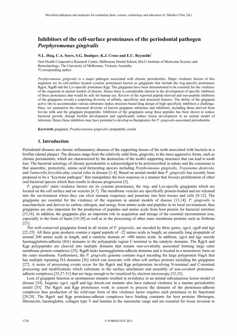
Many cysteine proteases including the gingipains are synthesized as inactive forms or zymogens with N-terminal propeptide regions. These propeptides may have multiple functions including inhibiting the proteolytic activity of the mature enzyme, folding of the precursor enzyme, and mediating membrane association [76]. Recent studies have highlighted that protease propeptides are a promising source of inhibitors for the cognate protease [77,78]. Although cysteine protease propeptides range from 30-250 amino acids, most are less than 100 amino acids [76,79]. The gingipain catalytic-domain propeptides are unusually long, being ~200 residues. Comparison of the sequences of the RgpA/B and Kgp propeptides and the catalytic domains from the known P. gingivalis strains (W50, W83, ATCC 33277, TDC60, 381, W12) reveals that the sequence conservation between the homologs is much greater than between the paralogs. The RgpA/B and Kgp propeptides from the known P. gingivalis strains are all highly conserved with a calculated percentage identity (%ID) of 98-100% between the propeptide homologs. However sequence conservation is less between the RgpA and RgpB propeptide paralogs (75-76% ID ) and between the RgpA/B and Kgp propeptide paralogs (20-22% ID). Similarly the sequences of the catalytic domains of RgpA/B and Kgp are also highly conserved (94-100% ID) between the homologs with less conservation between the paralogs. Neverthless, the overall topologies of the Kgp and RgpB catalytic domain paralogs are predicted to be similar (Fig 1).
Microbial pathogens and strategies for combating them: science, technology and education (A. Méndez-Vilas, Ed.)
© FORMATEX 2013
____________________________________________________________________________________________
1730
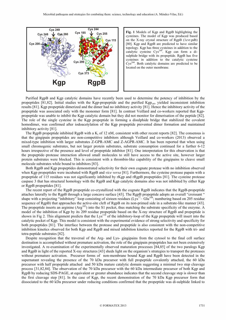
Fig. 1 Models of Kgp and RgpB highlighting the cysteines. The model of Kgp was produced based on the X-ray crystal structure of RgpB (1cvr.pdb) [80]. Kgp and RgpB are predicted to have similar topology. Kgp has three cysteines in addition to the catalytic cysteine Cys249. Kgp can form a di-sulphide bridge with its propeptide. RgpB has five cysteines in addition to the catalytic cysteine Cys244. Both catalytic domains are predicted to be located on the outer membrane.
Purified RgpB and Kgp catalytic domains have recently been used to determine the potency of inhibition by the propeptides [81,82]. Initial studies with the Kgp-propeptide and the purified Kgpcat, yielded inconsistent inhibition results [81]. Kgp propeptide dimerized and the dimer had no inhibitory activity [81]. Hence the inhibitory activity of the propeptide was associated only with the monomer form [81]. In contrast Veillard and co-workers reported that Kgp-propeptide was unable to inhibit the Kgp catalytic domain but they did not monitor for dimerisation of the peptide [82]. The role of the single cysteine in the Kgp propeptide in forming a disulphide bridge that stabilized the covalent homodimer, was confirmed after iodoacetylation of the Kgp propeptide prevented dimer formation and maintained inhibitory activity [81]. The RgpB propeptide inhibited RgpB with a Ki of 12 nM, consistent with other recent reports [82]. The consensus is that the gingipain propeptides are non-competitive inhibitors although Veillard and co-workers (2013) observed a mixed-type inhibition with larger substrates Z-GPR-AMC and Z-AGPR-AMC. It has been reported that when using small chromogenic substrates, but not larger protein substrates, substrate consumption continued for a further 6-12 hours irrespective of the presence and level of propeptide inhibitor [81]. One interpretation for this observation is that the propeptide–protease interaction allowed small molecules to still have access to the active site, however larger protein substrates were blocked. This is consistent with a thrombin-like capability of the gingipains to cleave small molecule substrates while bound to inhibitors [83]. Both RgpB and Kgp propeptides demonstrated selectivity for their own cognate protease with no inhibition observed when Kgp propeptides were incubated with RgpB and vice versa [81]. Furthermore, the cysteine protease papain with a propeptide of 115 residues was not significantly inhibited by rKgp and rRgpB propeptides [81]. The cysteine protease caspase 3 that has structural homology with the RgpB and Kgp catalytic domains also was not inhibited by either Kgp or RgpB propeptides [81]. The recent report of the RgpB propeptide co-crystallized with the cognate RgpB indicates that the RgpB-propeptide attaches laterally to the RgpB through a large concave surface [43]. The RgpB propeptide adopts an overall “croissant “ shape with a projecting “inhibitory” loop consisting of sixteen residues (Lys77– Glu104; numbering based on 205 residue sequence of RgpB) that approaches the active-site cleft of RgpB on its non-primed side in a substrate-like manner [43]. The propeptide inserts an arginine (Arg102) into the S1 pocket, thus matching the substrate specificity of the enzyme. A model of the inhibition of Kgp by its 209 residue propeptide based on the X-ray structure of RgpB and propeptide is shown in Fig 2. This alignment predicts that the Lys110 of the inhibitory-loop of the Kgp propeptide will insert into the catalytic pocket of Kgp. This model is consistent with the experimental evidence of strong selectivity and specificity for both propeptides [81]. The interface between the protease and propeptide is also consistent with the non-competitive inhibition kinetics observed for both Kgp and RgpB and mixed inhibition kinetics reported for the RgpB with tri- and tetra-peptide substrates [82]. Despite recognition that the traversal of the Arg- and Lys- gingipains from the cytosol to the final cell surface destination is accomplished without premature activation, the role of the gingipain propeptides has not been extensively investigated. A re-examination of the experimentally observed maturation processes [84,85] of the two paralogs Kgp and RgpB in light of the reported X-ray structures [43] sheds light on the organism’s strategies to transport the proteases without premature activation. Precursor forms of non-membrane bound Kgp and RgpB have been detected in the supernatant revealing the presence of the 70 kDa precursor with full propeptide covalently attached, the 60 kDa precursor with half propeptide attached and 50 kDa mature catalytic domain suggesting a minimal two step cleavage process [31,82,84]. The observation of the 70 kDa precursor with the 60 kDa intermediate precursor of both Kgp and RgpB by reducing SDS-PAGE, at equivalent or greater abundance indicates that the second cleavage step is slower than the first cleavage step [81]. In the case of Kgp, the recent demonstration of the 70 kDa Kgp precursor form that dissociated to the 60 kDa precursor under reducing conditions confirmed that the propeptide was di-sulphide linked to
Cys 249Cys 260Cys 248
Cys 200
CysCys 244
Cys 255
Cys 115
Cys 185
Cys 373
Cys 299
Kgp RgpB
Microbial pathogens and strategies for combating them: science, technology and education (A. Méndez-Vilas, Ed.)
© FORMATEX 2013
____________________________________________________________________________________________
1731

the catalytic domain through one of the surface exposed cysteine residues [Fig. 1]. These observations emphasize that rate of detachment is slower than the rate of the first cleavage. Although RgpB maturation in vivo has been shown to be dependent on the function of the gingipains, for either of RgpA, RgpB, or Kgp [85] the detachment process of the propeptide fragments has not been elucidated. The X-ray structure of RgpB with its propeptide (4ief.pdb) reveals 82 contacts (<0.4A) between 11 propeptide and 11 catalytic domain residues [43]. The participation of residues of both N-terminal and C-terminal halves of the long propeptide forming salt-bridges, hydrogen bonds, and hydrophobic interactions with the catalytic domain is consistent with the slow detachment of the propeptide fragments during the maturation process observed in vitro. The proteolytic capability of these intermediate precursor forms has been examined in several studies [31,84]. Recombinant RgpB produced in yeast as an inactive zymogen was shown to be processed in a stepwise manner with incremental increases in activity. The initial cleavage in the propeptide was identified at the Arg102-Ala-103 bond. This Arg102 residue represents the inhibitory arginine that inserts into the S1 pocket [43]. Mutation of Arg102 to Ala102 prevented hydrolysis of the propeptide and activation [84]. Unexpectedly, it did not prevent affinity labelling using Ac-biotinyl-Lys-Tyr-6-aminohexanoic-Arg-acyloxymethyl ketone indicating that the active site was accessible to small molecule, irreversible inhibitors [84] despite the association of the propeptide with the RgpB in the zymogen. While the X-ray structure of RgpB with its propeptide (4ief.pdb) reveals a number of contacts between the propeptide and the catalytic domain, very few are associated with the inhibitory loop, residues Lys99 to Glu104, apart from those involving Arg102 [43]. Inspection of the temperature factors associated with the inhibitory loop show a minimum in the region around the inhibitory Arg102. These observations suggest that the interactions between Arg102 and the catalytic domain play an important role in stabilizing the inhibitory loop in its experimentally observed position. Furthermore, they are consistent with the observation that mutating Arg102 allows the inhibitory loop to move away from the catalytic site and permit access to small molecule substrates and inhibitors. The affinity labelling was also reported for the 60 kDa precursor form of Kgp that includes the C-terminal half of the propeptide, as observed from SDS-PAGE under reduced conditions [31]. In that study, the affinity labelling ability of the precursor forms was not monitored under non-reducing conditions. However it can be anticipated that the first cleavage step, without the immediate detachment of the first half of the propeptide, will allow accessibility to small molecule, irreversible inhibitors, or small substrates. Hence while the bulk of the 200 residue propeptide can remain covalently or non-covalently attached to the catalytic domain, substitution of the inhibitory Arg or Lys, or cleavage at those residues allows access to the active site by small molecules. Consequently, the exquisite specificity and selectivity demonstrated by the gingipain propeptide for its cognate protease is attributable to the flexible inhibitory loop projecting from a rampart-like domain attached to the mature protease. This architectural design prevents access to larger natural substrates that would be encountered during the maturation pathway and has inbuilt specific and separate cleavage and detachment steps to prevent premature activation. The high affinity and unique specificity of the gingipain propeptides makes them exciting candidates for the development of a new class of therapeutics for P. gingivalis-associated periodontal disease.
Fig. 2 Schematic representation of the inhibition of Kgp by its propeptide. Kgp was modelled based on the X-ray crystal structure of RgpB 1cvr.pdb [80]. The propeptide is based on the A chain of the X-ray crystal structure of RgpB interacting with its propeptide 4ief.pdb [43].
5. Conclusion
Inhibitors of the cell surface proteinases (gingipains) have been identified, including the cognate propeptides, which have the specificity and potency to be considered as promising candidates for the development of novel therapeutics for the treatment of P. gingivalis-associated periodontitis.
Kgp-Propeptide
Kgpcatalytic domain
Inhibitoryloop
Microbial pathogens and strategies for combating them: science, technology and education (A. Méndez-Vilas, Ed.)
© FORMATEX 2013
____________________________________________________________________________________________
1732

Acknowledgements The support of the Australian Federal Government’s Cooperative Research Centre Scheme is gratefully acknowledged.
References
[1] Socransky SS, Haffajee AD, Cugini MA, Smith C, Kent RL, Jr. Microbial complexes in subgingival plaque. J Clin Periodontol. 1998;25:134-144.
[2] Darveau RP. Periodontitis: a polymicrobial disruption of host homeostasis. Nat Rev Microbiol. 2010;8:481-490. [3] Hajishengallis G, Lambris JD. Microbial manipulation of receptor crosstalk in innate immunity. Nat Rev Immunol.
2011;11:187-200. [4] Hajishengallis G, Lamont RJ. Beyond the red complex and into more complexity: the polymicrobial synergy and dysbiosis
(PSD) model of periodontal disease etiology. Mol Oral Microbiol. 2012;27:409-419. [5] Hajishengallis G. Immune evasion strategies of Porphyromonas gingivalis. J Oral Biosci. 2011;53:233-240. [6] Holt SC, Bramanti TE. Factors in virulence expression and their role in periodontal disease pathogenesis. Crit Rev Oral Biol
Med. 1991;2:177-281. [7] O'Brien-Simpson NM, Paolini RA, Hoffmann B, Slakeski N, Dashper SG, Reynolds EC. Role of RgpA, RgpB, and Kgp
proteinases in virulence of Porphyromonas gingivalis W50 in a murine lesion model. Infect Immun. 2001;69:7527-7534. [8] Furuta N, Takeuchi H, Amano A. Entry of Porphyromonas gingivalis outer membrane vesicles into epithelial cells causes
cellular functional impairment. Infect Immun. 2009;77:4761-4770. [9] Haurat MF, Aduse-Opoku J, Rangarajan M, Dorobantu L, Gray MR, Curtis MA et al. Selective sorting of cargo proteins into
bacterial membrane vesicles. J Biol Chem. 2011;286:1269-1276. [10] Nakao R, Hasegawa H, Ochiai K, Takashiba S, Ainai A, Ohnishi M et al. Outer membrane vesicles of Porphyromonas
gingivalis elicit a mucosal immune response. PLoS One. 2011;6:e26163. [11] Grenier D. Porphyromonas gingivalis outer membrane vesicles mediate coaggregation and piggybacking of Treponema
denticola and Lachnoanaerobaculum saburreum. Int J Dent. 2013;2013:305476. [12] Inagaki S, Onishi S, Kuramitsu HK, Sharma A. Porphyromonas gingivalis vesicles enhance attachment, and the leucine-rich
repeat BspA protein is required for invasion of epithelial cells by "Tannerella forsythia". Infect Immun. 2006;74:5023-5028. [13] O'Brien-Simpson N, Veith PD, Dashper SG, Reynolds EC. Porphyromonas gingivalis gingipains: the molecular teeth of a
microbial vampire. Curr Protein Pept Sci. 2003;4:409-426. [14] Guo Y, Nguyen K-A, Potempa J. Dichotomy of gingipains action as virulence factors: from cleaving substrates with the
precision of a surgeon’s knife to a meat chopper-like brutal degradation of proteins. Periodontology 2000. 2010;54:15-44. [15] Shi Y, Ratnayake DB, Okamoto K, Abe N, Yamamoto K, Nakayama K. Genetic analyses of proteolysis, hemoglobin binding,
and hemagglutination of Porphyromonas gingivalis. Construction of mutants with a combination of rgpA, rgpB, kgp, and hagA. J Biol Chem. 1999;274:17955-17960.
[16] Dashper SG, Cross KJ, Slakeski N, Lissel P, Aulakh P, Moore C et al. Hemoglobin hydrolysis and heme acquisition by Porphyromonas gingivalis. Oral Microbiol Immunol. 2004;19:50-56.
[17] DeCarlo AA, Paramaesvaran M, Yun PL, Collyer C, Hunter N. Porphyrin-mediated binding to hemoglobin by the HA2 domain of cysteine proteinases (gingipains) and hemagglutinins from the periodontal pathogen Porphyromonas gingivalis. J Bacteriol. 1999;181:3784-3791.
[18] Lewis JP, Dawson JA, Hannis JC, Muddiman D, Macrina FL. Hemoglobinase activity of the lysine gingipain protease (Kgp) of Porphyromonas gingivalis W83. J Bacteriol. 1999;181:4905-4913.
[19] Nakayama K, Ratnayake DB, Tsukuba T, Kadowaki T, Yamamoto K, Fujimura S. Haemoglobin receptor protein is intragenically encoded by the cysteine proteinase-encoding genes and the haemagglutinin-encoding gene of Porphyromonas gingivalis. Mol Microbiol. 1998;27:51-61.
[20] O'Brien-Simpson NM, Pathirana RD, Walker GD, Reynolds EC. Porphyromonas gingivalis RgpA-Kgp proteinase-adhesin complexes penetrate gingival tissue and induce proinflammatory cytokines or apoptosis in a concentration-dependent manner. Infect Immun. 2009;77:1246-1261.
[21] Nakayama K, Yoshimura F, Kadowaki T, Yamamoto K. Involvement of arginine-specific cysteine proteinase (Arg-gingipain) in fimbriation of Porphyromonas gingivalis. J Bacteriol. 1996;178:2812-2824.
[22] Curtis MA, Kuramitsu HK, Lantz M, Macrina FL, Nakayama K, Potempa J et al. Molecular genetics and nomenclature of proteases of Porphyromonas gingivalis. J Periodontal Res. 1999;34:464-472.
[23] Potempa J, Nguyen KA. Purification and characterization of gingipains. Curr Protoc Protein Sci. 2007;Chapter 21:Unit 21 20. [24] Slakeski N, Bhogal PS, O'Brien-Simpson NM, Reynolds EC. Characterization of a second cell-associated Arg-specific cysteine
proteinase of Porphyromonas gingivalis and identification of an adhesin-binding motif involved in association of the prtR and prtK proteinases and adhesins into large complexes. Microbiology. 1998;144:1583-1592.
[25] Bhogal PS, Slakeski N, Reynolds EC. A cell-associated protein complex of Porphyromonas gingivalis W50 composed of Arg- and Lys-specific cysteine proteinases and adhesins. Microbiol. 1997;143 ( Pt 7):2485-2495.
[26] Kozarov E, Whitlock J, Dong H, Carrasco E, Progulske-Fox A. The number of direct repeats in hagA is variable among Porphyromonas gingivalis strains. Infect Immun. 1998;66:4721-4725.
[27] Frazer LT, O'Brien-Simpson NM, Slakeski N, Walsh KA, Veith PD, Chen CG et al. Vaccination with recombinant adhesins from the RgpA-Kgp proteinase-adhesin complex protects against Porphyromonas gingivalis infection. Vaccine. 2006;24:6542-6554.
[28] Pathirana RD, O'Brien-Simpson NM, Veith PD, Riley PF, Reynolds EC. Characterization of proteinase-adhesin complexes of Porphyromonas gingivalis. Microbiology. 2006;152:2381-2394.
[29] Veith PD, Talbo GH, Slakeski N, Dashper SG, Moore C, Paolini RA et al. Major outer membrane proteins and proteolytic processing of RgpA and Kgp of Porphyromonas gingivalis W50. Biochem J. 2002;363:105-115.
Microbial pathogens and strategies for combating them: science, technology and education (A. Méndez-Vilas, Ed.)
© FORMATEX 2013
____________________________________________________________________________________________
1733

[30] Slakeski N, Cleal SM, Bhogal PS, Reynolds EC. Characterization of a Porphyromonas gingivalis gene prtK that encodes a lysine-specific cysteine proteinase and three sequence-related adhesins. Oral Microbiol Immunol. 1999;14:92-97.
[31] Sztukowska M, Veillard F, Potempa B, Bogyo M, Enghild JJ, Thogersen IB et al. Disruption of gingipain oligomerization into non-covalent cell-surface attached complexes. Biol Chem. 2012;393:971-977.
[32] Takii R, Kadowaki T, Baba A, Tsukuba T, Yamamoto K. A functional virulence complex composed of gingipains, adhesins, and lipopolysaccharide shows high affinity to host cells and matrix proteins and escapes recognition by host immune systems. Infect Immun. 2005;73:883-893.
[33] Chen YY, Peng B, Yang Q, Glew MD, Veith PD, Cross KJ et al. The outer membrane protein LptO is essential for the O-deacylation of LPS and the co-ordinated secretion and attachment of A-LPS and CTD proteins in Porphyromonas gingivalis. Mol Microbiol. 2011;79:1380-1401.
[34] McKee AS, McDermid AS, Wait R, Baskerville A, Marsh PD. Isolation of colonial variants of Bacteroides gingivalis W50 with a reduced virulence. J Med Microbiol. 1988;27:59-64.
[35] Pathirana RD, O'Brien-Simpson NM, Brammar GC, Slakeski N, Reynolds EC. Kgp and RgpB, but not RgpA, are important for Porphyromonas gingivalis virulence in the murine periodontitis model. Infect Immun. 2007;75:1436–1442.
[36] Schechter I, Berger A. On the size of the active site in proteases. I. Papain. Biochem Biophys Res Commun. 1967;27:157-162. [37] Rawlings ND, Barrett AJ, Bateman A. MEROPS: the database of proteolytic enzymes, their substrates and inhibitors. Nucleic
Acids Res. 2012;40:D343-D350. . [38] Imamura T, Potempa J, Travis J. Activation of the kallikrein-kinin system and release of new kinins through alternative
cleavage of kininogens by microbial and human cell proteinases. Biol Chem. 2004;385:989-996. [39] O'Brien-Simpson NM, Dashper SG, Reynolds EC. Histatin 5 is a substrate and not an inhibitor of the Arg- and Lys-specific
proteinases of Porphyromonas gingivalis. Biochem Biophys Res Commun. 1998;250:474-478. [40] van der Post S, Subramani DB, Backstrom M, Johansson ME, Vester-Christensen MB, Mandel U et al. Site-specific O-
glycosylation on the MUC2 mucin inhibits cleavage by the Porphyromonas gingivalis secreted cysteine protease (RgpB). J Biol Chem. 2013.
[41] Ally N, Whisstock JC, Sieprawska-Lupa M, Potempa J, Le Bonniec BF, Travis J et al. Characterization of the specificity of arginine-specific gingipains from Porphyromonas gingivalis reveals active site differences between different forms of the enzymes. Biochemistry. 2003;42:11693-11700.
[42] Bialas A, Grembecka J, Krowarsch D, Otlewski J, Potempa J, Mucha A. Exploring the Sn binding pockets in gingipains by newly developed inhibitors: structure-based design, chemistry, and activity. J Med Chem. 2006;49:1744-1753.
[43] de Diego I, Veillard FT, Guevara T, Potempa B, Szutkowska M, Potempa J et al. Porphyromonas gingivalis virulence factor gingipain RgpB shows a unique zymogenic mechanism for cysteine peptidases. J Biol Chem. 2013.
[44] Potempa J, Pike R, Travis J. Titration and mapping of the active site of cysteine proteinases from Porphyromonas gingivalis (gingipains) using peptidyl chloromethanes. Biol Chem. 1997;378:223-230.
[45] Curtis MA, Aduse Opoku J, Rangarajan M, Gallagher A, Sterne JAC, Reid CR et al. Attenuation of the virulence of Porphyromonas gingivalis by using a specific synthetic Kgp protease inhibitor. Infect Immun. 2002;70:6968-6975.
[46] Hara T, Yokoyama A, Ishihara H, Yokoyama Y, Nagahara T, Iwamoto M. DX-9065a, a new synthetic, potent anticoagulant and selective inhibitor for factor Xa. Thromb Haemost. 1994;71:314-319.
[47] Matsushita K, Imamura T, Tancharoen S, Tatsuyama S, Tomikawa M, Travis J et al. Selective inhibition of Porphyromonas gingivalis growth by a factor Xa inhibitor, DX-9065a. J Periodontal Res. 2006;41:171-176.
[48] Kadowaki T, Baba A, Abe N, Takii R, Hashimoto M, Tsukuba T et al. Suppression of pathogenicity of Porphyromonas gingivalis by newly developed gingipain inhibitors. Mol Pharmacol. 2004;66:1599-1606.
[49] Davies GE, Francis J, Martin AR, Rose FL, Swain G. 1:6-Di-4'-chlorophenyldiguanidohexane (hibitane); laboratory investigation of a new antibacterial agent of high potency. Br J Pharmacol Chemother. 1954;9:192-196.
[50] Rangarajan M, Smith SJ, U S, Curtis MA. Biochemical characterization of the arginine-specific proteases of Porphyromonas gingivalis W50 suggests a common precursor. Biochem J. 1997;323:701-709.
[51] Cronan C, Potempa J, Travis J, Mayo J. Inhibition of Porphyromonas gingivalis proteinases (gingipains) by chlorhexidine: synergistic effect of Zn(II). Oral Microbiol Immunol. 2006;21:212-217.
[52] Imamura T, Matsushita K, Travis J, Potempa J. Inhibition of trypsin-like cysteine proteinases (gingipains) from Porphyromonas gingivalis by tetracycline and its analogues. Antimicrob Agents Chemother. 2001;45:2871-2876.
[53] Okamoto M, Sugimoto A, Leung KP, Nakayama K, Kamaguchi A, Maeda N. Inhibitory effect of green tea catechins on cysteine proteinases in Porphyromonas gingivalis. Oral Microbiology and Immunology. 2004;19:118-120.
[54] Bakri IM, Douglas CW. Inhibitory effect of garlic extract on oral bacteria. Arch Oral Biol. 2005;50:645-651. [55] Bodet C, Piche M, Chandad F, Grenier D. Inhibition of periodontopathogen-derived proteolytic enzymes by a high-molecular-
weight fraction isolated from cranberry. J Antimicrob Chemother. 2006;57:685-690. [56] McConnell RM, York JL, Frizzell D, Ezell C. Inhibition studies of some serine and thiol proteinases by new leupeptin
analogues. J Med Chem. 1993;36:1084-1089. [57] Kitano S, Irimura K, Sasaki T, Abe N, Baba A, Miyake Y et al. Suppression of gingival inflammation induced by
Porphyromonas gingivalis in rats by leupeptin. Jpn J Pharmacol. 2001;85:84-91. [58] Bania J, Kubiak A, Wojtachnio K, Polanowski A. Pancreatic secretory trypsin inhibitor acts as an effective inhibitor of cysteine
proteinases gingipains from Porphyromonas gingivalis. J Periodontal Res. 2007;43:232-236. [59] Kadowaki T, Yamamoto K. Suppression of virulence of Porphyromonas gingivalis by potent inhibitors specific for gingipains.
Curr Protein Pept Sci. 2003;4:451-458. [60] Snipas SJ, Stennicke HR, Riedl S, Potempa J, Travis J, Barrett AJ et al. Inhibition of distant caspase homologues by natural
caspase inhibitors. Biochem J. 2001;357:575-580. [61] Gusman H, Travis J, Helmerhorst EJ, Potempa J, Troxler RF, Oppenheim FG. Salivary histatin 5 is an inhibitor of both host and
bacterial enzymes implicated in periodontal disease. Infect Immun. 2001;69:1402-1408.
Microbial pathogens and strategies for combating them: science, technology and education (A. Méndez-Vilas, Ed.)
© FORMATEX 2013
____________________________________________________________________________________________
1734

[62] Simpson K, Shaw D, Nicholas K. Developmentally-regulated expression of a putative protease inhibitor gene in the lactating mammary gland of the tammar wallaby, Macropus eugenii. Comp Biochem Physiol B Biochem Mol Biol. 1998;120:535-541.
[63] Nicholas KR, Fisher JA, Muths E, Trott J, Janssens PA, Reich C et al. Secretion of whey acidic protein and cystatin is down regulated at mid-lactation in the red kangaroo (Macropus rufus). Comp Biochem Physiol A Mol Integr Physiol. 2001;129:851-858.
[64] Toh EC, Dashper SG, Huq NL, Attard TJ, O'Brien-Simpson NM, Chen YY et al. Porphyromonas gingivalis cysteine proteinase inhibition by κ−casein peptides. Antimicrob Agents Chemother. 2011;55:1155-1161.
[65] Meisel H, Walsh DJ, Murray B, Fitzgerald RJ. ACE Inhibitory Peptides. Florida: Taylor and Francis Group; 2006. [66] Hill RD, Lahav E, Givol D. A rennin-sensitive bond in α-s1 β-casein. J Dairy Res. 1974;41:147-153. [67] Malkoski M, Dashper SG, O'Brien-Simpson N, Talbo GH, Macris M, Cross KJ et al. Kappacin, a novel antibacterial peptide
from bovine milk. Antimicrob Agents Chemother. 2001;45:2309-2315. [68] Toh ECY, Dashper SG, Huq NL, Attard TJ, Cross KJ, Stanton DP et al. Inhibition of proteolytic activity of periodontal
pathogens by casein-derived peptides. Int Dairy J. 2012;24:22-26. [69] Kesavalu L, Holt SC, Ebersole JL. Virulence of a polymicrobic complex, Treponema denticola and Porphyromonas gingivalis,
in a murine model. Oral Microbiol Immunol. 1998;13:373-377. [70] Farnaud S, Evans RW. Lactoferrin--a multifunctional protein with antimicrobial properties. Mol Immunol. 2003;40:395-405. [71] Kanyshkova TG, Buneva VN, Nevinsky GA. Lactoferrin and its biological functions. Biochemistry (Mosc). 2001;66:1-7. [72] Lonnerdal B. Nutritional and physiologic significance of human milk proteins. Am J Clin Nutr. 2003;77:1537S-1543S. [73] Pan Y, Wan J, Roginski H, Lee A, Shiell B, Michalski WP et al. Comparison of the effects of acylation and amidation on the
antimicrobial and antiviral properties of lactoferrin. Lett Appl Microbiol. 2007;44:229-234. [74] Dashper SG, Pan Y, Veith PD, Chen YY, Toh EC, Liu SW et al. Lactoferrin inhibits Porphyromonas gingivalis proteinases and
has sustained biofilm inhibitory activity. Antimicrob Agents Chemother. 2012;56:1548-1556. [75] Wakabayashi H, Yamauchi K, Kobayashi T, Yaeshima T, Iwatsuki K, Yoshie H. Inhibitory effects of lactoferrin on growth and
biofilm formation of Porphyromonas gingivalis and Prevotella intermedia. Antimicrob Agents Chemother. 2009;53:3308-3316. [76] Wiederanders B, Kaulmann G, Schilling K. Functions of propeptide parts in cysteine proteases. Curr Protein Pept Sci.
2003;4:309-326. [77] Guay J, Falgueyret JP, Ducret A, Percival MD, Mancini JA. Potency and selectivity of inhibition of cathepsin K, L and S by
their respective propeptides. Eur J Biochem. 2000;267:6311-6318. [78] Demidyuk IV, Shubin AV, Gasanov EV, Kostrov SV. Propeptides as modulators of functional activity of proteases.
BioMolecular Concepts. 2010;1:305-322. [79] Rawlings ND, Tolle DP, Barrett AJ. MEROPS: the peptidase database. Nucleic Acids Res. 2004;32:D160-164. [80] Eichinger A, Beisel H-G, Jacob U, Huber R, Medrano F-J, Banbula A et al. Crystal structure of gingipain R: an Arg-specific
bacterial cysteine proteinase with a caspase-like fold. EMBO J. 1999;18:5453-5462. [81] Huq NL, Seers CA, Toh ECY, Dashper.S.G., Slakeski N, Zhang L et al. Propeptide-mediated inhibition of cognate gingipain
proteinases. PLos One. 2013;accepted Jan 28, 2013. [82] Veillard F, Sztukowska M, Mizgalska D, Ksiazek Ma, Houston J, Potempa B et al. Inhibition of gingipains by their
profragments as the mechanism protecting Porphyromonas gingivalis against premature activation of secreted proteases. Biochim Biophys Acta. 2013;doi:pii: S0304-4165(13)00133-5.
[83] Wilkens M, Krishnaswamy S. The contribution of factor Xa to exosite-dependent substrate recognition by prothrombinase. J Biol Chem. 2002;277:9366-9374.
[84] Mikolajczyk J, Boatright KM, Stennicke HR, Nazif T, Potempa J, Bogyo M et al. Sequential autolytic processing activates the zymogen of Arg-gingipain. J Biol Chem. 2003;278:10458-10464.
[85] Seers CA, Slakeski N, Veith PD, Nikolof T, Chen YY, Dashper SG et al. The RgpB C-terminal domain has a role in attachment of RgpB to the outer membrane and belongs to a novel C-terminal-domain family found in Porphyromonas gingivalis. J Bacteriol. 2006;188:6376-6386.
Microbial pathogens and strategies for combating them: science, technology and education (A. Méndez-Vilas, Ed.)
© FORMATEX 2013
____________________________________________________________________________________________
1735