Embed Size (px)
Citation preview
Initial Treatment of Fractures in Primary Care Setting
Introduction Pediatric fractures are common. Most are non-displaced and are treated non-operatively. Initial treatment is generally with a splint, elevation, rest, and pain medications. The diagnosis and initial management of fractures has typically been provided in acute care centers where diagnosis and treatment can be initiated. Unfortunately, our office is not equipped or staffed to provide acute care for all patients who may or may not have a fracture. For patients with an acute injury who are seen in a primary care office, X-rays can be obtained as an outpatient, but care of that injury is often inconvenient. This guide is intended to provide helpful information to primary care providers for initial management of these injuries.
Note: This guide is intended for fractures that are… 1) closed (no laceration communicating with fracture site), and 2) intact neurovascular status (no clear nerve compromise), and 3) non- or minimally displaced fractures (see details below).
Fractures that do not meet these criteria should be sent to the emergency department at Eastern Maine Medical Center (there is little or no pediatric surgical and/or inpatient care available at other acute care centers). The emergency department is the best place to send them as other options (our office or direct admit to the pediatric floor) will delay care and limit availability for full evaluation, additional imaging/testing, coordination of care, wound management, splinting, , etc. Cautions - Diagnosis is made by the history, exam, and imaging. - X-rays reviewed online (even by us) are not “diagnostic quality” resolution. - Anatomic and developmental variations are common. - Normal X-rays do not exclude a non-displaced physeal fracture. - Treat the patient, not the X-rays. - It is better to over-treat a sprain than to under-treat a fracture. Reassurances - It is our hope that we can accommodate seeing all these patients in our office within 3-5
days. Usually, swelling has decreased and a definitive cast can be applied. - Generally, one of us is in the office for clinic during the day. Clinics are busy, but if you
leave a message, we can usually reply between patients. Also, one of us is always on call. - Worst case scenarios are very very uncommon. If a fracture is closed with intact
neurovascular status, almost all of these fractures, even if displaced, can be effectively treated on delayed fashion
Basic Treatment Common to All Fractures
Check status of skin, circulation, and nerve function. - Open fractures have high risk of infection and generally need surgical treatment. Bruising
and superficial abrasions are not concerning.
- Pulses and capillary refill should be intact. Some temperature variation (coolness), swelling, even minimal cyanosis (blueness) are okay if pulses and cap refill are good.
Hint: True ischemia hurts a lot.
- Nerve function should be intact. Check sensation at thumb web space (radial), radial side of index finder (median), and ulnar side of little finger (ulnar). Check strength with finger extension (radial), finger flexion (median), and finger abduction (ulnar). If nerve exam is unclear, observation is appropriate.
Hint: Diffuse numbness is probably pain response.
Splint or protect the fracture site for pain control and to prevent additional injury.
� Splint should be worn full time.
� Instruct for pain control (see below).
� Out of gym and sports until cleared.
� Keep splint clean and dry. I recommend sponge baths only. Use full compliment of options for pain control initially, and decrease if doing well.
� Splint and protect fracture – decreases local pain stimulus.
� Rest the whole patient at home until severe pain resolves. Increase activity (i.e. return to school) after 48 hours if off narcotic pain medication.
� Elevate affected limb to decrease swelling for 24-48 hours, and as needed for swelling.
� Use ice, if helpful. If splinted, true cooling to skin or fracture site may be minimal, but
risk and cost are low, and there may be an indirect beneficial effect.
� Use distraction to keep attention off pain with activities like TV, music, talking, etc.
� Medicate pain. o Ibuprofen every 8 hours is good first choice (Clark et al: Pediatrics 2007 119:460) o If needed, supplement with TylCod or Lortab elixir every 4-6 hours for 24-48 hours. o If doing well, decrease to half dose TylCod or Lortab on second day. o Continue ibuprofen until pain free. o If nausea with narcotic, stop or decrease and try Tylenol as supplement to ibuprofen.
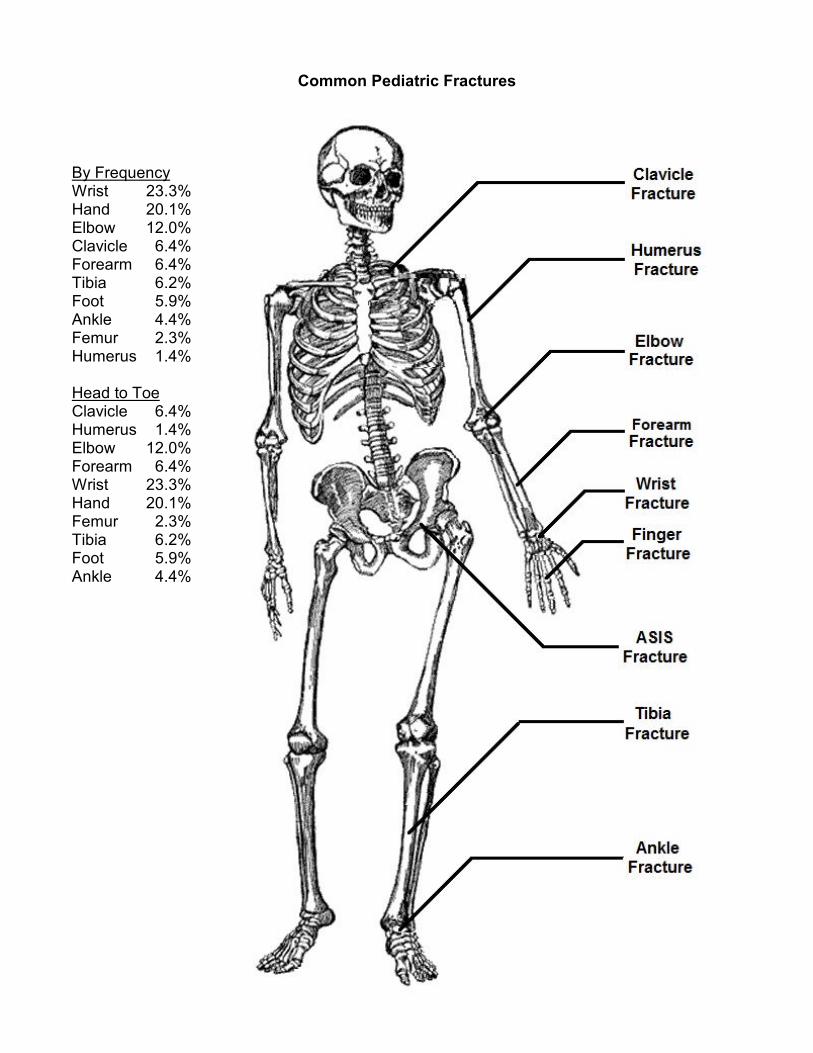
Common Pediatric Fractures By Frequency Wrist 23.3% Hand 20.1% Elbow 12.0% Clavicle 6.4% Forearm 6.4% Tibia 6.2% Foot 5.9% Ankle 4.4% Femur 2.3% Humerus 1.4% Head to Toe Clavicle 6.4% Humerus 1.4% Elbow 12.0% Forearm 6.4% Wrist 23.3% Hand 20.1% Femur 2.3% Tibia 6.2% Foot 5.9% Ankle 4.4%
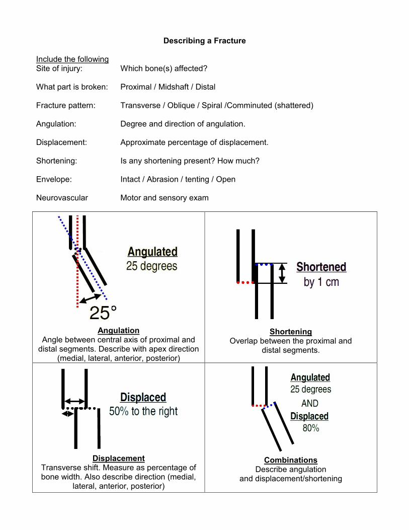
Describing a Fracture Include the following Site of injury: Which bone(s) affected? What part is broken: Proximal / Midshaft / Distal Fracture pattern: Transverse / Oblique / Spiral /Comminuted (shattered) Angulation: Degree and direction of angulation. Displacement: Approximate percentage of displacement. Shortening: Is any shortening present? How much? Envelope: Intact / Abrasion / tenting / Open Neurovascular Motor and sensory exam
Angulation
Angle between central axis of proximal and distal segments. Describe with apex direction
(medial, lateral, anterior, posterior)
Shortening
Overlap between the proximal and distal segments.
Displacement
Transverse shift. Measure as percentage of bone width. Also describe direction (medial,
lateral, anterior, posterior)
Combinations Describe angulation
and displacement/shortening
Upper Extremity Fracture Care Guidelines
Clavicle Fracture Mechanism – Often a fall onto the shoulder. Exam – Pain and swelling over clavicle. Adjacent areas,
including scapula and upper arm okay. X-rays – AP and axial view of injured clavicle. In children, mild
to moderate displacement is common, but generally acceptable… conservatively, acceptable to have 100% translation, but should have less than 40 degrees angulation and less than 2 cm shortening. Any more deformity should be discussed with orthopedist.
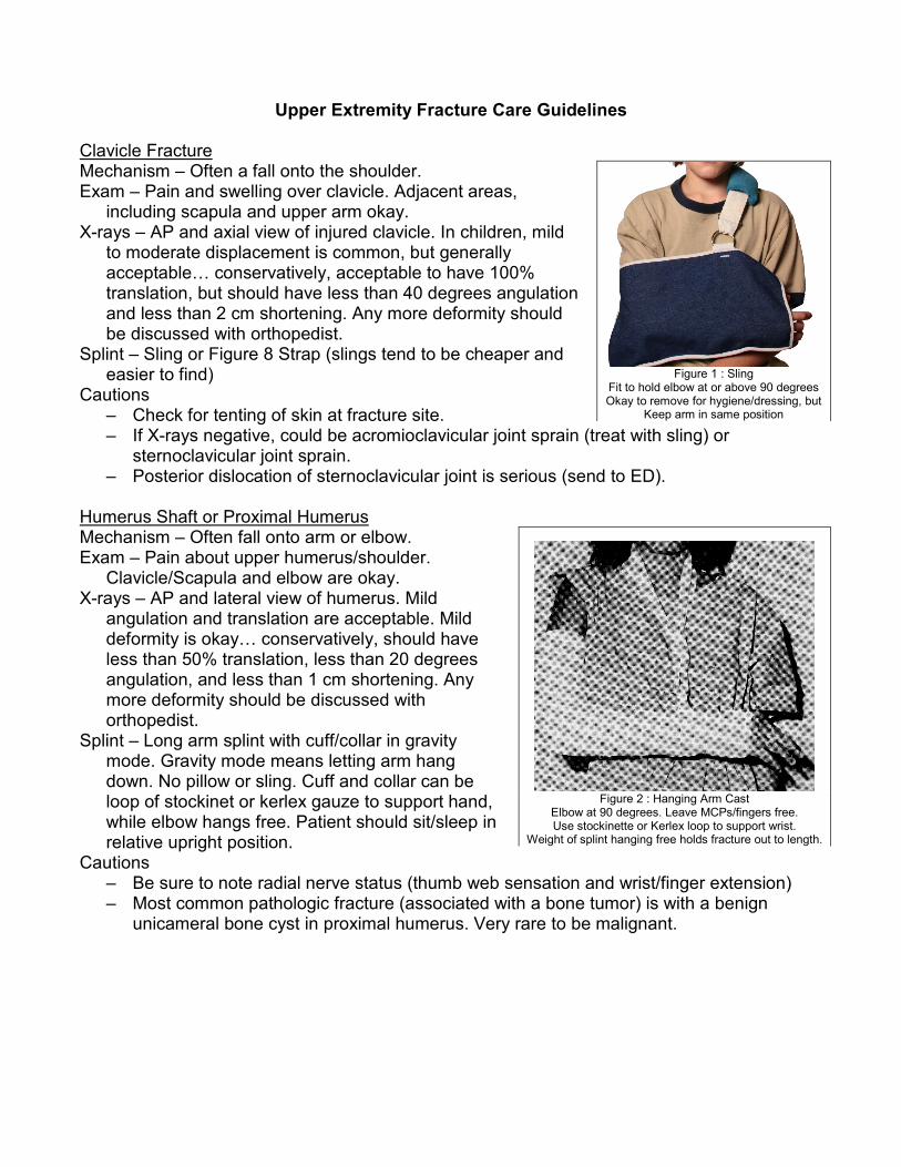
Splint – Sling or Figure 8 Strap (slings tend to be cheaper and easier to find)
Cautions – Check for tenting of skin at fracture site. – If X-rays negative, could be acromioclavicular joint sprain (treat with sling) or
sternoclavicular joint sprain. – Posterior dislocation of sternoclavicular joint is serious (send to ED).
Humerus Shaft or Proximal Humerus Mechanism – Often fall onto arm or elbow. Exam – Pain about upper humerus/shoulder.
Clavicle/Scapula and elbow are okay. X-rays – AP and lateral view of humerus. Mild
angulation and translation are acceptable. Mild deformity is okay… conservatively, should have less than 50% translation, less than 20 degrees angulation, and less than 1 cm shortening. Any more deformity should be discussed with orthopedist.
Splint – Long arm splint with cuff/collar in gravity mode. Gravity mode means letting arm hang down. No pillow or sling. Cuff and collar can be loop of stockinet or kerlex gauze to support hand, while elbow hangs free. Patient should sit/sleep in relative upright position.
Cautions – Be sure to note radial nerve status (thumb web sensation and wrist/finger extension) – Most common pathologic fracture (associated with a bone tumor) is with a benign
unicameral bone cyst in proximal humerus. Very rare to be malignant.
Figure 1 : Sling
Fit to hold elbow at or above 90 degrees Okay to remove for hygiene/dressing, but
Keep arm in same position
Figure 2 : Hanging Arm Cast
Elbow at 90 degrees. Leave MCPs/fingers free. Use stockinette or Kerlex loop to support wrist.
Weight of splint hanging free holds fracture out to length.
Elbow Mechanism – Often fall onto hand or
elbow Exam – Pain and swelling at elbow.
Joint effusion palpable posteriorly adjacent to olecranon.
X-rays – AP, lateral, and oblique views of elbow (important to get true AP and true lateral views of distal humerus and good view of radial head/neck). Must be truly non-displaced or review with orthopedist.
Splint – Long arm splint. Cautions
– Elbow fractures in children have many variations, most common is in the supracondylar region of distal humerus.
– If Xrays show joint effusion (i.e. elevation of fat pads or “sail” sign), treat as fracture. – Be wary of radial head dislocation
Forearm Mechanism – Often fall onto hand. Exam – Pain and swelling at fracture
site. X-rays – AP and lateral views of
forearm. Must include elbow and wrist on same film. Mild deformity is okay… conservatively angulation should be less than 5 degrees, translation less than 50%, and no shortening. Double check elbow alignment.
Splint – Sugar tong splint. Cautions – Check neurovascular status. Wrist Mechanism – Often fall onto hand. Exam – Pain and swelling at fracture
site. X-rays – PA, lateral, and oblique
views of wrist. Mild deformity is okay… conservatively, angulation should be less than 10 degrees, translation less than 50%, and no shortening.
Splint – volar wrist splint. Cautions – Check neurovascular status.
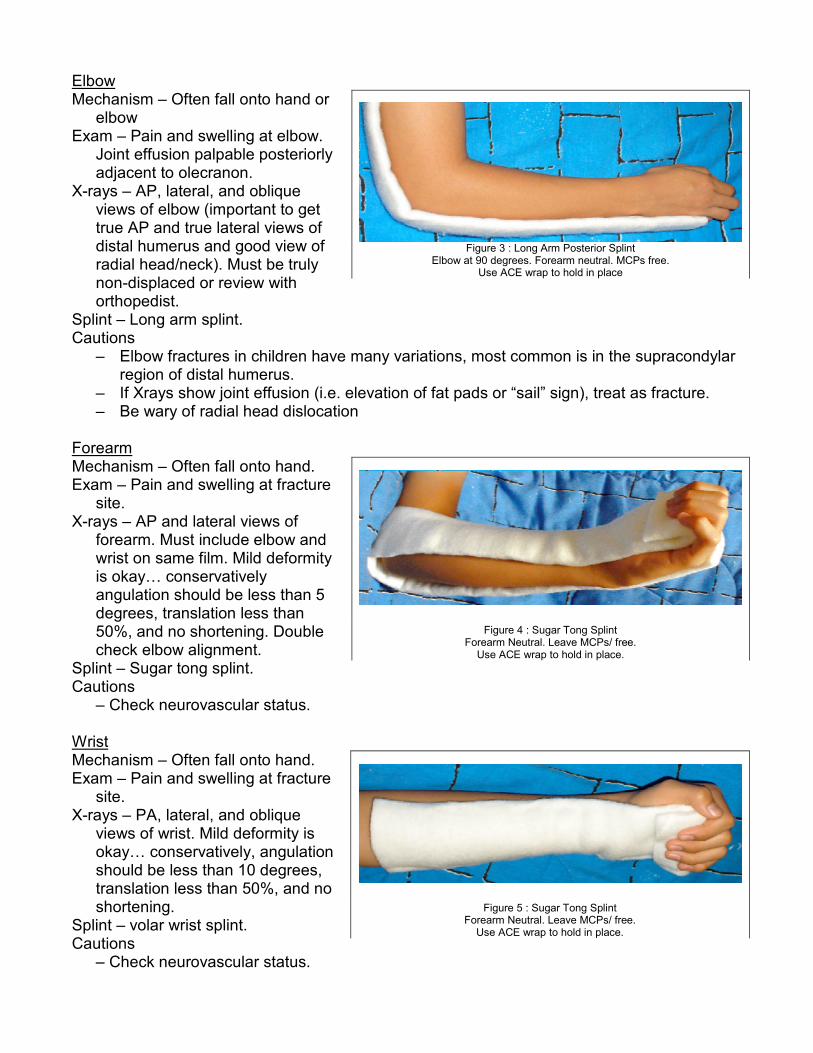
Figure 3 : Long Arm Posterior Splint
Elbow at 90 degrees. Forearm neutral. MCPs free. Use ACE wrap to hold in place
Figure 4 : Sugar Tong Splint Forearm Neutral. Leave MCPs/ free. Use ACE wrap to hold in place.
Figure 5 : Sugar Tong Splint Forearm Neutral. Leave MCPs/ free. Use ACE wrap to hold in place.
Scaphoid/Thumb Mechanism – Often fall onto the thumb or
thumb hit by a ball. Exam – Pain and swelling at wrist. Tender
with palpation in anatomic snuff box. X-rays – PA, lateral, and scaphoid views of
wrist Splint – Thumb spica wrist splint. Cautions
– Fracture is often subtle. – Double check carpal alignment.
Other Carpal/Metacarpal/Finger Mechanism – Often fall onto or other impact
to hand/finger. Exam – Pain and swelling at fracture site.
Check for rotational deformity with gentle flexion.
X-rays – PA, lateral, and oblique views of hand/fingers
Splint – for Index finger use radial gutter or
clam digger volar wrist/hand splint. – for long/ring/little fingers use ulnar
gutter splint Cautions
– Alumifoam splints acceptable for mature and cooperative patients. – Fingers are valuable, don’t undertreat.
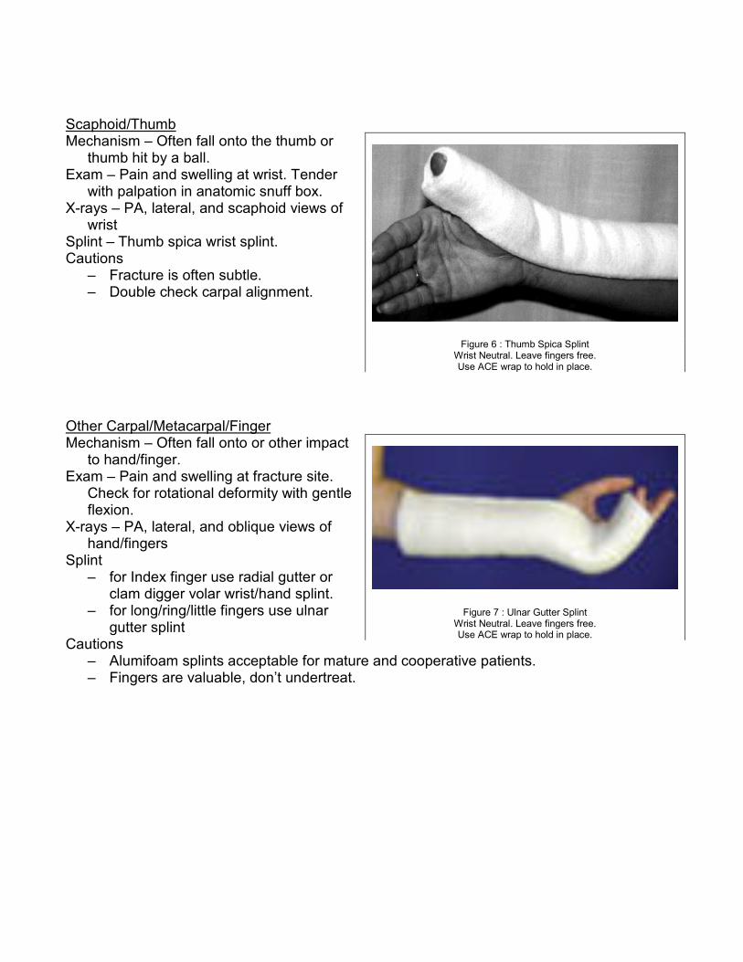
Figure 6 : Thumb Spica Splint Wrist Neutral. Leave fingers free. Use ACE wrap to hold in place.
Figure 7 : Ulnar Gutter Splint Wrist Neutral. Leave fingers free. Use ACE wrap to hold in place.
Lower Extremity Fracture Care Guidelines
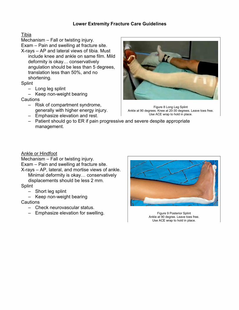
Tibia Mechanism – Fall or twisting injury. Exam – Pain and swelling at fracture site. X-rays – AP and lateral views of tibia. Must
include knee and ankle on same film. Mild deformity is okay… conservatively angulation should be less than 5 degrees, translation less than 50%, and no shortening.
Splint – Long leg splint – Keep non-weight bearing
Cautions – Risk of compartment syndrome,
generally with higher energy injury. – Emphasize elevation and rest. – Patient should go to ER if pain progressive and severe despite appropriate
management. Ankle or Hindfoot Mechanism – Fall or twisting injury. Exam – Pain and swelling at fracture site. X-rays – AP, lateral, and mortise views of ankle.
Minimal deformity is okay… conservatively displacements should be less 2 mm.
Splint – Short leg splint – Keep non-weight bearing
Cautions – Check neurovascular status. – Emphasize elevation for swelling.
Figure 8 Long Leg Splint Ankle at 90 degrees. Knee at 20-30 degrees. Leave toes free.
Use ACE wrap to hold in place.
Figure 9 Posterior Splint Ankle at 90 degree. Leave toes free. Use ACE wrap to hold in place.
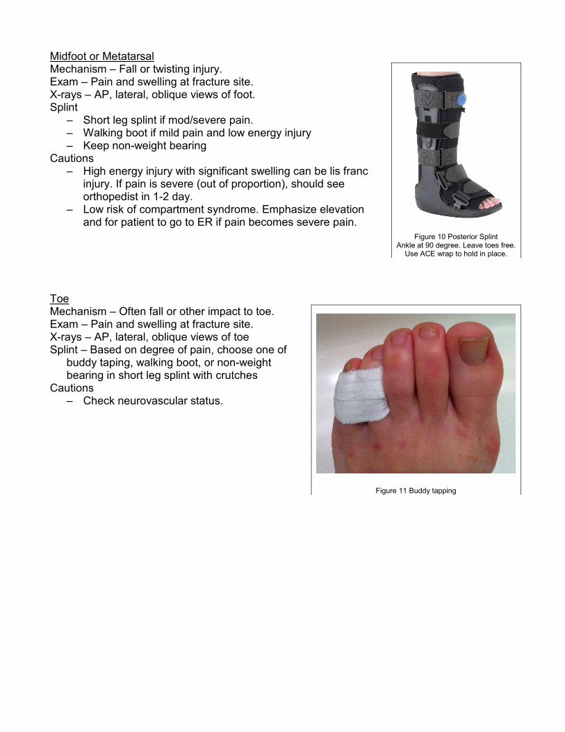
Midfoot or Metatarsal Mechanism – Fall or twisting injury. Exam – Pain and swelling at fracture site. X-rays – AP, lateral, oblique views of foot. Splint
– Short leg splint if mod/severe pain. – Walking boot if mild pain and low energy injury – Keep non-weight bearing
Cautions – High energy injury with significant swelling can be lis franc
injury. If pain is severe (out of proportion), should see orthopedist in 1-2 day.
– Low risk of compartment syndrome. Emphasize elevation and for patient to go to ER if pain becomes severe pain.
Toe Mechanism – Often fall or other impact to toe. Exam – Pain and swelling at fracture site. X-rays – AP, lateral, oblique views of toe Splint – Based on degree of pain, choose one of
buddy taping, walking boot, or non-weight bearing in short leg splint with crutches
Cautions – Check neurovascular status.
Figure 10 Posterior Splint Ankle at 90 degree. Leave toes free. Use ACE wrap to hold in place.
Figure 11 Buddy tapping


![Fifth metatarsal fractures and current treatment · the foot[1]. Approximately five to six percent of fractures encountered in the primary care setting are metatarsal fractures[2].In](https://img.pdfslide.net/doc/110x75/5e7eff3a09763541940e650a/fifth-metatarsal-fractures-and-current-treatment-the-foot1-approximately-five.jpg)