Embed Size (px)
Citation preview

Initiation factor 2 stabilizes the ribosome in asemirotated conformationClarence Ling and Dmitri N. Ermolenko1
Department of Biochemistry and Biophysics and Center for RNA Biology, School of Medicine and Dentistry, University of Rochester, Rochester, NY 14642
Edited by Peter B. Moore, Yale University, New Haven, CT, and approved November 17, 2015 (received for review October 15, 2015)
Intersubunit rotation and movement of the L1 stalk, a mobile domainof the large ribosomal subunit, have been shown to accompany theelongation cycle of translation. The initiation phase of protein synthesisis crucial for translational control of gene expression; however, incontrast to elongation, little is known about the conformationalrearrangements of the ribosome during initiation. Bacterial initiationfactors (IFs) 1, 2, and 3mediate the binding of initiator tRNA andmRNAto the small ribosomal subunit to form the initiation complex, whichsubsequently associates with the large subunit by a poorly understoodmechanism. Here, we use single-molecule FRET to monitor intersubunitrotation and the inward/outward movement of the L1 stalk of thelarge ribosomal subunit during the subunit-joining step of translationinitiation. We show that, on subunit association, the ribosome adopts adistinct conformation in which the ribosomal subunits are in a semi-rotated orientation and the L1 stalk is positioned in a half-closed state.The formation of the semirotated intermediate requires the presenceof an aminoacylated initiator, fMet-tRNAfMet, and IF2 in the GTP-boundstate. GTP hydrolysis by IF2 induces opening of the L1 stalk and thetransition to the nonrotated conformation of the ribosome. Our resultssuggest that positioning subunits in a semirotated orientation facili-tates subunit association and support a model in which L1 stalk move-ment is coupled to intersubunit rotation and/or IF2 binding.
ribosome | translation initiation | single-molecule FRET | initiation factor 2
The coordinated structural rearrangements of the ribosomeand protein factors underlie the mechanism of translation.
During the elongation phase of protein synthesis, the movementof tRNAs through the ribosome is accompanied by large-scaleconformational changes, such as intersubunit rotation (1), theswiveling of the 30S subunit head (2), and the movement of amobile domain of the large ribosomal subunit, the L1 stalk (3).Although the elongating ribosome likely samples a number oftransient conformations, it predominantly adopts two mainstructural states: the nonrotated, classical state and the rotated,hybrid state (4). Translocation of tRNAs from the A and P to theP and E sites occurs through the formation of the intermediatehybrid A/P and P/E states, in which anticodon stem-loops oftRNAs are bound to the A and P site of the small subunit,whereas the acceptor ends are bound to the P and E sites of thelarge subunit, respectively (5). Hybrid state formation is coupledto a ∼7°–∼10° rotation of the body and platform of the smallribosomal subunit relative to the large ribosomal subunit and theinward movement of the 50S L1 stalk (6). Blocking intersubunitrotation by a covalent cross-link between subunits abolishestRNA translocation (7). Furthermore, the antibiotics viomycinand neomycin inhibit tRNA translocation while trapping the ri-bosome in the rotated and semirotated conformations, respectively(8, 9). Hence, rearrangements of the ribosome are essential fortranslation elongation. However, the role of ribosomal structuraldynamics in the other phases of protein synthesis such as initiation,termination, and recycling is less well understood.Translation initiation is a key regulatory step in protein synthesis in
all organisms. Initiation of protein synthesis in bacteria is controlledby initiation factors (IFs), IF1, 2, and 3, which promote the binding ofinitiator tRNA, fMet-tRNAfMet, and mRNA to the small (30S) ri-bosomal subunit to form the 30S initiation complex (30S IC). IF1, 2,
and 3 cooperatively maintain the fidelity of start codon selection andinitiator tRNA binding (10–12). IF2 is a translational GTPase thatfacilitates the association of the large (50S) ribosomal subunit withthe 30S IC. GTP hydrolysis stimulated by the large ribosomal subunittriggers the release of IF2 (13, 14), whereas IF1 and IF3 likely dis-associate from the ribosome concurrently (11) or shortly after subunitjoining, but before the release of IF2 (12, 15). Although the specificfunctions of each initiation factor, as well as the order and kinetics ofthe different steps of initiation, have been extensively studied(12, 16–20), many molecular details of this process including theconformational rearrangements of the ribosome remain unclear.Previous cryo-EM and FRET studies have suggested that rotation
between ribosomal subunits may be involved in the transition fromthe initiation to the elongation phase of protein synthesis (21–24).However, these studies have not unambiguously determined whichconformation is adopted by the ribosome on IF2-mediated subunitjoining. Relatively low-resolution (>11 Å) cryo-EM reconstructionsof a late intermediate of initiation, the 70S IC bound with IF2,suggested that the small ribosomal subunit in 70S•IF2 ICs is rotated∼4–5° relative to the large ribosomal subunit, which is less than thedegree of rotation observed in hybrid, fully rotated ribosomes (21–23).Furthermore, in the Escherichia coli 70S•IF2 IC, the initiator tRNAwas observed in an intermediate position between the classical P/Pand hybrid P/E states that was named the P/I state (22). By contrast,the cryo-EM reconstruction of the Thermus thermophilus 70S•IF2IC showed the ribosome in a conformation that was similar to thenonrotated state containing initiator tRNA bound in the classicalP/P state (21). Moreover, the cryo-EM reconstruction of IF2 boundto the rotated, hybrid state ribosome has also been determined (23).Additionally, a single-molecule FRET (smFRET) study, whichused energy transfer between fluorophores attached to the smalland large subunits, suggested that, on subunit joining during initi-ation, the ribosome adopts a conformation that is indistinguishablefrom the rotated, hybrid state (24). The discrepancy betweenaforementioned studies may be due to differences in experimental
Significance
Translation initiation is a key step for the regulation of proteinsynthesis. Initiation factors mediate the binding of initiator tRNAand mRNA to the small ribosomal subunit and subsequent bindingof the large ribosomal subunit. The molecular mechanism of sub-unit association is poorly understood. Here, we show that bacterialinitiation factor 2 (IF2) positions ribosomal subunits in a distinctrotational orientation during the subunit-joining step of initiation.IF2 also stabilizes the mobile domain of the large subunit namedthe L1 stalk in a unique conformation. Our studies provide insightsinto how IF2 promotes the association of ribosomal subunits.
Author contributions: C.L. and D.N.E. designed research; C.L. performed research; C.L.analyzed data; and C.L. and D.N.E. wrote the paper.
The authors declare no conflict of interest.
This article is a PNAS Direct Submission.1To whom correspondence should be addressed. Email: [email protected].
This article contains supporting information online at www.pnas.org/lookup/suppl/doi:10.1073/pnas.1520337112/-/DCSupplemental.
www.pnas.org/cgi/doi/10.1073/pnas.1520337112 PNAS Early Edition | 1 of 6
BIOCH
EMISTR
Y

conditions, low resolution, and ensemble averaging of cryo-EMreconstructions. Nevertheless, structural features of the 70S IC re-main elusive. In addition, a thermodynamic description of ribosomedynamics during initiation is also lacking.Here, using previously established smFRET assays (25, 26), we
follow the orientation of ribosomal subunits and the position of theL1 stalk during the subunit association step of translation initiation.We show that IF2 stabilizes an intermediate of initiation where theribosomal subunits adopt a semirotated conformation and the 50S L1stalk is in a half-closed position. We also demonstrate that the for-mation of the semirotated 70S IC requires the presence of an ami-noacylated initiator, fMet-tRNAfMet, implicating the IF2-mediatedsubunit joining step in the preservation of initiation fidelity. Finally,we show that GTP hydrolysis by IF2 controls the transition of the70S IC into the nonrotated conformation of the ribosome and, thus,to the elongation phase of protein synthesis.
ResultsThe Ribosome Adopts a Semirotated Conformation During the Subunit-Joining Step of Initiation. To follow subunit joining and intersubunitrotation during initiation, we used FRET between a fluorophoreattached to protein L9 of the large ribosomal subunit and a fluo-rophore attached to protein S6 located on the platform of the smallsubunit (25). This smFRET assay has previously demonstrated thatelongation-like complexes fluctuate between the nonrotated, clas-sical state and the rotated, hybrid conformations of the ribosome,which correspond to 0.6 and 0.4 FRET states, respectively (27)(Fig. S1 A and B). Because cryo-EM reconstructions suggested thatthe 70S IC might adopt an intermediate conformation between thenonrotated and fully rotated states of the ribosome, we first testedwhether the S6/L9 FRET assay could detect a median rotationalorientation between ribosomal subunits. X-ray crystallography andsmFRET between fluorophores attached to ribosomal proteins L1and S13 were previously used to demonstrate that the antibioticneomycin stabilizes 70S ribosomes in a partially rotated confor-mation, in which the small ribosomal subunit was rotated by ∼6°relative to the large subunit (9). Consistent with this report, whenwe incubated S6/L9-labeled ribosomes containing a deacylatedtRNAfMet in the P site with 100 μM neomycin and imaged ribo-somes using total internal reflection fluorescence (TIRF) micros-copy, a predominant 0.5 FRET state was observed (Fig. S1 C andD). Thus, the S6/L9 FRET assay can detect the formation of apartially rotated neomycin-stabilized intermediate, which is differ-ent from the nonrotated and rotated conformations of the ribo-some corresponding to the 0.6 and 0.4 FRET states, respectively.We next sought to determine which rotational orientation the
ribosomal subunits adopt during the subunit-joining step oftranslation initiation. We tested activity of purified recombinantIF1, IF2, and IF3 from E. coli in ensemble stopped-flow kineticexperiments in which subunit association was detected by theincrease in light scattering. Our kinetic data validated activity ofIFs used in this work and showed that, consistent with previousreports (10, 11), IF2 accelerated subunit joining, whereas IF1and IF3 significantly slowed down subunit association in theabsence of IF2 (Fig. S2). For smFRET measurements, 30S ICswere assembled in the presence of IF1, IF2, IF3, GTP, Cy3-S630S subunits, fMet-tRNAfMet, and mRNA m291, which werethen tethered to the microscope slide using a biotinylated DNAoligonucleotide (Fig. 1A). 30S ICs were imaged for 10 s, and thenCy5-L9 50S subunits were injected into the sample chamber. Theappearance of Cy5 fluorescence indicated the joining of the 50Ssubunit (Fig. S3A). The FRET distribution histogram assembledfrom hundreds of individual traces showed a predominant 0.6FRET value corresponding to the nonrotated conformation(Fig. 2 A and B and Table S1). This observation is consistent withpreviously published smFRET data demonstrating that followingthe release of initiation factors, postinitiation 70S ribosomes con-taining fMet-tRNAfMet in the P site are fixed in the nonrotated
conformation (24, 27). A contour plot showing the evolution ofpopulation FRET over time suggests the presence of a populationof ribosomes exhibiting a 0.5 FRET value in the first ∼100–500 msafter subunit joining (Fig. 2A) that then transition into the 0.6FRET state. Consistent with contour plot analysis, apparent tran-sient sampling of 0.5 FRET before the transition into the 0.6FRET state can be seen in a number of individual smFRET traces(Fig. S3 B–D). Hence, on subunit joining, at least a fraction of the70S ICs transiently adopt a semirotated conformation, which isdistinct from the nonrotated and rotated conformations corre-sponding to the 0.6 and 0.4 FRET states, respectively, beforetransitioning into the nonrotated conformation of the ribosome.Previous smFRET studies suggested that the transition of the 70S
IC into the postinitiation 70S complex after subunit joining is trig-gered by IF2-catalyzed GTP hydrolysis at the rate of 30–50 s−1 (24).The average dwell time of 70S•IF2 IC in the prehydrolysis state(τ = 1/k) is expected to be 20–30 ms, which is below the time res-olution of our smFRET measurements (100 ms). Thus, 0.5 FRET islikely not detected in a majority of traces showing subunit joining(Fig. S3A) because of the 100-ms time resolution limit of oursmFRET experiments. To extend the lifetime of the 70S•IF2 IC inthe prehydrolysis state, we replaced GTP with a nonhydrolysableanalog, β,γ-methyleneguanosine 5′-triphosphate (GDPCP). Subunitjoining in the presence of GDPCP and all three initiation factorsresulted in the appearance of a predominant 0.5 FRET value (Fig.2 C and D, Fig. S3E, and Table S1), suggesting the 70S IC adopts asemirotated conformation. Likewise, a predominant 0.5 FRET valuewas observed in subunit joining experiments when GTP was replacedwith other nonhydrolysable analogs, either β,γ-imidoguanosine 5′-triphosphate (GDPNP) or guanosine 5′-O-(gamma-thio) triphosphate(GTPγS) (Fig. S4). These results show that the identity of the GTPanalog does not influence the ability of IF2 to trap the ribosome in thesemirotated conformation and, thus, it is likely that IF2•GDPCP au-thentically recapitulates the function of IF2•GTP. Furthermore, theseresults suggested that the transition from the semirotated to thenonrotated conformation is triggered by GTP hydrolysis/inorganicphosphate release or following IF2 disassociation from the ribosome.Subunit association in the presence of IF2•GDPCP and in the
absence of IF1 and IF3 also resulted in the appearance of a pre-dominant 0.5 FRET state (Fig. 2 E and F and Fig. S3F), indicatingthat IF2 alone is able to induce the semirotated conformation ofthe ribosome. The apparent bimolecular rate for subunit joiningdetermined from smFRET data (Fig. 2 A and B) in the presenceof all three initiation factors and GTP was 9 μM−1·s−1, which is
Fig. 1. Experimental design. (A) Following intersubunit movement duringsubunit joining by FRET. Cy3-labeled 30S initiation complexes (IC) formed in thepresence of mRNA m291, fMet-tRNAfMet, IF1, IF2, IF3, and GTP (or GDPCP) wereimmobilized to a quartz slide by NeutrAvidin and a biotinylated DNA primerannealed to the mRNA. Cy5-labeled 50S subunits were delivered to the 30S ICsduring imaging and subunit joining was detected by the appearance of FRETbetween Cy3 and Cy5. (B) L1/L33 FRET pair designed to follow the movement ofthe 50S L1 stalk (26). Fluorescent dyes were attached to residues 88 and 29(yellow spheres) of proteins L1 (red) and L33 (in green), respectively. Helixes 76,77, and 78 of 23S rRNA comprising the 50S L1 stalk are shown in blue. The restof the 50S subunit is shown in gray. The large subunit is viewed from thesubunit interface (Protein Data Bank ID code 4V51) (45).
2 of 6 | www.pnas.org/cgi/doi/10.1073/pnas.1520337112 Ling and Ermolenko

very similar to the rate of 11 μM−1·s−1 previously determined bysmFRET for subunit joining in the presence of all initiation factors(15). The rate of subunit joining in the presence of all three initiationfactors and GDPCP was 3 μM−1·s−1 (Fig. 2C andD), whereas subunitassociation in the presence of GDPCP and IF2 alone (i.e., withoutIF1 and IF3; Fig. 2 E and F) was threefold faster (9 μM−1·s−1). Hence,consistent with previous reports (15, 28), IF1 and IF3 moderately slowdown IF2-mediated subunit association.
IF2 Induces the Semirotated Conformation of the Ribosome. Tofurther elucidate the effect of IFs binding on ribosome structuraldynamics, preassociated 70S S6-Cy5/L9-Cy3 ribosomes containingfMet-tRNAfMet in the P site were imaged in the presence of GDPCPand various concentrations of IF2. In contrast to the subunit joiningexperiments, this approach allows for the examination of the relativestability of different ribosomal conformations in equilibrium. Ap-proximately 80% of 70S•fMet-tRNAfMet ribosomes imaged in theabsence of IF2 were observed in the 0.6 FRET state (Fig. 3A). Asmall fraction of ribosomes (∼20%) was observed in the 0.4 FRETstate. These ribosomes likely contain tRNAfMet that sponta-neously deacylated during nonenzymatic loading to the ribosomalP site and subsequent data acquisition, allowing the ribosome totransition into the hybrid, rotated state. Incubation of ribosomescontaining fMet-tRNAfMet in the P site with IF2•GDPCP resulted ina leftward shift and broadening of the high FRET peak that
indicated the appearance of an additional (0.5) FRET state (Fig. 3and Figs. S3 G–I and S5). Indeed, FRET distribution histogramswere best fit by the sum of three Gaussians corresponding to the 0.4,0.5, and 0.6 FRET states (Fig. 3 B and C and Fig. S5). Noteworthy,the 0.5 FRET state was observed in both possible orientations ofdonor and acceptor (S6-Cy5/L9-Cy3 and S6-Cy3/L9-Cy5), suggestingthat the appearance of the 0.5 FRET state is not likely the result ofsite-specific perturbation of the fluorescent properties of the dyesdue to local environmental effects (Figs. 2 and 3).The fraction of ribosomes in the semirotated conformation, deter-
mined by the area under the 0.5 FRET peak, increased with higherIF2 concentrations (Fig. 3D) and reached a maximum at 500 nMIF2. A population of ribosomes containing deacylated tRNAfMet
in the P site likely make up the ∼20% of traces that fluctuated be-tween 0.4 and 0.6 FRET, even in the presence of saturating con-centrations of IF2 (Fig. S3G). Fitting the fraction of semirotatedribosomes as a function of IF2 concentration to a hyperbola gave anapparent KD for IF2 binding of 80± 20 nM. These experiments showthat IF2 binding shifts the equilibrium between different structuralstates of the ribosome toward the semirotated conformation.We next tested whether IF1 and IF3 stabilize the semirotated
conformation of the 70S•IF2 IC. S6-Cy5/L9-Cy3 70S ribosomescontaining initiator fMet-tRNAfMet in the P site were incubated with100 nM IF2, 1 mM GDPCP, and 2 μM of either IF1 or IF3. Weused the 100 nM concentration of IF2, which is near the apparentKD of IF2 binding to the 70S ribosome (Fig. S6A). Under theseconditions both destabilizing and stabilizing effects of IF1 and IF3 onsemirotated conformation of the ribosome can be detected with highsensitivity. Neither IF1 nor IF3 shifted the equilibrium between thenonrotated, semirotated, and rotated conformations toward thesemirotated state (Fig. S6 B and C). Likewise, the combination ofIF1 and IF3 did not enrich the fraction of ribosomes in the 0.5FRET state (Fig. S6D). Thus, neither IF1 nor IF3 contribute to thestabilization of the semirotated conformation of the 70S•IF2 IC.
IF2 Requires GTP and an Aminoacylated Initiator tRNA to Stabilize theSemirotated State. Some reports suggested that IF2 in the GDP-bound, posthydrolysis state can catalyze subunit association (16)
Fig. 2. The ribosome adopts a semirotated conformation upon subunit joiningduring initiation. L9-Cy5 50S subunits were added to S6-Cy3 30S ICs assembledwith IF1, IF2, and IF3 (A–D) or IF2 alone (E and F) in the presence of GTP (A andB) or GDPCP (C–F). Surface contour plots (A, C, and E) generated by superim-position of hundreds of FRET traces postsynchronized at the time of subunitjoining show the frequency of sampled FRET values as a function of time.Surface contour plots were plotted from white (<10% of counts in the mostpopulated FRET vs. time bin) to red (≥60% of counts in the most populatedFRET vs. time bin). Dashed lines indicate FRET values corresponding to rotated(R), semirotated (SR), and nonrotated (NR) states. (B, D, and F) Histogramscompiled from hundreds of traces show the distribution of FRET values in 70Sribosomes associated under respective conditions. N, number of traces used toassemble each histogram; black lines, Gaussian fits.
Fig. 3. IF2 stabilizes the 70S ribosome in a semirotated conformation.(A–C) FRET distribution histograms of S6-Cy5/L9-Cy3 70S ribosomes containingP-site fMet-tRNAfMet in the absence of IF2 (A) or in the presence of 125 nM (B) or1 μM (C) IF2 and GDPCP. Yellow lines represent Gaussian fits centered at 0.4, 0.5,and 0.6 FRET efficiency; the black line represents the sum of two (A) or three(B and C) Gaussians. (D) The fraction of S6-Cy5/L9-Cy3 70S ribosomes observed in0.5 FRET state vs. IF2 concentration was fit to a hyperbola (black line).
Ling and Ermolenko PNAS Early Edition | 3 of 6
BIOCH
EMISTR
Y
and induce intersubunit rotation in the 70S ribosome (21),prompting us to test whether IF2•GDP can stabilize the semi-rotated conformation of the ribosome. However, neither in theabsence of nucleotides (Fig. S7A) nor in the presence of GDP(Fig. S7B) did the addition of IF2 (up to 2 μM) to 70S ribosomescontaining fMet-tRNAfMet in the P site lead to the appearance ofthe 0.5 FRET state, which corresponds to the semirotated con-formation of the ribosome. These results are consistent withreports demonstrating that IF2 dissociates from the ribosome onGTP hydrolysis and promotes subunit joining in the presence ofGTP or nonhydrolysable analogs of GTP much more efficientlythan in the presence of GDP (14).We next elucidated the contribution of initiator fMet-tRNAfMet
to the stabilization of the semirotated conformation of the ribosomeby IF2. No 0.5 FRET state was observed in vacant S6-Cy5/L9-Cy370S ribosomes incubated with 2 μM IF2 and GDPCP (Fig. S7C andD). Likewise, IF2·GDPCP failed to induce the 0.5 FRET state in70S ribosomes containing a deacylated initiator tRNAfMet in the Psite (Fig. S7 E and F), suggesting that the formation of the semi-rotated conformation of the ribosome requires the presence of anaminoacylated initiator fMet-tRNAfMet. These results are consistentwith cryo-EM reconstructions suggesting that the C-terminal do-main of IF2 interacts with the acceptor end of initiator tRNA(21, 22, 29, 30) and biochemical experiments showing that IF2 re-quires the formyl-methionyl moiety to catalyze efficient subunit
joining (10). Noteworthy, virtually no rotated conformation corre-sponding to the 0.4 FRET state is observed in 70S ribosomes formedin the presence of fMet-tRNAfMet and IF2 (Fig. 2). This result in-dicates that no subunit joining with spontaneously deacylatedtRNAfMet occurs in the presence of IF2. Otherwise ribosomes con-taining a deacylated tRNA in the P site frequently sample the ro-tated conformation (Fig. S1A). Hence, IF2-mediated subunit joiningstep likely contributes to assuring fidelity of initiation via discrimi-nation against deacylated tRNA.Interestingly, the fraction of S6-Cy5/L9-Cy3 70S ribosomes
containing a deacylated tRNAfMet in the P site observed in therotated (0.4 FRET) state reproducibly increased in the presence ofIF2·GDPCP from ∼50% to ∼60% (Fig. S7 E and F). The slightstabilization of the 0.4 FRET state by IF2 is similar to some degreeto the observation that other translational GTPases, such as EF-Gand RF3, stabilize the rotated state in ribosomes containingdeacylated P-site tRNAs (3, 31). This finding prompted us to askwhether stabilization of the semirotated conformation could be aproperty of the aminoacylated initiator fMet-tRNAfMet, whichprevents translational GTPases from inducing the hybrid, fullyrotated state, or if induction of the semirotated state is a specificproperty of IF2. S6-Cy5/L9-Cy3 70S ribosomes containing P-sitefMet-tRNAfMet were incubated with RF3, which is involved in thetermination phase, or EF-G, which is involved in the elongationand recycling phases of translation in the presence of the non-hydrolysable analog of GTP, GDPNP. Neither translationalGTPase induced the appearance of the 0.5 FRET state (Fig. S7 Gand H), suggesting that stabilization of the semirotated state is aspecific property of IF2.
IF2 Stabilizes the L1 Stalk in a Half-Closed Conformation. During theelongation phase of protein synthesis, translocation of tRNAs andintersubunit rotation are accompanied by movements of the L1stalk of the large subunit that are thought to facilitate the bindingand release of deacylated tRNA in the 50S E site (3). Structuraland FRET studies suggest that the L1 stalk samples at least threeconformations: open, half-closed, and fully closed (2, 3, 26, 32, 33).In the half-closed and fully closed conformations, the L1 stalk in-teracts with the elbow of deacylated tRNAs bound in E/E and P/Estates, respectively (33, 34). The open conformation of the L1 stalkcorresponds to the ribosome with a vacant 50S E site (2, 34).However, the conformation of the L1 stalk was not unambiguouslyresolved in previous cryo-EM reconstructions of the IF2•ribosomecomplex (21, 22); smFRET studies of L1-stalk dynamics duringinitiation are also lacking.We followed L1-stalk movements during subunit joining and
the transition to the postinitiation 70S complex using smFRETbetween acceptor-labeled protein L1 on the L1 stalk and donor-labeled protein L33 in the static core of the 50S subunit (26)(Fig. 1B). This assay allows for the detection of the three previouslydescribed distinct conformations of the L1 stalk. Consistent withprevious data (26), 70S L1-Cy5/L33-Cy3 ribosomes containingdeacylated tRNAfMet in the P site showed spontaneous fluctuationsbetween 0.2 and 0.5 FRET states (Fig. S8 A and B), correspondingto the open and fully closed conformations of the L1 stalk on thenonrotated, classical and rotated, hybrid states of the ribosome,respectively (26). The half-closed conformation of the L1 stalkinduced by the binding of a deacylated tRNA (tRNALys) to the Esite of the nonrotated, classical-state ribosome containing anaminoacylated tRNA (fMet-tRNAfMet) in the P site was man-ifested by the appearance of a 0.3 FRET state (Fig. S8 C and D).Apparent FRET values observed in this work for the open, half-closed, and closed conformations (0.2, 0.3, and 0.5 FRET) wereslightly lower than the apparent FRET values previously detectedusing this smFRET assay (0.25, 0.4 and 0.55 FRET) (26), which islikely due to minor differences in the efficiencies of Cy3 and Cy5detection between the two optical setups for TIR microscopy thatwere used in current and previous works.
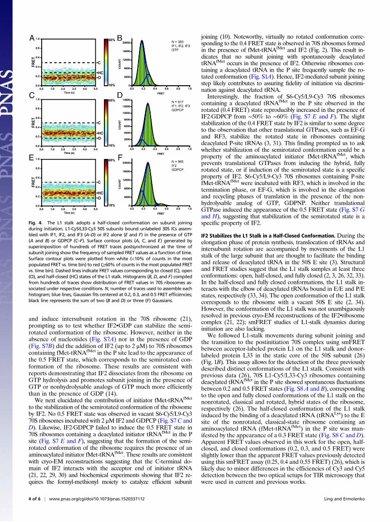
Fig. 4. The L1 stalk adopts a half-closed conformation on subunit joiningduring initiation. L1-Cy5/L33-Cy3 50S subunits bound unlabeled 30S ICs assem-bled with IF1, IF2, and IF3 (A–D) or IF2 alone (E and F) in the presence of GTP(A and B) or GDPCP (C–F). Surface contour plots (A, C, and E) generated bysuperimposition of hundreds of FRET traces postsynchronized at the time ofsubunit joining show the frequency of sampled FRET values as a function of time.Surface contour plots were plotted from white (<10% of counts in the mostpopulated FRET vs. time bin) to red (≥60% of counts in the most populated FRETvs. time bin). Dashed lines indicate FRET values corresponding to closed (C), open(O), and half-closed (HC) states of the L1 stalk. Histograms (B, D, and F) compiledfrom hundreds of traces show distribution of FRET values in 70S ribosomes as-sociated under respective conditions. N, number of traces used to assemble eachhistogram; blue lines, Gaussian fits centered at 0.2, 0.3, and 0.5 FRET efficiencies;black line represents the sum of two (B and D) or three (F) Gaussians.
4 of 6 | www.pnas.org/cgi/doi/10.1073/pnas.1520337112 Ling and Ermolenko
We next examined L1-stalk movements during the subunit-joiningstep of translation initiation. Unlabeled 30S subunits were pre-incubated with mRNA, initiator fMet-tRNAfMet, IF1, IF2, IF3, andGTP. After 5 s of imaging, L1-Cy5/L33-Cy3 50S subunits were in-jected into the sample chamber. The burst of Cy3 and Cy5 fluo-rescence denoted subunit joining (Fig. S9A). The majority (∼80%)of traces showed a single 0.2 FRET value (Fig. 4 A and B), sug-gesting that in postinitiation ribosomes, the L1 stalk is predominatelyin the open position. However, a small number of traces began at 0.2FRET and then show a transition to 0.3 FRET followed by a quicktransition to 0.2 FRET (Fig. S9 B and C). These traces suggest thatat the moment of subunit joining the L1 stalk may be open (0.2FRET) and then transiently sample an intermediate conformationcorresponding to 0.3 FRET before transitioning into the openconformation (0.2 FRET). The transient 0.3 FRET state is likelymasked in the contour plot of evolution of FRET distribution(Fig. 4A) because of averaging over traces that begin at 0.2 thenasynchronously transition between the 0.3 and 0.2 FRET values.To further test whether the L1 stalk transiently samples a half-
closed conformation during subunit joining, we replaced GTP withGDPCP to trap the 70S IC in the semirotated conformation. WhenL1-Cy5/L33-Cy3 50S subunits were added to unlabeled 30S ICs as-sembled in the presence of GDPCP, a predominant 0.3 FRET valuewas observed, indicating that the L1 stalk adopts an intermediateposition between the open and fully closed conformation (Fig. 4 Cand D). Likewise, a predominant 0.3 FRET value was observedwhen IF1 and IF3 were omitted, demonstrating that IF2•GDPCPalone is sufficient to induce the half-closed conformation of the L1stalk (Fig. 4 E and F and Fig. S9 F and G). In the presence ofIF2•GDPCP, 70S ribosomes containing a deacylated initiatortRNAfMet in the P site fluctuated between 0.2 and 0.5 FRET states(Fig. S8 E and F). However, these ribosomes do not sample the 0.3FRET state corresponding to the half-closed conformation of the L1stalk. Hence, stabilization of the half-closed conformation of the L1stalk by IF2 requires the presence of an N-formylated and amino-acylated initiator fMet-tRNAfMet. Taken together, experiments withthe L1/L33 FRET pair suggest that the 50S L1 stalk adopts the half-closed conformation in the late intermediate of translation initiationtrapped in the 70S•IF2•GDPCP complex.
DiscussionOur smFRET data provide independent evidence that, on subunitjoining during translation initiation, the ribosome adopts a distinctconformation in which ribosomal subunits are positioned in anintermediate, semirotated orientation relative to the nonrotated,classical and rotated, hybrid states. The 0.5 FRET detected duringsubunit joining using the S6/L9 intersubunit FRET pair is in-distinguishable from the 0.5 FRET observed when ribosomescontaining a deacylated P-site tRNA were incubated with neo-mycin (compare Fig. S1D with Figs. 2 and 3). Hence, the degree ofintersubunit rotation in the 70S IC is similar to the ∼6° rotationobserved in the 70S•neomycin crystal structure containing adeacylated tRNA bound in an intermediate position between theclassical P/P and hybrid P/E state, i.e., the P/pe state (9). In addi-tion, the semirotated intermediate of initiation detected in ourFRET experiments is likely similar to the conformation of the ri-bosome seen in cryo-EM reconstructions of the E. coli 70S•IF2complex (22), in which the platform and body of the 30S subunitare rotated by ∼4–5° relative to the large subunit and the initiatortRNA is bound in the P/I state, which resembles the P/pe stateobserved in in the 70S•neomycin crystal structure (9).Our experiment with the L1/L33 FRET pair revealed a pre-
viously unobserved structural feature of the 70S•IF2 IC: the L1stalk was detected in an intermediate position relative to theopen and closed conformations. The 0.3 FRET state observedin semirotated 70S•IF2•GDPCP ICs is indistinguishable fromFRET seen in nonrotated ribosomes containing a deacylatedtRNA bound in the classical E/E state (Fig. S8 C and D). However,
the strict specificity of the 50S E site for a deacylated tRNAacceptor end (35) excludes the possibility of initiator fMet-tRNAfMet binding to the E site of the 70S•IF2•GDPCP IC.Furthermore, the L1 stalk in the half-closed position is likely tobe too distant to interact with the initiator tRNA bound in P/Istate. Indeed, in the crystal structure of the 70S•neomycin complex,which shows a similar degree of intersubunit rotation to the70S•IF2 complex (Fig. S1 C and D), the L1 stalk does not make acontact with P/pe-site tRNA despite being in the fully closedconformation (9). Therefore, coupling between the formation ofthe half-closed conformation of the L1 stalk and IF2-inducedstabilization of the semirotated conformation of the ribosome islikely not mediated by the interaction between the L1 stalk andtRNA bound either in the P/I or E/E state. Consistent with ourresults, recent smFRET data showed that coupling betweenintersubunit rotation and L1 stalk movement can occur in vacantribosomes (36), further supporting the idea that the inwardmovement of the L1 stalk does not require interaction betweenthe L1 stalk and tRNA.GTP hydrolysis by IF2 was previously observed to occur at the
rate of ∼30 s−1 (16), whereas IF2 dissociation from the ribosome issignificantly slower and occurs ∼1 s−1 (37). There are conflictingreports on whether the release of inorganic phosphate from IF2 veryrapidly follows GTP hydrolysis (38) or is slower by nearly one orderof magnitude (16, 17). In our subunit-joining smFRET experimentsperformed at the 100-ms time resolution, the semirotated in-termediate of initiation was stabilized when GTP was replaced withGDPCP, whereas it was undetected in the majority of tracesobtained in the presence of GTP. Therefore, the rate of the tran-sition from the semirotated to the nonrotated conformation of the70S IC correlates with the rate of GTP hydrolysis (and, possibly, therate of inorganic phosphate release) rather than with IF2 dissocia-tion. The fraction of smFRET traces obtained in the presence ofGTP that showed transient sampling of 0.5 FRET (Fig. 2A and Fig.S3) likely corresponds to the tail of the dwell-time distribution of theprehydrolysis state of the IF2-ribosome complex. Consistent withearly proposals (24), GTP hydrolysis likely triggers conformationalchanges in IF2 that result in the transition of the ribosomal subunitsto the nonrotated orientation and the outward movement of the L1stalk into the open conformation. It is possible that GTP hydrolysisby IF2 creates a proofreading step for the 70S IC formation similarto the EF-Tu–mediated proofreading mechanism of tRNA accom-modation into the A site during translation elongation.Interestingly, IF1 and IF3 did not affect the stability of the IF2-
induced semirotated conformation of the ribosome in equilibriumexperiments. Consistent with published reports (10, 12, 15, 28), IF1and IF3 slowed IF2-mediated subunit association by approximatelythreefold in both single-molecule (Fig. 2) and ensemble kineticmeasurements (Fig. S2). By contrast, IF1 and IF3 dramaticallyinhibited subunit association in the absence of IF2 (Fig. S2), sup-porting the model that IF1 and IF3 play important roles in main-taining the fidelity of initiation by preventing premature subunitassociation in the absence of IF2, the start codon and initiator tRNA.IF2 was shown to accelerate subunit association by one to
three orders of magnitude depending on experimental conditions(10, 11). IF2 possibly aids subunit joining by spanning the smalland large subunits through specific interactions that IF2 makeswith both subunits and the initiator tRNA. Our results suggest anadditional mechanism by which IF2 may enhance subunit asso-ciation: IF2-mediated positioning of ribosomal subunits in thesemirotated orientation may be required to facilitate the dockingof intersubunit bridges that stabilize the 70S ribosome. Impor-tantly, the majority of intersubunit bridges, notably the bridges inthe core of the ribosome near the tRNA and mRNA bindingsites, are conserved between eukaryotic and bacterial ribosomes(39, 40). A cryo-EM reconstruction of the 80S IC from Saccha-romyces cerevisiae containing the eukaryotic initiator tRNA, Met-tRNAi
Met and bound to the eukaryotic ortholog of IF2, eIF5B,
Ling and Ermolenko PNAS Early Edition | 5 of 6
BIOCH
EMISTR
Y

shows the tRNA in a P/I state similar to that observed in re-constructions of the bacterial ICs along with a modest, 3.4°rotation of the small ribosomal subunit relative to the large ribo-somal subunit (41). Thus, although translation initiation is regulatedby different mechanisms in bacteria and eukaryotes, the mechanismof subunit association may be conserved throughout all domainsof life.
MethodsMaterials and methods are described in detail in SI Methods. The mRNA m291,IFs, aminoacylated fMet-tRNAfMet, and reconstituted ribosomes were prepared
as previously described (42–44). Single-molecule FRET measurements weretaken using a prism-type TIR microscope as previously described (27). Ap-parent FRET efficiencies (Eapp) were calculated from the emission intensitiesof donor (ICy3) and acceptor (ICy5) as follows: Eapp = ICy5/[ICy5 + ICy3].
ACKNOWLEDGMENTS. We thank Harry Noller for providing plasmids for IF1,IF2, and IF3 expression; Laura Lancaster for advice on IF1 purification; Jillian Dannfor assistance with IFs purification; Peter Cornish for sharing MatLab scripts;Stanislav Bellaousov for the contour plot analysis script; and Gloria Culver andAndrei Korostelev for helpful discussions. These studies were supported by USNational Institute of Health (NIH) Grant GM-099719 (to D.N.E.). C.L. was partiallysupported by NIH Training Grant in Cellular, Biochemical, and Molecular Sciences5T32 GM-068411.
1. Frank J, Agrawal RK (2000) A ratchet-like inter-subunit reorganization of the ribo-some during translocation. Nature 406(6793):318–322.
2. Schuwirth BS, et al. (2005) Structures of the bacterial ribosome at 3.5 A resolution.Science 310(5749):827–834.
3. Valle M, et al. (2003) Locking and unlocking of ribosomal motions. Cell 114(1):123–134.
4. Frank J, Gonzalez RL, Jr (2010) Structure and dynamics of a processive Brownianmotor: The translating ribosome. Annu Rev Biochem 79(1):381–412.
5. Moazed D, Noller HF (1989) Intermediate states in the movement of transfer RNA inthe ribosome. Nature 342(6246):142–148.
6. Voorhees RM, Ramakrishnan V (2013) Structural basis of the translational elongationcycle. Annu Rev Biochem 82:203–236.
7. Horan LH, Noller HF (2007) Intersubunit movement is required for ribosomal trans-location. Proc Natl Acad Sci USA 104(12):4881–4885.
8. Ermolenko DN, et al. (2007) The antibiotic viomycin traps the ribosome in an in-termediate state of translocation. Nat Struct Mol Biol 14(6):493–497.
9. Wang L, et al. (2012) Allosteric control of the ribosome by small-molecule antibiotics.Nat Struct Mol Biol 19(9):957–963.
10. Antoun A, Pavlov MY, Lovmar M, Ehrenberg M (2006) How initiation factors maxi-mize the accuracy of tRNA selection in initiation of bacterial protein synthesis. MolCell 23(2):183–193.
11. Antoun A, Pavlov MY, Lovmar M, Ehrenberg M (2006) How initiation factors tune therate of initiation of protein synthesis in bacteria. EMBO J 25(11):2539–2550.
12. Milón P, Konevega AL, Gualerzi CO, Rodnina MV (2008) Kinetic checkpoint at a latestep in translation initiation. Mol Cell 30(6):712–720.
13. Luchin S, et al. (1999) In vitro study of two dominant inhibitory GTPase mutants ofEscherichia coli translation initiation factor IF2. Direct evidence that GTP hydrolysis isnecessary for factor recycling. J Biol Chem 274(10):6074–6079.
14. Antoun A, Pavlov MY, Andersson K, Tenson T, Ehrenberg M (2003) The roles of ini-tiation factor 2 and guanosine triphosphate in initiation of protein synthesis. EMBO J22(20):5593–5601.
15. MacDougall DD, Gonzalez RL, Jr (2015) Translation initiation factor 3 regulatesswitching between different modes of ribosomal subunit joining. J Mol Biol 427(9):1801–1818.
16. Tomsic J, et al. (2000) Late events of translation initiation in bacteria: A kineticanalysis. EMBO J 19(9):2127–2136.
17. Grigoriadou C, Marzi S, Kirillov S, Gualerzi CO, Cooperman BS (2007) A quantitativekinetic scheme for 70 S translation initiation complex formation. J Mol Biol 373(3):562–572.
18. Fabbretti A, et al. (2007) The real-time path of translation factor IF3 onto and off theribosome. Mol Cell 25(2):285–296.
19. Wang J, Caban K, Gonzalez RL, Jr (2015) Ribosomal initiation complex-driven changesin the stability and dynamics of initiation factor 2 regulate the fidelity of translationinitiation. J Mol Biol 427(9):1819–1834.
20. Elvekrog MM, Gonzalez RL, Jr (2013) Conformational selection of translation initia-tion factor 3 signals proper substrate selection. Nat Struct Mol Biol 20(5):628–633.
21. Myasnikov AG, et al. (2005) Conformational transition of initiation factor 2 from theGTP- to GDP-bound state visualized on the ribosome. Nat Struct Mol Biol 12(12):1145–1149.
22. Allen GS, Zavialov A, Gursky R, Ehrenberg M, Frank J (2005) The cryo-EM structure of atranslation initiation complex from Escherichia coli. Cell 121(5):703–712.
23. Simonetti A, et al. (2013) Involvement of protein IF2 N domain in ribosomal subunitjoining revealed from architecture and function of the full-length initiation factor.Proc Natl Acad Sci USA 110(39):15656–15661.
24. Marshall RA, Aitken CE, Puglisi JD (2009) GTP hydrolysis by IF2 guides progression ofthe ribosome into elongation. Mol Cell 35(1):37–47.
25. Ermolenko DN, et al. (2007) Observation of intersubunit movement of the ribosomein solution using FRET. J Mol Biol 370(3):530–540.
26. Cornish PV, et al. (2009) Following movement of the L1 stalk between three func-tional states in single ribosomes. Proc Natl Acad Sci USA 106(8):2571–2576.
27. Cornish PV, Ermolenko DN, Noller HF, Ha T (2008) Spontaneous intersubunit rotationin single ribosomes. Mol Cell 30(5):578–588.
28. Liu Q, Fredrick K (2015) Roles of helix H69 of 23S rRNA in translation initiation. ProcNatl Acad Sci USA 112(37):11559–11564.
29. Simonetti A, et al. (2009) A structural view of translation initiation in bacteria. CellMol Life Sci 66(3):423–436.
30. Julián P, et al. (2011) The Cryo-EM structure of a complete 30S translation initiationcomplex from Escherichia coli. PLoS Biol 9(7):e1001095.
31. Zhou J, Lancaster L, Trakhanov S, Noller HF (2012) Crystal structure of release factorRF3 trapped in the GTP state on a rotated conformation of the ribosome. RNA 18(2):230–240.
32. Fei J, Kosuri P, MacDougall DD, Gonzalez RL, Jr (2008) Coupling of ribosomal L1 stalkand tRNA dynamics during translation elongation. Mol Cell 30(3):348–359.
33. Korostelev A, Trakhanov S, Laurberg M, Noller HF (2006) Crystal structure of a 70Sribosome-tRNA complex reveals functional interactions and rearrangements. Cell126(6):1065–1077.
34. Dunkle JA, et al. (2011) Structures of the bacterial ribosome in classical and hybridstates of tRNA binding. Science 332(6032):981–984.
35. Lill R, Robertson JM, Wintermeyer W (1986) Affinities of tRNA binding sites of ribo-somes from Escherichia coli. Biochemistry 25(11):3245–3255.
36. Ning W, Fei J, Gonzalez RL, Jr (2014) The ribosome uses cooperative conformationalchanges to maximize and regulate the efficiency of translation. Proc Natl Acad SciUSA 111(33):12073–12078.
37. Tsai A, et al. (2012) Heterogeneous pathways and timing of factor departure duringtranslation initiation. Nature 487(7407):390–393.
38. Pavlov MY, Sanyal S, Ehrenberg M (2011) Initiation of bacterial protein synthesis withwild type and mutated variants of initiation factor 2. Ribosomes Structure, Function,and Dynamics, eds Rodnina MV, Wintermeyer W, Green R (Spinger-Verlag, Wien), pp129–141.
39. Yusupov MM, et al. (2001) Crystal structure of the ribosome at 5.5 A resolution.Science 292(5518):883–896.
40. Ben-Shem A, Jenner L, Yusupova G, Yusupov M (2010) Crystal structure of the eu-karyotic ribosome. Science 330(6008):1203–1209.
41. Fernández IS, et al. (2013) Molecular architecture of a eukaryotic translational initi-ation complex. Science 342(6160):1240585–1240585.
42. Joseph S, Noller HF (1998) EF-G-catalyzed translocation of anticodon stem-loop an-alogs of transfer RNA in the ribosome. EMBO J 17(12):3478–3483.
43. Dallas A, Noller HF (2001) Interaction of translation initiation factor 3 with the 30Sribosomal subunit. Mol Cell 8(4):855–864.
44. Lancaster L, Noller HF (2005) Involvement of 16S rRNA nucleotides G1338 and A1339in discrimination of initiator tRNA. Mol Cell 20(4):623–632.
45. Selmer M, et al. (2006) Structure of the 70S ribosome complexed with mRNA andtRNA. Science 313(5795):1935–1942.
46. Hickerson R, Majumdar ZK, Baucom A, Clegg RM, Noller HF (2005) Measurement ofinternal movements within the 30 S ribosomal subunit using Förster resonance energytransfer. J Mol Biol 354(2):459–472.
47. Svidritskiy E, Ling C, Ermolenko DN, Korostelev AA (2013) Blasticidin S inhibitstranslation by trapping deformed tRNA on the ribosome. Proc Natl Acad Sci USA110(30):12283–12288.
48. McKinney SA, Joo C, Ha T (2006) Analysis of single-molecule FRET trajectories usinghidden Markov modeling. Biophys J 91(5):1941–1951.
49. Acker MG, et al. (2009) Kinetic analysis of late steps of eukaryotic translationinitiation. J Mol Biol 385(2):491–506.
6 of 6 | www.pnas.org/cgi/doi/10.1073/pnas.1520337112 Ling and Ermolenko