Embed Size (px)
Citation preview

Inkjet Printing of Bioadhesives
Anand Doraiswamy,1 Timothy M. Dunaway,2 Jonathan J. Wilker,2 Roger J. Narayan1
1 Joint Department of Biomedical Engineering, University of North Carolina at Chapel Hill, Chapel Hill,North Carolina 27599-7575
2 Department of Chemistry, Purdue University, West Lafayette, Indiana 47907-2084
Received 6 February 2008; revised 24 April 2008; accepted 3 June 2008Published online 19 August 2008 in Wiley InterScience (www.interscience.wiley.com). DOI: 10.1002/jbm.b.31183
Abstract: Over the past century, synthetic adhesives have largely displaced their natural
counterparts in medical applications. However, rising concerns over the environmental and
toxicological effects of the solvents, monomers, and additives used in synthetic adhesives have
recently led the scientific community to seek natural substitutes. Marine mussel adhesive
protein is a formaldehyde-free natural adhesive that demonstrates excellent adhesion to
several classes of materials, including glasses, metals, metal oxides, and polymers. In this
study, we have demonstrated computer aided design (CAD) patterning of various biological
adhesives using piezoelectric inkjet technology. A MEMS-based piezoelectric actuator was
used to control the flow of the mussel adhesive protein solution through the ink jet nozzles.
Fourier transform infrared spectroscopy (FTIR), microscopy, and adhesion studies were
performed to examine the chemical, structural, and functional properties of these patterns,
respectively. FTIR revealed the piezoelectric inkjet technology technique to be nondestructive.
Atomic force microscopy was used to determine the extent of chelation caused by Fe(III). The
adhesive strength in these materials was correlated with the extent of chelation by Fe(III).
Piezoelectric inkjet printing of naturally-derived biological adhesives may overcome several
problems associated with conventional tissue bonding materials. This technique may
significantly improve wound repair in next generation eye repair, fracture fixation, wound
closure, and drug delivery devices. ' 2008 Wiley Periodicals, Inc. J Biomed Mater Res Part B: Appl
Biomater 89B: 28–35, 2009
Keywords: biomaterials; thin film; bioadhesive; microfabrication
INTRODUCTION
Suturing is the ‘‘gold standard’’ joining technique for many
medical procedures. Unfortunately, the use of suture mate-
rials requires long operating times as well as significant
surgical skill. In addition, use of sutures is associated with
several complications, including granulomas, postoperative
epithelial ingrowth, postoperative discomfort, infection, and
inflammation. Furthermore, sutures may place excess ten-
sion on tissues, leading to tissue warping. An alternative
joining technique that has gained support over the past few
years involves the use of adhesives, which hold tissue to-
gether for several weeks while inflammatory and tissue
regrowth processes allow the defect to heal. Medical adhe-
sives must perform several functions, which include
degrading in order to allow complete healing at the lesion
site and providing sufficient tensile strength for the
intended application.
Conventional adhesives and techniques suffer from bio-
compatibility and safety issues. For example, tissue sealants
derived from cyanoacrylate esters (Dermabond1, Inder-
mil1, Nexaband1, and Vetbond1) are used in repairing
tendon, tooth enamel, cornea, and skin tissues.1–5 Unfortu-
nately, cyanoacrylate adhesives are nonbiodegradable and
permanently remain at the treatment site. As a result, these
materials have the potential to induce local inflammation,
neovascularization, foreign body reaction, and necrosis.6–8
In addition, these materials can demonstrate dose-depend-
ent carcinogenic and toxic properties. Fibrin sealants
(derived from human blood coagulation factors) have also
been considered for use in a variety of surgical and endo-
scopic applications.9–11 For example, Beriplast1 P is a
fibrin sealant that contains Combiset-1 [aprotinin (bovine),
factor XIII (human), and fibrinogen (human)] and Combi-
set-2 (calcium chloride and thrombin (human)); these com-
ponents are mixed in the operating room. The components
obtained from pooled human plasma (fibrinogen, factor
XIII, and thrombin) undergo various sterilization, manufac-
Correspondence to: Prof. R. J. Narayan (e-mail: [email protected])Contract grant sponsors: National Science Foundation; National Institutes of
Health; Office of Naval Research
' 2008 Wiley Periodicals, Inc.
28

turing, and pasteurization measures. However, there are
several safety issues that have limited the use of these
materials, including the possibility of disease transmission.
For example, the risk of HIV in blood-derived materials
screened with the p24 HIV-1 antigen test is currently esti-
mated at 1:700,000.12 Concerns also exist regarding the
transmission of human T-cell lymphotropic virus-1, hepati-
tis A virus, hepatitis B virus, hepatitis C virus, Parvovirus
B19, and spongiform encephalopathy agents from blood-
derived materials. Surgeons and their patients require
improved tissue joining materials and methods.
Mussel adhesive proteins are natural adhesives secreted
by sedentary mollusks (mussels) that inhibit intertidal and
subtidal areas. An attachment plaque known as a byssus
allows mussels to form strong attachments to underwater
surfaces. Mytilus edulis (common blue mussel) is one of
the most widely studied mussels.14–23 It produces several
unique adhesive proteins, including Mytilus edulis foot pro-tein-1 (Mefp-1), Mytilus edulis foot protein-2 (Mefp-2),
Mytilus edulis foot protein-3 (Mefp-3), Mytilus edulis foot
protein-4 (Mefp-4), and Mytilus edulis foot protein-5
(Mefp-5). These proteins contain up to 30 mole percent
3,4-dihydroxyphenyl-L-alanine (DOPA), which is a mole-
cule created by hydroxylation of the aromatic ring in the
amino acid tyrosine. It has been suggested that DOPA
drives the adhesion of a mussel plaque to an environmental
surface by means of hydogen bonding, metal-mediated cat-
echol complexation, and/or weak physical interactions.14–23
Rapid prototyping is a technology originally developed
approximately thirty years ago for the preparation of
machine tool prototypes. One possible application for rapid
prototyping technology is microscale processing of bioma-
terials. Computer aided design (CAD) rapid prototyping
techniques such as inkjet printing may allow for high
throughput patterning of biological materials for medical
applications.24–26 In piezoelectric inkjet printers, the print
head consist of a piezoelectric transducer, nozzle, manifold,
pumping chamber, and inlet passage. Piezoelectric printers
are categorized based on the deformation mode of the lead
zirconate titanate piezoelectric crystal (e.g., squeeze, bend,
push or shear). When a voltage is applied to the lead
zirconate titanate piezoelectric transducer, the transducer
deforms. Mechanical vibrations and acoustic waves are
generated. When a given linear velocity is reached by the
fluid, it is ejected from the orifice as a droplet. Ink jet
printers can dispense fluid droplets with volumes in the
picoliter to microliter range. The resolution of patterns fab-
ricated using piezoelectric ink jet printing is determined by
several factors, including ink viscosity, ink surface tension,
ink droplet size, and printerhead resolution. Unlike thermal
inkjet printers, the ink used in piezoelectric inkjet printers
does not undergo heating and cooling cycles. We have
recently demonstrated that piezoelectric inkjet deposition is
a powerful, noncontact, and nondestructive technique for
patterning many biological materials, including streptavidin
protein, monofunctional acrylate esters, sinapinic acid,
deoxyribonucleic acid, and multiwalled carbon nanotube/
DNA hybrid materials.27
In this study, we have demonstrated CAD patterning of
various biological adhesives using piezoelectric inkjet tech-
nology. Fourier transform infrared spectroscopy (FTIR),
atomic force microscopy, and adhesion studies were per-
formed to examine the chemical, structural, and functional
properties of these patterns, respectively. This technique
may significantly improve wound repair in next generation
eye repair, fracture fixation, wound closure, and drug deliv-
ery devices.
MATERIALS AND METHODS
Mussel adhesive proteins (Mefp-1 and Mefp-2) were
extracted from marine mussel feet as described in28 with
slight modification. The protein pellets were extracted with
water, rather than the reported acetic acid.28 The extract
yields a solution that contains predominantly two proteins,
�80% Mefp-1 and �20% Mefp-2.28 The final DOPA con-
centration in this solution was 0.16 mM, the total protein
concentration in this solution was �2 lM, and the viscosity
of this solution was similar to water (density �1 g/mL). To
study iron-induced cross-linking, a FeCl3 solution was pre-
pared in series dilution to obtain ratios of 1:1 Fe:DOPA,
10:1 Fe:DOPA, and 100:1 Fe:DOPA. N-Butyl cyanoacry-
late (Vetbond1 Tissue Adhesive) was obtained from a
commercial source (3M, St. Paul, MN). 2-Octyl cyanoacry-
late (Nexaband1 Liquid Topical Tissue Adhesive) was
obtained from a commercial source (Abbott Laboratories,
North Chicago, IL). Ethyl cyanoacrylate (Loctite1 Quick
Set Adhesive) was obtained from a commercial source
(Ted Pella, Redding, CA).
The DMP 2800 piezoelectric inkjet printer (FujiFilm
Dimatix, Santa Clara, CA) is based on a cartridge print-
head system. Fluid was injected into the fluid module. The
fluid module was then attached to the jetting module to
form a sealed cartridge. The inkjet print head itself consists
of a silicon die with sixteen individually addressable jets,
which are spaced 254 lm apart. The effective nozzle diam-
eter is 21.5 lm, which provides droplets that are �10 pL
in volume. The waveform pulse shape (amplitude, slew
rate, and duration), frequency and voltage were optimized
for each adhesive solution independently. The droplet flight
(distance traveled) from the nozzle was recorded using an
ultra-fast camera. The protein solution was also inkjetted at
several voltage values (10, 20, 30, and 40 V) in order to
study the effect of voltage on protein structure. The images
were recorded at 30 ls intervals at several voltage values.
Approximately 10 lL of the adhesives were inkjetted at
a temperature of 258C and at 40% relative humidity into
uniform 1 cm2 patterns; dispensed volume was determined
using the DMP2800 software based on pattern and drop pa-
rameters. Adhesives were deposited on Si(111) substrates
for optical, AFM, XPS, and contact angle measurements;
on full-thickness porcine skin substrates for adhesion test-
29INKJET PRINTING OF BIOADHESIVES
Journal of Biomedical Materials Research Part B: Applied Biomaterials

ing; and on KCl and AgCl substrates for FTIR measure-
ments. To examine iron-induced cross-linking, mussel ad-
hesive protein solution was inkjetted. A layer of FeCl3solution (at varying concentrations to reach 1:1, 10:1,
100:1 Fe: DOPA, respectively) was subsequently inkjetted
using the identical pattern. Dropcast samples were prepared
for the FITR studies.
Fourier transform infrared (FTIR) was performed using
a Mattson 5000s spectrometer with 4 cm21 resolution,
which was operated in transmission mode. Atomic force
microscopy (AFM) was performed using a N-scriptor sys-
tem (Nanoink, Chicago, IL). Three-dimensional analysis
was performed using SPM Nanorule1 software (Nanoink,
Chicago, IL). X-ray photoelectron spectra was acquired
using an LAS-3000 spectrometer (Riber, Rueil-Malmaison,
France) with a Mg Ka source (k 5 1254 eV) and a 1 mm
spot size. The take off angle was 758 from the surface, the
X-ray incidence angle was 208, and the X-ray source-
analyzer angle was 558. The base pressure in the analysis
chamber was �10210 Torr. Contact-angle studies were per-
formed using a goniometer consisting of a syringe, an
aligned digital zoom camera, and an illumination source.
Adhesion studies were performed with butt joints on full-
thickness porcine skin substrates using an 8501 uniaxial
tensile test system (Instron, Norwood, MA), which has a
load range of load range 610 kN. The adhesion testing
was performed on porcine full thickness skin (North
Carolina State University College of Veterinary Medicine,
Raleigh, NC), which was inkjetted with adhesive over
1 cm2 area. Loading rates of 0.6 mm/s and sampling rates
of 20 s21 were utilized in this study. The tests were carried
out six times for each sample, and a statistical analysis was
performed using Student’s t-test.
RESULTS AND DISCUSSION
The piezoelectric print head utilizes a voltage waveform
input that allows control over volume of solution that is
dispensed. The waveform varies as a function of viscosity,
surface tension and temperature of the jetted solution. The
print head moves the ink solution from the cartridge to the
channel. The impedance matching unit allows the solution
to move through the descender, where it is ejected through
a nozzle. In this study, the mussel adhesive protein solution
was processed using piezoelectric inkjetting and dropcast-
ing. Images of the drop dispensed at various voltages
(Figure 1) were used to estimate the velocity and volume
dispensed. For the mussel adhesive protein solution, an
increase in firing voltage resulted in a linear increase in jet-
ting velocity. Contact angle measurements (Figure 2) per-
formed on the mussel adhesive protein solutions that were
patterned on Si(111) substrates revealed hydrophilic (con-
tact angle \458) behavior (Table I). No significant differ-
ence in contact angle values (p \ 0.05) for 1:1 Fe:DOPA,
10:1 Fe:DOPA, and 100:1 Fe:DOPA was observed.
Figure 1. Optical micrograph of mussel adhesive protein solution
inkjetted at several voltages (15–40 V) and captured at 30 ls timedelay. A corresponding plot of mean velocity of inkjetted solution
plotted versus firing voltage is also shown. Bar indicates standard
deviation of mean velocity (p\ 0.05).
Figure 2. Contact angle image of mussel adhesive protein solution
containing 80% Mefp-1 and 20% Mefp-2.
TABLE I. Contact Angle Measurements for Mussel AdhesiveProtein Solutions (With Varying Iron Concentration) Examinedon Si (111) Substrates
Solution on Si (111) Substrate Contact Anglea
Mefp (0.16 mM DOPA) 208 6 2.281:1 Fe:DOPA 18.68 6 2810:1 Fe:DOPA 21.38 6 1.68100:1 Fe:DOPA 22.28 6 1.98Deionized water (Control) 13.28 6 1.68
a Values are expressed as mean 6 SD.
30 DORAISWAMY ET AL.
Journal of Biomedical Materials Research Part B: Applied Biomaterials
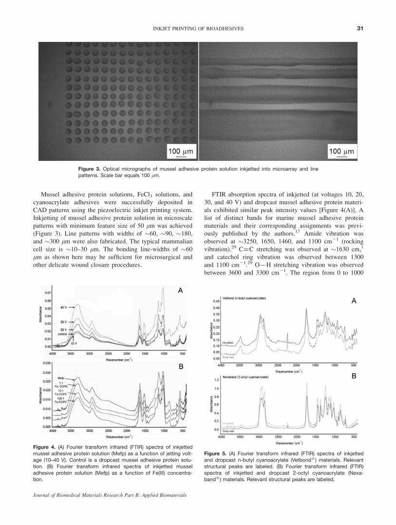
Mussel adhesive protein solutions, FeCl3 solutions, and
cyanoacrylate adhesives were successfully deposited in
CAD patterns using the piezoelectric inkjet printing system.
Inkjetting of mussel adhesive protein solution in microscale
patterns with minimum feature size of 50 lm was achieved
(Figure 3). Line patterns with widths of �60, �90, �180,
and �300 lm were also fabricated. The typical mammalian
cell size is �10–30 lm. The bonding line-widths of �60
lm as shown here may be sufficient for microsurgical and
other delicate wound closure procedures.
FTIR absorption spectra of inkjetted (at voltages 10, 20,
30, and 40 V) and dropcast mussel adhesive protein materi-
als exhibited similar peak intensity values [Figure 4(A)]. A
list of distinct bands for marine mussel adhesive protein
materials and their corresponding assignments was previ-
ously published by the authors.17 Amide vibration was
observed at �3250, 1650, 1460, and 1100 cm21 (rocking
vibration).29 C¼¼C stretching was observed at �1630 cm,1
and catechol ring vibration was observed between 1300
and 1100 cm21.29 O��H stretching vibration was observed
between 3600 and 3300 cm21. The region from 0 to 1000
Figure 3. Optical micrographs of mussel adhesive protein solution inkjetted into microarray and linepatterns. Scale bar equals 100 lm.
Figure 4. (A) Fourier transform infrared (FTIR) spectra of inkjettedmussel adhesive protein solution (Mefp) as a function of jetting volt-
age (10–40 V). Control is a dropcast mussel adhesive protein solu-
tion. (B) Fourier transform infrared spectra of inkjetted mussel
adhesive protein solution (Mefp) as a function of Fe(III) concentra-tion.
Figure 5. (A) Fourier transform infrared (FTIR) spectra of inkjetted
and dropcast n-butyl cyanoacrylate (Vetbond1) materials. Relevant
structural peaks are labeled. (B) Fourier transform infrared (FTIR)
spectra of inkjetted and dropcast 2-octyl cyanoacrylate (Nexa-band1) materials. Relevant structural peaks are labeled.
31INKJET PRINTING OF BIOADHESIVES
Journal of Biomedical Materials Research Part B: Applied Biomaterials

cm21 contains low-frequency skeletal vibrations, out-of-
plane ring deformation, wagging modes of hydrogen atoms,
wagging modes of hydroxyl groups on the catechol ring,
and wagging modes of carboxylate groups.29 Significant
differences in the absorption peak intensities were observed
in materials that were inkjetted at 10, 20, 30, and 40 V.
This result may be attributed to the increase in jetting vol-
ume that results from an increase in jetting voltage. Simi-
larly, FTIR spectra of inkjetted and dropcast (control) n-butyl cyanoacrylate [Figure 5(A)] and 2-octyl cyanoacrylate
[Figure 5(B)] revealed similar peak intensity values. FTIR
spectra of inkjetted mussel adhesive protein solutions con-
taining iron (in Fe:DOPA ratios of 1:1, 10:1, and 100:1)
and as-prepared mussel adhesive protein solution are shown
in Figure 4(B). No significant differences in peak intensity
values among these materials were observed. The FTIR
spectroscopy data suggest that piezoelectric inkjet printing
does not significantly alter the structure of the marine mus-
sel adhesive protein or cyanoacrylate adhesives.
X-ray photoelectron spectra of mussel adhesive protein
solutions (Figure 6) revealed C��C bonding (corresponding
to 285 eV), C��N bonding (corresponding to 286.1 eV),
and N��C¼¼O bonding (corresponding to 288.2 eV). The
peaks may be attributed to aliphatic and aromatic carbons
in the marine mussel adhesive protein material. Deconvolu-
tion of the C 1s peak revealed the concentration of various
Figure 6. C 1s spectra of inkjetted mussel adhesive protein solutions cured with Fe(III). X-ray photo-
electron spectra shown for (A) Mefp (0.16 mM DOPA), (B) 1:1 Fe:DOPA, (C) 10:1 Fe:DOPA, and (D) 100:1
Fe: DOPA. [Color figure can be viewed in the online issue, which is available at www.interscience.wiley.
com.]
TABLE II. C 1s Peak Deconvolution from X-Ray PhotoelectronSpectra
Assignment C��Ca C��Nb N��C¼¼Oc
Mefp (0.16 Mm DOPA) 46% 35% 19%
1:1 Fe:DOPA 44 38 18
10:1 Fe:DOPA 42 37 21
100:1 Fe:DOPA 50 36 14
a C��C peak corresponds to 285 eV.b C��N peak corresponds to 286.1 eV.c N��C¼¼O peak corresponds to 288.2 eV.
32 DORAISWAMY ET AL.
Journal of Biomedical Materials Research Part B: Applied Biomaterials

functional groups in inkjetted Fe:DOPA materials (Table II).
1:1 Fe:DOPA and 10:1 Fe:DOPA materials exhibited less
C��C bonding than as-prepared Mefp (0.16 mM DOPA)
solution. On the other hand, the 100:1 Fe:DOPA material
revealed more C��C bonding and less N��C¼¼O bonding
than as-prepared Mefp (0.16 mM DOPA) solution. X-ray
photoelectron spectra of the inkjetted protein solutions were
inconclusive in determining the role of Fe(III) in complex
formation. However, the distribution of X-ray photoelectron
peaks in the inkjet printed materials was similar to that pre-
viously observed in spectra of dropcast mussel adhesive pro-
teins (Mefp) materials.30
Atomic force microscopy has previously been used to
examine the morphology of DOPA-containing residues.31
An atomic force microscopy of inkjetted mussel adhesive
protein solution (Figure 7) revealed cross-linking upon
addition of Fe(III). In the absence of iron, mussel adhesive
protein (Mefp) [Figure 7(A)] revealed cross-linking and the
presence of some fibrous networks. Precipitation of more
complex fiber networks was observed in 1:1 Fe:DOPA and
10:1 Fe:DOPA materials [Figure 7(B,C)]. A higher degree
Figure 7. Topography-flattened atomic force micrograph of inkjetted mussel adhesive protein, 1:1
Fe:DOPA, 10:1 Fe:DOPA, and 100:1 Fe:DOPA structures. Scale bar equals 10 lm.
Figure 8. Three-dimensional representation of the surface of mussel
adhesive protein solution containing Fe(III) (1:1 Fe:DOPA) obtained
using atomic force microscopy. Image was obtained twenty-four
hours after curing. [Color figure can be viewed in the online issue,which is available at www.interscience.wiley.com.]
33INKJET PRINTING OF BIOADHESIVES
Journal of Biomedical Materials Research Part B: Applied Biomaterials

of cross-linking was observed in the 10:1 Fe:DOPA mate-
rial than in the 1:1 Fe:DOPA material. Three-dimensional
imaging of the surface of the 1:1 Fe:DOPA material
revealed high-aspect ratio fibrous network structures
(Figure 8). The height of the fibrous networks varied from
�400 to �800 nm, while the width of fibrous networks
varied from �500 to �5 lm. The 100:1 Fe:DOPA material
revealed islands of cross-linked mussel adhesive protein
material (Figure 7D). Small regions of fibrous networks
were observed within the inkjetted pattern, which result
from nonuniform distribution of mussel adhesive protein in
the inkjetted solution. Previous studies have demonstrated
iron-induced cross-linking of mussel adhesive proteins
using electron paramagnetic, infrared, and ultraviolet-visi-
ble absorption spectroscopies.22,33
Adhesion characteristics of pure marine mussel extracts
have been previously examined under different curing con-
ditions (Figure 9).34,35 Low humidity and nonoxidative con-
ditions have been shown to be critical in obtaining strong
adhesion properties in mussel adhesive proteins. Tensile
testing of inkjetted materials on full-thickness porcine skin
revealed that mussel adhesive proteins exhibit significantly
lower adhesion strength values than cyanoacrylate adhe-
sives (Figure 9). Inkjetted ethyl cyanoacrylate (Quick SetTM
Loctite1) showed highest strength among tested adhesives.
Inkjetted n-butyl cyanoacrylate (Vetbond1) and 2-octyl
cyanoacrylate (Nexaband1) patterns exhibited similar adhe-
sion strength values; however, the toxic effects of these
materials are well-documented.6–8 Addition of Fe(III) to
Mefp (0.16 mM DOPA) in 1:1 Fe:DOPA improved adhe-
sion strength. The 10:1 Fe:DOPA and 1:1 Fe:DOPA mate-
rials did not exhibit significant differences in adhesion
strength. However, the 100:1 Fe:DOPA material exhibited
lower adhesion strength values than the 1:1 Fe:DOPA and
10:1 Fe:DOPA materials. This finding suggests that the rel-
atively low adhesion strength of the 100:1 Fe:DOPA mate-
rial results from a high degree of cross-linking within the
mussel adhesive protein material, which limits interaction
with the porcine skin substrate. In addition, two different
ferric catecholate complexes may be formed at low and
high Fe:DOPA ratios.21 Previous studies have shown that
iron (III) can serve as a cross-linking agent for mussel ad-
hesive protein.36–38 For example, previous electron para-
magnetic resonance studies have confirmed iron-induced
cross-linking in precursor proteins, which is similar to that
observed in intact mussel plaques.22 The atomic force mi-
croscopy images for the 1:1 and 10:1 Fe:DOPA materials
contain a relatively high density of fibrous networks. On
the other hand, a relatively low density of fibrous networks
is observed in the Mefp (0.16 mM DOPA) and 100:1
Fe:DOPA materials. The density of fibrous networks may
be correlated with the adhesion strength observed in mussel
adhesive protein materials. These results suggest that the
extent of cross-linking and precipitation in these inkjetted
mussel adhesive protein patterns may be correlated with
iron concentration. As discussed earlier, metal-mediated
catechol complexation is thought to be responsible for the
adhesion properties of mussel adhesive proteins.
CONCLUSIONS
Mussel adhesive proteins could serve as environmentally
friendly alternatives to synthetic adhesives in biomedical,
electronics, and marine-equipment applications. Fourier
transform infrared spectra and X-ray photoelectron spectra
have shown that piezoelectric inkjetting is a nondestructive
technique that may be successfully used to dispense picoli-
ter amounts of mussel adhesive proteins and other adhe-
sives. Atomic force microscopy and adhesion testing have
demonstrated that the adhesive strength in these materials
may be correlated with the amount of iron-induced cross-
linking. CAD ink-jetting of naturally-derived mussel adhe-
sive proteins such as Mytilus edulis foot proteins may over-
come several problems associated with conventional
medical adhesives. This technology may greatly improve
wound repair in next generation eye repair, fracture fixa-
tion, wound closure, and drug delivery devices.
REFERENCES
1. Ginsberg SP, Polack FM. Cyanoacrylate tissue adhesive in oc-ular disease. Ophthalmic Surg 1972;3:126–132.
2. Landegren T, Risling M, Persson JKE. Local tissue reactionsafter nerve repair with ethyl-cyanoacrylate compared withepineural sutures. Scand J Plast Reconstr 2007;41:217–227.
3. Eskandari MM, Ozturk OG, Eskandari HG, Balli E, YilmazC. Cyanoacrylate adhesive provides efficient local drug deliv-ery. Clin Orthop Relat Res 2006;451:242–250.
4. Applebaum JS, Zalut T, Applebaum D. The use of tissueadhesion for traumatic laceration repair in the emergencydepartment. Ann Emerg Med 1993;22:1190–1192.
Figure 9. Average strength of bioadhesives inkjetted on full thick-
ness porcine skin. All samples were cured for 24 h. The difference
in strength was statistically significant for all except those indicated
by ‘‘*’’, ‘‘^’’, and ‘‘#’’ (p < 0.05). Bars indicate standard deviation ofmean strength.
34 DORAISWAMY ET AL.
Journal of Biomedical Materials Research Part B: Applied Biomaterials

5. Vanholder R, Misotten A, Roels H, Matton G. Cyanoacrylatetissue adhesive for closing skin wounds- a double-blind random-ized comparison with sutures. Biomaterials 1993;14:737–742.
6. Hauptmann M, Lubin JH, Stewart PA, Hayes RB, Blair A. Mortal-ity from lymphohematopoietic malignancies among workers informaldehyde industries. J Nat Cancer Inst 2003;95:1615–1623.
7. Pinkerton LE, Hein MJ, Stayner LT. Mortality among acohort of garment workers exposed to formaldehyde: anupdate. Occup Environ Med 2004;61:193–200.
8. Leggat PA, Smith DR, Kedjarune U. Surgical applications ofcyanoacrylate adhesives: A review of toxicity. Aust NZ JSurg 2007;77:209–213.
9. Dunn CJ, Goa KL. Fibrin sealant—A review of its use in sur-gery and endoscopy. Drugs 1999;58:863–886.
10. Sierra DH. Fibrin sealant adhesive systems: A review of theirchemistry, material properties, and clinical applications. J Bio-mater Appl 1993;7:309–352.
11. Clark RA. Fibrin glue for wound repair: Facts and fancy.Thromb Haemost 2003;90:1003–1006.
12. Schreiber GB, Busch MP, Kleinman SH, Korelitz JJ. The riskof transfusion-transmitted viral infections. New Engl J Med1996;334:1685–1690.
13. Lin Q, Gourdon D, Sun C, Holten-Andersen N, Anderson TH,Waite JH, Israelachvili JN. Adhesion mechanisms of the mus-sel foot proteins mfp-1 and mfp-3. Proc Natl Acad Sci USA2008;104:3782–3786.
14. Waite JH, Housley TJ, Tanzer ML. Peptide repeats in amussel glue protein- theme and variattions. Biochemistry1985;24:5010–5014.
15. Waite JH. Natures underwater adhesive specialist. Int J AdhesAdhes 1987;7:9–14.
16. Olivieri MP, Baier RE, Loomis RE. Surface properties ofmussel adhesive protein-component films. Biomaterials 1992;13:1000–1008.
17. Doraiswamy A, Narayan RJ, Cristescu R, Mihailescu IN,Chrisey DB. Laser processing of natural mussel adhesive pro-tein thin films. Mater Sci Eng C. 2007;27:409–413.
18. Waite JH. Adhesion a la Moule. Integr Comp Biol2002;42:1172–1180.
19. Wiegemann M. Adhesion in blue mussels (Mytilus edulis)and barnacles (genus Balanus): Mechanisms and technicalapplications. Aquat Sci 2005;67:166–176.
20. Taylor SW, Chase DB, Emptage MH, Nelson MJ, Waite JH.Ferric ion complexes of a DOPA-containing adhesive proteinfrom Mytilus edulis. Inorg Chem 1996;35:7572–7577.
21. Monahan J, Wilker JJ. Specificity of metal ion cross-linking inmarine mussel adhesives. Chem Commun 2003;14:1672–1673.
22. Sever MJ, Weisser JT, Monahan J, Srinivasan S, Wilker JJ.Metal-mediated cross-linking in the generation of a marine-mussel adhesive. Angew Chem Int Ed 2004;43:448–450.
23. Waite JH, Andersen NH, Jewhurst S, Sun CJ. Mussel adhe-sion: Finding the tricks worth mimicking. J Adhes 2005;81:297–314.
24. Lemmo AV, Rose DJ, Tisone TC. Inkjet dispensing technol-ogy: applications in drug discovery. Curr Opin Biotechnol1998;9:615–617.
25. Calvert P. Inkjet printing for materials and devices. ChemMater 2001;13:3299–3305.
26. Roth EA, Xu T, Das M, Gregory C, Hickman JJ, Boland T.Inkjet printing for high-throughput cell patterning. Biomateri-als 2004;25:3707–3715.
27. Sumerel J, Lewis J, Doraiswamy A, Deravi LF, Sewell SL,Gerdon AE, Wright DW, Narayan RJ. Piezoelectric ink jetprocessing of materials for medical and biological applica-tions. Biotechnol J 2006;1:976–987.
28. Waite JH. Precursors of quinone tanning-dopa-containing pro-teins. Method Enzymol 1995;258:1–20.
29. Weinhold M, Soubatch S, Temirov R, Rohlfing M, Jastorff B,Tautz FS, Doose C. Structure and bonding of the multifunc-tional amino acid L-DOPA on Au(110). J Phys Chem B2006;110:23756–23769.
30. Baty AM, Suci PA, Tyler BJ, Geesey GG. Investigationof mussel adhesive protein adsorption on polystyrene andpoly(octadecyl methacrylate) using angle dependent XPS.ATR-FTIR, and AFM. J Colloid Interface Sci 1996;177:307–315.
31. Lee H, Scherer NF, Messersmith PB. Single-moleculemechanics of mussel adhesion. Proc Natl Acad Sci USA2006;103:12999–13003.
32. Weisser JT, Nilges MJ, Sever MJ, Wilker JJ. EPR investiga-tion and spectral simulations of iron-catecholate complexesand iron-peptide models of marine adhesive cross-links. InorgChem 2006;45:7736–7747.
33. Sever MJ, Wilker JJ. Absorption spectroscopy and bindingconstants for first row transition metal complexes of a DOPA-containing peptide. Dalton Trans 2006;6:813–822.
34. Ninan L, Monahan J, Stroshine RL, Wilker JJ, Shi RY. Adhe-sive strength of marine mussel extracts on porcine skin. Bio-materials 2003;24:4091–4099.
35. Schnurrer J, Lehr CM. Mucoadhesive properties of the musseladhesive protein. Int J Pharm 1996;141:251–256.
36. Monahan J, Wilker JJ. Reagents for cross-linking the proteinprecursor of a marine mussel adhesive. Langmuir 2004;20:3724–3729.
37. Loizou E, Weisser JT, Schmidt G, Wilker JJ. Effects of ironion cross-linking on biopolymer hydrogels. Macromol Biosci2006;6:711–718.
38. Hight LM, Wilker JJ. Synergestic effects of metals and oxida-tion in the curing of marine mussel adhesive. J Mater Sci2007;42:8934–8942.
35INKJET PRINTING OF BIOADHESIVES
Journal of Biomedical Materials Research Part B: Applied Biomaterials