Embed Size (px)
Citation preview

RESEARCH Open Access
Inositol pyrophosphates mediated theapoptosis induced by hypoxic injury inbone marrow-derived mesenchymal stemcells by autophagyJingyu Deng1†, Chao Yang3†, Yong Wang4†, Ming Yang5, Haixu Chen6, Hongjuan Ning7, Chengzhu Wang2,Yanjun Liu2, Zheng Zhang2* and Taohong Hu2*
Abstract
Objective: To investigate the potential effect of IP7 on the autophagy and apoptosis of bone marrowmesenchymal stem cells (BM-MSCs) caused by hypoxia.
Methods: BM-MSCs isolated from adult male C57BL/6 mice were exposed to normoxic condition and hypoxicstress for 6 h, 12 h, and 24 h, respectively. Then, flow cytometry detected the characteristics of BM-MSCs.Furthermore, N6-(p-nitrobenzyl) purine (TNP) was administrated to inhibit inositol pyrophosphates (IP7). TUNELassay determined the apoptosis in BM-MSCs with hypoxia. Meanwhile, RFP-GFP-LC3 plasmid transfection andtransmission microscope was used for measuring autophagy. In addition, Western blotting assay evaluated proteinexpressions.
Results: Hypoxic injury increased the autophagy and apoptosis of BM-MSCs. At the same time, hypoxic injuryenhanced the production of IP7. Moreover, hypoxia decreased the activation of Akt/mTOR signaling pathway. Atlast, TNP (inhibitor of IP7) repressed the increased autophagy and apoptosis of BM-MSCs under hypoxia.
Conclusion: The present study indicated that hypoxia increased autophagy and apoptosis via IP7-mediated Akt/mTORsignaling pathway of BM-MSCs. It may provide a new potential therapy target for myocardial infarction (MI).
Keywords: Hypoxia, Bone marrow mesenchymal stem cells (BM-MSCs), Inositol pyrophosphates (IP7), Autophagy,Apoptosis, Akt/mTOR signaling pathway
IntroductionAlthough the treatment of coronary heart disease hasmade rapid progress, ischemic heart disease induced bycoronary artery occlusion is a key cause of morbidityand mortality around the world [1–4]. At present, treat-ment with stem cell has a potential effect on cardiovas-cular regeneration of ischemic heart disease. And it hasbeen considered as a promising therapeutic method.Bone marrow mesenchymal stem cells (BM-MSCs) arewidely applied for regenerative medicine because of their
plasticity and effectiveness [5–8]. However, in practice,the poor activity and function of donor cells greatly limitthe efficiency of stem cell transplantation [9]. Thus,excessive apoptosis, caused by harsh microenvironment,has been regarded as the main reason for the donor celldeath [8].Autophagy, a self-degrading process that responds to
stress, plays a key effect on balancing sources of energyand resetting misfolded proteins. Therefore, autophagyis considered as a protective and adaptive mechanism topromote cell survival under normal conditions [10, 11].However, most autophagosomes (over-autophagosomeformation) are associated with autophagic cell death[12]. In the heart, a certain degree of autophagy is ob-served at baseline, and its expression is upregulated by
© The Author(s). 2019 Open Access This article is distributed under the terms of the Creative Commons Attribution 4.0International License (http://creativecommons.org/licenses/by/4.0/), which permits unrestricted use, distribution, andreproduction in any medium, provided you give appropriate credit to the original author(s) and the source, provide a link tothe Creative Commons license, and indicate if changes were made. The Creative Commons Public Domain Dedication waiver(http://creativecommons.org/publicdomain/zero/1.0/) applies to the data made available in this article, unless otherwise stated.
* Correspondence: [email protected];[email protected]†Jingyu Deng, Chao Yang and Yong Wang contributed equally to this work.2Department of Cardiology, The Rocket Army Special Medical Center of thePLA, Beijing 100088, ChinaFull list of author information is available at the end of the article
Deng et al. Stem Cell Research & Therapy (2019) 10:159 https://doi.org/10.1186/s13287-019-1256-3
pathological stimuli such as myocardial ischemia [13, 14].Currently, our previous study demonstrated that hypoxiaincreased the autophagy and apoptosis of BM-MSCstime-dependently. Furthermore, autophagy regulated theincreased apoptosis in BM-MSCs under hypoxia viaAMPK/mTOR signal pathway [8].Inositol polyphosphates are a key signaling molecules in
cells [15]. Although most of the more than 30 inositolpolyphosphates in mammalian cells have unknown physio-logical functions, a group of higher inositol phosphatesincluding energetic pyrophosphate bonds and inositol py-rophosphates (IP7) has been identified [16–18]. IP7, gener-ated by inositol hexakisphosphate (IP6) through Inositolhexakisphosphate kinases (IP6Ks), is related to diversefunctions containing vesicle transport and chemotaxis[19–24]. Recently, Chakraborty et al. [25] found thatIP6k1 gene knockout can increase Akt activity, con-firming that IP7 is a physiological inhibitor of Aktsignal pathway. In addition, study has also showedthat hypoxic injury increased IP7 formation in MSCs,which inhibits Akt activation [26].Previous research has found an increase in the inositol
pyrophosphate signaling induces the increased autopha-gosomes. Furthermore, they thought that IP7 plays a keyrole in regulating autophagy [15]. However, the specificmechanism of IP7 and autophagy that contributes toapoptosis in MSCs under hypoxic injury is still unclear.Thus, our present study aimed to elucidate the potentialeffect of IP7 on the autophagy with hypoxic injury,which may provide optimal approaches to improve thetherapeutic effect of MSCs for ischemic heart disease.
MethodsAnimalsWe selected adult male C57BL/6 mice as experimental an-imals (from the Laboratory Animal Research Center ofRocket Army Special Medical Center of Chinese People’sLiberation Army) to isolate BM-MSCs. Before any experi-ment, mice were placed into temperature-controlled ani-mal facilities for 12 h of light/dark cycle (light cycle, 8:00a.m. to 8:00 p.m.), with tap water and rodent chowprovided 2 weeks ad libitum. The Animal Care and UseCommittee of the Rocket Army Special Medical Center ofthe PLA (ID: 5034) approved all the experimental proce-dures. And experimental procedures were conformed tothe Guidelines for the Nursing and Use of ExperimentalAnimals published by the Press of the National Academyof Sciences.
Isolation and culture of BM-MSCsWe used a modified procedure to isolate and expand ofBM-MSCs [27]. In brief, we flushed bone marrow fromthe femoral and tibia with phosphate-buffered saline(PBS). Dulbecco’s modified Eagle’s medium (DMEM)
supplemented with 20% fetal bovine serum (FBS) and1% penicillin/streptomycin medium re-suspended cellgranules after being passed through a 70-mm filter andcentrifugation at 1200 rpm for 5 min. Different treat-ments with third-passage MSCs avoided contaminationwith other types of cell.
Characteristics of BM-MSCsFlow cytometry tested characteristics of BM-MSCs, suchas CD34-, CD45-, CD44+, and CD90+. And we performedin vitro differentiation assay [26, 27]. Briefly, a FACSCalibur system (BD) treated BM-MSCs according to themanufacturer’s guideline after being incubated withmonoclonal PE-binding antibodies against special CDmarkers (BD, San Jose, CA, USA) for 1 h.The in vitro differentiation assays were used for perform-
ing MSCs [26, 27]. Adipogenic media (aMEM containing10% FCS, 50mM indomethacin, 1% antibiotics, 1 mMdexamethasone, and 0.5mM IBMX) were added into theconfertus layer of MSCs for 21 days in order to per-form adipogenic differentiation. Then, a working solu-tion of Oil Red O stained the adipogenic cells for 15min at room temperature. Twenty-one-day osteogenicdifferentiation of MSCs was induced concurrently by osteo-genic medium (OM, 10% FBS, 10mM b-glycerophosphate,1000 nM dexamethasone, and 0.2mM ascorbic acid ina-MEM). Alizarin red S staining was used for estimatingthe degree of extracellular matrix calcification. Further-more, we induced chondrogenic differentiation of BM-MSCs as described previously [28]. In brief, chondrogenicmedium (containing 50 mg/l ascorbic acid, 100 nmol/ldexamethasone, 1% fetal bovine serum, low-glucoseDMEM, 100 mg/l sodium pyruvate, 40 mg/l L-proline,and 1.0% indometacin) was added into BM-MSCs for21 days. Meanwhile, Alcian blue stain (0.5 ml, 30 minat room temperature) was performed to evaluate thechondrogenic differentiation.
Hypoxia/serum deprivation injuryHypoxia/ serum deprivation injury was used to performthe hypoxic stress of BM-MSCs as described previously[26]. In short, BM-MSCs were cultivated with 20% FBS.After being replaced in Hanks buffer, BM-MSCs weretreated with hypoxic condition (94% N2–5% CO2–1%O2) with an anaerobic system (Thermo Forma) at 37 °Cfor 6, 12, and 24 h, respectively. BM-MSCs in the controlgroup were maintained at normal (95% air–5% CO2)condition for equal periods.
Treatment of cellsTo regulate the autophagy level in BM-MSCs, IP7 in-hibitor N6-(p-nitrobenzyl) purine (10 μM, TNP) wasadded to further explore the molecular mechanism of
Deng et al. Stem Cell Research & Therapy (2019) 10:159 Page 2 of 13

autophagy in BM-MSCs with normal and hypoxic treat-ments. Treatments of cells were done in duplicate [27].
Determination of apoptosis in BM-MSCsTerminal-deoxynucleotidyl transferase mediated-dUTPnick-end labeling (TUNEL) assay confirmed the apop-tosis of MSCs with an assay kit (In Situ Cell DeathDetection Kit; Roche Diagnostics) according to the man-ufacturer’s guideline [29]. In brief, BM-MSCs were incu-bated with TdT and fluorescein-labeled dUTP for 45 minat 37 °C after different treatments. Then, 4,6-diamidi-no-2-phenylindole (DAPI) was used for identifying nu-cleus. We took photographs with confocal microscopy(Olympus Fluoview 2000). At the same time, we calcu-lated the percentage of apoptotic cells. And then, wecounted five random fields for analysis in each group.We did all assays blindly.
Measurement of autophagy in BM-MSCsFor assessing the autophagy of MSCs, we transfected themRFP-GFP-LC3 plasmids (Hanbio biotechnology Co., Ltd.,Shanghai, China) into BM-MSCs as described previously[30, 31]. In brief, we took microphotographs of BM-MSCsafter different treatments. Furthermore, we counted fiverandom fields and calculated the percentages of cells withRFP-GFP-LC3 punctate. We did all assays blindly. At thesame time, Western blotting detected the expressions ofLC-3 and autophagy-associated protein (P62 and Beclin-1).
Western blot assayWe harvested and dissolved BM-MSCs in protein lysisbuffer (Sigma). Equivalent protein (50 mg/lane) was sep-arated by electrophoresis on 12% SDS-PAGE gels at 120V for 90 min and then electrophoretically transferredonto PVDF membranes at 100 mV for 1.5 h. Cellularmembranes were subjected to immune-blotting with pri-mary antibodies overnight at the temperature 4 °C afterblocked in 5% nonfat dry milk (BD Biosciences) at roomtemperature for 1 h. After incubation with appropriatesecondary antibody binding to horseradish peroxidase,we used an enhanced chemiluminescene system (Amer-sham Bioscience) to visualize blots bands. Furthermore,we used VisionWorks LS, version 6.7.1, to determinedensitometric analysis of Western blots [8].The primary antibodies were used: rabbit anti-mouse
LC-3 (1:500, Cellular Signal Technology), rabbit anti-mouseBeclin-1 (1:500, Cellular Signaling Technology), rabbitanti-mouse P62 (1:500, Cellular Signal Technology), rabbitanti-mouse phosphorylated (Thr172) and rabbit anti-mousetotal AKt (1:200, Abcam), rabbit anti-mice mTOR (1:500,Abcam), rabbit anti-mouse p-mTOR (1:200, Abcam),rabbit anti-mouse p70S6k (1:500, Abcam), rabbitanti-mouse S6 (1:500, Abcam), rabbit anti-micep-p70S6k (1:200, Abcam), rabbit anti-mouse p-S6
(1:200, Abcam), rabbit anti-mouse caspase-3 and cleavedcaspase-3 (1:500, Abcam), and rabbit anti-mouse β-actin(12,000, Abcam).
Statistics analysisOur results were shown with the mean ± SEM. Prism 5.0(GraphPad Software Inc., San Diego, CA, USA) was usedfor the statistical analyses. The different groups of thisstudy were compared by the homogeneity tests andone-way ANOVA. P value < 0.05 was considered as astatistical significance.
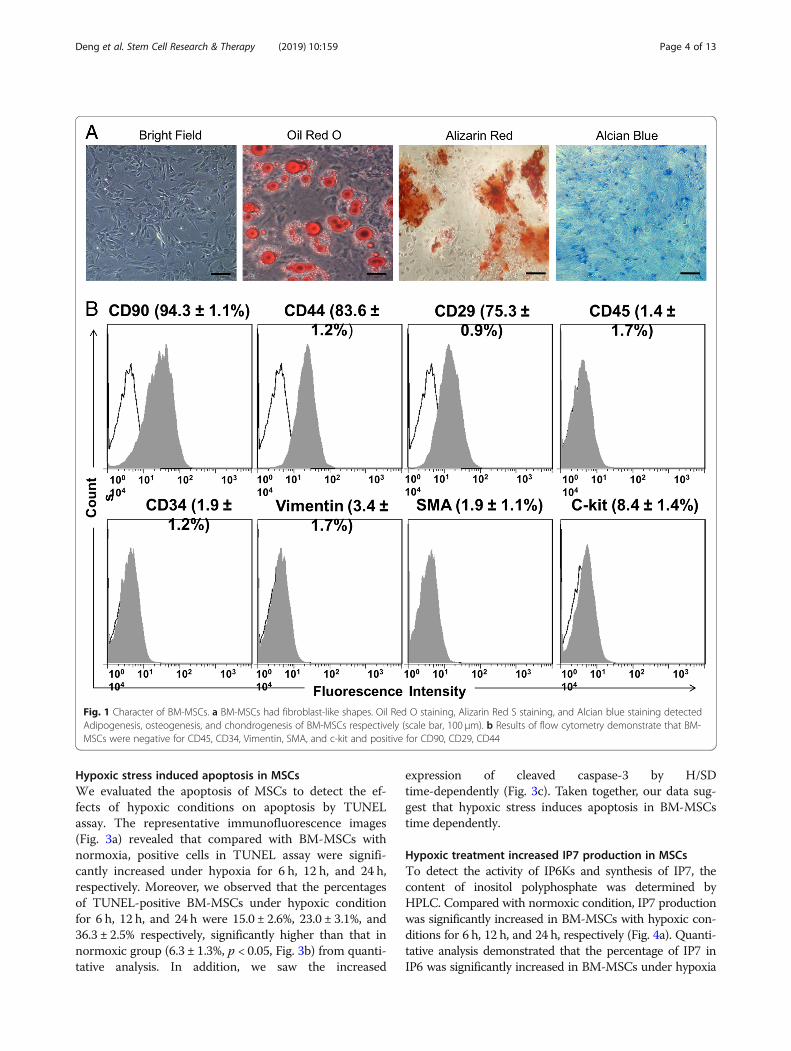
ResultsBiologic character of BM-MSCsWe used in vitro multi-lineage differentiation and flowcytometry analysis to identify and analyze the characterof BM-MSCs. We observed BM-MSCs of fibroblast-likeshapes (Fig. 1a). To set up the multi-lineage differenti-ation proficiency of BM-MSCs, we used adipogenic andosteoblastogenic media to incubate cells for 21 days re-spectively. The result of Oil Red O staining revealed thatabout 70% of BM-MSCs possessed an adipocytes pheno-type. Moreover, we observed BM-MSCs differentiatinginto osteogenic cells from the images of alizarin red Sstaining for calcium deposit. Furthermore, Alcian bluestaining indicated the chondrogenic differentiation ofBM-MSCs (Fig. 1a). All of these results demonstrated thatBM-MSC had proficiency of multi-lineage differentiation.Results of flow cytometry analysis revealed that MSCswere homogeneously positive for BM-MSC markersCD29, CD44, and CD90 and negative for Vimention,c-Kit, SMA, CD45, and CD34 (Fig. 1b).
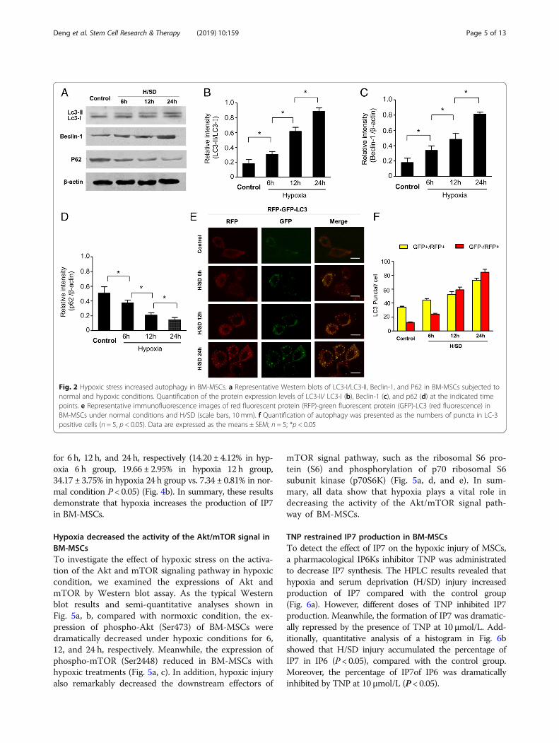
Hypoxia increased autophagy of MSCsWe assessed the expressions of autophagy regulating signalproteins to explore the autophagy level under hypoxic con-dition. Western blot revealed that the expression of LC3-IIand Beclin-1 was increased significantly in BM-MSCs withhypoxia time-dependently, while the expressions of p62were decreased (Fig. 2a–d). Moreover, we transfectedBM-MSCs with RFP-GFP-LC3 and tracked the expressionsof LC3. After hypoxic treatment, not only were the num-bers of green and red puncta both significantly higher(Fig. 2e), but the yellow dots were also typically increasedin the merged images, which show that autophago-somes were increased. These results imply that autoph-agic flux of BM-MSCs was enhanced in hypoxiccondition. Meanwhile, semi-quantitative analysis re-vealed that the numbers of puncta in LC-3-positivecells gradually increased along with the exposure timeto hypoxic treatment compared with the control group(Fig. 2f ). Taken together, results suggest that hypoxiaincreased the autophagy in BM-MSCs.
Deng et al. Stem Cell Research & Therapy (2019) 10:159 Page 3 of 13
Hypoxic stress induced apoptosis in MSCsWe evaluated the apoptosis of MSCs to detect the ef-fects of hypoxic conditions on apoptosis by TUNELassay. The representative immunofluorescence images(Fig. 3a) revealed that compared with BM-MSCs withnormoxia, positive cells in TUNEL assay were signifi-cantly increased under hypoxia for 6 h, 12 h, and 24 h,respectively. Moreover, we observed that the percentagesof TUNEL-positive BM-MSCs under hypoxic conditionfor 6 h, 12 h, and 24 h were 15.0 ± 2.6%, 23.0 ± 3.1%, and36.3 ± 2.5% respectively, significantly higher than that innormoxic group (6.3 ± 1.3%, p < 0.05, Fig. 3b) from quanti-tative analysis. In addition, we saw the increased
expression of cleaved caspase-3 by H/SDtime-dependently (Fig. 3c). Taken together, our data sug-gest that hypoxic stress induces apoptosis in BM-MSCstime dependently.
Hypoxic treatment increased IP7 production in MSCsTo detect the activity of IP6Ks and synthesis of IP7, thecontent of inositol polyphosphate was determined byHPLC. Compared with normoxic condition, IP7 productionwas significantly increased in BM-MSCs with hypoxic con-ditions for 6 h, 12 h, and 24 h, respectively (Fig. 4a). Quanti-tative analysis demonstrated that the percentage of IP7 inIP6 was significantly increased in BM-MSCs under hypoxia
Fig. 1 Character of BM-MSCs. a BM-MSCs had fibroblast-like shapes. Oil Red O staining, Alizarin Red S staining, and Alcian blue staining detectedAdipogenesis, osteogenesis, and chondrogenesis of BM-MSCs respectively (scale bar, 100 μm). b Results of flow cytometry demonstrate that BM-MSCs were negative for CD45, CD34, Vimentin, SMA, and c-kit and positive for CD90, CD29, CD44
Deng et al. Stem Cell Research & Therapy (2019) 10:159 Page 4 of 13
for 6 h, 12 h, and 24 h, respectively (14.20 ± 4.12% in hyp-oxia 6 h group, 19.66 ± 2.95% in hypoxia 12 h group,34.17 ± 3.75% in hypoxia 24 h group vs. 7.34 ± 0.81% in nor-mal condition P < 0.05) (Fig. 4b). In summary, these resultsdemonstrate that hypoxia increases the production of IP7in BM-MSCs.
Hypoxia decreased the activity of the Akt/mTOR signal inBM-MSCsTo investigate the effect of hypoxic stress on the activa-tion of the Akt and mTOR signaling pathway in hypoxiccondition, we examined the expressions of Akt andmTOR by Western blot assay. As the typical Westernblot results and semi-quantitative analyses shown inFig. 5a, b, compared with normoxic condition, the ex-pression of phospho-Akt (Ser473) of BM-MSCs weredramatically decreased under hypoxic conditions for 6,12, and 24 h, respectively. Meanwhile, the expression ofphospho-mTOR (Ser2448) reduced in BM-MSCs withhypoxic treatments (Fig. 5a, c). In addition, hypoxic injuryalso remarkably decreased the downstream effectors of
mTOR signal pathway, such as the ribosomal S6 pro-tein (S6) and phosphorylation of p70 ribosomal S6subunit kinase (p70S6K) (Fig. 5a, d, and e). In sum-mary, all data show that hypoxia plays a vital role indecreasing the activity of the Akt/mTOR signal path-way of BM-MSCs.
TNP restrained IP7 production in BM-MSCsTo detect the effect of IP7 on the hypoxic injury of MSCs,a pharmacological IP6Ks inhibitor TNP was administratedto decrease IP7 synthesis. The HPLC results revealed thathypoxia and serum deprivation (H/SD) injury increasedproduction of IP7 compared with the control group(Fig. 6a). However, different doses of TNP inhibited IP7production. Meanwhile, the formation of IP7 was dramatic-ally repressed by the presence of TNP at 10 μmol/L. Add-itionally, quantitative analysis of a histogram in Fig. 6bshowed that H/SD injury accumulated the percentage ofIP7 in IP6 (P < 0.05), compared with the control group.Moreover, the percentage of IP7of IP6 was dramaticallyinhibited by TNP at 10 μmol/L (P < 0.05).
Fig. 2 Hypoxic stress increased autophagy in BM-MSCs. a Representative Western blots of LC3-I/LC3-II, Beclin-1, and P62 in BM-MSCs subjected tonormal and hypoxic conditions. Quantification of the protein expression levels of LC3-II/ LC3-I (b), Beclin-1 (c), and p62 (d) at the indicated timepoints. e Representative immunofluorescence images of red fluorescent protein (RFP)-green fluorescent protein (GFP)-LC3 (red fluorescence) inBM-MSCs under normal conditions and H/SD (scale bars, 10 mm). f Quantification of autophagy was presented as the numbers of puncta in LC-3positive cells (n = 5, p < 0.05). Data are expressed as the means ± SEM; n = 5; *p < 0.05
Deng et al. Stem Cell Research & Therapy (2019) 10:159 Page 5 of 13
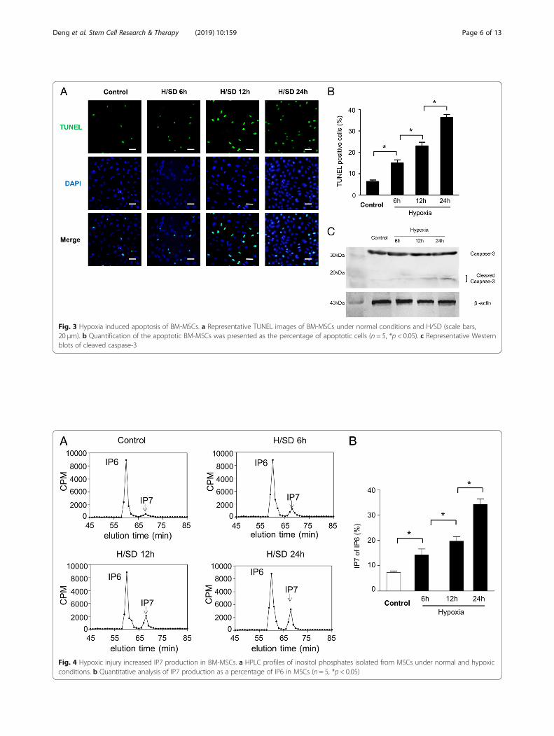
Fig. 3 Hypoxia induced apoptosis of BM-MSCs. a Representative TUNEL images of BM-MSCs under normal conditions and H/SD (scale bars,20 μm). b Quantification of the apoptotic BM-MSCs was presented as the percentage of apoptotic cells (n = 5, *p < 0.05). c Representative Westernblots of cleaved caspase-3
Fig. 4 Hypoxic injury increased IP7 production in BM-MSCs. a HPLC profiles of inositol phosphates isolated from MSCs under normal and hypoxicconditions. b Quantitative analysis of IP7 production as a percentage of IP6 in MSCs (n = 5, *p < 0.05)
Deng et al. Stem Cell Research & Therapy (2019) 10:159 Page 6 of 13
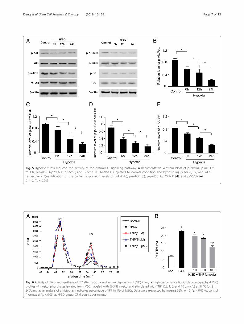
Fig. 5 Hypoxic stress reduced the activity of the Akt/mTOR signaling pathway. a Representative Western blots of p-Akt/Ak, p-mTOR/mTOR, p-p70S6 K/p70S6 K, p-S6/S6, and β-actin in BM-MSCs subjected to normal condition and hypoxic injury for 6, 12, and 24 h,respectively. Quantification of the protein expression levels of p-Akt (b), p-mTOR (c), p-p70S6 K/p70S6 K (d), and p-S6/S6 (e)(n = 5, *p < 0.05)
Fig. 6 Activity of IP6Ks and synthesis of IP7 after hypoxia and serum deprivation (H/SD) injury. a High-performance liquid chromatography (HPLC)profiles of inositol phosphates isolated from MSCs labeled with [2-3H] inositol and stimulated with TNP (0.5, 1, 5, and 10 μmol/L) at 37 °C for 2 h.b Quantitative analysis of a histogram indicates percentage of IP7 in IP6 of MSCs. Data were expressed by mean ± SEM. n = 5, *p < 0.05 vs. control(normoxia), #p < 0.05 vs. H/SD group. CPM counts per minute
Deng et al. Stem Cell Research & Therapy (2019) 10:159 Page 7 of 13
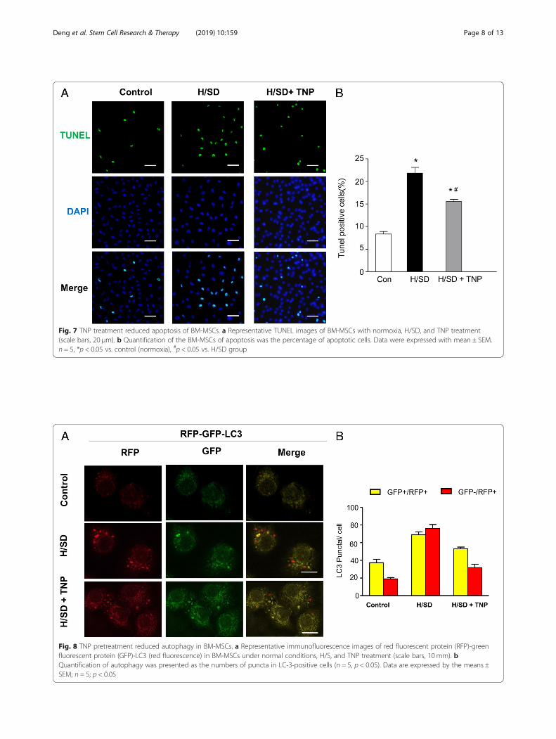
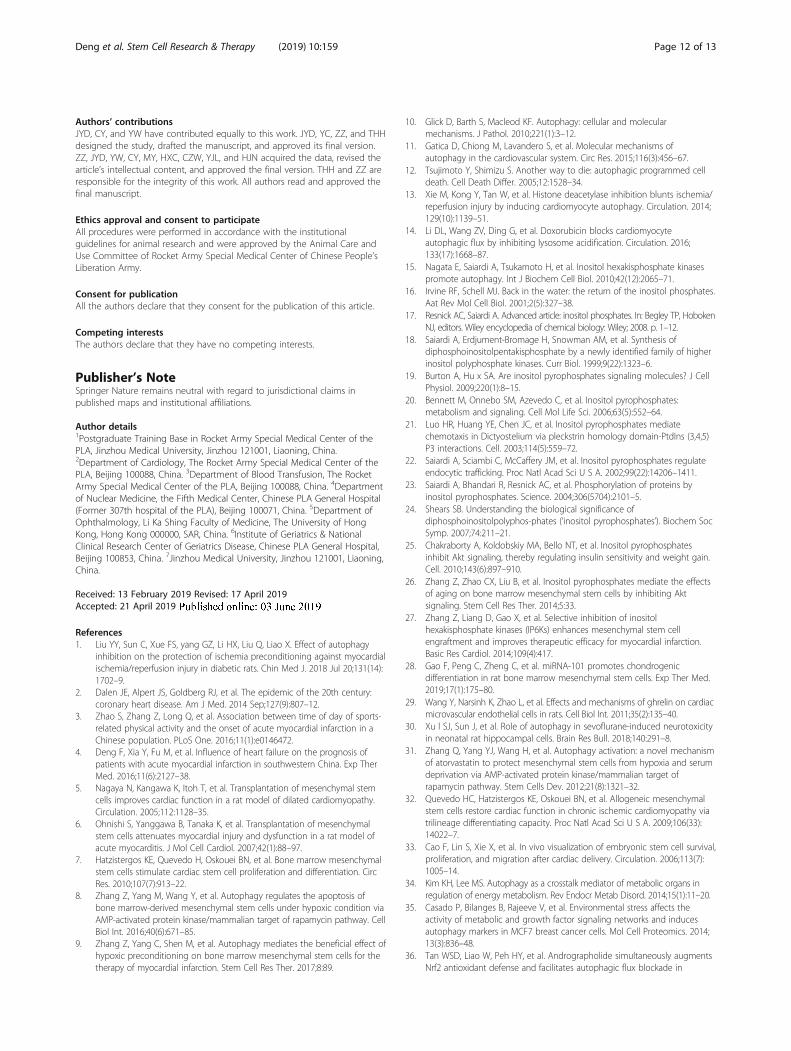
Fig. 8 TNP pretreatment reduced autophagy in BM-MSCs. a Representative immunofluorescence images of red fluorescent protein (RFP)-greenfluorescent protein (GFP)-LC3 (red fluorescence) in BM-MSCs under normal conditions, H/S, and TNP treatment (scale bars, 10 mm). bQuantification of autophagy was presented as the numbers of puncta in LC-3-positive cells (n = 5, p < 0.05). Data are expressed by the means ±SEM; n = 5; p < 0.05
Fig. 7 TNP treatment reduced apoptosis of BM-MSCs. a Representative TUNEL images of BM-MSCs with normoxia, H/SD, and TNP treatment(scale bars, 20 μm). b Quantification of the BM-MSCs of apoptosis was the percentage of apoptotic cells. Data were expressed with mean ± SEM.n = 5, *p < 0.05 vs. control (normoxia), #p < 0.05 vs. H/SD group
Deng et al. Stem Cell Research & Therapy (2019) 10:159 Page 8 of 13

TNP decreased apoptosis in BM-MSCsTo discover the effects of IP7 on the apoptosis inducedby hypoxia, we evaluated the apoptosis of BM-MSCsafter TNP administration by the TUNEL assay. Therepresentative immunofluorescence images of TUNEL(Fig. 7a) demonstrated that compared with BM-MSCsin control, TUNEL-positive cells were significantlyincreased under hypoxia. Moreover, TUNEL-positivecells were reduced after TNP treatment compared withthat in H/SD group. In addition, the quantitative ana-lysis indicated that H/SD significantly induced apop-tosis of MSCs (21.87 ± 2.21% vs. 8.37 ± 1.02% in normalcondition, p < 0.01). However, TNP at 10 μM signifi-cantly decreased the percentage of apoptotic MSCscompared with H/SD group (15.53 ± 0.84% vs. 21.87 ±2.21%, p < 0.05) (Fig. 7b).
TNP pre-treated reduced BM-MSC autophagyTo investigate the effects of IP7 on the apoptosisinduced by hypoxia, we evaluated the autophagy ofBM-MSCs after TNP administration. Immunofluores-cence images in Fig. 8a showed that the numbers ofgreen and red dots both reduced after TNP pre-treatment and the yellow puncta were also typicallydecreased after TNP group vs. H/SD group in the
merged images. Furthermore, the result of quantita-tive analysis revealed that compared with that in H/SD group, the numbers of puncta in LC-3-positivecells decreased in TNP treatment (Fig. 8b). Overall,our results demonstrated that TNP restrained au-tophagy in BM-MSCs.
TNP treatment increased the activity of the Akt/mTORsignal in BM-MSCsWe detected the effect of TNP pre-treatment on theactivity of the Akt and mTOR signal with Westernblot assays. The results of Western blot and semi-quantitative analyses (Fig. 9a, b) showed that the ex-pression levels of phospho-Akt (Ser473) were signifi-cantly decreased under H/SD condition. However, theexpression levels of phospho-Akt (Ser473) were re-markably increased in BM-MSCs with TNP treatmentcompared to that in H/SD BM-MSCs. Meanwhile, theexpression of phospho-mTOR (Ser2448) reduced inBM-MSCs with hypoxia. However, we observed the in-creased expression of phospho-mTOR (Ser2448) inBM-MSCs with TNP treatments (Fig. 9a, c). In addition,hypoxic injury also significantly depressed the phosphory-lations of the downstream effectors of mTOR signal, suchas ribosomal S6 and p70S6K. Nevertheless, TNP
Fig. 9 TNP enhanced the activity of the Akt/mTOR signaling pathway. a Western blots of p-Akt/Ak, p-mTOR/mTOR, p-p70S6 K/p70S6 K, p-S6/S6,and β-actin of BM-MSCs subjected to normal condition, H/SD injury, and TNP treatment. Quantification of the protein expression levels of p-Akt(b), p-mTOR (c), p-p70S6 K/p70S6 K (d), and p-S6/S6 (e) (n = 5, *p < 0.05)
Deng et al. Stem Cell Research & Therapy (2019) 10:159 Page 9 of 13

pre-treated increased the expressions of p70S6K and S6 ofBM-MSCs (Fig. 9a, d, and e). Taken together, these dataindicated that TNP improved the activation of the Akt/mTOR signal in BM-MSCs.
DiscussionAlthough bone marrow mesenchymal stem cells(BM-MSCs) were considered as a potential cellularsource for therapy of myocardial infarction (MI) [32],the poor survival rate of transplanted cells was a majorchallenge for therapeutic efficacy [33]. Previous studieshave found that autophagy may be a mechanism to regu-late the death of MSCs after transplantation. Thus, thekey to improve the treatment of MSCs is to regulate
autophagy and promote the survival of MSCs aftertransplantation. IP7 is a newly discovered upstream sig-naling molecule regulating Akt, which play a crucialrole in regulating autophagy. In our present study, weobserved that hypoxia increased the autophagy andapoptosis of BM-MSCs time-dependently. Moreover,hypoxic stress also increased IP7 production in MSCs.Furthermore, restraining IP7 production by TNP re-duced the autophagy and apoptosis of BM-MSCscaused by hypoxia. In short, our results for the firsttime implied that selective inhibition of IP6Ks has theprotective effects of MSCs from hypoxic injury presum-ably via activity of the Akt signal. Furthermore, IP6Ksinhibition may be a potential strategy of optimizing
Fig. 10 Proposed mechanism of autophagy and apoptosis of BM-MSCs caused by hypoxic stress. Hypoxia increases the production of IP7, whichinhibits activation of Akt and subsequently restrains mTOR. Moreover, repressed mTOR directly enhances autophagy. Therefore, hypoxic injuryinduces autophagy regulating apoptosis via Akt/mTOR signaling pathway mediated by IP7 in BM-MSCs
Deng et al. Stem Cell Research & Therapy (2019) 10:159 Page 10 of 13

mesenchymal stem cells therapy for patients with MI(Fig. 10).Though autophagy plays an essential role in maintaining
homeostasis or normal function in basic catabolic mech-anism, it also can be dramatically induced and upregulatedby unfavorable stimulus, such as hypoxia [34, 35]. There-fore, autophagy, a paradox that protects and damages cellsurvival, depends on the environment. Our previous stud-ies demonstrated that autophagy was increased inBM-MSCs under hypoxic injury [8, 36]. In our study, wetransfected BM-MSCs with RFP-GFP-LC3 to evaluate theautophagy of MSCs. The red LC3B puncta indicates asuccessful fusion among autophagosomes and lysosomes,because the acidic pH of the lysosome inhibits theacid-sensitive GFP (green color) on LC3B. Moreover, theyellow LC3B points show an impaired fusion betweenautophagosome and lysosome, because the acid-sensitiveGFP will remain intact together with the acid-insensitiveRFP (red color) [36]. Our results revealed that hypoxicstress increased the punctate LC3 in BM-MSCs, whichenhances the expression of LC3-I/LC3-II and Beclin-1.Moreover, previous studies demonstrated that the p62protein becomes incorporated into the completed autopha-gosome and is degraded in autolysosomes. Therefore, theexpression of p62 is negatively correlated with autophagicactivity [37, 38]. Our result showed that the expression ofp62 was decreased under hypoxia. Taken together, theseresults indicated that hypoxia increased the autophagy ofMSCs. Furthermore, the TUNEL assay showed that apop-tosis in BM-MSCs was significantly increased underhypoxia time-dependently. Meanwhile, we also observedthe increased expression of cleaved caspase-3 by H/SDtime-dependently. In summary, these data suggested thathypoxia increased autophagy and apoptosis of BM-MSCs,which shows no difference with previous study [8, 39].Akt signal pathway played an important role in regulat-
ing energy metabolism, cell survival, and oxidative stress[40]. Studies showed that mTOR repressed the cellularcatabolic pathway, containing autophagy [41]. Similarly,our results demonstrated that hypoxia decreased signifi-cantly the expressions of pAkt and pmTOR in BM-MSCs.Meanwhile, hypoxic injury decreased the phosphorylationof mTOR substrates, such as p70S6K and S6, which showsno difference with previous study [8]. Overall, these re-sults suggested that hypoxia downregulated the activationof Akt and mTOR signal pathways.Inositol polyphosphates are a diverse group of signal
molecules, produced through the sequential phosphory-lations with inositol polyphosphate kinases (IPKs) from IP3[42]. IP3, regulating intracellular calcium release, sequen-tially phosphorylated to generate IP6 and 5-diphospho-ino-sitolpentakisphosphate (IP7). IP6 produces IP7 catalyzed byinositol hexakiphosphate kinase (IP6Ks) [16, 18]. Al-though the physiological functions of IPs remain
poorly clear, studies confirmed that IP7 regulatedmany physiological functions, including apoptosis [43,44]. In addition, previous study revealed that increasedIP7 signaling induces the emergence of autophago-somes, indicating that IP7 promoted autophagy [15].Our data also indicated that hypoxic injury increasedproduction of IP7, autophagy, and apoptosis. Further-more, we found that TNP, a selective inhibition ofIP6Ks, reduced the information of IP7 and decreasedautophagy and apoptosis. Taken together, all data sug-gested that hypoxia upregulated IP7, which activatesautophagy and apoptosis of BM-MSCs.IP7 seems to inhibit Akt signaling and regulate apop-
tosis. Chakraborty also confirmed that IP7 is a physio-logical inhibitor of Akt signaling pathway [25]. Thus,our results showed that IP7 mediated the effects onBM-MSCs with hypoxia by inhibiting Akt/mTOR signal-ing, which was in accordance with previous studies [8].Although present study has some clinical significance,
this also includes a lot of limitations. The cellular H/SDmodel was considered to be useful for excluding the in-fluence of neural and humoral factors in vivo; the modelwas limited as an artificial experimental model that can-not fully simulate the ischemic and inflammatory envir-onment in vivo. In addition, the physiological functionof IP7 has not been fully clear. Therefore, future studiesare essential to determine the exact mechanism in orderto understand the hypoxic process of BM-MSCs.
ConclusionsIn a word, the current study demonstrated that hypoxicinjury increased autophagy and apoptosis by IP7, whichinhibit Akt/mTOR signaling pathway in BM-MSCs,which may provide new ideas for the treatment of MI.
AbbreviationsAkt: Protein kinase B; BM-MSCs: Bone marrow mesenchymal stem cells;DAPI: 4,6-Diamidino-2-phenylindole; FBS: Fetal bovine serum; H/SD: Hypoxiaand serum deprivation; IP6: Inositol hexakisphosphate; IP6KI: Inositolhexakisphosphate kinases; IP7: Inositol pyrophosphates; MI: Myocardialinfarction; mTOR: Mammalian target of rapamycin; p70S6K: p70 ribosomal S6subunit kinase; RFP-GFP-LC3: Red fluorescent protein-microtubule-Greenfluorescent protein-microtubule associated protein lightchain3; TNP: N6-(p-nitrobenzyl)purine; TUNEL: Terminal deoxynucleotidy transferase-mediateddUTP nick-end labeling
AcknowledgementsNot applicable.
FundingThis work was supported by the National Nature Science Foundation ofChina (No.31600681), Military Medical Science and Technology YouthTraining Program (No. 17QNP029), Beijing Nova Program of Science andTechnology (No. xx2017103), and the Translational Medicine Project of theChinese PLA General Hospital (No. 2016TM-012).
Availability of data and materialsThe data sets supporting the results of this article are included within thearticle and its additional files.
Deng et al. Stem Cell Research & Therapy (2019) 10:159 Page 11 of 13
Authors’ contributionsJYD, CY, and YW have contributed equally to this work. JYD, YC, ZZ, and THHdesigned the study, drafted the manuscript, and approved its final version.ZZ, JYD, YW, CY, MY, HXC, CZW, YJL, and HJN acquired the data, revised thearticle’s intellectual content, and approved the final version. THH and ZZ areresponsible for the integrity of this work. All authors read and approved thefinal manuscript.
Ethics approval and consent to participateAll procedures were performed in accordance with the institutionalguidelines for animal research and were approved by the Animal Care andUse Committee of Rocket Army Special Medical Center of Chinese People’sLiberation Army.
Consent for publicationAll the authors declare that they consent for the publication of this article.
Competing interestsThe authors declare that they have no competing interests.
Publisher’s NoteSpringer Nature remains neutral with regard to jurisdictional claims inpublished maps and institutional affiliations.
Author details1Postgraduate Training Base in Rocket Army Special Medical Center of thePLA, Jinzhou Medical University, Jinzhou 121001, Liaoning, China.2Department of Cardiology, The Rocket Army Special Medical Center of thePLA, Beijing 100088, China. 3Department of Blood Transfusion, The RocketArmy Special Medical Center of the PLA, Beijing 100088, China. 4Departmentof Nuclear Medicine, the Fifth Medical Center, Chinese PLA General Hospital(Former 307th hospital of the PLA), Beijing 100071, China. 5Department ofOphthalmology, Li Ka Shing Faculty of Medicine, The University of HongKong, Hong Kong 000000, SAR, China. 6Institute of Geriatrics & NationalClinical Research Center of Geriatrics Disease, Chinese PLA General Hospital,Beijing 100853, China. 7Jinzhou Medical University, Jinzhou 121001, Liaoning,China.
Received: 13 February 2019 Revised: 17 April 2019Accepted: 21 April 2019
References1. Liu YY, Sun C, Xue FS, yang GZ, Li HX, Liu Q, Liao X. Effect of autophagy
inhibition on the protection of ischemia preconditioning against myocardialischemia/reperfusion injury in diabetic rats. Chin Med J. 2018 Jul 20;131(14):1702–9.
2. Dalen JE, Alpert JS, Goldberg RJ, et al. The epidemic of the 20th century:coronary heart disease. Am J Med. 2014 Sep;127(9):807–12.
3. Zhao S, Zhang Z, Long Q, et al. Association between time of day of sports-related physical activity and the onset of acute myocardial infarction in aChinese population. PLoS One. 2016;11(1):e0146472.
4. Deng F, Xia Y, Fu M, et al. Influence of heart failure on the prognosis ofpatients with acute myocardial infarction in southwestern China. Exp TherMed. 2016;11(6):2127–38.
5. Nagaya N, Kangawa K, Itoh T, et al. Transplantation of mesenchymal stemcells improves cardiac function in a rat model of dilated cardiomyopathy.Circulation. 2005;112:1128–35.
6. Ohnishi S, Yanggawa B, Tanaka K, et al. Transplantation of mesenchymalstem cells attenuates myocardial injury and dysfunction in a rat model ofacute myocarditis. J Mol Cell Cardiol. 2007;42(1):88–97.
7. Hatzistergos KE, Quevedo H, Oskouei BN, et al. Bone marrow mesenchymalstem cells stimulate cardiac stem cell proliferation and differentiation. CircRes. 2010;107(7):913–22.
8. Zhang Z, Yang M, Wang Y, et al. Autophagy regulates the apoptosis ofbone marrow-derived mesenchymal stem cells under hypoxic condition viaAMP-activated protein kinase/mammalian target of rapamycin pathway. CellBiol Int. 2016;40(6):671–85.
9. Zhang Z, Yang C, Shen M, et al. Autophagy mediates the beneficial effect ofhypoxic preconditioning on bone marrow mesenchymal stem cells for thetherapy of myocardial infarction. Stem Cell Res Ther. 2017;8:89.
10. Glick D, Barth S, Macleod KF. Autophagy: cellular and molecularmechanisms. J Pathol. 2010;221(1):3–12.
11. Gatica D, Chiong M, Lavandero S, et al. Molecular mechanisms ofautophagy in the cardiovascular system. Circ Res. 2015;116(3):456–67.
12. Tsujimoto Y, Shimizu S. Another way to die: autophagic programmed celldeath. Cell Death Differ. 2005;12:1528–34.
13. Xie M, Kong Y, Tan W, et al. Histone deacetylase inhibition blunts ischemia/reperfusion injury by inducing cardiomyocyte autophagy. Circulation. 2014;129(10):1139–51.
14. Li DL, Wang ZV, Ding G, et al. Doxorubicin blocks cardiomyocyteautophagic flux by inhibiting lysosome acidification. Circulation. 2016;133(17):1668–87.
15. Nagata E, Saiardi A, Tsukamoto H, et al. Inositol hexakisphosphate kinasespromote autophagy. Int J Biochem Cell Biol. 2010;42(12):2065–71.
16. Irvine RF, Schell MJ. Back in the water: the return of the inositol phosphates.Aat Rev Mol Cell Biol. 2001;2(5):327–38.
17. Resnick AC, Saiardi A. Advanced article: inositol phosphates. In: Begley TP, HobokenNJ, editors. Wiley encyclopedia of chemical biology: Wiley; 2008. p. 1–12.
18. Saiardi A, Erdjument-Bromage H, Snowman AM, et al. Synthesis ofdiphosphoinositolpentakisphosphate by a newly identified family of higherinositol polyphosphate kinases. Curr Biol. 1999;9(22):1323–6.
19. Burton A, Hu x SA. Are inositol pyrophosphates signaling molecules? J CellPhysiol. 2009;220(1):8–15.
20. Bennett M, Onnebo SM, Azevedo C, et al. Inositol pyrophosphates:metabolism and signaling. Cell Mol Life Sci. 2006;63(5):552–64.
21. Luo HR, Huang YE, Chen JC, et al. Inositol pyrophosphates mediatechemotaxis in Dictyostelium via pleckstrin homology domain-PtdIns (3,4,5)P3 interactions. Cell. 2003;114(5):559–72.
22. Saiardi A, Sciambi C, McCaffery JM, et al. Inositol pyrophosphates regulateendocytic trafficking. Proc Natl Acad Sci U S A. 2002;99(22):14206–1411.
23. Saiardi A, Bhandari R, Resnick AC, et al. Phosphorylation of proteins byinositol pyrophosphates. Science. 2004;306(5704):2101–5.
24. Shears SB. Understanding the biological significance ofdiphosphoinositolpolyphos-phates (‘inositol pyrophosphates’). Biochem SocSymp. 2007;74:211–21.
25. Chakraborty A, Koldobskiy MA, Bello NT, et al. Inositol pyrophosphatesinhibit Akt signaling, thereby regulating insulin sensitivity and weight gain.Cell. 2010;143(6):897–910.
26. Zhang Z, Zhao CX, Liu B, et al. Inositol pyrophosphates mediate the effectsof aging on bone marrow mesenchymal stem cells by inhibiting Aktsignaling. Stem Cell Res Ther. 2014;5:33.
27. Zhang Z, Liang D, Gao X, et al. Selective inhibition of inositolhexakisphosphate kinases (IP6Ks) enhances mesenchymal stem cellengraftment and improves therapeutic efficacy for myocardial infarction.Basic Res Cardiol. 2014;109(4):417.
28. Gao F, Peng C, Zheng C, et al. miRNA-101 promotes chondrogenicdifferentiation in rat bone marrow mesenchymal stem cells. Exp Ther Med.2019;17(1):175–80.
29. Wang Y, Narsinh K, Zhao L, et al. Effects and mechanisms of ghrelin on cardiacmicrovascular endothelial cells in rats. Cell Biol Int. 2011;35(2):135–40.
30. Xu l SJ, Sun J, et al. Role of autophagy in sevoflurane-induced neurotoxicityin neonatal rat hippocampal cells. Brain Res Bull. 2018;140:291–8.
31. Zhang Q, Yang YJ, Wang H, et al. Autophagy activation: a novel mechanismof atorvastatin to protect mesenchymal stem cells from hypoxia and serumdeprivation via AMP-activated protein kinase/mammalian target ofrapamycin pathway. Stem Cells Dev. 2012;21(8):1321–32.
32. Quevedo HC, Hatzistergos KE, Oskouei BN, et al. Allogeneic mesenchymalstem cells restore cardiac function in chronic ischemic cardiomyopathy viatrilineage differentiating capacity. Proc Natl Acad Sci U S A. 2009;106(33):14022–7.
33. Cao F, Lin S, Xie X, et al. In vivo visualization of embryonic stem cell survival,proliferation, and migration after cardiac delivery. Circulation. 2006;113(7):1005–14.
34. Kim KH, Lee MS. Autophagy as a crosstalk mediator of metabolic organs inregulation of energy metabolism. Rev Endocr Metab Disord. 2014;15(1):11–20.
35. Casado P, Bilanges B, Rajeeve V, et al. Environmental stress affects theactivity of metabolic and growth factor signaling networks and inducesautophagy markers in MCF7 breast cancer cells. Mol Cell Proteomics. 2014;13(3):836–48.
36. Tan WSD, Liao W, Peh HY, et al. Andrographolide simultaneously augmentsNrf2 antioxidant defense and facilitates autophagic flux blockade in
Deng et al. Stem Cell Research & Therapy (2019) 10:159 Page 12 of 13
cigarette smoke-exposed human bronchial epithelial cells. Toxicol ApplPharmacol. 2018;360:120–30.
37. Jaakkola PM, Pursiheimo JP. P62 degradation by autophagy: another wayfor cancer cells to survive under hypoxia. Autophagy. 2009;5(3):410–2.
38. Rusten TE, Stenmark H. P62, an autophagy hero or culprit? Nat Cell Biol.2010;12(3):207–9.
39. Liu J, Hao H, Huang H et al. Hypoxia regulates the therapeutic potential ofmesenchymal stem cells through enhanced autophagy. Int J Low ExtremWounds2015;14(1):63–72.
40. Shiojima I, Walsh K. Regulation of cardiac growth and coronaryangiogenesis by the Akt/PKB signaling pathway. Genes Dev. 2006;20(24):3347–65.
41. Sarkar S, Ravikumar B, Floto RA, et al. Rapamycin and mTOR-independentautophagy inducers ameliorate toxicity of polyglutamine-expandedhuntingtin and related proteinopathies. Cell Death Differ. 2009;16(1):46–56.
42. Bhandari R, Saiardi A, Ahmadibeni Y, et al. Protein pyrophosphorylation byinositol pyrophosphates is a posttranslational event. Proc Natl Acad Sci U SA. 2007;104(39):15305–10.
43. Morrison BH, Haney R, Lamarre E, et al. Gene deletion of inositolhexakisphosphate kinase 2 predisposes to aerodigestive tract carcinoma.Oncogene. 2009;28:2383–92.
44. Nagata E, Luo RH, Saiardi A, et al. Inositol hexakisphosphate kinase-2, aphysiologic mediator of cell death. J Biol Chem. 2005;280:1634–40.
Deng et al. Stem Cell Research & Therapy (2019) 10:159 Page 13 of 13


![Inositol Pyrophosphates: Energetic, Omnipresent and ...J. Indian Inst. Sci. | VOL 97:1 | 23]40 March 2017| journal.iisc.ernet.in 1 3 Inositol Pyrophosphates: Energetic, Omnipresent](https://img.pdfslide.net/doc/110x75/603319ed228e525579130834/inositol-pyrophosphates-energetic-omnipresent-and-j-indian-inst-sci-vol.jpg)