Embed Size (px)
Citation preview
Inscuteable Regulates the Pins-Mud Spindle OrientationPathwayJonathon F. Mauser, Kenneth E. Prehoda*
Institute of Molecular Biology and Department of Chemistry, University of Oregon, Eugene, Oregon, United States of America
Abstract
During asymmetric cell division, alignment of the mitotic spindle with the cell polarity axis ensures that the cleavage furrowseparates fate determinants into distinct daughter cells. The protein Inscuteable (Insc) is thought to link cell polarity andspindle positioning in diverse systems by binding the polarity protein Bazooka (Baz; aka Par-3) and the spindle orientingprotein Partner of Inscuteable (Pins; mPins or LGN in mammals). Here we investigate the mechanism of spindle orientationby the Insc-Pins complex. Previously, we defined two Pins spindle orientation pathways: a complex with Mushroom bodydefect (Mud; NuMA in mammals) is required for full activity, whereas binding to Discs large (Dlg) is sufficient for partialactivity. In the current study, we have examined the role of Inscuteable in mediating downstream Pins-mediated spindleorientation pathways. We find that the Insc-Pins complex requires Gai for partial activity and that the complex specificallyrecruits Dlg but not Mud. In vitro competition experiments revealed that Insc and Mud compete for binding to the Pins TPRmotifs, while Dlg can form a ternary complex with Insc-Pins. Our results suggest that Insc does not passively couple polarityand spindle orientation but preferentially inhibits the Mud pathway, while allowing the Dlg pathway to remain active. Insc-regulated complex assembly may ensure that the spindle is attached to the cortex (via Dlg) before activation of spindlepulling forces by Dynein/Dynactin (via Mud).
Citation: Mauser JF, Prehoda KE (2012) Inscuteable Regulates the Pins-Mud Spindle Orientation Pathway. PLoS ONE 7(1): e29611. doi:10.1371/journal.pone.0029611
Editor: Cayetano Gonzalez, Institute for Research in Biomedicine, Spain
Received September 22, 2011; Accepted December 1, 2011; Published January 10, 2012
Copyright: � 2012 Mauser, Prehoda. This is an open-access article distributed under the terms of the Creative Commons Attribution License, which permitsunrestricted use, distribution, and reproduction in any medium, provided the original author and source are credited.
Funding: This work was funded by NIH Grants GM068032 and GM087457 to KEP. The funders had no role in study design, data collection and analysis, decisionto publish, or preparation of the manuscript.
Competing Interests: The authors have declared that no competing interests exist.
* E-mail: [email protected]
Introduction
Precise positioning of the mitotic spindle is critical for a broad
range of processes, including cell type differentiation and tissue
organization [1,2]. For example, in the asymmetric division of
Drosophila neuroblasts proper segregation of fate determinants
requires that the spindle align with the axis of apical/basal cell
polarity [3,4]. Incorrect spindle orientation has been implicated in
a number of pathologies, including tumorigenesis [5].
During the neuroblast asymmetric division, cell fate determi-
nants become polarized by metaphase. Factors important for
differentiation of the basal daughter cell localize to the basal cell
cortex, whereas factors that maintain neuroblast identity localize
to the apical cortex [5]. During cytokinesis, the two polarity
domains become separated by the cleavage furrow such that the
apical daughter cell retains the neuroblast identity and the basal
cell differentiates into a neuron or glial cell. The mitotic spindle
plays a crucial role in specifying the position of the cleavage furrow
[6–8] and thus proper fate determinant segregation requires
alignment of the spindle with the polarity axis.
Coupling of polarity and spindle orientation is thought to be
mediated by the protein Inscuteable (Insc) because of its ability to
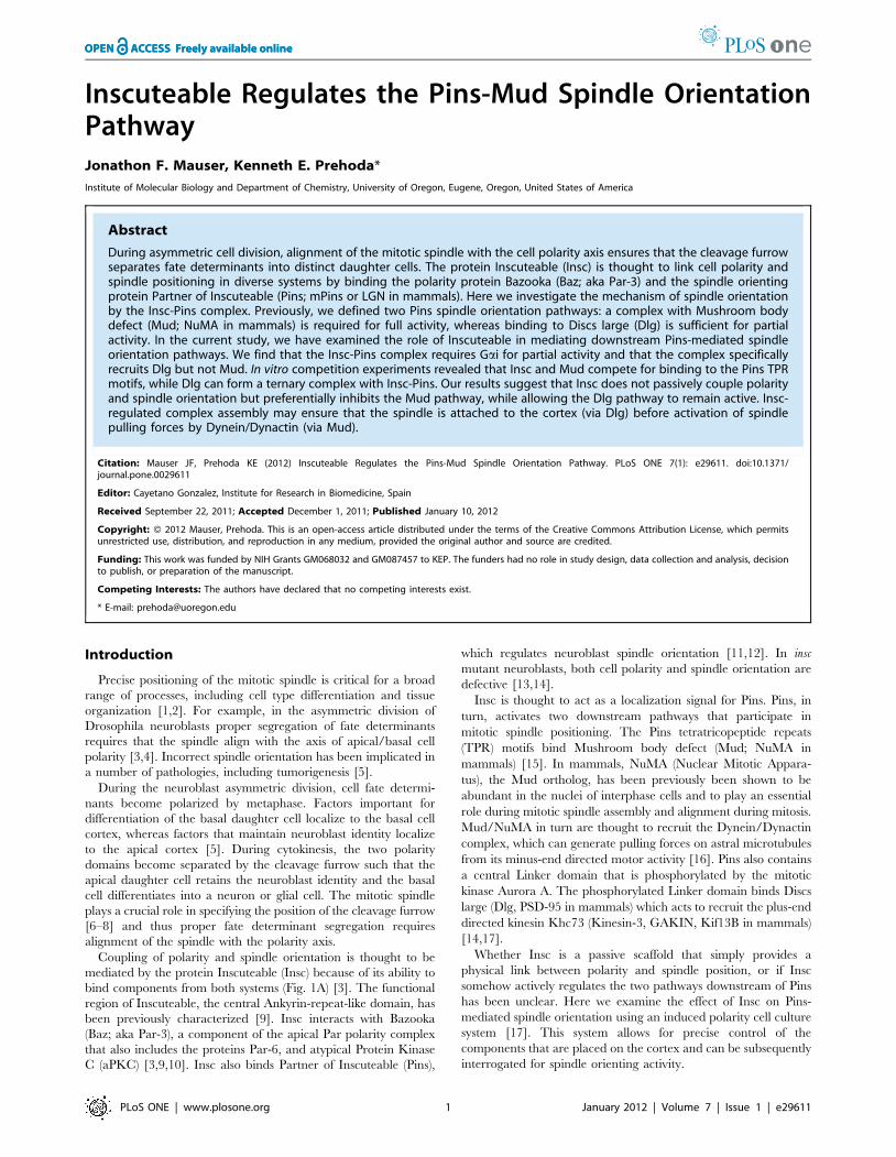
bind components from both systems (Fig. 1A) [3]. The functional
region of Inscuteable, the central Ankyrin-repeat-like domain, has
been previously characterized [9]. Insc interacts with Bazooka
(Baz; aka Par-3), a component of the apical Par polarity complex
that also includes the proteins Par-6, and atypical Protein Kinase
C (aPKC) [3,9,10]. Insc also binds Partner of Inscuteable (Pins),
which regulates neuroblast spindle orientation [11,12]. In insc
mutant neuroblasts, both cell polarity and spindle orientation are
defective [13,14].
Insc is thought to act as a localization signal for Pins. Pins, in
turn, activates two downstream pathways that participate in
mitotic spindle positioning. The Pins tetratricopeptide repeats
(TPR) motifs bind Mushroom body defect (Mud; NuMA in
mammals) [15]. In mammals, NuMA (Nuclear Mitotic Appara-
tus), the Mud ortholog, has been previously been shown to be
abundant in the nuclei of interphase cells and to play an essential
role during mitotic spindle assembly and alignment during mitosis.
Mud/NuMA in turn are thought to recruit the Dynein/Dynactin
complex, which can generate pulling forces on astral microtubules
from its minus-end directed motor activity [16]. Pins also contains
a central Linker domain that is phosphorylated by the mitotic
kinase Aurora A. The phosphorylated Linker domain binds Discs
large (Dlg, PSD-95 in mammals) which acts to recruit the plus-end
directed kinesin Khc73 (Kinesin-3, GAKIN, Kif13B in mammals)
[14,17].
Whether Insc is a passive scaffold that simply provides a
physical link between polarity and spindle position, or if Insc
somehow actively regulates the two pathways downstream of Pins
has been unclear. Here we examine the effect of Insc on Pins-
mediated spindle orientation using an induced polarity cell culture
system [17]. This system allows for precise control of the
components that are placed on the cortex and can be subsequently
interrogated for spindle orienting activity.
PLoS ONE | www.plosone.org 1 January 2012 | Volume 7 | Issue 1 | e29611
Results
Polarized Inscuteable recruits Pins but lacks spindleorientation activity
In the current model for Insc-based coupling of polarity and
spindle orientation, Insc recruits Pins, which in turn recruits the
downstream effectors Mud and Dlg (Fig. 1A) [11,15,17,18]. In
previous work, we found that Insc lacked activity in an induced
polarity spindle orientation assay [17]. In this assay, proteins are
fused to the cytoplasmic domain of the adhesion protein Echinoid
(Ed) and transiently transfected into cultured Drosophila S2 cells.
Cell clustering leads to polarization of the Ed fusion protein at sites
of cell-cell contact and the angle of the spindle to the center of the
induced crescent can be measured. Although we have observed
that the spindle aligns with polarized Ed-PinsTPR-LINKER
fusions, the spindle is randomly oriented in cells with polarized Ed-
Insc fusions (Fig. 1B, D).
To investigate why Ed-PinsTPR-LINKER orients the spindle
but Ed-Insc fails to do so, we first determined if Pins is recruited to
Ed-Insc. Endogenous Pins protein strongly colocalizes with Ed-
Insc (Fig. 1E). However in cells with polarized Ed alone Pins
remains in the cytoplasm (Fig. 1F). Thus, we conclude that Ed-Insc
recruits Pins, yet is unable to orient the spindle. Pins is known to be
autoinhibited for Mud-binding by an intramolecular interaction
between its NH2- and COOH termini [19,20]. This autoinhibi-
tion is relieved by binding of the heterotrimeric G-protein subunit,
Gai. Ed-Insc may not exhibit spindle orientation activity because
the Pins that it recruits has not been activated. We expressed Gai
with Ed-Insc to ensure that Insc-bound Pins is activated. Ed-Insc
and Gai co-expression leads to the formation of an Insc-Pins-Gai
complex at the crescent, but only a moderate amount of spindle
orienting activity, similar to cells with polarized Pins in which only
the downstream Dlg pathway, but not the Mud pathway, has been
activated [17] (Fig. 1C, D, G).
Insc-Pins recruits Dlg but not MudThe Insc-Pins-Gai complex may not fully orient the mitotic
spindle because of failure to recruit downstream effectors that are
normally brought to the cortex by Pins. We tested for recruitment
of the two known Pins spindle orientation pathways, Dlg and Mud.
In cells expressing Ed-Pins, Dlg is robustly recruited to the cell-cell
contacts (Fig. 1H) and co-expression of Ed-Pins with Gai results in
strong Mud recruitment (Fig. 1I). Dlg and Mud recruitment is
specific as it is not observed in cells expressing Ed-GFP (Fig. 1 J,
K).
We next examined whether Dlg and Mud are recruited to Ed-
Insc. Dlg is recruited to Ed-Insc in a similar manner as Ed-Pins
(Fig. 1L). However, while Pins-Gai can recruit Mud, Insc-Pins-Gai
is unable to do so (Fig. 1M). Thus, Insc appears to regulate Pins
complex assembly, leading to preferential activation of only one of
the two spindle orientation pathways, with the effect of an overall
reduction in spindle orientation activity.
Insc represses Pins-mediated spindle orientationEd-Insc cannot fully orient the spindle even though it recruits
Pins and Gai. Polarized Ed-Pins coexpressed with Gai, however,
Figure 1. Inscuteable-mediated orientation of the mitotic spindle requires Gai. a, current model of Inscuteable function. Insc serves as alink between the apical PAR complex and the spindle-orienting Pins-Gai complex. b, Ed-Insc (green) transfected S2 cells randomly orient the mitoticspindle (red) with respect to the region that is enriched in Ed. Spindle alignment is measured by drawing a vector from the center of the crescent(arrow) to the center of the mitotic spindle and then along the axis (dashes). c, Expression of Gai (blue) with Ed-Insc (green)is able to confer moderatespindle orienting activity. d, Cumulative percentage plot of spindle angles measured in the S2 Echinoid induced-polarity assay for Ed-Insc and Ed-Insc+Gai compared to previously-published data [17]. In these plots, the cumulative percentage of cells with a spindle angle below a particular value(x-axis) is shown. High spindle orienting activity corresponds to a deflection to lower spindle angles whereas no activity is a line across the diagonal.e, Ed-Insc expression in S2 cells is sufficient to robustly recruit endogenous Pins from the cytoplasm to the region of Ed enrichment. f, Ed alone isunable to polarize endogenous Pins. e, g, Ed-Insc induces colocalization of endogenous Pins with overexpressed Gai. h, Ed-Pins is able to recruitendogenous Dlg. i, Co-expression of Gai with Ed-Pins results in robust recruitment of endogenous Mud. j,k Ed-GFP is unable to recruit endogenousDlg or Mud to the induced-polarity cortical domains. l, Ed-Insc is able to recruit Dlg to the cortex, similar to cells expressing Ed-GFP-Pins. m, Ed-Insc isnot able to recruit Mud (red) to the Ed-crescent, even in the presence of Gai. Scale bars for all panels represent 5 mm.doi:10.1371/journal.pone.0029611.g001
Regulation of Spindle Orientation by Inscuteable
PLoS ONE | www.plosone.org 2 January 2012 | Volume 7 | Issue 1 | e29611

has full spindle-orienting activity [17]. These data suggest that Insc
preferentially inhibits Pins spindle orienting activity. To further
investigate if Insc inhibits Pins-mediated spindle orientation, we
expressed Insc in cells with polarized Ed-PinsTPR-LINKER, a
construct lacking autoinhibition that, when expressed on its own,
fully aligns the mitotic spindle (Fig. 2A) [17]. We observed that
Insc is recruited to Ed-PinsTPR-LINKER crescents (Fig. 2A, inset)
and that the presence of Insc reduces its spindle orienting activity
to a level indistinguishable from the Dlg pathway alone (Fig. 2B).
Thus, we conclude that Insc inhibits the spindle-orienting activity
of PinsTPR-LINKER.
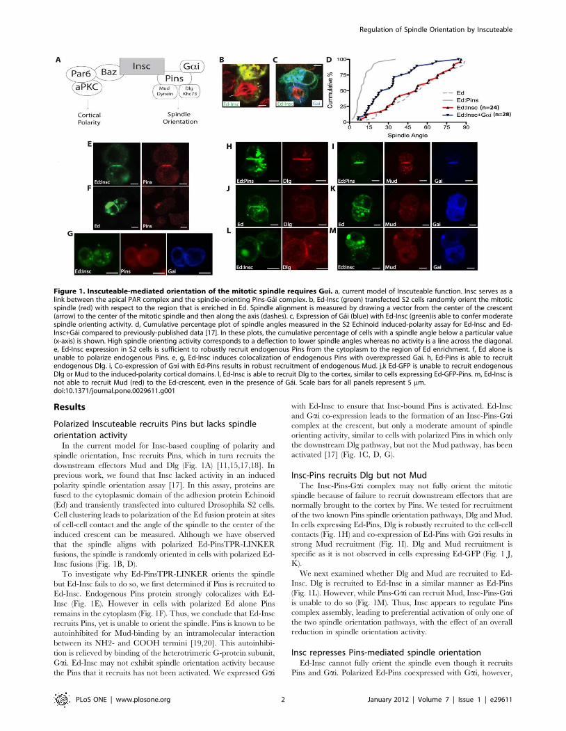
The Pins TPR domains bind InscuteableWhy might Insc-Pins recruit Dlg, but not Mud? One possible
explanation is that Insc and Mud compete for binding to Pins.
Mud is known to bind the Pins tetratricopeptide repeats (TPRs)
[15,19,20]. To identify the Pins region responsible for binding
Insc, we performed a deletion analysis using affinity pulldowns
with purified proteins. Pins contains seven TPR repeats followed
by a flexible Linker domain and three GoLoco motifs that bind
Gai. GST-fusions of full-length Pins, the GoLoco region, and the
TPR region were generated and incubated with a purified MBP-
fusion of the central Ankyrin-repeat containing domain of
Inscuteable (MBP-Insc) [9].
All constructs containing the full set of 7 Pins TPRs are able to
bind Insc, whereas those lacking these repeats, such as the
COOH-terminal GoLoco domains, are unable to bind Insc
(Fig. 3A). Further TPR truncations were also performed to find the
minimal TPR region required for binding to Insc. While binding
of Insc to Pins is detectable using a constructs consisting of the full
set of TPRs as well as TPRs 1–5, all seven TPRs are required for
high-affinity association with Insc (Fig. 3A). Since Mud/NuMA
have also been shown to require a full array of TPRs for high-
affinity binding [15] both Insc and Mud bind to the Pins TPR
motifs.
Mud and Insc compete for binding to PinsAs Insc and Mud both bind the Pins TPRs, we examined
whether Insc and Mud could bind simultaneously to Pins. Extracts
were prepared from the brains of wild-type third-instar larvae and
complex formation was examined by immunoprecipitation of the
endogenous components. As expected, we observed both Insc and
Mud in Pins immunoprecipitates. However, in Insc immunopre-
cipitates, we observed Pins but Mud was not present (Fig. 3B). The
lack of Mud in Insc immunoprecipitates suggested that Pins forms
mutually exclusive complexes with Insc and Mud. Likewise Insc
was also not observed in Mud immunoprecipitates (Fig. 3B). Thus,
Insc and Mud appear to form mutually exclusive complexes with
Pins.
We further tested for competition between Insc and Mud for
Pins using qualitative pull-downs with purified proteins. The Mud-
Pins complex can be readily formed on glutathione agarose, using
GST-PinsTPR and a purified Mud fragment containing the
minimal TPR binding domain (Fig. 3C) [15]. Introduction of
MBP-Insc to these reactions dissociates Pins from Mud, resulting
in switching to the Pins-Insc complex, a result consistent with
competition between Insc and Mud for Pins. This effect is not
observed with identical concentrations of MBP alone (Fig. 3D).
GST- pulldowns with pre-formed complexes of GST-Mud and
Pins TPR were likewise disrupted by addition of MBP-Insc
(Fig. 3E). This effect is not observed when MBP alone is titrated
into identical reactions (Fig. 3F).
Finally, we examined Insc and Mud competition using
fluorescence anisotropy. We labeled a peptide representing the
minimal region of Mud that binds Pins with the fluorophore
tetramethylrhodamine (TMR-Mud). Binding of Pins causes a
significant increase in TMR-Mud anisotropy due to complex
assembly (Fig. 3G). Insc addition to a pre-formed complex of Pins
& Mud leads to a decrease in TMR-Mud anisotropy to a value
consistent with free peptide. No effect of Insc was observed when
Pins is not present, indicating that TMR-Mud does not bind
directly to Insc (Fig. 3H).
The decrease in anisotropy is a further indication that Insc
competes for Mud binding and allows for calculation of the Insc
affinity for Pins of Kd = 5 mM. Interestingly, this affinity is
somewhat lower than the Pins-Mud interaction (Kd = 1.1 mM).
Together, the immunoprecipitation, pull-down, and fluorescence
anisotropy results indicate that Mud and Insc compete for Pins
binding.
Discs large, Inscuteable, and Pins form a stable ternarycomplex
Pins can also bind the downstream effector Dlg through its
phosphorylated Linker domain. Activation of the Dlg pathway
leads to partial spindle orienting activity, similar to that observed
for the Insc-Pins complex. To determine if Insc-Pins can bind Dlg,
we examined their binding in a pull-down experiment. Interaction
of Pins with Dlg requires Aurora-A phosphorylation of the Pins
Figure 2. Expression of Inscuteable in cells expressing constitutively-active Pins reduces spindle orientation to Dlg-like levels. a, Co-expression of Ed-Pins 1–466 (green), which robustly orients the mitotic spindle, with Inscuteable (inset), reduces the levels of spindle orientation inadherent, polarized S2 cells. b, Cumulative percentage plot of spindle angles measured in cells co-expressing Ed-Pins 1–466 and Inscuteablecompared to published data [17].doi:10.1371/journal.pone.0029611.g002
Regulation of Spindle Orientation by Inscuteable
PLoS ONE | www.plosone.org 3 January 2012 | Volume 7 | Issue 1 | e29611

Linker domain and we observed phosphorylation-dependent
formation of an Insc-Pins-Dlg ternary complex (Fig. 3I). This
result indicates that while Insc represses Mud binding to the Pins
TPRs, it has no effect on regulating the downstream Dlg pathway.
Discussion
Spindle positioning is important in many physiological contexts
[21–23]. At a fundamental level, spindle orientation determines
the placement of the resulting daughter cells in the developing
tissue, which is important for correct morphogenesis and tissue
organization [24–25]. In other contexts, such as asymmetric cell
division, spindle position ensures proper segregation of fate
determinants and subsequent differentiation of daughter cells.
We have examined the function of a protein thought to provide a
‘‘passive’’ mark on the cortex for subsequent recruitment of the
spindle orientation machinery. During neuroblast asymmetric cell
division, Insc has been thought to mark the cortex based on the
location of the Par polarity complex.
Ectopic expression of Insc in cells that normally do not express
the protein has revealed that it is sufficient to induce cell divisions
oriented perpendicular to the tissue layer, reminiscent of
neuroblast divisions [13,26,27]. Expression of the mammalian
Figure 3. Inscuteable competes with Mud, but not Dlg, for binding to Pins. a, GST-pulldowns of Inscuteable with different Pins constructsreveals that Inscuteable binds specifically to constructs containing the full array of Pins TPRs. b, Coimmunoprecipitations of endogenous proteinsfrom wild-type L3 brain extracts demonstrate that Inscuteable and Mud form exclusive complexes with Pins. c, GST-pulldown using GST-Pins TPRsincubated with a constant amount of Mud and increasing MBP-Insc reveals effective competition between Mud & Insc for binding to Pins. d, A controltitrations of MBP alone do not result in dissociation of Mud from GST-Pins. e, GST-pulldown using GST-Mud incubated with constant 2 uM Pins TPRand increasing amounts of MBP-Insc results in an approximately 1:1 stoichiometric dissociation of Pins TPRs from GST-Mud. f, A control titration ofMBP alone does not result in disruption of Pins-Mud binding. g, Fluorescence anisotropy of TMR-Mud with increasing amounts of Pins TPRs exhibits arobust association profile. h, Addition of Inscuteable to a pre-formed complex of 100 nM TMR-Mud & 1 uM Pins causes a dissociation of the Mud-Pinscomplex & reduction of TMR-Mud anisotropy. i, GST-pulldown using in vitro Aurora-A phosphorylated Pins 1–466 results in complex formation withthe Dlg GK domain. AurA treatment of a pre-formed MBP-Insc/Pins 1–466 complex likewise is able to form a complex with the Dlg GK domain.doi:10.1371/journal.pone.0029611.g003
Regulation of Spindle Orientation by Inscuteable
PLoS ONE | www.plosone.org 4 January 2012 | Volume 7 | Issue 1 | e29611

ortholog of Inscuteable, mInsc, in epidermal progenitors has
shown that this phenotype is not completely penetrant over time
[27]. Expression of mInsc leads to a transient re-orientation of
mitotic spindles, in which mInsc and NuMA initially co-localize at
the apical cortex. After prolonged expression, however, the
epidermal progenitors return to dividing along the tissue polarity
axis, a scheme in which mInsc and NuMA no longer co-localize.
These results indicate that Insc and Mud can be decoupled from
one another.
We have examined the effect of Insc-Pins complex formation
both in an induced polarity spindle orientation assay and in in vitro
binding assays. Our results indicate that Insc plays a more active
role in spindle positioning than previously appreciated. Rather
than passively coupling polarity and spindle positioning systems,
Insc acts to regulate the activity of downstream Pins pathways. We
have shown that the Dlg pathway is unaffected by Inscuteable
expression while the Mud pathway is inhibited by Insc binding.
Recent work on the mammalian versions of these proteins
explains the structural mechanism for competition between the
Insc-Pins and Pins-Mud complexes [28]. The binding sites on Pins
for these two proteins overlap making binding mutually exclusive
because of steric considerations. The observation of Insc
dissociation of the Pins-Mud complex in Drosophila (this work)
and mammalian proteins (LGN-NuMA) [28] suggests that Insc
regulation of Mud-binding is a highly conserved behavior.
This competition between Mud and Insc for Pins binding is
consistent with previous work done with a chimeric version of
Inscuteable/Pins [29]. This protein, in which the Pins TPR
domain was replaced with the Inscuteable Ankyrin-repeat domain,
bypasses the Insc-Pins recruitment step of apical complex
formation. In these cells, the chimeric Insc-Pins protein was able
to rescue apical/basal polarity and spindle orientation in
metaphase pins mutant neuroblasts. As this protein lacks the
Mud-binding TPR domain, Mud binding to Pins is not absolutely
necessary for spindle alignment. Importantly, the PinsLINKER
domain is still intact in the Insc-Pins fusion, implying that Dlg, not
Mud, function is sufficient for partial activity, as observed in the S2
system [17].
The Mud and Dlg pathways may play distinct roles in spindle
positioning. The Dlg pathway, through the activity of the plus-end
directed motor Khc73, may function to attach the cortex to the
spindle through contacts with astral microtubules [14]. In contrast,
the Mud pathway, through the minus-end directed Dynein/
Dynactin generates force to draw the centrosome towards the
center of the cortical crescent [16]. Fusion of the Pins TPR motifs,
which recruit Mud, to Echinoid does not lead to spindle
alignment, indicating that the Mud pathway is not sufficient for
spindle alignment. The PinsLINKER domain does have partial
activity on its own, however, and when placed in cis with the TPRs
leads to full alignment [17]. In this framework, the function of Insc
may be temporal control, ensuring that microtubule attachment
by the Dlg pathway occurs before the force generation pathway is
activated.
In the temporal model of Insc function, what might cause the
transition from the Insc-Pins-Dlg complex, which mediates astral
microtubule attachment, to the Mud-Pins-Dlg complex, which
generates spindle pulling forces? By early prophase, Inscuteable
recruits Pins and Gai to the apical cortex [14]. During this phase
of the cell cycle, Mud is localized to the nucleus in high
concentration [30,31]. Apically-localized Pins binds Dlg, creating
an apical target for astral microtubules (Fig. 4A). During early
phases of mitosis, Inscuteable would serve to inhibit binding of low
concentrations of cytoplasmic Mud to the Pins TPRs to prevent
spurious activation of microtubule shortening pathways. After
nuclear envelope breakdown, Mud enters the cytoplasm in greater
concentrations [31] and could then act to compete with Insc for
binding to Pins (Fig. 4B), allowing Pins output to be directed into
microtubule-shortening pathways. Future work will be directed
towards testing additional aspects of this model.
Materials and Methods
Molecular cloning, protein expression and purificationConstructs encoding Drosophila Pins, Inscuteable, and Mud
have been described [9,18]. Residues 252–600 of Inscuteable,
including the central Ankyrin-repeat containing region, were used
for all experiments. Residues 1–466 of Pins, corresponding to the
TPR+LINKER domains, 42–398, corresponding to the TPR
domain, and 372–658, corresponding to the three GoLoco
domains were used for Inscuteable binding studies. Mud residues
1825–2016, which includes the minimal Pins-binding domain,
were also amplified for binding assays.
Echinoid (Ed) fusion constructs were made in pMT-V5
(Invitrogen, Carlsbad, CA), replacing the Ed cytoplasmic domain
with a visualization tag and the protein of interest at the COOH
terminus (e.g., Ed-GFP-Insc). Proteins for pull down and
anisotropy experiments were expressed in Escherichia coli strain
BL21(DE3) using pGEX 4T-1-based vectors for GST fusions,
Figure 4. Proposed model for Inscuteable regulation of spindle orientation. a, In early interphase, Inscuteable recruits cortical Gai-Pins tothe apical cortex. Insc-bound Pins can scaffold for Dlg, allowing for early microtubule attachment, but inhibits binding of Mud, preventing ectopicmicrotubule shortening. b, after nuclear envelope breakdown and trafficking along the mitotic spindle, Mud from astral microtubules competes Pinsaway from Insc and allows for microtubule shortening.doi:10.1371/journal.pone.0029611.g004
Regulation of Spindle Orientation by Inscuteable
PLoS ONE | www.plosone.org 5 January 2012 | Volume 7 | Issue 1 | e29611

pMAL-c2 based-vectors for MBP fusions, and pBH-based vectors
for hexahistidine fusions. GST-fusion proteins were purified on
glutathione-agarose resin and washed with a large excess of GST
pulldown buffer (10 mM HEPES pH 7.5/100 mM NaCl/1 mM
DTT). The resin was then used for subsequent GST-pulldowns.
MBP-fusion proteins were purified on amylose resin (New England
Biolabs), washed with three bed-volumes of PBS+1% Triton X-
100 and one bed-volume of PBS. Proteins were eluted using
PBS+1M Methyl-a-D-glucopyranoside (Sigma-Aldrich). Hexahis-
tidine-fusion proteins were purified on Ni-NTA agarose resin
(Qiagen). The incubated resin was then washed with a large excess
of cell lysis buffer (50 mM NaPO4/150 mM NaCl/10 mM
imidazole). Samples were then eluted with elution buffer
(50 mM NaPO4/150 mM NaCl/300 mM imidazole).
Transfection and S2 Cell ExperimentS2 cells were grown and cultured at room temperature in
Schneider’s Insect Media (Sigma) supplemented with 10% fetal
bovine serum. Echinoid polarity assays were carried out as
described previously [14]. In short, 1610‘6 cells were transiently
transfected with pMT-V5 fusion constructs (400 ng each) using
Effectene (QIAGEN) reagent according to manufacturer protocol.
24–48 hrs after transfection, protein expression was induced by
incubation with CuSO4 (500 mM) for 24 hr. Cells were harvested
by centrifugation and the media was replaced. These cells were
then shaken (175 RPM) for 2–3 hr to induce Ed-mediated cell-cell
clusters. These cells were then were plated on glass coverslips and
allowed to incubate for 3 hr to allow for cell divisions to occur.
ImmunostainingFor immunostaining, S2 cells were fixed for 20 min in 4%
paraformaldehyde, stained, and imaged on a Leica SP2 confocal
microscope with a 6361.4 NA lens. Antibodies and dilutions were
as follows: rabbit Gai, 1:1000 [17], mouse Dlg, 1:250 (Develop-
mental Studies Hybridoma Bank, Iowa); rabbit Mud 1:1000 (gift
from Y. Bellaiche); rat Pins, 1:500 [10]; rat tubulin, 1:1000
(Abcam); rabbit Insc 1:1000 (gift from W.Chia), rabbit HA, 1:1000
(Covance).
Immunoprecipitation and Western BlotsFor western blot lysate inputs, 20 mg of total protein from brain
extracts were used per lane. Immunoprecipitation from larval
brain extracts was carried out using antibodies bound to protein G
sepharose (GE Healthcare) according to the manufacturer’s
instructions. 40 brains from L3 larvae were dissected and
homogenized by douncing in 300 uL sample buffer (50 mM
HEPES pH 7.5/150 mM NaCl/1 mM DTT/0.1% Triton X-
100/EDTA-free Protease Inhibitors (Roche)). Extracts were then
centrifuged twice for 10 minutes each at 10,000 rpm to pellet
insoluble cell debris. The resulting supernatant was then
precleared with protein G sepharose and incubated with
antibody-bound resin. Following three washes in sample buffer,
the resin was heated to 95uC in SDS loading buffer (1% SDS/
100 mM DTT/50 mM Tris pH. 7.5/0.003% bromophenol blue).
Immunoprecipitates were resolved on SDS-PAGE followed by
western blotting.
Measuring Cortical Polarity, Spindle Orientation, andCentrosome Alignment
Spindle alignment measurements were made as described
previously [17]. Briefly, spindle angles were measured with a
vector perpendicular to the center of the Ed crescent and a vector
matching the spindle or connecting the spindle poles. The angle
between these two vectors was then assessed.
In Vitro Binding AssaysGST pull-down assays have been described [19]. Briefly, ligands
were added to glutathione agarose with adsorbed GST fusion
proteins in binding buffer (10 mM Hepes/100 mM NaCl/1 mM
DTT) at the indicated concentrations to a final reaction volume of
50 ml and incubated at room temperature for 15 min before
washing, elution, and analysis by gel electrophoresis.
Fluorescence anisotropy binding assays were as described [19].
A peptide containing the sequence of Mud residues 1955–1970
and an NH2-terminal cysteine was labeled with tetramethylrho-
damine maleimide (Life Technologies) according to the manufac-
turer’s instructions. The labeled protein was purified by reverse-
phase HPLC. For binding experiments, solutions were prepared
with increasing amount of ligand and constant dye-labeled
component (100 nM) in binding buffer with the temperature
maintained at 20uC by using a circulating water bath. Data series
were fit to an equation describing 1:1 binding.
In Vitro Kinase AssaysRecombinant Aurora-A kinase was purchased from Millipore
(Billerica, MA). Pins constructs (10 mg) and Aurora-A (100 ng)
were diluted in ice-cold assay buffer (20 mM Tris [pH 7.4],
100 mM NaCl, 1 mM DTT, 10 mM MgCl2, and 10 mM ATP).
These reactions were then moved to room temperature for
30 minutes. Reactions were then chilled on ice and added to
affinity pulldown resin for pulldown experiments.
Acknowledgments
We thank Rhonda Newman, Nick Smith, and Rick Nipper for their
assistance at the inception of this project. We also thank members of the
Prehoda and Doe laboratories for providing reagents and for helpful
discussions and questions.
Author Contributions
Conceived and designed the experiments: JFM KEP. Performed the
experiments: JFM. Analyzed the data: JFM KEP. Contributed reagents/
materials/analysis tools: JFM. Wrote the paper: JFM KEP.
References
1. Cabernard C, Doe CQ (2009) Apical/basal spindle orientation is required for neuroblast
homeostasis and neuronal differentiation in Drosophila. Dev Cell 17: 134–141.
2. Baena-Lopez LA, Baonza A, Garcıa-Bellido A (2005) The orientation of cell
divisions determines the shape of Drosophila organs. Curr Biol 15: 1640–1644.
3. Siller KH, Doe CQ (2009) Spindle orientation during asymmetric cell division.
Nature Cell Biol 11: 365–374.
4. Knoblich JA (2010) Asymmetric cell division: recent developments and their
implications for tumour biology. Nat Rev Mol Cell Biol 11: 849–860.
5. Prehoda KE (2009) Polarization of Drosophila neuroblasts during asymmetric
division. Cold Spring Harb Perspect Biol 1: a001388.
6. Chia W, Somers WG, Wang H (2008) Drosophila neuroblast asymmetric
divisions: cell cycle regulators, asymmetric protein localization, and tumorigen-
esis. J Cell Biol 180: 267–272.
7. Glotzer M (2004) Cleavage furrow positioning. J Cell Biol 164(3): 347–351.
8. Cabernard C, Prehoda KE, Doe CQ (2010) A spindle-independent cleavage
furrow positioning pathway. Nature 467: 91–94.
9. Schober M, Schaefer M, Knoblich JA (1999) Bazooka recruits Inscuteable to
orient asymmetric cell divisions in Drosophila neuroblasts. Nature 402: 548–551.
10. Wodarz A, Ramrath A, Kuchinke U, Knust E (1999) Bazooka provides an apical
cue for Inscuteable localization in Drosophila neuroblasts. Nature 402: 544–547.
Regulation of Spindle Orientation by Inscuteable
PLoS ONE | www.plosone.org 6 January 2012 | Volume 7 | Issue 1 | e29611

11. Yu F, Morin X, Cai Y, Yang X, Chia W (2000) Analysis of partner of
inscuteable, a novel player of Drosophila asymmetric divisions, reveals twodistinct steps in inscuteable apical localization. Cell 100: 399–409.
12. Schaefer M, Shevchenko A, Shevchenko A, Knoblich JA (2000) A protein
complex containing Inscuteable and the Galpha-binding protein Pins orientsasymmetric cell divisions in Drosophila. Current Biol 10: 353–362.
13. Kraut R, Chia W, Jan LY, Jan YN, Knoblich JA (1996) Role of inscuteable inorienting asymmetric cell divisions in Drosophila. Nature 383: 50–55.
14. Siegrist SE, Doe CQ (2005) Microtubule-induced Pins/Galphai cortical polarity
in Drosophila neuroblasts. Cell 123: 1323–1335.15. Siller KH, Cabernard C, Doe CQ (2006) The NuMA-related Mud protein binds
Pins and regulates spindle orientation in Drosophila neuroblasts. Nature CellBiol 8: 594–600.
16. Siller KH, Doe CQ (2008) Lis1/dynactin regulates metaphase spindleorientation in Drosophila neuroblasts. Dev Biol 319: 1–9.
17. Johnston CA, Hirono K, Prehoda KE, Doe CQ (2009) Identification of an
Aurora-A/PinsLINKER/Dlg spindle orientation pathway using induced cellpolarity in S2 cells. Cell 138: 1150–1163.
18. Tio M, Zavortink M, Yang X, Chia W (1999) A functional analysis ofinscuteable and its roles during Drosophila asymmetric cell divisions. J Cell Sci
112: 1541–1551.
19. Nipper RW, Siller KH, Smith NR, Doe CQ, Prehoda KE (2007) Galphaigenerates multiple Pins activation states to link cortical polarity and spindle
orientation in Drosophila neuroblasts. Proc Natl Acad Sci U S A 104:14306–14311.
20. Du Q, Macara IG (2004) Mammalian Pins is a conformational switch that linksNuMA to heterotrimeric G proteins. Cell 119: 503–516.
21. Moore JK, Cooper JA (2010) Coordinating mitosis with cell polarity: Molecular
motors at the cell cortex. Semin Cell Dev Biol 21: 283–289.
22. Lechler T, Fuchs E (2005) Asymmetric cell divisions promote stratification and
differentiation of mammalian skin. Nature 437: 275–280.
23. Reinsch S, Karsenti E (1994) Orientation of spindle axis and distribution of
plasma membrane proteins during cell division in polarized MDCKII cells. J Cell
Biol 126: 1509–1526.
24. Gray RS, Cheung KJ, Ewald AJ (2010) Cellular mechanisms regulating
epithelial morphogenesis and cancer invasion. Curr Opin Cell Biol 22: 640–650.
25. Pease JC, Tirnauer JS (2011) Mitotic spindle misorientation in cancer–out of
alignment and into the fire. J Cell Sci 124: 1007–1016.
26. Egger B, Boone JQ, Stevens NR, Brand AH, Doe CQ (2007) Regulation of
spindle orientation and neural stem cell fate in the Drosophila optic lobe. Neural
Dev 2: 1.
27. Poulson N, Lechler T (2010) Robust control of mitotic spindle orientation in the
developing epidermis. J Cell Biol 191: 915–922.
28. Zhu J, Wen W, Zheng Z, Shang Y, Wei Z, et al. (2011) LGN/mInsc and LGN/
NuMA complex structures suggest distinct functions in asymmetric cell division
for the Par3/mInsc/LGN and Gai/LGN/NuMA pathways. Mol Cell 43:
418–431.
29. Yu F, Ong CT, Chia W, Yang X (2000) Membrane targeting and asymmetric
localization of Drosophila partner of inscuteable are discrete steps controlled by
distinct regions of the protein. Mol Cell Biol 22: 4230–4240.
30. Du Q, Stukenberg PT, Macara IG (2001) A mammalian Partner of inscuteable
binds NuMA and regulates mitotic spindle organization. Nature Cell Biol 3:
1069–1075.
31. Kisurina-Evgenieva O, Mack G, Du Q, Macara I, Khodjakov A, et al. (2004)
Multiple mechanisms regulate NuMA dynamics at spindle poles. J Cell Sci 117:
6391–6400.
Regulation of Spindle Orientation by Inscuteable
PLoS ONE | www.plosone.org 7 January 2012 | Volume 7 | Issue 1 | e29611