Embed Size (px)
Citation preview

Biomech Model Mechanobiol (2012) 11:689–702DOI 10.1007/s10237-011-0343-x
ORIGINAL PAPER
Insights into interstitial flow, shear stress, and mass transporteffects on ECM heterogeneity in bioreactor-cultivatedengineered cartilage hydrogels
Tony Chen · Mark Buckley · Itai Cohen ·Lawrence Bonassar · Hani A. Awad
Received: 7 March 2011 / Accepted: 1 August 2011 / Published online: 19 August 2011© Springer-Verlag 2011
Abstract Interstitial flow in articular cartilage is secondaryto compressive and shear deformations during joint motionand has been linked with the well-characterized heterogene-ity in structure and composition of its extracellular matrix.In this study, we investigated the effects of introducing gra-dients of interstitial flow on the evolution of compositionalheterogeneity in engineered cartilage. Using a parallel-platebioreactor, we observed that Poiseuille flow stimulation ofchondrocyte-seeded agarose hydrogels led to an increase inglycosaminoglycan and type II collagen deposition in thesurface region of the hydrogel exposed to flow. Experimentalmeasurements of the interstitial flow fields based on the fluo-rescence recovery after photobleaching technique suggestedthat the observed heterogeneity in composition is associatedwith gradients in interstitial flow in a boundary layer at thehydrogel surface. Interestingly, the interstitial flow velocity
Electronic supplementary material The online version of thisarticle (doi:10.1007/s10237-011-0343-x) contains supplementarymaterial, which is available to authorized users.
T. Chen · H. A. AwadDepartment of Biomedical Engineering, University of Rochester,Rochester, NY, USA
T. Chen · H. A. Awad (B)Department of Biomedical Engineering,The Center for Musculoskeletal Research,University of Rochester Medical Center,Box 665, 601 Elmwood Avenue,Rochester, NY 14642, USAe-mail: [email protected]
M. Buckley · I. CohenDepartment of Physics, Cornell University, Ithaca, NY, USA
L. BonassarDepartment of Biomedical Engineering, Cornell University,Ithaca, NY, USA
profiles were nonlinearly influenced by flow rate, which uponcloser examination led us to the original observation that theapparent hydrogel permeability decreased exponentially withincreased interfacial shear stress. We also observed that inter-stitial flow enhances convective mass transport irrespectiveof molecular size within the boundary layer near the hydro-gel surface and that the convective contribution to transportdiminishes with depth in association with interstitial flow gra-dients. The implications of the nonlinearly inverse relation-ship between the interfacial shear stress and the interstitialflux and permeability and its consequences for convectivetransport are important for tissue engineering, since porousscaffolds comprise networks of Poiseuille channels (pores)through which interstitial flow must navigate under mechan-ical stimulation or direct perfusion.
Keywords Tissue engineering · Bioreactors · Cartilage ·Interstitial flow · Shear Stress · Permeability ·Fluorescence recovery after photobleaching (FRAP) ·Computational fluid dynamics (CFD)
1 Introduction
Articular cartilage is characterized by anisotropy in com-position and architecture that affects its biomechanics andmechanobiology. This anisotropy plays important roles insupporting “physiologic” loading and facilitating the nearlyfrictionless articular motion throughout the healthy life of thejoint (Carter et al. 2004; Carter and Wong 2003; Lammi 2004;Wong and Carter 2003). Physiologic loading affects the chon-drocytes’ metabolic activity and cartilage composition, struc-ture, and function through several mechano-electrochemicaltransduction mediators including compressive, shear, andtensile strains, hydrostatic pressure, interstitial fluid flow,streaming potentials, and convective molecular transport
123
690 T. Chen et al.
(Gray et al. 1988). Therefore, the intimate coupling betweencomposition, structure, and biomechanical function hasimportant implications in functional tissue engineering ofcartilage (Butler et al. 2000).
Functional tissue engineering is a scientific paradigm thataddresses the influence of biomechanical and physical factorsin bioreactor systems on the differentiation and metabolicactivity of cells and engineered tissues and organs. Early gen-erations of cartilage bioreactors were designed to overcomediffusion limitation in clinical scale constructs, by improv-ing mixing of media and convective transport through varioushydrodynamic designs (Freed et al. 1998, 1993). Others havedemonstrated that applying dynamic compressive deforma-tions that mimic in vivo joint loads stimulate the biosyntheticactivity of chondrocytes in cartilage explants (Bonassar et al.2001; Sah et al. 1989) and recognized that this can be rep-licated in tissue engineering bioreactors (Buschmann et al.1995; Mauck et al. 2000) to improve the biochemical com-position and biomechanical properties of tissue engineeredcartilage constructs (e.g. agarose hydrogels) over time (Hunget al. 2004). Interestingly, in this unconfined compressionsetup, the observed enhancement in chondrocyte biosyntheticactivity has been co-localized with increased interstitial fluidflow on the periphery of the constructs and did not seem to belocally associated with fluid pressure or compressive strainsin these peripheral regions, suggesting an important mech-anotransduction role for interstitial fluid flow (Buschmannet al. 1995, 1999). While the mechanism of flow-inducedmechanotransduction may vary in different cell types, as ithas been reported to involve integrin-mediated and stretch-activated ion channel signaling in chondrocytes (Lee et al.2000), heparin-sulfate-mediated FAK signaling in smoothmuscle cells (Shi et al. 2011), and primary cilia-mediatedsignaling in bone cells (Malone et al. 2007), it is widely rec-ognized that the flow-related mechanobiology has importantimplications in cell differentiation, tissue morphogenesis,and tissue engineering. In addition to its mechanotransduc-tion role, it has also been long recognized that interstitialflow leads to compaction of the extracellular matrix andconsequently to heterogeneity in the structure and compo-sition of the extracellular matrix (Mow and Wang 1999).Furthermore, interstitial flow has been shown to create gra-dients of soluble signals, which could alter cell behaviorsuch as migration, as has been demonstrated in the tumormicroenvironment (Shieh and Swartz 2011). Consistent withthese observation, perfusion bioreactors that induce inter-stitial fluid flow through tissue engineered constructs havebeen shown to increase chondrogenic matrix production inengineered cartilage in the absence of compressive strains,presumably through convective transport and shear stressstimulation of the cells (Bujia et al. 1995; Pazzano et al.2000; Sittinger et al. 1994). Despite the reported beneficialeffects of bioreactor culture on the composition and bio-
mechanical integrity of engineered cartilage, reproducingthe native tissue’s heterogeneous zonal organization and theassociated functional anisotropy in engineered cartilage con-structs remains an unsolved challenge for tissue engineers.
To address this challenge, we previously reported usingcomputational fluid dynamics (CFD) modeling that Poiseu-ille flow over a hydrogel construct induces depth-dependentgradients in interstitial fluid flow velocity in agreement withtheory (Beavers and Joseph 1967; Hou et al. 1989), andwe hypothesized that this flow would induce compositionalheterogeneity and differentially affect collagen alignment(Lamkin-Kennard et al. 2005). Gemmiti and Guldberg (2006)experimentally demonstrated that Poiseuille flow-inducedshear stress magnitude and duration in a parallel-plate bio-reactor modulate a heterogenous matrix composition, anddifferentially align the collagen fibers in the extracellularmatrix in engineered cartilaginous tissue, thereby influenc-ing its tensile mechanical properties (Gemmiti and Guldberg2006, 2009). In subsequent work, Pierre et al. (2008) dem-onstrated, using finite element modeling, that the shear stresscould modulate the level and distribution of tissue oxygena-tion (Pierre et al. 2008). However, whether the reported floweffects on the compositional and structural inhomogeneityare due to interstitial flow and shear stress stimulation orconvective transport of nutrients such as glucose and var-ious growth factors remains to be determined. To addressthis question, an accurate mapping of the interstitial flowfields and mass transport within the engineered cartilage con-structs needs to be determined to help model the hydrody-namics and mass transport effects. To that end, this study wasdesigned to microscopically characterize the interstitial flowfields and their effects on convective transport within aga-rose hydrogels in a Poiseuille flow parallel-plate bioreactorsystem. We first evaluated the effects of Poiseuille flow stim-ulation on matrix composition in chondrocyte-seeded aga-rose hydrogels. We then used a flow visualization techniquebased on the Fluorescent Recovery After Photobleaching(FRAP) method to measure the interstitial fluid flow velocityand evaluate its effects on mass transport at different depthswithin the agarose hydorgel. The nonlinear coupling betweenthe flow rate, shear stress, apparent permeability, and masstransport was evaluated through a combination of theoreticalconsiderations of interstitial flow in biphasic mixtures underPoiseuille flow (Hou et al. 1989) and Computational FluidDynamics (CFD) modeling.
2 Methods
2.1 Bioreactor design
The parallel-plate bioreactor used in chondrocyte-seededhydrogel experiments consists of three main compartments
123

Interstitial flow in engineered cartilage hydrogels 691
Fig. 1 A 2D schematic of the Poiseuille flow bioreactor showing: a Theupper rectangular flow channel through which media is perfused witha pump, the rectangular cavity to hold the tissue engineered cartilage(TEC) hydrogel (2% agarose seeded with 40 million chondrocytes/ml),and the bottom compartment containing an acellular hydrogel to main-tain the TEC hydration. b The experimental setup in which a syringe
pump drives flow in the parallel-plate bioreactor in a cyclic fashion(1 Hz) at ±5 ml/min estimated to induce a nominal shear stress of±0.012 Pa at the interface between the culture medium in the flow chan-nel and the surface of the TEC hydrogel. The flow regimen is repeatedover 7 days following an initial static culture period of 14 days
(Fig. 1a). The upper compartment comprises a rectangularflow channel (1.25 mm height) with inlet and outlet ports thatconnect via silastic tubing to a programmable syringe pump(New Era Pump Systems Inc, Wantagh NY) and a culturemedia reservoir, respectively (Fig. 1b). The middle compart-ment comprises a rectangular cavity (2 mm deep) into whichthe chondrocyte-seeded 2% agarose (tissue engineered car-tilage or TEC) hydrogel is cast. A cell-free 1.25% agarosehydrogel is cast within the bottom compartment (4 mm deep)to provide hydration to the bottom of the TEC hydrogel andanchor it in place.
The nominal shear stress across the surface of the TEChydrogel is approximated by
τ = 6μQ
bh2 (1)
where τ is the shear stress, μ is the dynamic viscosity, Q isthe flow rate, and b and h are the width and height of theflow channel, respectively. Based on the dimensions of thechambers (Table 1), a flow rate (Q) of 5 ml/min results ina nominal shear stress of 0.012 Pa across the surface of theTEC hydrogel.
2.2 Chondrocyte isolation and tissue engineered cartilage(TEC) hydrogels
All reagents were purchased from Invitrogen (Carlsbad, CA)unless otherwise noted. Chondrocytes were isolated from the
elbow joints of pigs within 24 h after slaughter. The jointspace was opened using aseptic technique; cartilage bits wereshaved from the articulating ends of the ulna, radius, andhumerus of the elbow joint and placed into wash media con-sisting of DMEM (high glucose), 20% Fetal Bovine Serum(FBS), 1% Penicillin/Streptomycin (P/S), Kanamycin Sul-fate (Sigma-Aldrich, St. Louis MO), and Fungizone and Gen-tamicin. The cartilage was then minced into finer (2 mm ×2 mm) pieces and enzymatically digested using pronase (Cal-BioChem, San Diego CA) for 1.5 h followed by type-2 colla-genase (Worthington Biochemical, Lakewood NJ) for 16 h.The digested tissue suspension was then strained through a40 µm cell strainer, and the retrieved cells were washed twiceand then suspended in complete media (DMEM/F-12, 10%FBS, 1% P/S) until used to create the TEC hydrogels.
Isolated cells were suspended in complete media andmixed with molten (∼ 40◦C) type VII agarose (Sigma-Aldrich) in PBS to create a 40 × 106 cells/ml 2% aga-rose solution. The chondrocyte/agarose solution was castin the hydrogel cavity of the middle bioreactor compart-ment and allowed to polymerize at room temperature for20 min to form the TEC hydrogel. Following 14 days ofpreculture in an incubator, the bioreactor containing theTEC hydrogel was assembled, placed in an incubator at37◦C and 5% CO2, and connected to the syringe pumpand media reservoir using flexible tubing. Since intermit-tent loading has been shown to induce anabolic effects inchondrocyte-seeded agarose hydrogels (Mauck et al. 2003),
123
692 T. Chen et al.
Table 1 Bioreactor design and operational parameters
Large cell culture bioreactor Scaled-down FRAP bioreactor
Nominal bioreactor dimensions
Media flow channel
Height (h1) 1.25 mm 0.2 mm
Width (w1) 18 mm 20 mm
Length (L1) 50 mm 46 mm
TE hydrogel (2% agarose) compartment
Height (h2) 2 mm 0.32 mm
Width (w2) 15 mm 2.4 mm
Length (L2) 30 mm 4.8 mm
Bottom hydration (1.25% agarose) compartment
Height (h3) 4 mm 0.64 mm
Width (w3) 20 mm 3.2 mm
Length (L3) 65 mm 10.4 mm
Flow media properties
Media density ρ 1,000 kg/m3
Media viscosity μ 6.9 × 10⁻⁴ Ns/m2
Media flow rate Q Oscillatory (1 Hz) flow ±5 ml/min∗ Steady flow 0.014–0.14 ml/min
Resting (steady) flow ∼0.167 ml/min∗
Nominal interfacial shear stress τhs = 6μQ/w1h21 Oscillatory (1 Hz) flow ±0.012 Pa 0.0012–0.012 Pa
Resting (steady) flow ∼ 0.0004 Pa
TE hydrogel properties
Chondrocytes number density 40 × 106 cells/ml N/A
Fluid volume fraction φ f 0.98
* See Fig. 1 for flow regimen
the syringe pump, controlled externally via custom-writtenLabview (National Instruments, Austin TX) software, wasprogrammed to perfuse culture media at ±5 ml/min in afull 1 Hz sinusoidal wave for 30 min once daily, followedby steady flow at 0.167 ml/min for the remainder of theday (alternating steady-state infusion and withdrawal every2 h) (Fig. 1c). This regimen was repeated over for 7 days.Control TEC hydrogels were created similarly and culturedin a petri dish in the incubator at 37◦C for 21 days. Inboth the control and the bioreactor-cultivated hydrogels,the media was supplemented with 189 mM fresh ascor-bate every 3 days. At the end of 21 days, specimens (5 mmdiameter) were collected from the control and bioreactor-cultivated TEC hydrogels and used for biochemistry andhistology.
2.3 Biochemical and histological analyses
For biochemical analyses, specimens were digested using0.5 Units/ml of papain (Sigma-Aldrich) with 2 mM L-cys-teine (Sigma-Aldrich) and 2 mM ethylenediaminetetraaceticacid (EDTA; Sigma-Aldrich) for 16 h. The dissolved spec-imens were stored frozen at −80◦C until assayed using
Hydroxyproline, Dimethylmethylene Blue (DMMB), andPicogreen assays for collagen, sulfated-glycosaminogly-can (s-GAG), and DNA content, respectively (Awad et al.2004).
For histology, specimens were fixed in 10% Neutral Buf-fered Formalin (NBF) at room temperature for 24 h. Sampleswere then removed from the NBF and dehydrated under vac-uum through graded alcohol steps (30, 50, 70, 80, 95 and100% ethanol) for 1 h each, followed by two 15 min washesin Xylenes (Sigma-Aldrich, St. Louis MO). Samples werethen infiltrated with paraffin at 60◦C for 2 h under vacuumbefore embedding. After sections were embedded in paraffin,5 µm sections were cut and stained with Toluidine Blue orprocessed for immunohistochemistry. Briefly, sections werequenched of endogenous peroxidases using 3% hydrogenperoxide (MediChoice, Mechanicsville VA). Sections werethen blocked for an hour with a 2% solution of Normal HorseSerum (Vector Labs, Burlingame CA) followed by a 1:100dilution of mouse anti-Collagen II (II-II6B3; DevelopmentalStudies Hybridoma Bank, The University of Iowa) in NormalHorse Serum. Biotinylated horse anti-mouse secondary anti-body (Vector Labs) was added for 30 min followed by addi-tion of HRP (Thermo Scientific, Rockford IL) for 20 min.
123
Interstitial flow in engineered cartilage hydrogels 693
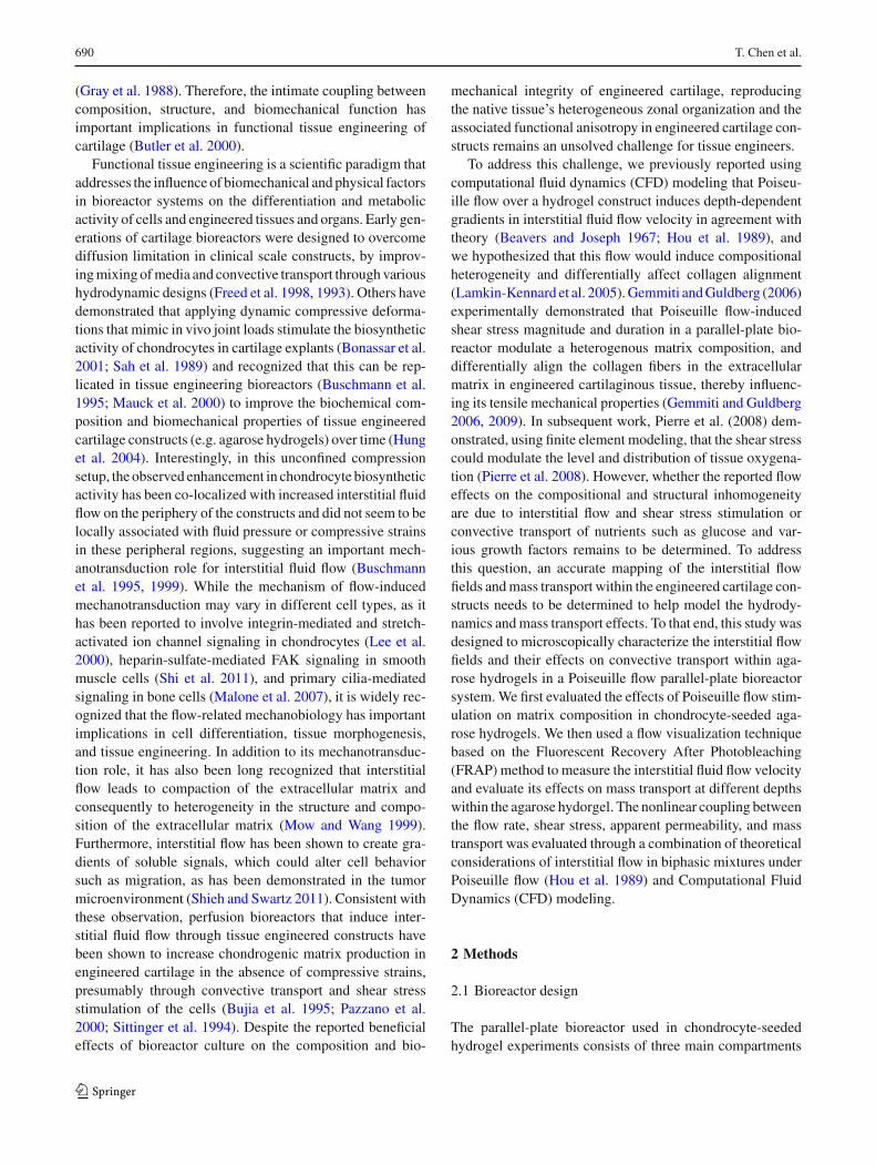
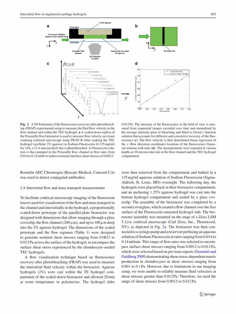
Fig. 2 A 2D Schematic of the fluorescence recovery after photobleach-ing (FRAP) experimental setup to measure the fluid flow velocity in theflow channel and within the TEC hydrogel. a A scaled-down replica ofthe Poiseuille flow bioreactor is used to measure flow velocity on a laserscanning confocal microscope using FRAP. b After soaking the TEChydrogel (acellular 2% agarose) in Sodium Fluorescein (0.125 mg/ml)for 24 h, a 12.4-micron thick line is photobleached. A Fluorescein solu-tion is then pumped in the Poiseuille flow channel at flow rates from0.014 to 0.14 ml/hr to induce nominal interface shear stresses of 0.0012–
0.012 Pa. The intensity of the fluorescence in the field of view is mea-sured from sequential images recorded over time and normalized bythe average intensity prior to bleaching and fitted to Green’s functionsolution that accounts for diffusive and convective recovery of the fluo-rescence (c). The flow velocity is then determined linear regression ofthe y (flow direction coordinate) locations of the fluorescence Gauss-ian minima with time (d). The measurements were repeated at variousdepths at 10-micron intervals in the flow channel and the TEC hydrogelcompartment
Romulin AEC Chromogen (Biocare Medical, Concord CA)was used to detect conjugated antibodies.
2.4 Interstitial flow and mass transport measurements
To facilitate confocal microscopy imaging of the fluorescenttracers used for visualization of the flow and mass transport inthe channel and interstitially in the hydrogel, a proportionallyscaled-down prototype of the parallel-plate bioreactor wasdesigned with dimensions that allow imaging through a glasscoverslip, the flow channel (200 µm), and up to 100 µm deepinto the 2% agarose hydrogel. The dimensions of the scaledprototype and the flow regimen (Table 1) were designedto generate nominal shear stresses ranging from 0.0012 to0.012 Pa across the surface of the hydrogel, to encompass thesurface shear stress experienced by the chondrocyte-seededTEC hydrogels.
A flow visualization technique based on fluorescencerecovery after photobleaching (FRAP) was used to measurethe interstitial fluid velocity within the bioreactor. Agarosehydrogels (2%) were cast within the TE hydrogel com-partment of the scaled-down bioreactor and allowed 20 minat room temperature to polymerize. The hydrogel slabs
were then removed from the compartment and bathed in a125 mg/ml aqueous solution of Sodium Fluorescein (Sigma-Aldrich, St. Louis, MO) overnight. The following day, thehydrogels were placed back in their bioreactor compartment,and an anchoring 1.25% agarose hydrogel was cast into thebottom hydrogel compartment and sealed by a glass cov-erslip. The assembly of the bioreactor was completed by asecond coverglass, which created a flow channel over the freesurface of the Fluorescein-saturated hydrogel slab. The bio-reactor assembly was mounted on the stage of a Zeiss LSM5 Live confocal microscope (Carl Zeiss, Inc., Thornwood,NY), as depicted in Fig. 2a. The bioreactor was then con-nected to a syringe pump and a reservoir perfusing an aqueoussolution of Sodium Fluorescein at rates ranging from 0.014 to0.14 ml/min. This range of flow rates was selected to encom-pass surface shear stresses ranging from 0.0012 to 0.012 Pa,which were selected based on previous reports (Gemmiti andGuldberg 2009) demonstrating shear stress-dependent matrixproduction in chondrocytes at shear stresses ranging from0.001 to 0.1 Pa. However, due to limitations in our imagingsetup, we were unable to reliably measure fluid velocities atshear stresses greater than 0.012 Pa. Therefore, we used therange of shear stresses from 0.0012 to 0.012 Pa.
123
694 T. Chen et al.
The confocal microscope was configured to captureimages at a rate of 100 frames per second. Starting approxi-mately at the surface of the hydrogel, twenty frames werecaptured before a 12.4 µm thick line was photobleachedin the center of the imaging frame, after which another 80frames were captured for imaging and analysis of FRAP(Fig. 2b). Three images were captured at each depth withinthe hydrogel up to a depth of 100 µm at 10 µm intervals. Sim-ilarly, sequences of FRAP images were also taken throughthe flow channel (∼200 µm) at 10 µm intervals. This processwas repeated for each of the flow rates in three independentexperiments. The image sequences were exported as AVImovie files using LSM explorer software (Carl Zeiss, Inc)for analysis.
Fluorescence recovery after photobleaching under uni-axial flow can be modeled by the one-dimensional diffu-sion equation with a uniaxial drift term v∂C(y, t)/∂y and anadditional term ρ(y, t) representing the production of photo-bleached molecules by the line-scanning laser. Taking Q0 tobe the laser’s bleaching power (Leddy et al. 2006) and assum-ing that photobleaching occurs at a single instant (t = 0)
and location (y = 0), ρ(y, t) can be written as Q0δ(y)δ(t).Therefore, the experiment can be modeled using the follow-ing partial differential equation (PDE):
∂C(y, t)
∂t= D
∂C2(y, t)
∂y− v
∂C(y, t)
∂y+ Q0δ(y)δ(t) (2)
where C(y, t) is the concentration of photobleached fluoro-phores at location y and time t, v is the flow velocity in they direction, and D is the diffusion constant for the photo-bleached molecules. This PDE is solved by employing thesubstitution (Polianin 2002)
C(y, t) = exp(βt + μy)u(y, t) (3)
where β = − v2
4D and μ = v2D , yielding
du
dt= D
d2u
dt2 + exp (−βt − μy) Q0δ(y)δ(t) (4)
with the initial condition C(y, 0) = 0 or u(y, 0) = 0.Equation (4) is the nonhomogeneous heat equation, whose
solution is given by the integral (Polianin 2002)
u =t∫
0
∞∫
−∞Q0δ(τ )δ(y) exp(−βτ − μq)
G(y, q, t − τ)dq dτ (5)
where G(y, q, t) = 12√
π Dtexp
(− (y−q)2
4Dt
).
Integrating Eq. (4) and solving for C(y, t) yield
C(y, t) = Q0
2√
π Dtexp
(− (y − vt)2
4Dt
)(6)
Since regions with high concentrations C(y, t) of photo-bleached molecules appear brightest, the fluorescence inten-sity profile of the collected images may be written as
I (y, t) ∝ − 1
2√
π Dtexp
(− (y − vt)2
4Dt
)(7)
The acquired image sequences were analyzed using a cus-tom Matlab (Mathworks, Natick MA) algorithm to trackthe bleached region in sequential images recorded overtime by fitting the fluorescence intensity profile to Eq. (6)(Fig. 2c). This equation describes a Gaussian curve whosewidth and height increase and decrease in time, respectively,while the global minimum translates in the y-direction withvelocity v. Therefore, the flow velocity at each depth wasdetermined from the slope of the plot of the y-coordinateof the minima of the fluorescence intensity versus time(Fig. 2d).
The experimental velocity measurements were fit usinga nonlinear least squares analysis to a theoretical biphasicsolution of an idealized form of this problem, which waspreviously described by Hou et al. (1989) based on the clas-sical Beavers-Joseph problem of viscous flow over a rigidporous-permeable medium such as a hydrogel (Beavers andJoseph 1967). This solution predicts that the interstitial flowis enhanced in a boundary layer beneath the hydrogel sur-face and is influenced by the solid/fluid volume fraction,drag coefficient, the apparent viscosity of the fluid in thehydrogel, and characteristic lengths defining the height andthickness of the flow channel and hydrogel, respectively. Thecurve fit allows us to eliminate experimental uncertainty indetermining the z-coordinate of the interface between theflow channel and the TEC hydrogel (Sect. 1, SupplementalData).
To investigate Poiseuille-driven interstitial flow effectson mass transport, FRAP-based measurements under flow(0.07 ml/min) or no flow were repeated for different depthswithin 2% agarose hydrogel slabs saturated in aqueous solu-tions of various molecular weight FITC- dextran (4, 10,40, and 70 kDa). The mean fluorescence intensity withinthe bleached region (normalized to the background inten-sity prior to photobleaching) was calculated for each imagetaken at various depths within the hydrogel over the recov-ery period (0.8 s) and plotted versus time for the variousmolecular sizes. The recovery of fluorescence within thebleach region over time was used to characterize changesin dye transport. The area under the normalized fluo-rescence recovery curve was computed to compare therates of recovery as a function of hydrogel depth andmolecular size. Duplicate measurements were performed in
123

Interstitial flow in engineered cartilage hydrogels 695
Fig. 3 Effects of the bioreactor cultivation and induced interstitial flowon aggrecan and type II collagen accumulation in the chondrocyte-seeded TEC hydrogel. The constructs were cultured in static (no-flow)conditions for 14 days followed by 7 days of no flow (a, b) or underPoiseuille flow stimulation in the bioreactor, in which they were sub-jected to oscillatory flow ±5 ml/min in a full 1 Hz sinusoidal wave for
30 min once daily, followed by steady flow at ±0.167 ml/min for theremainder of the day (alternating steady-state infusion and withdrawalevery 2 h) (c, d). Note the depth-dependent heterogeneity in composi-tion and collagen alignment in the constructs subjected to Poiseuilleflow
each of the two independent experiments for each molecularsize.
3 Results
3.1 Poiseuille flow induces spatial heterogeneityin the ECM distribution of s-GAG and collagen
Biochemical analysis of the extracellular cellular matrix(ECM) demonstrated that Poiseuille flow in the bioreactor didnot affect the total s-GAG or collagen content in the chondro-cyte/agarose hydrogel (38.56 ± 4.61 µg s-GAG/µgDNAvs. 46.67 ± 14.27 µg s-GAG/µgDNA and 6.18 ± 4.84 µgcollagen/µg DNA vs. 7.07 ± 1.86 µg collagen/µg DNA,in the bioreactor-cultivated and control specimens respec-tively, mean ± standard deviation, n = 3). However,while histological sections of control TEC hydrogel sam-ples showed uniform staining for Toluidine Blue and typeII collagen, sections sampled from the bioreactor-cultivatedTEC hydrogels were consistently characterized by the pres-ence of a ∼ 0.24- mm thick layer (about 13% of con-struct thickness) that stained intensely for Toluidine Blueand type II collagen near the top surface of the construct,which reflects the enhanced metabolic activity of the cellsexposed to Poiseuille flow (Fig. 3a–d). It is also worthnoting that the cells in the center of the flow-exposedconstructs appear to have migrated toward the surfaceexposed to flow, presumably under the influence of gradi-ents of soluble factors created by the interstitial flow bound-
ary layer, as has been described (Rutkowski and Swartz2007).
3.2 Interstitial flow gradients and hydrogel permeabilityare nonlinearly influenced by flow rate
To formally test the hypothesis that the bioreactor inducedheterogeneity in s-GAG and type II collagen distributionis associated with interstitial flow fields, the flow velocityprofiles within the flow chamber and the TEC hydrogelswere experimentally measured as a function of flow ratethrough a scaled-down parallel-plate (Poiseuille flow) bio-reactor and fitted to the Hou and Mow equation of Poiseu-ille flow over a biphasic mixture (Hou et al. 1989) (dashedlines in Fig. 4a, b). As theoretically predicted, Poiseuille flowhad the characteristic parabolic profile in the flow chamberand induced measurable interstitial fluid flow in a bound-ary layer beneath the TEC hydrogel surface (Fig. 4a). Theinterstitial fluid velocity at the interface ranged from 26.6to 65.8 µm/s (Fig. 4b, Table 2). Furthermore, with increas-ing flow rate, the boundary layer thickness decreased whilethe flow velocity gradients increased. To better visualize theeffects of flow rate on interstitial flow gradients, the fluidvelocity in the flow channel and within the hydrogel werenormalized by the maximum flow velocity in the channelfor each of the flow rates and plotted as a function of depth(Fig. 4c). This plot suggests that the thickness of the inter-stitial boundary layer was increased at the lower flow rates,implying an increased apparent permeability of the hydrogel.This observation agrees with theory which suggests that thethickness of the boundary layer is influenced by the ratio of
123

696 T. Chen et al.
Fig. 4 Flow velocity profiles within the flow chamber and the TEChydrogels were experimentally measured as a function of flow ratethrough the scaled-down parallel-plate (Poiseuille flow) bioreactor andfitted to theoretical predictions of Poiseuille flow over a biphasic mix-ture (Hou et al. 1989). a Experimental measurements (symbols, mean± SEM, n = 3 independent experiments per flow rate) and theoreti-cal fits (lines) of the flow velocity profile at pump rates of 0.014, 0.07,
and 0.14 ml/min. b Depth-dependent interstitial flow velocity gradi-ents within a boundary layer near the flow-exposed surface of the TEChydrogel. c Velocity profiles normalized by the maximum velocity inthe flow channel demonstrate that the thickness of the boundary layerwith enhanced interstitial flow decreases with increasing flow rate andsurface shear stress
Table 2 Interstitial flow parameters determined from fitting the experimentally measured flow velocities to the theoretical biphasic model (Houet al. 1989)
Poiseuille flow rate (ml/min) Interfacial flow velocity (µm/s) Interfacial shear stress (×10−3 Pa) Apparent Darcy’s permeability (m2)
0.014 26.59 (5.98) 0.60 (0.117) 3.79 × 10⁻¹⁰ (9.92 × 10⁻¹¹)
0.07 65.79 (5.26) 3.6 (0.28) 7.13 × 10⁻¹¹ (1.50 × 10⁻¹¹)
0.14 42.59 (6.84) 8.2 (0.86) 6.17 × 10⁻¹² (2.52 × 10⁻¹²)
Values presented are mean (SEM) of triplicate independent measurements
viscous to drag forces, μa/K h2, where μa is the apparentviscosity of the interstitial fluid within the hydrogel, K isthe interstitial drag coefficient, and h is the hydrogel thick-ness (Hou et al. 1989). The drag coefficient was calculatedfrom fitting the experimental data to the theoretical solution(Sect. 1, Supplemental Data), and the apparent Darcian per-meability (κ) of the hydrogel was estimated from the dragcoefficient using the equation κ = φ f 2
μ f /K , where φ f isthe interstitial fluid volume fraction (Hou et al. 1989), whichconfirmed the qualitative observation of decreased hydrogelpermeability with increased flow rate (Table 2).
3.3 Apparent hydrogel permeability decreasesexponentially with increased interfacial shear stress
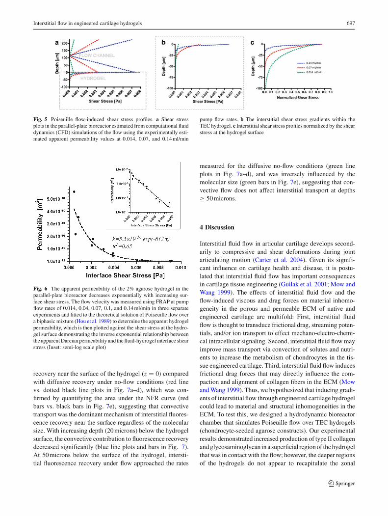
The shear stress profiles in the flow channel and withinthe hydrogels (Fig. 5a, b) were determined using computa-tional fluid dynamics (CFD) modeling (see Sect. 2 in thesupplemental data file) based on the experimentally esti-mated hydrogel permeability. The interfacial (hydrogel sur-face) shear stress increased linearly from 0.6 × 10⁻³ to8.2 × 10−3 Pa when the flow rate was increased from 0.014to 0.14 ml/min (Table 2). Interestingly, the interstitial shearstress gradients within the boundary layer also increased withincreasing flow rate (Fig. 5b, c). Furthermore, we observed
that in this flow configuration, the increase in interfacial shearstress was associated with an exponential reduction in theapparent permeability of the hydrogel (Fig. 6). Interestingly,the relationship between the apparent permeability and theinterfacial shear stress fits an exponential function of the formk = ko × exp(−Mτ), where M is an empirical shear stress-dependent permeability factor (Pa−1).
3.4 Convective mass transport enhancement correlateswith interstitial flow gradients
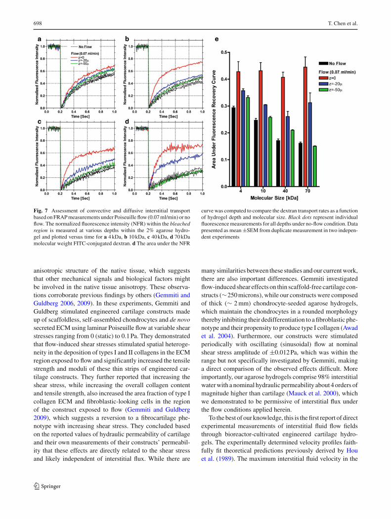
To formally evaluate whether Poiseuille-driven flow improvesinterstitial mass transport, FRAP-based measurements underflow (0.07 ml/min) or no flow were repeated for differentdepths within 2% agarose hydrogel slabs saturated in aque-ous solutions of various molecular weight FITC-dextran(4, 10, 40, and 70 kDa). Under no-flow conditions where dif-fusion is the only mode of interstitial fluorescence recov-ery, there were no depth-dependent effects regardless of thedextran molecular size (dotted black line plots—Fig. 7a–d).Furthermore, quantification of the area under the normal-ized fluorescence recovery (NFR) curve demonstrated theexpected inverse effects of molecular size on the diffusiverecovery of fluorescence (black bars in Fig. 7e). On the otherhand, Poiseuille flow significantly accelerated fluorescence
123
Interstitial flow in engineered cartilage hydrogels 697
Fig. 5 Poiseuille flow-induced shear stress profiles. a Shear stressplots in the parallel-plate bioreactor estimated from computational fluiddynamics (CFD) simulations of the flow using the experimentally esti-mated apparent permeability values at 0.014, 0.07, and 0.14 ml/min
pump flow rates. b The interstitial shear stress gradients within theTEC hydrogel. c Interstitial shear stress profiles normalized by the shearstress at the hydrogel surface
Fig. 6 The apparent permeability of the 2% agarose hydrogel in theparallel-plate bioreactor decreases exponentially with increasing sur-face shear stress. The flow velocity was measured using FRAP at pumpflow rates of 0.014, 0.04, 0.07, 0.1, and 0.14 ml/min in three separateexperiments and fitted to the theoretical solution of Poiseuille flow overa biphasic mixture (Hou et al. 1989) to determine the apparent hydrogelpermeability, which is then plotted against the shear stress at the hydro-gel surface demonstrating the inverse exponential relationship betweenthe apparent Darcian permeability and the fluid-hydrogel interface shearstress (Inset: semi-log scale plot)
recovery near the surface of the hydrogel (z = 0) comparedwith diffusive recovery under no-flow conditions (red linevs. dotted black line plots in Fig. 7a–d), which was con-firmed by quantifying the area under the NFR curve (redbars vs. black bars in Fig. 7e), suggesting that convectivetransport was the dominant mechanism of interstitial fluores-cence recovery near the surface regardless of the molecularsize. With increasing depth (20 microns) below the hydrogelsurface, the convective contribution to fluorescence recoverydecreased significantly (blue line plots and bars in Fig. 7).At 50 microns below the surface of the hydrogel, intersti-tial fluorescence recovery under flow approached the rates
measured for the diffusive no-flow conditions (green lineplots in Fig. 7a–d), and was inversely influenced by themolecular size (green bars in Fig. 7e), suggesting that con-vective flow does not affect interstitial transport at depths≥ 50 microns.
4 Discussion
Interstitial fluid flow in articular cartilage develops second-arily to compressive and shear deformations during jointarticulating motion (Carter et al. 2004). Given its signifi-cant influence on cartilage health and disease, it is postu-lated that interstitial fluid flow has important consequencesin cartilage tissue engineering (Guilak et al. 2001; Mow andWang 1999). The effects of interstitial fluid flow and theflow-induced viscous and drag forces on material inhomo-geneity in the porous and permeable ECM of native andengineered cartilage are multifold: First, interstitial fluidflow is thought to transduce frictional drag, streaming poten-tials, and/or ion transport to effect mechano-electro-chemi-cal intracellular signaling. Second, interstitial fluid flow mayimprove mass transport via convection of solutes and nutri-ents to increase the metabolism of chondrocytes in the tis-sue engineered cartilage. Third, interstitial fluid flow inducesfrictional drag forces that may directly influence the com-paction and alignment of collagen fibers in the ECM (Mowand Wang 1999). Thus, we hypothesized that inducing gradi-ents of interstitial flow through engineered cartilage hydrogelcould lead to material and structural inhomogeneities in theECM. To test this, we designed a hydrodynamic bioreactorchamber that simulates Poiseuille flow over TEC hydrogels(chondrocyte-seeded agarose constructs). Our experimentalresults demonstrated increased production of type II collagenand glycosaminoglycan in a superficial region of the hydrogelthat was in contact with the flow; however, the deeper regionsof the hydrogels do not appear to recapitulate the zonal
123
698 T. Chen et al.
Fig. 7 Assessment of convective and diffusive interstitial transportbased on FRAP measurements under Poiseuille flow (0.07 ml/min) or noflow. The normalized fluorescence intensity (NFR) within the bleachedregion is measured at various depths within the 2% agarose hydro-gel and plotted versus time for a 4 kDa, b 10 kDa, c 40 kDa, d 70 kDamolecular weight FITC-conjugated dextran. d The area under the NFR
curve was computed to compare the dextran transport rates as a functionof hydrogel depth and molecular size. Black dots represent individualfluorescence measurements for all depths under no-flow condition. Datapresented as mean ±SEM from duplicate measurement in two indepen-dent experiments
anisotropic structure of the native tissue, which suggeststhat other mechanical signals and biological factors mightbe involved in the native tissue anisotropy. These observa-tions corroborate previous findings by others (Gemmiti andGuldberg 2006, 2009). In these experiments, Gemmiti andGuldberg stimulated engineered cartilage constructs madeup of scaffoldless, self-assembled chondrocytes and de novosecreted ECM using laminar Poiseuille flow at variable shearstresses ranging from 0 (static) to 0.1 Pa. They demonstratedthat flow-induced shear stresses stimulated spatial heteroge-neity in the deposition of types I and II collagens in the ECMregion exposed to flow and significantly increased the tensilestrength and moduli of these thin strips of engineered car-tilage constructs. They further reported that increasing theshear stress, while increasing the overall collagen contentand tensile strength, also increased the area fraction of type Icollagen ECM and fibroblastic-looking cells in the regionof the construct exposed to flow (Gemmiti and Guldberg2009), which suggests a reversion to a fibrocartilage phe-notype with increasing shear stress. They concluded basedon the reported values of hydraulic permeability of cartilageand their own measurements of their constructs’ permeabil-ity that these effects are directly related to the shear stressand likely independent of interstitial flux. While there are
many similarities between these studies and our current work,there are also important differences. Gemmiti investigatedflow-induced shear effects on thin scaffold-free cartilage con-structs (∼250 microns), while our constructs were composedof thick (∼ 2 mm) chondrocyte-seeded agarose hydrogels,which maintain the chondrocytes in a rounded morphologythereby inhibiting their dedifferentiation to a fibroblastic phe-notype and their propensity to produce type I collagen (Awadet al. 2004). Furthermore, our constructs were stimulatedperiodically with oscillating (sinusoidal) flow at nominalshear stress amplitude of ±0.012 Pa, which was within therange but not specifically investigated by Gemmiti, makinga direct comparison of the observed effects difficult. Moreimportantly, our agarose hydrogels comprise 98% interstitialwater with a nominal hydraulic permeability about 4 orders ofmagnitude higher than cartilage (Mauck et al. 2000), whichwe demonstrated to be permissive of interstitial flux underthe flow conditions applied herein.
To the best of our knowledge, this is the first report of directexperimental measurements of interstitial fluid flow fieldsthrough bioreactor-cultivated engineered cartilage hydro-gels. The experimentally determined velocity profiles faith-fully fit theoretical predictions previously derived by Houet al. (1989). The maximum interstitial fluid velocity in the
123

Interstitial flow in engineered cartilage hydrogels 699
hydrogel was about 1 to 11/2 orders of magnitude higher thanthe values reported for compression-induced interstitial flowin cartilage (Buschmann et al. 1995; Mow and Wang 1999),which seems intuitively reasonable given the differences inhydraulic permeability between the native tissue and the aga-rose hydrogel.
Based on the interstitial fluid velocity measurements andthe theory, we estimated the apparent hydrogel permeabil-ity under the various flow conditions and made the observa-tion that the apparent hydrogel permeability decreases withincreasing flow rate and hence shear stress at the hydro-gel surface. The permeability of hydrated mixtures such asbiological tissues and engineered hydrogels is an intrinsicproperty that governs interstitial fluid flow and is typicallyestimated using Darcy’s Law, which for a unidirectional flowin a hydrogel column with cross-sectional area (A) predictsthat the volumetric flow rate (Q) or the average velocity (v)
of a Newtonian fluid through the hydrogel is directly propor-tional to the pressure drop (P) across the column height(h).
v = Q
A= κ
μ
P
h(8)
The proportionality constant is defined as the hydraulicpermeability (k = κ/μ), which is the ratio of Darcy’spermeability of the hydrogel (κ) to the dynamic viscos-ity of the fluid (μ). The permeability is often reportedfor native and engineered cartilage as a material prop-erty and has significant implications for tissue biome-chanics and interstitial transport (diffusion and convection)(Mow and Wang 1999).
We observed that our 2% agarose gel permeability pre-dictions were several orders of magnitude higher than therange of intrinsic permeability values reported in the lit-erature from permeation and confined compression experi-ments (Gu et al. 2003; Johnson and Deen 1996; Mauck et al.2000). One potential cause is that our FRAP-based measure-ments were done in a hyposmotic aqueous solution and couldnot, due to technical difficulties, be reproducibly performedat physiologic ionic strength that would replicate the cul-ture conditions. However, it has been recently shown thatdecreasing the ionic strength of negatively-charged agarose-GAG gels by 2 orders of magnitude results in only a two-fold decrease in Darcy’s permeability (Mattern et al. 2008).In our electrically neutral agarose hydrogels, the oppositeeffects might be expected due to the lack of charged spe-cies within the gel, although the magnitude of the hypotoniceffect is expected to be similarly small, hence suggestingthis mechanism is not the reason behind the observed dis-crepancy in permeability values. On the other hand, it ispossible that small reductions in agarose concentration (e.g.increases in the water fraction and porosity) could resultin more pronounced permeability increases, compared with
changes in ionic strength, as has been experimentally deter-mined (Gu et al. 2003; Maroudas et al. 1969). However, basedon the empirical relationship between hydraulic permeabil-ity and the water/solid fraction of the agarose gel describedby Gu et al. (2003), the potential inaccuracies in preparingthe agarose hydrogel at the nominal concentration are notsufficient to explain the observed discrepancy in the perme-ability values. The more plausible explanation has in factbeen described by Beavers and Joseph (1967) in the con-text of deriving and experimentally validating the boundaryconditions at a permeable construct interface subject to freeflow, in which they argued that enforcing conservation ofmass and linear momentum of the fluid phase at the inter-face between the free flow and the agarose hydrogel inducesa “boundary layer” in the hydrogel within which the inter-stitial fluid flow velocity departs from Darcian predictions(e.g. significantly higher). Furthermore, within this bound-ary layer, the velocity changes rapidly from its “slip” value atthe interface to the Darcy’s value deeper within the hydrogel,giving rise to the observed flow gradients. Thus, the bound-ary layer phenomenon, which does not conform to Darcy’sestimation, results in an increase in the interstitial flow nearthe hydrogel interface with the free flow in the Poiseuillechannel, consequently leading to an increase in the apparentpermeability of the hydrogel. In other words, strictly usingthe Darcian permeability values in simulating this problemfails to predict this boundary layer phenomenon and leadsto the wrong conclusion that interstitial fluid flow near theinterface is negligible.
Moreover, the velocity gradients within the boundary layerappear to be proportional to the shear stresses at the inter-face, such that the thickness of this interstitial boundarylayer decreases with increasing flow rate and interfacial shearstress, which explains the inverse exponential relationshipbetween the interface shear stress and the apparent hydro-gel permeability. The original observation that the apparentpermeability of the hydrogel decreases exponentially withincreasing shear stress at the free fluid-hydrogel interfacebears striking similarity to the well-described phenomenonof deformation-dependent permeability under compressiveloading, which is empirically modeled as an inverse expo-nential relationship (Mow et al. 1984). Previous reports cor-roborate our findings and suggest that the permeability ofbiological tissues or engineered scaffolds can be an effectiveindicator of shear stresses in these systems (Vossenberg et al.2009; Wang and Tarbell 1995). In agreement with our obser-vation, Vossenberg et al. (2009) recently demonstrated usinga finite element model that the average and maximum shearstress in perfused printed scaffolds increases significantlywith decreased porosity (and hence reduced permeability)and appears insensitive to pore size (Vossenberg et al. 2009).While the permeability data empirically fit an inverse expo-nential function of the interface shear stress, this curve fit and
123
700 T. Chen et al.
the continuum assumption used for predicting the interstitialflow fields and permeability do not explain this phenome-non. Under compression, the reduction in permeability isattributed to reductions in the effective porosity and the poresize, which contributes to fluid pressurization and enablescartilage to support large compressive loads. The mecha-nism of shear stress-dependent permeability in our systemis less clear, but could theoretically arise from compaction ofthe pores at the interface that effectively reduces their open-ing size and provides greater resistance to interstitial flow,similar to the mechanism observed under compression. Wehypothesize that there exists a flow threshold below which nosignificant deformations are experienced by the pore wallsat the hydrogel surface, and therefore, the interstitial veloc-ity at the interface increases with flow rate, which explainsthe increased interstitial velocity observed at the intermediateflow rate. When the flow rate increases beyond this threshold,the pore walls at the surface deform substantially and theiropenings reduce, which explains the reduction in the inter-stitial fluid velocity at the higher flow rate. This suggeststhat the interstitial boundary layer flow depends not only onintrinsic material properties such as the permeability but alsoon the structural nature of the porous interface (Beavers andJoseph 1967).
It should be noted that in scaling the bioreactor to enablethe experimental flow measurements, we maintained the ratioof the flow channel height to the biphasic layer thicknessλ = h1/h2. We also varied the pump flow rate to maintain thesame nominal interfacial shear stress range in the two experi-ments. Given that the material properties of the hydrogel andfluid are initially the same in the two systems, we believethat the observations from the experimental measurementsof interstitial flow are representative of the scaled-up bio-reactor used to cultivate the chondrocyte-seeded hydrogels.There are, however, important differences including the factthat flow in the cell culture experiments was oscillatory, whilethe flow in the FRAP experiments was steady for practicalreasons. Furthermore, the accrual of the ECM in the largerchondrocyte-seeded engineered cartilage hydrogel was notaccounted for in the scaled-down FRAP bioreactor measure-ments, which were done in a cell-free 2% agarose hydrogelrepresenting the initial material property of the chondrocyte-seeded hydrogel at day 0. We hypothesize that as new ECMis produced, the permeability of the engineered constructs,and consequently the interstitial fluid flow, would decrease.Formal investigation of this hypothesis will be the subject offuture studies.
The inverse nonlinear relationship between the interfacialshear stress and the apparent gel permeability has significantimplications for cartilage tissue engineering. While shearstress has important mechanostimulatory consequences forcartilage tissue engineering (Gemmiti and Guldberg 2006,2009; Pazzano et al. 2000; Saini and Wick 2003; Stoddart
et al. 2006), its effects on permeability and consequentlyon interstitial nutrient transport should not be overlooked.We demonstrated in this experiment that Poiseuille-inducedinterstitial flow enhances convective transport of FITC-conjugated, neutral dextran molecules in a boundary layer(∼ 50 microns) of high fluid flow near the surface of thehydrogel, independent of molecular size (for the range of3–70 kDa). These results correlate with interstitial flow mea-surements, which were negligible at depths ≥ 50 microns,approximately the thickness of the boundary layer at a flowrate of 0.07 ml/min (Fig. 4b, c). Theoretically, the thicknessof the boundary layer of enhanced flow in the larger “cell cul-ture” bioreactor should scale-up in proportion to the thick-ness of the hydrogel (Sect. 1, Supplemental Data), whichmight explain the thicker region (240 microns) of enhancedcell metabolic activity (Fig. 3). Additionally, gradients ofsoluble factors created as a result of the enhanced trans-port and mechanostimulation in the boundary layer mightlead to paracrine signal effects that extend beyond that layer.The convective transport decreased with depth through thehydrogel in association with the measured interstitial flowgradients, such that at depths where fluid flow was negli-gible the transport was dominated by passive diffusion. Weposit that increasing the interface shear stress at the interfacewith the hydrogel (by increasing the Poiseuille flow rate)and the associated decrease in apparent hydrogel permeabil-ity will hinder convective transport and affect the materialinhomogeneity within the engineered TEC hydrogels. Thishypothesis warrants formal investigation in future studies,especially since the broader significance of the inverse rela-tionship between the interfacial shear stress on one handand interstitial permeability and convective transport on theother is not limited to our experimental setup. Indeed, avariety of scaffolds for cartilage tissue engineering com-prise interconnected porous channels whose walls are linedwith seeded cells and their newly secreted ECM. Theseporous networks can be approximated as Poiseuille flowchannels through which fluid (i.e. culture media) flows pastthe neotissue lining the pores as a result of dynamic com-pressive stimulation or direct perfusion. Therefore, strik-ing a balance between the mechanostimulatory effects ofthe flow-induced shear stress and the potential hindranceof permeability and interstitial convective transport to theneotissue lining the porous scaffold walls should be an impor-tant design consideration in tissue engineering bioreactorsystems.
Acknowledgments We would like to thank Ryan Tierney and thehistology core at the Center for Musculoskeletal Research Center fortheir excellent technical assistance. We would like to thank ProfessorLori Setton for past discussions during class that inspired this work. Wewould also like to thank Professors Richard Waugh, J. Edward Puzas,Michael Zuscik, Edward Brown, and Michael King for valuable discus-sions throughout the evolution of this work. This work was supported by
123

Interstitial flow in engineered cartilage hydrogels 701
grants from the Empire State Stem Cell Board (NYSTEM N08G-019)and the NIH (AR056696 and AR054041).
References
Awad HA, Wickham MQ, Leddy HA, Gimble JM, Guilak F (2004)Chondrogenic differentiation of adipose-derived adult stem cellsin agarose, alginate, and gelatin scaffolds. Biomaterials 25(16):3211–3222
Beavers GS, Joseph DD (1967) Boundary conditions at a naturally per-meable wall. J Fluid Mech 30:197–207
Bonassar LJ, Grodzinsky AJ, Frank EH, Davila SG, Bhaktav NR,Trippel SB (2001) The effect of dynamic compression on theresponse of articular cartilage to insulin-like growth factor-I.J Orthop Res 19(1):11–17
Bujia J, Sittinger M, Minuth WW, Hammer C, Burmester G,Kastenbauer E (1995) Engineering of cartilage tissue using biore-sorbable polymer fleeces and perfusion culture. Acta Otolaryngol115(2):307–310
Buschmann MD, Gluzband YA, Grodzinsky AJ, Hunziker EB (1995)Mechanical compression modulates matrix biosynthesis in chon-drocyte/agarose culture. J Cell Sci 108(Pt 4):1497–1508
Buschmann MD, Kim YJ, Wong M, Frank E, Hunziker EB,Grodzinsky AJ (1999) Stimulation of aggrecan synthesis in car-tilage explants by cyclic loading is localized to regions of highinterstitial fluid flow. Arch Biochem Biophys 366(1):1–7
Butler DL, Goldstein SA, Guilak F (2000) Functional tissue engineer-ing: the role of biomechanics. J Biomech Eng 122(6):570–575
Carter DR, Beaupre GS, Wong M, Smith RL, Andriacchi TP, Schur-man DJ (2004) The mechanobiology of articular cartilage devel-opment and degeneration. Clin Orthop Relat Res (427 Suppl):S69–S77
Carter DR, Wong M (2003) Modelling cartilage mechanobiology. Phi-los Trans R Soc Lond B Biol Sci 358(1437): 1461–1471. doi:10.1098/rstb.2003.1346
Freed LE, Hollander AP, Martin I, Barry JR, Langer R,Vunjak-Novakovic G (1998) Chondrogenesis in a cell-polymer-bioreactor system. Exp Cell Res 240(1):58–65
Freed LE, Vunjak-Novakovic G, Langer R (1993) Cultivation ofcell-polymer cartilage implants in bioreactors. J Cell Biochem51(3):257–264
Gemmiti CV, Guldberg RE (2006) Fluid flow increases type II collagendeposition and tensile mechanical properties in bioreactor-growntissue-engineered cartilage. Tissue Eng 12(3):469–479
Gemmiti CV, Guldberg RE (2009) Shear stress magnitude and durationmodulates matrix composition and tensile mechanical properties inengineered cartilaginous tissue. Biotechnol Bioeng 104(4):809–820
Gray ML, Pizzanelli AM, Grodzinsky AJ, Lee RC (1988) Mechanicaland physiochemical determinants of the chondrocyte biosyntheticresponse. J Orthop Res 6(6):777–792
Gu WY, Yao H, Huang CY, Cheung HS (2003) New insight intodeformation-dependent hydraulic permeability of gels and carti-lage, and dynamic behavior of agarose gels in confined compres-sion. J Biomech 36(4):593–598
Guilak F, Butler DL, Goldstein SA (2001) Functional tissue engineer-ing: the role of biomechanics in articular cartilage repair. ClinOrthop Relat Res (391 Suppl):S295–S305
Hou JS, Holmes MH, Lai WM, Mow VC (1989) Boundary conditionsat the cartilage-synovial fluid interface for joint lubrication andtheoretical verifications. J Biomech Eng 111(1):78–87
Hung CT, Mauck RL, Wang CC, Lima EG, Ateshian GA (2004) Aparadigm for functional tissue engineering of articular cartilage
via applied physiologic deformational loading. Ann Biomed Eng32(1):35–49
Johnson EM, Deen WM (1996) Hydraulic permeability of agarose gels.Aiche J 42(5):1220–1224
Lamkin-Kennard KA, King MR, Awad HA (2005) A novel lid-driven cavity flow bioreactor for cartilage tissue engineering.In: Proceedings of the 2005 summer bioengineering conference,Vail, CO, USA. American Society of Mechanical Engineers,pp 1060–1061
Lammi MJ (2004) Current perspectives on cartilage and chondrocytemechanobiology. Biorheology 41(3–4):593–596
Leddy HA, Haider MA, Guilak F (2006) Diffusional anisotropy in col-lagenous tissues: fluorescence imaging of continuous point photo-bleaching. Biophys J 91(1):311–316
Lee HS, Millward-Sadler SJ, Wright MO, Nuki G, Salter DM (2000)Integrin and mechanosensitive ion channel-dependent tyrosinephosphorylation of focal adhesion proteins and beta-catenin inhuman articular chondrocytes after mechanical stimulation. J BoneMiner Res 15(8):1501–1509
Malone AM, Anderson CT, Tummala P, Kwon RY, Johnston TR,Stearns T, Jacobs CR (2007) Primary cilia mediate mechanosen-sing in bone cells by a calcium-independent mechanism. Proc NatlAcad Sci USA 104(33):13325–13330
Maroudas A, Muir H, Wingham J (1969) The correlation of fixed neg-ative charge with glycosaminoglycan content of human articularcartilage. Biochim Biophys Acta 177(3):492–500
Mattern KJ, Nakornchai C, Deen WM (2008) Darcy permeability ofagarose-glycosaminoglycan gels analyzed using fiber-mixture anddonnan models. Biophys J 95(2):648–656
Mauck RL, Soltz MA, Wang CC, Wong DD, Chao PH, Valhmu WB,Hung CT, Ateshian GA (2000) Functional tissue engineering ofarticular cartilage through dynamic loading of chondrocyte-seededagarose gels. J Biomech Eng 122(3):252–260
Mauck RL, Wang CC, Oswald ES, Ateshian GA, Hung CT (2003) Therole of cell seeding density and nutrient supply for articular car-tilage tissue engineering with deformational loading. OsteoarthrCartil 11(12):879–890
Mow VC, Wang CC (1999) Some bioengineering considerations for tis-sue engineering of articular cartilage. Clin Orthop Relat Res (367Suppl):S204–S223
Mow VC, Holmes MH, Lai WM (1984) Fluid transport and mechanicalproperties of articular cartilage: a review. J Biomech 17(5):377–394
Pazzano D, Mercier KA, Moran JM, Fong SS, DiBiasio DD, Rulfs JX,Kohles SS, Bonassar LJ (2000) Comparison of chondrogensis instatic and perfused bioreactor culture. Biotechnol Prog 16(5):893–896
Pierre J, Gemmiti CV, Kolambkar YM, Oddou C, Guldberg RE (2008)Theoretical analysis of engineered cartilage oxygenation: influ-ence of construct thickness and media flow rate. Biomech ModelMechanobiol 7(6):497–510
Polianin AD (2002) Handbook of linear partial differential equationsfor engineers and scientists. Chapman & Hall/CRC, Boca Raton
Rutkowski JM, Swartz MA (2007) A driving force for change:interstitial flow as a morphoregulator. Trends Cell Biol 17(1):44–50
Sah RL, Kim YJ, Doong JY, Grodzinsky AJ, Plaas AH, Sandy JD(1989) Biosynthetic response of cartilage explants to dynamiccompression. J Orthop Res 7(5):619–636
Saini S, Wick TM (2003) Concentric cylinder bioreactor for produc-tion of tissue engineered cartilage: effect of seeding density andhydrodynamic loading on construct development. Biotechnol Prog19(2):510–521
Shi ZD, Wang H, Tarbell JM (2011) Heparan sulfate proteoglycansmediate interstitial flow mechanotransduction regulating MMP-13
123
702 T. Chen et al.
expression and cell motility via FAK-ERK in 3D collagen. PloSone 6(1):e15956
Shieh AC, Swartz MA (2011) Regulation of tumor invasion by intersti-tial fluid flow. Phys Biol 8(1):015012
Sittinger M, Bujia J, Minuth WW, Hammer C, Burmester GR (1994)Engineering of cartilage tissue using bioresorbable polymer carri-ers in perfusion culture. Biomaterials 15(6):451–456
Stoddart MJ, Ettinger L, Hauselmann HJ (2006) Enhanced matrix syn-thesis in de novo, scaffold free cartilage-like tissue subjected tocompression and shear. Biotechnol Bioeng 95(6):1043–1051
Vossenberg P, Higuera GA, van Straten G, van Blitterswijk CA,van Boxtel AJ (2009) Darcian permeability constant as indicatorfor shear stresses in regular scaffold systems for tissue engineering.Biomech Model Mechanobiol 8(6):499–507
Wang DM, Tarbell JM (1995) Modeling interstitial flow in an arterywall allows estimation of wall shear stress on smooth muscle cells.J Biomech Eng 117(3):358–363
Wong M, Carter DR (2003) Articular cartilage functional histomor-phology and mechanobiology: a research perspective. Bone33(1):1–13
123