Embed Size (px)
Citation preview
**Vermont Genetics Network University of Vermont Microarray Outreach Program**
Instruction Team: Scott Tighe, Tim Hunter, Pat Reed, and Janet Murray Manual: Version cerevisiae 12/09
**Vermont Genetics Network University of Vermont Microarray Outreach Program**
2
Overview
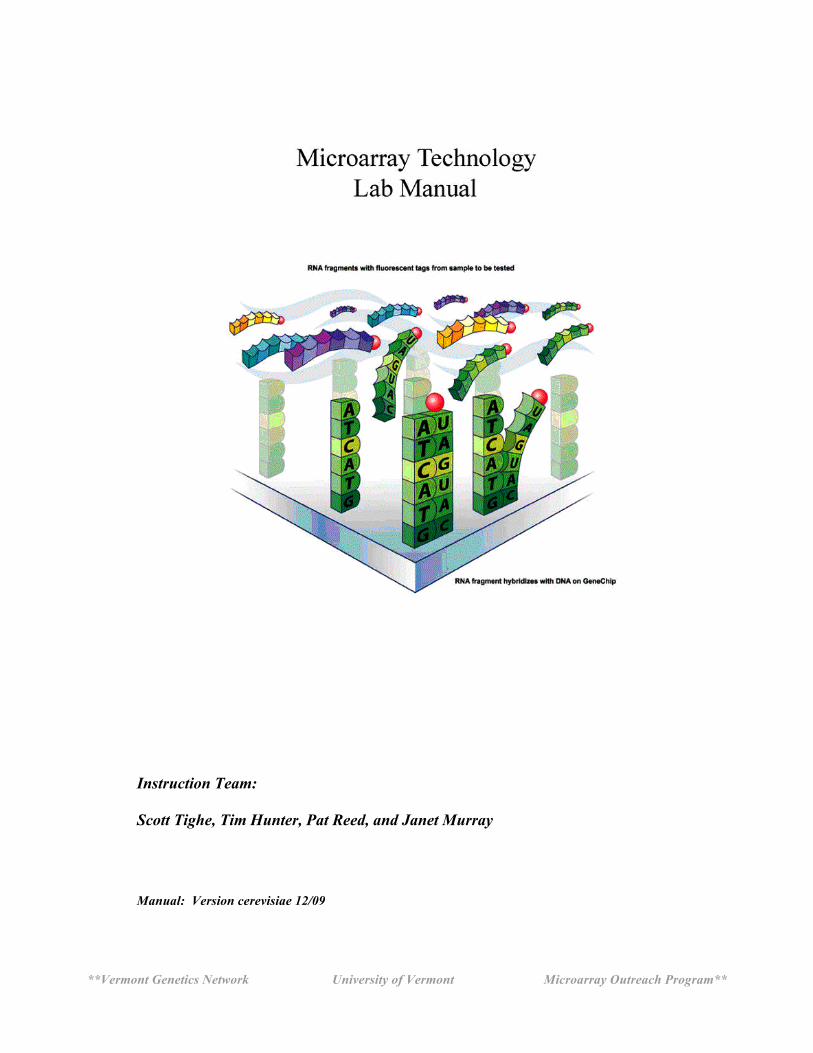
In this module, you will learn how gene expression in yeast (Saccharomyces cerevisiae) is changed after exposure to oxidative stress induced by the addition of hydrogen peroxide (H2O2), an oxidizing agent. In the experiment, yeast are grown for 48 hours in 1/2x YPD broth containing 3x glucose. The culture is split into a control and treated group. The experiment culture is treated with 0.5mM H2O2 in Hanks Buffered Saline (HBSS) for 1 hour. The control culture is treated with HBSS only. Total RNA is extracted from both cultures and is converted to a labeled cRNA product through a multi-step process. The final synthesis product is taken back to the UVM Microarray Core Facility and hybridized to the Affymetrix yeast GeneChips. The resulting gene expression data are uploaded into bioinformatics data analysis software. You will select specific genes whose expressions have changed as a result of the exposure to H2O2 and generate a report to the class.
**Vermont Genetics Network University of Vermont Microarray Outreach Program**
3
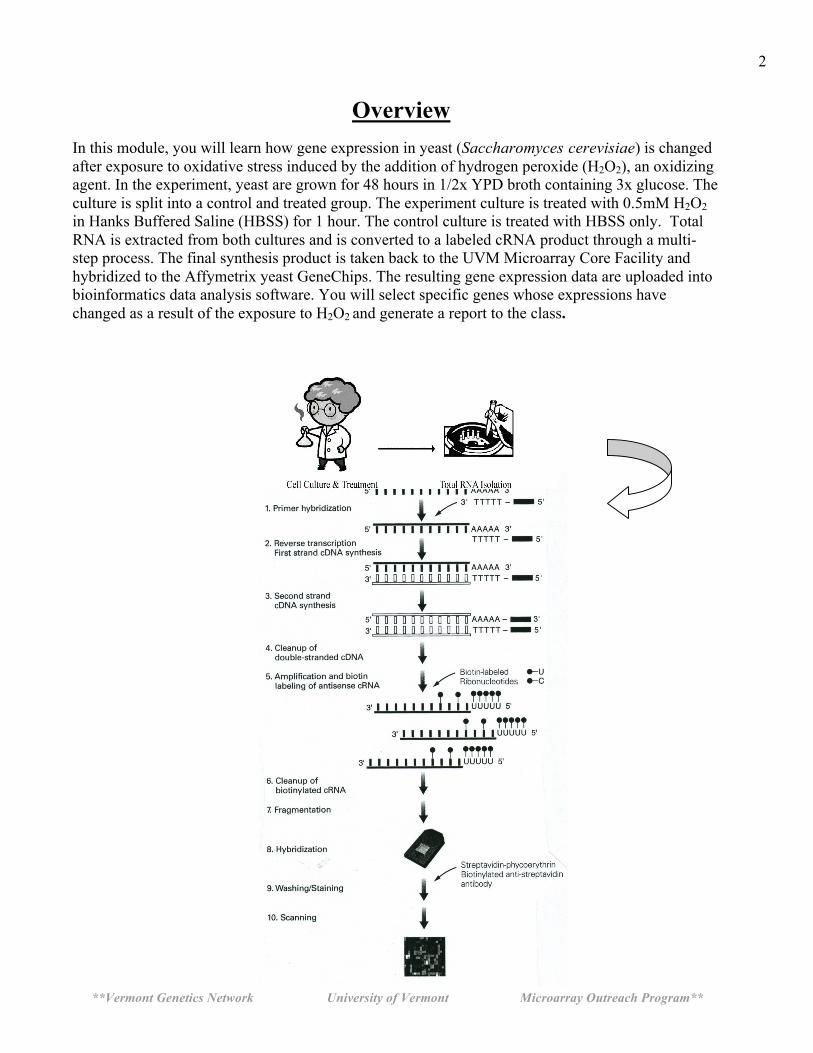
Why Use Yeast (S. cerevisiae)??
• easily manipulated in the laboratory • simple eukaryote, unicellular • rapid growth (doubling 1.5 - 2.5 hours) • non-pathogenic • stable haploid and diploid states • complete genome sequenced • very well characterized • many pathways are conserved Yeast Life cycle
http://www.bath.ac.uk/bio-sci/images/profiles/wheals2.gif

**Vermont Genetics Network University of Vermont Microarray Outreach Program**
Special Notes- Important!
1] RNase-free water is DEPC water and the terms maybe used interchangeably throughout the protocol.
2] Record all data in notebooks. 3] Label the tops and sides of tubes with: Sample ID/Initials Date What is in the tube…ie cDNA, RNA, etc. Concentration 4] Check off each step in the lab manual as you complete it. 5] Read the Technical Discussion section before each day. This is fair game for quiz questions. 6] MSDS safety sheets are available for each chemical in the front of the room. 7] RPM on a Centrifuge does NOT equal G-force. See the conversion chart below
8] Use RNase Zap anytime you want, better safe then sorry!
**Vermont Genetics Network University of Vermont Microarray Outreach Program**
5
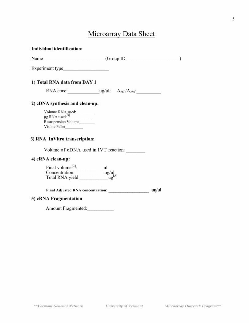
Microarray Data Sheet Individual identification: Name _________________________ (Group ID ______________________) Experiment type___________________
1) Total RNA data from DAY 1
RNA conc:_____________ug/ul: A260/A280:__________
2) cDNA synthesis and clean-up:
Volume RNA used: _________ µg RNA used[B] ___________ Resuspension Volume________ Visible Pellet_________
3) RNA InVitro transcription:
Volume of cDNA used in IVT reaction: ________ 4) cRNA clean-up:
Final volume[C]: __________ ul Concentration: ____________ug/ul Total RNA yield ____________ug[A]
Final Adjusted RNA concentration: _________________ ug/ul
5) cRNA Fragmentation: Amount Fragmented:___________
**Vermont Genetics Network University of Vermont Microarray Outreach Program**
6
Attach Spectrophometer data
Attach gel images
**Vermont Genetics Network University of Vermont Microarray Outreach Program**
7
Set-up before day 1: For course Instructor
Note: The broth culture must be inoculated about 48 hours before the
treatment procedure of day 1 with a log phase culture of the yeast [See appendix A].
Note : The H2O2 treatment procedure must be performed 0.5 hour before class begins: total treatment time is 60 minutes.
Protocol for Preparing Yeast Cultures for Microarray Analysis (3-4 days prior to day 1)
Necessary Supplies Autoclave (tape, tinfoil) Inoculating loop (or sterile swabs) and Bunsen burner Sterile flasks containing stir bars that are the same size: (1) 500 ml flask, (2)125 ml flasks (2) Stir plates 5 and 25 ml pipettes (sterile) Tape and sharpies for labeling
Supplies provided by VGN Yeast Stain Saccharomyces cerevisiae [NRRLY-12632 or ATCC 18824]` 1/2xYPD+3x glucose nutrient media (modified powder –Fisher Scientific-This is pre-made by VGN staff. (12.5gram YPD powder+10g glucose+500ml water) 30% Hydrogen Peroxide (H2O2) Sterile DI Water HBSS without Ca, Mg, or dye
The broth culture must be inoculated 48 hours before the treatment procedure of day 1. Incubation should be conducted in a room that is 70-80°F. It is best to inoculate this culture from a pre-culture that is in log phase.
Plan the treatment of the cultures so that the 1 hour treatment is complete 30 minutes AFTER the start of class on Day 1. Check off each step in the protocol after completing it.
Please read through all instructions before proceeding.
**Vermont Genetics Network University of Vermont Microarray Outreach Program**
8
A) Making the growth media and sterilizing flasks with stir bars. ____1) Prepare 250 ml of 1/2x YPD-3x Glu broth. Weigh 22.5 gm of the media and transfer to
a 500 ml flask. Transfer 250 ml dd water to the flask. Place a stir bar in the flask, wrap tin foil loosely around the top, label the flask, and autoclave under standard conditions (15 psig at 121°C) for 15 minutes.
____2) Also autoclave (2) empty 125 ml flasks with the same size stir bar in each. Wrap
top in foil. It is important not to autoclave LONGER than 15 minutes as the sugars will caramelize and degrade.
B) Inoculating the parent culture
____1) Use sterile technique throughout this procedure. Bacterial contamination will be
undetectable throughout the rest of the experiment and will adversely affect the final results.
____2) The parent culture should be inoculated 48 hrs before class of day 1 from a log phase
pre-culture. See appendix A. Use a good size inoculum!! ____3 Transfer a colony of an actively growing yeast using an inoculating loop to the broth.
Alternatively, if a liquid pre-culture is used, a 500 µl aliquot can be transferred to the new media.
____4) Uncap and tilt the 500 ml flask at a 30 degree angle [or so] and inoculate the 1/2x YPD-3x
Glu broth .
Notice: you want to minimize the time the flask is open AND you do not want your hands or sleeves over the flask opening. Broth is easily contaminated and it will not be possible to detect this until the very end of the experiment. ____5) Flame the mouth of the flask and place the tinfoil cap back on the flask. Replace the
tinfoil around the mouth of the flask, but not too tightly as it is necessary for oxygen to get in. It is important that the yeast grow aerobically as respiration is the metabolic process that builds cell mass.
____6) Place the flask on stir plate and stir approximately 48 hours at a medium speed at
room temp (22-25°C). Do not have the speed so high that there is foaming. The culture should be aerated to ensure the yeast are not in a fermentation metabolism.
**Vermont Genetics Network University of Vermont Microarray Outreach Program**
9
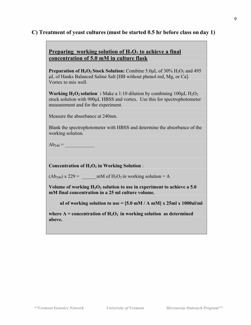
C) Treatment of yeast cultures (must be started 0.5 hr before class on day 1) Preparing working solution of H2O2 to achieve a final concentration of 5.0 mM in culture flask Preparation of H2O2 Stock Solution: Combine 5.0µL of 30% H2O2 and 495 µL of Hanks Balanced Saline Salt [HB without phenol red, Mg, or Ca]. Vortex to mix well. Working H2O2 solution : Make a 1:10 dilution by combining 100µL H2O2 stock solution with 900µL HBSS and vortex. Use this for spectrophotometer measurement and for the experiment. Measure the absorbance at 240nm. Blank the spectrophotometer with HBSS and determine the absorbance of the working solution. Ab240 = ____________
Concentration of H2O2 in Working Solution :
(Ab240) x 229 = ______mM of H2O2 in working solution = A
Volume of working H2O2 solution to use in experiment to achieve a 5.0 mM final concentration in a 25 ml culture volume.
ul of working solution to use = [5.0 mM / A mM] x 25ml x 1000ul/ml
where A = concentration of H2O2 in working solution as determined above.
**Vermont Genetics Network University of Vermont Microarray Outreach Program**
10
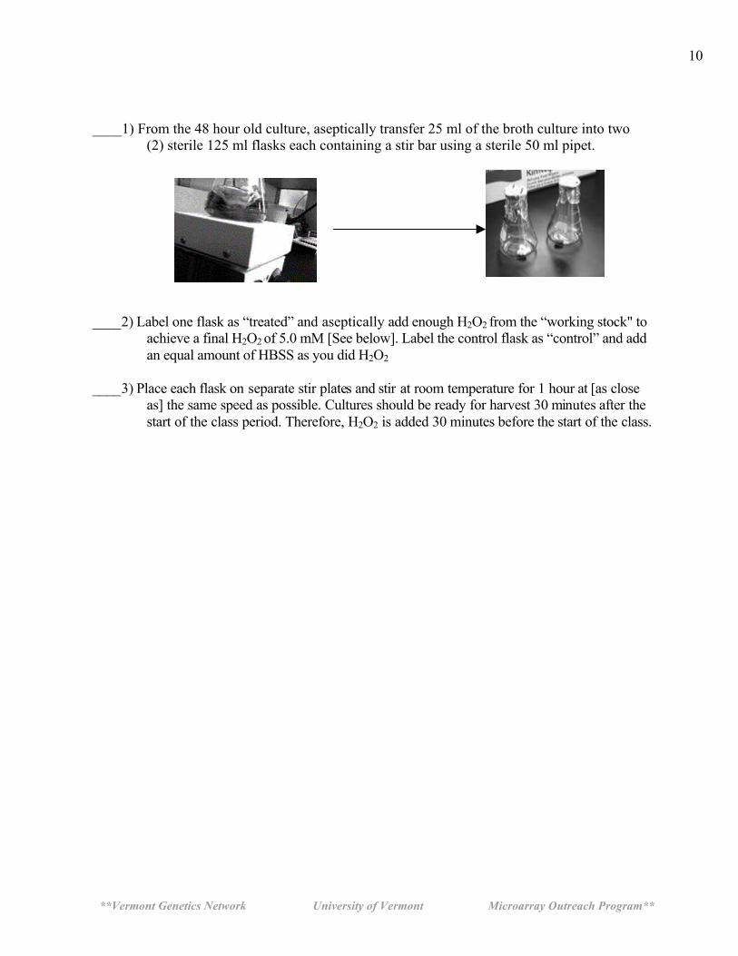
____1) From the 48 hour old culture, aseptically transfer 25 ml of the broth culture into two
(2) sterile 125 ml flasks each containing a stir bar using a sterile 50 ml pipet.
____2) Label one flask as “treated” and aseptically add enough H2O2 from the “working stock" to
achieve a final H2O2 of 5.0 mM [See below]. Label the control flask as “control” and add an equal amount of HBSS as you did H2O2
____3) Place each flask on separate stir plates and stir at room temperature for 1 hour at [as close
as] the same speed as possible. Cultures should be ready for harvest 30 minutes after the start of the class period. Therefore, H2O2 is added 30 minutes before the start of the class.
**Vermont Genetics Network University of Vermont Microarray Outreach Program**
11
DAY 1
Isolating Total RNA from the Yeast Using Enzymatic Lysis
Protocol. Technical Overview:
You will be treating the yeast, Saccharomyces cerevisiae, with hydrogen peroxide [H2O2] to
determine the effects on gene expression during oxidative stress conditions. After one hour of
treatment the treated and control yeast cultures are pelleted by centrifugation. The yeast are
treated with lyticase, an enzyme that degrades the poly-β-1,3 glucan bonds in the cell wall,
resulting in the formation of spheroplasts (a yeast cell without a cell wall). The second step
ruptures the spheoplasts using a buffer containing the chaotropic agent, guanidinium
isothiocyanate, which dissolves the cell membrane. All the RNA is collected using a silica gel
spin column manufactured by Qiagen. (RNeasy system) Please read through all laboratory procedures prior to lab.
Check off each step in the protocol as you complete it.
**Vermont Genetics Network University of Vermont Microarray Outreach Program**
12
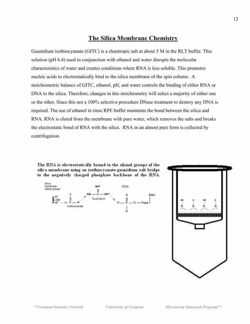
The Silica Membrane Chemistry
Guanidium isothiocyanate (GITC) is a chaotropic salt at about 5 M in the RLT buffer. This
solution (pH 6.4) used in conjunction with ethanol and water disrupts the molecular
characteristics of water and creates conditions where RNA is less soluble. This promotes
nucleic acids to electrostatically bind to the silica membrane of the spin column. A
stoichiometric balance of GITC, ethanol, pH, and water controls the binding of either RNA or
DNA to the silica. Therefore, changes in this stoichiometry will select a majority of either one
or the other. Since this not a 100% selective procedure DNase treatment to destroy any DNA is
required. The use of ethanol in rinse RPE buffer maintains the bond between the silica and
RNA. RNA is eluted from the membrane with pure water, which removes the salts and breaks
the electrostatic bond of RNA with the silica. RNA in an almost pure form is collected by
centrifugation.
**Vermont Genetics Network University of Vermont Microarray Outreach Program**
13
Isolating total RNA from Saccharomyces cerevisiae using Enzymatic Lysis
Materials: Axygen MCT150C 1.7ml tubes P1000, P200, and P20 pipets Yeast Lytic Enzyme-VWR # IC15352710-MP Biomedical 153527-10,000 units) 100 % Ethanol DEPC water (aka RNase-free water) Qiagen Mini RNeasy kit Qiagen DNase Kit Vortex Microcentrifuge Spectrophotometer Eppendorf UVettes 2 ml microcentrifuge tubes 2% SDS
____1. Prepare an RNase-free work zone using RNase ZAP. This will be demonstrated by the instructor.
____ 2a. Prepare fresh working lyticase reagent by adding 1 ml of DEPC water directly to the
lyticase vial. Vortex well and invert several times to insure complete mixing. This 10 units/ul solution is stable for 12 hours.
10,000 U Lyticase 1 ml DEPC Water
_____ 2b. Prepare fresh DNase I solution using the Qiagen DNase kit and store on ice.
10 µl DNase I
70 µl Buffer RDD 80 µl per reaction
____3. Label a 1.7 ml microcentrifuge tube with your identification information. Transfer
1.5 ml of the appropriate yeast culture the tube.
Note: The Instructor will perform step 2 tasks.
**Vermont Genetics Network University of Vermont Microarray Outreach Program**
14
____4. Centrifuge the tube at 5000x g (about 3/4 full speed) for 2 minutes at room temperature. Carefully remove the supernatant (without disturbing pellet) using a micropipette and discard the supernatant.
. **** Repeat step 4 if pellet size is too small or if indicated by instructor.****
____5. Add 1000 ul DEPC water to the pellet, vortex, and centrifuge at 5000 x g for 2 minutes.
Discard the supernatant. Remove as much of the liquid as possible from the yeast pellet.
____ 6. Add the following to the yeast pellet and vortex to mix. SG buffer 10 µl
Lyticase solution (10u/µl) 30 µl ____7. Incubate for 30 min at room temperature.
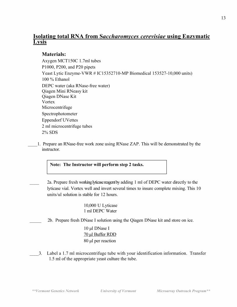
____8. Gently swirl the tube every 10 minutes to generate spheroplasts. Spheroplasts must
be handled gently. [optional] …Check spheroplast formation by placing 10ul of cells on a slide with
10ul of 2% SDS. These and can be swirl mixed with a pipet tip right on the slide. ____9. Add 350 µl b-RLT buffer to the tube and vortex vigorously for 1 minute. Ensure your
tube is tightly capped by holding the lid closed while vortexing. This procedure will lyse the spheroplasts.
____10. Add 250 µl 100% ethanol to the tube and briefly vortex. A precipitate may form after the addition of ethanol, but this will not affect the collection of RNA.
____11. Collecting the RNA: Assemble an RNeasy spin column with a 2 ml collection
tube. Label the spin column with your identification information. Carefully transfer all of the solution from step 10 to the spin column using a micropipette. Be careful not to touch the silica membrane with the pipette tip. Close the tube and centrifuge for 15 seconds at full speed. The RNA in the sample will adhere to the silica membrane of the spin column.
**Vermont Genetics Network University of Vermont Microarray Outreach Program**
15
____12. Remove the spin column from the collection tube. Pipet the pass through liquid (the
liquid in the collection tube) back onto the spin column and centrifuge again. Discard the collection tube (with the flow through fluid) and place the spin column into a new 2 ml collection tube.
____13. Add 350 µl RW1 buffer to the spin column. This solution is used to wash the RNA
and remove salts and dissolved cellular debris. Close the tube and centrifuge for 15 seconds at full speed.
____14. Degrading DNA: Remove the spin column from the centrifuge, open the
tube lid and add 80 µl of DNase I solution to the middle of the spin column membrane. This step will degrade any DNA in the sample and dissociate it from the silica membrane. Close the column lid and incubate at room temperature for 15 minutes.
____15. Transfer the spin column to a new 2 ml collection tube. ____16. Cleaning and washing the RNA: Add 350 µl RW1 buffer to the spin column and
centrifuge at full speed for 15 seconds. ____17. Discard the collection tube and place the spin column into a new 2 ml collection tube.
____18. Add 500 µl RPE buffer to the spin column to wash the RNA on the silica membrane. Close the tube and centrifuge for 15 seconds at full speed.
____19. Discard the collection tube and place the spin column into a new 2 ml collection tube.
____20. W a s h t h e silica membrane again by a dding another 500 µl RPE buffer to the spin
column. Close the tube and centrifuge for 15 seconds at full speed.
____21. Place the spin column in a new 2 ml collection tube and centrifuge in a microcentrifuge at full speed for 1 minute to ensure any residual liquid is removed from the silica membrane.
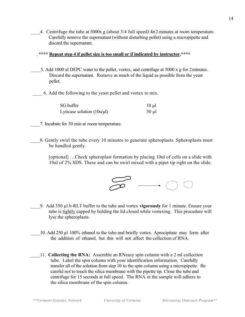
____22. Recovering the RNA: Label a 1.7 microcentrifuge tube with your identification
information. Transfer the spin column to this tube. The lid of the microcentrifuge tube will be off to the side. Carefully pipet 30 µl DEPC water directly onto the very center of the silica membrane. Do not touch the silica membrane with the pipet tip (see diagram). Look closely as you perform this step. Use both hands when pipetting, one on top and one down by the tip to guide the pipet. Make sure the water is evenly distributed on the membrane.
**Vermont Genetics Network University of Vermont Microarray Outreach Program**
16
____23. Incubate the spin column at room temperature for 1 minute. Centrifuge at full speed for 30 seconds.
____24. Carefully transfer the 30 µl that is recovered in the 1.7 ml tube back onto the center of
the silica membrane of the same spin column as in step 22 above. Place the column back in the same 1.7 ml collection tube and incubate for 1 minute at room temperature. Centrifuge for 1 minute at full speed. This double extraction ensures that the maximal amount of RNA is eluted from the membrane.
____25. Label two new 1.7 microcentrifuge tubes with your identification
information. Transfer a l l the recovered RNA s a m p l e to one of these tubes.
____26. Transfer 4 µl of this sample to the other labeled tube. This sample will be transport back to UVM for Nanodrop quantification and RNA assessment.
____27. Place all samples on ice. _____28. Quantify the RNA: Measure the absorbance of the RNA sample on the
spectrophotometer at a 1 to 50 dilution (1µl sample + 49µl H2O). This will be demonstrated by the instructor.
Absorbance of sample:_________________ A260/A280 ratio:_________________ Concentration of RNA in sample:_______________ug/ul Record these data on the data sheet provided on page 5 of the manual. ____28. Evaluate the RNA quality using agarose gel electrophoresis . This will be done as a
class.
_____29. Set up the E-gel electrophoresis apparatus. Pre-run the gel for 2 minutes by pressing the button continuously for 10 seconds. You will see the light start to blink indicating it is in a pre-run mode. It will beep and stop after 2 minutes.
Add 14 µl of water to each well on the E-gel. Add 1µl of each sample to each well and pipet up and down to mix well. Record which sample is in each lane. Run the gel for 20 minutes.
____ 30. Take a photo of the gel.
A sample of your RNA may be analyzed on the Agilent 2100 Bioanalyzer at UVM, which provides a move sensitive image of the RNA quality. (Described on the next page).
**Vermont Genetics Network University of Vermont Microarray Outreach Program**
17
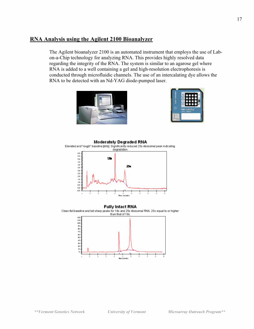
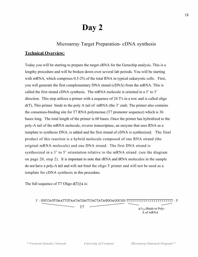
RNA Analysis using the Agilent 2100 Bioanalyzer
The Agilent bioanalyzer 2100 is an automated instrument that employs the use of Lab-on-a-Chip technology for analyzing RNA. This provides highly resolved data regarding the integrity of the RNA. The system is similar to an agarose gel where RNA is added to a well containing a gel and high-resolution electrophoresis is conducted through microfluidic channels. The use of an intercalating dye allows the RNA to be detected with an Nd-YAG diode-pumped laser.
**Vermont Genetics Network University of Vermont Microarray Outreach Program**
18
Day 2
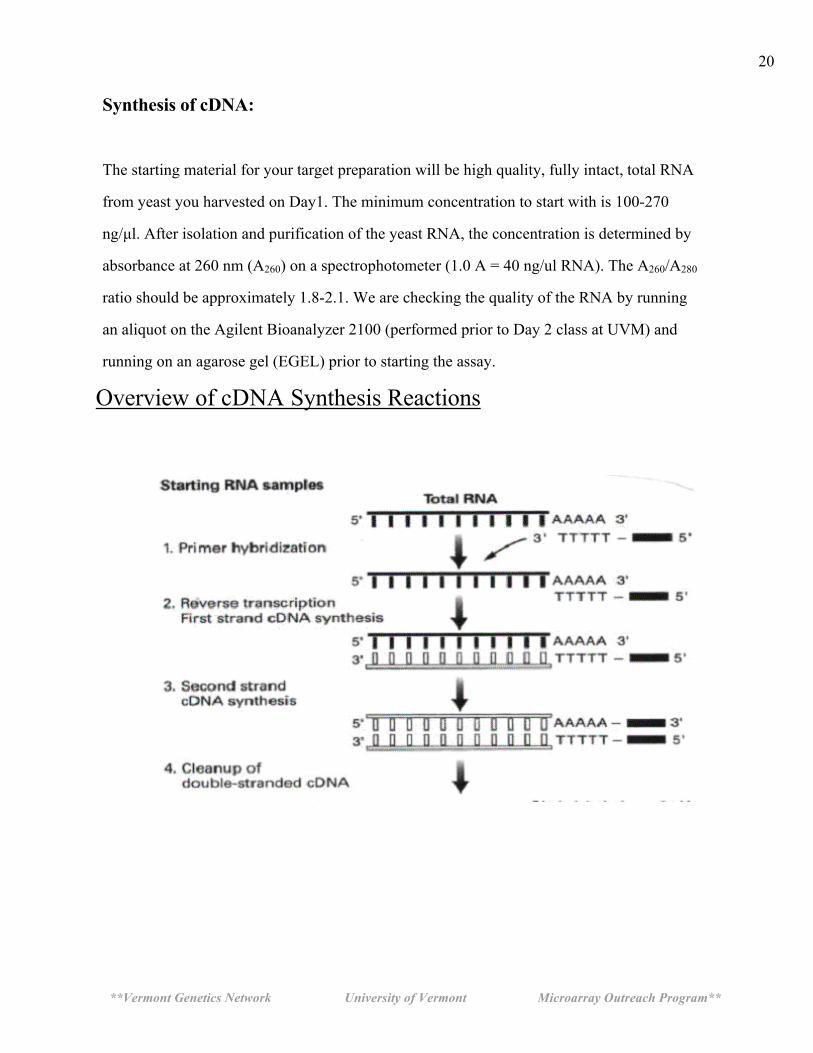
Microarray Target Preparation- cDNA synthesis Technical Overview:
Today you will be starting to prepare the target cRNA for the Genechip analysis. This is a
lengthy procedure and will be broken down over several lab periods. You will be starting
with mRNA, which comprises 0.5-2% of the total RNA in typical eukaryotic cells. First,
you will generate the first complementary DNA strand (cDNA) from the mRNA. This is
called the first strand cDNA synthesis. The mRNA molecule is oriented in a 5’ to 3’
direction. This step utilizes a primer with a sequence of 24 T's in a row and is called oligo
d(T), This primer binds to the poly A tail of mRNA (the 3’ end). The primer also contains
the consensus-binding site for T7 RNA polymerase (T7 promoter sequence) which is 36
bases long. The total length of the primer is 60 bases. Once the primer has hybridized to the
poly-A tail of the mRNA molecule, reverse transcriptase, an enzyme that uses RNA as a
template to synthesis DNA, is added and the first strand of cDNA is synthesized. The final
product of this reaction is a hybrid molecule composed of one RNA strand (the
original mRNA molecule) and one DNA strand. The first DNA strand is
synthesized in a 3’ to 5’ orientation relative to the mRNA strand (see the diagram
on page 20, step 2). It is important to note that rRNA and tRNA molecules in the sample
do not have a poly-A tail and will not bind the oligo T primer and will not be used as a
template for cDNA synthesis in this procedure.
The full sequence of T7 Oligo d(T)24 is:
**Vermont Genetics Network University of Vermont Microarray Outreach Program**
19
The next step in the cDNA reaction is the synthesis of the second cDNA strand from the first
cDNA strand. This is done by adding RNase H, DNA polymerase I, and DNA ligase. The
RNase H causes many nicks in the phosphodiester bonds (between the phosphate and the
ribose sugar) of the bound mRNA strand (not the cDNA stand already made) creating
exposed 3'OH groups. DNA polymerase I enzyme uses these 3’ OH groups as a priming site;
displacing the RNA in the strand and creating another cDNA strand using the first cDNA
strand as a template. It continues in this fashion until it reaches another nicked site, where it
stops. The DNA polymerase I can only displace RNA, not DNA. After cDNA has been
synthesized from all the nicks, the 3'OH nicks are reconnected or ligated. This is
accomplished using DNA ligase enzyme. After the two hour synthesis process, all the RNA
has been replaced with cDNA and ligated to form one double stranded cDNA molecule.
However, because the DNA polymerase does not synthesize completely to the end of the
strand, a T4 DNA polymerase is added. This enzyme ensures the full synthesis of the T7
promoter site from the original T7 oligo d(T) primer used in the first synthesis reaction. The
final product is a double stranded cDNA molecule which includes the original mRNA
sequence (in DNA form) on the second strand plus the full T7 RNA polymerase binding site
at the 3’ end of the molecule—the dark bar shown on step 3 on page 20.
**Vermont Genetics Network University of Vermont Microarray Outreach Program**
20
Synthesis of cDNA:
The starting material for your target preparation will be high quality, fully intact, total RNA
from yeast you harvested on Day1. The minimum concentration to start with is 100-270
ng/µl. After isolation and purification of the yeast RNA, the concentration is determined by
absorbance at 260 nm (A260) on a spectrophotometer (1.0 A = 40 ng/ul RNA). The A260/A280
ratio should be approximately 1.8-2.1. We are checking the quality of the RNA by running
an aliquot on the Agilent Bioanalyzer 2100 (performed prior to Day 2 class at UVM) and
running on an agarose gel (EGEL) prior to starting the assay.
Overview of cDNA Synthesis Reactions

**Vermont Genetics Network University of Vermont Microarray Outreach Program**
21
Day 2
Microarray Target Preparation- cDNA synthesis First Strand cDNA Synthesis Note: You will be measuring and transferring very small volumes. Centrifuge all reagent tubes before starting as some have only 1 µl in them. When pipetting , look closely at your pipet tip to ensure you have picked up the sample and look at the tube to which you are delivering it to ensure it has been added.
____1. Label a 0.5 ml PCR tube with your identification information. From the concentration of your RNA determined on Day 1, calculate the volume of the sample needed to contain 3.0 ug. Transfer this amount to the tube. Add DEPC water to bring the volume to 11.0 ul.
Example: RNA concentration of your sample = 800 ng/ul or .8ug/ul. 3.0 ug/.8 ug/ul = 3.75 ul. Transfer this amount to the tube. 11.0 ul – 3.75 ul = 7.25ul = volume of DEPC water to add to tube.
• RNA concentration of your sample: ________________ug/ul • 3.0 / (your RNA concentration) = _______________ul. This is the
volume of your sample you will add to the tube. • 11.0 – (the volume of your RNA sample) = ___________ul. This is the
volume of DEPC water to add to the tube.
____2. Add 1.0 ul T7 oligo d(T)24 reagent to the tube. Make sure you see the reagent transfer. Vortex the tube and centrifuge at full speed for 5 seconds. Place the tube in a thermocycler set at 70°C for 10 minutes.
____3. While the 70°C incubation is in progress prepare the following master mix
in a new tube. Add the following reagents IN ORDER to make the first strand master mix. Vortex and centrifuge at full speed for 5 second and place the tube on ice.
First strand master mix
First Strand Buffer 5X 4 µl 0.1M DTT 2 µl 10mM dNTP 1 µl Superscript II 1µl
Use a P2 micropipette to measure volumes of 2 ul or less.
**Vermont Genetics Network University of Vermont Microarray Outreach Program**
22
____4. After the 70°C incubation in step 2 is finished, add the first strand master mix
made in step 3 to the tube and incubate in a thermocycler at 42°C for 60 minutes. When first strand synthesis incubation is finished, place the tube on ice. During this incubation, prepare the second strand master mix.
Second Strand cDNA Synthesis
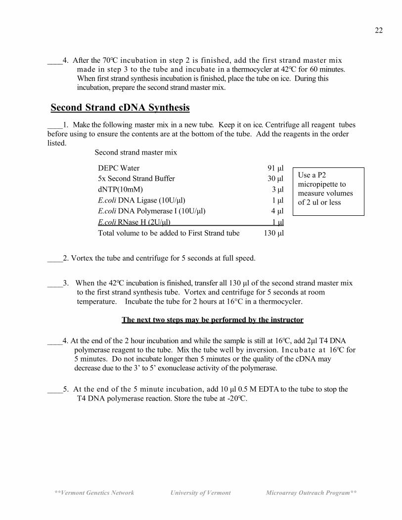
____1. Make the following master mix in a new tube. Keep it on ice. Centrifuge all reagent tubes before using to ensure the contents are at the bottom of the tube. Add the reagents in the order listed. Second strand master mix
DEPC Water 91 µl 5x Second Strand Buffer 30 µl dNTP(10mM) 3 µl E.coli DNA Ligase (10U/µl) 1 µl E.coli DNA Polymerase I (10U/µl) 4 µl E.coli RNase H (2U/µl) 1 µl Total volume to be added to First Strand tube 130 µl
____2. Vortex the tube and centrifuge for 5 seconds at full speed. ____3. When the 42°C incubation is finished, transfer all 130 µl of the second strand master mix
to the first strand synthesis tube. Vortex and centrifuge for 5 seconds at room temperature. Incubate the tube for 2 hours at 16°C in a thermocycler.
The next two steps may be performed by the instructor
____4. At the end of the 2 hour incubation and while the sample is still at 16°C, add 2µl T4 DNA
polymerase reagent to the tube. Mix the tube well by inversion. Incuba te a t 16°C for 5 minutes. Do not incubate longer then 5 minutes or the quality of the cDNA may decrease due to the 3’ to 5’ exonuclease activity of the polymerase.
____5. At the end of the 5 minute incubation, add 10 µl 0.5 M EDTA to the tube to stop the
T4 DNA polymerase reaction. Store the tube at -20°C.
Use a P2 micropipette to measure volumes of 2 ul or less
**Vermont Genetics Network University of Vermont Microarray Outreach Program**
23
Day 3
Technical Overview: Cleaning the cDNA:
The double stranded cDNA generated on Day 2 must be cleaned and purified before
proceeding to the next step. This is done using a solution of phenol-chloroform-isoamyl
alcohol (PCI) at a alkaline pH to denature proteins and trap them in the organic phase of the
reagent. cDNA remains in the aqueous phase and the two phases are separated using a phase
lock gel system. The phase lock gel tube is centrifuged causing the gel in the tube to migrate
between the aqueous phase (on the top and containing the cDNA) and the organic phase (on
the bottom and containing the denatured proteins). Recovery of cDNA is easy using this
method. The pH of the PCI solution is maintained by Tris buffer at pH 8.3 to ensure the
cDNA will dissolve in the aqueous layer.
cDNA is purified from the aqueous phase through a precipitation step using ethanol [an
alcohol] and ammonium acetate [a salt]. Because DNA is negatively charged, a high salt
concentration is used to mask the charge, decreasing the solubility of the cDNA. Ethanol is a
non-polar s o l v e n t and DNA will not readily dissolve in it in these conditions and
precipitates from the solution. This reaction is sometimes done at -20°C to encourage
maximum DNA precipitation. After precipitation, the cDNA is centrifuged at high speed to
form a pellet with a visualization agent called Pellet Paint, an additive that contains glycogen (a
co-precipitate) and a dye. This helps generate a high quality visible pellet on the bottom of
the tube. The cDNA pellet is washed several times with 80% ethanol, dried, and
resuspended in DEPC water.
**Vermont Genetics Network University of Vermont Microarray Outreach Program**
24
The In vitro Transcription (IVT) Reaction:
The IVT step uses the T7 promoter site that is attached to the T7 Oligo d(T)24 primer used in
the first strand cDNA synthesis. T7 RNA polymerase enzyme binds to this site and generates
cRNA molecules. In this arrangement the original mRNA sequence is the template strand and
the cRNA generated is antisense relative to this sequence. Two unique nucleotides are used
in the reaction. A select proportion of the C and U ribonucleotides have molecules of biotin
attached. A and G nucleotides are not biotinyated. As a result, the cRNA generated has
random amounts of biotin label incorporated into its structure. The biotin will be the
beginning of the signal generation process used on the gene chips based on a strepavidin-
phycoerytherin reaction. During the IVT reaction the T7RNA polymerase binds to the T7
promoter site and generates a molecule of cRNA. During the 16 hour incubation time for the
IVT, a single enzyme can generate many thousand cRNA molecules from the cDNA template.
The net result is a total increase in RNA at the end of the reaction. For a typical IVT reaction,
1.5 to 2.5 ug/ul yields are expected. 10 ug of cRNA is carried forward to Day 4 for
fragmentation and hybridization.
**Vermont Genetics Network University of Vermont Microarray Outreach Program**
25
Safety Considerations for Phenol Chloroform Isoamyl: Note: Phenol Chloroform Isoamyl Alcohol is at a ratio of 25:24:1 and saturated with 10 mM Tris- HCl pH 8.3. The Tris layer floats on top of the PCI layer. Phenol is an organic acid and care must be used when handling. Phenol can burn skin and be absorbed into the circulatory system and cause health problems. Chloroform is an organic solvent and should be used in a hood.
Phenol – Corrosive – Local anesthetic – Can severely damage eyes and skin – Can cause severe burns of the skin and eye – Poisonous if ingested-can be fatal
Chloroform – Can cause irreversible corneal injury – Fatal if swallowed – Flammable
Isoamyl Alcohol – Flammable – Poisonous
Note: PCI tends to leak out of the pipet tips so work quickly when measuring it or transferring it from one container to another.
**Vermont Genetics Network University of Vermont Microarray Outreach Program**
26
Day 3 Precipitating the cDNA:
____1. The total volume of the cDNA synthesis step from Day 2 is 162 ul. The
PCI step requires equal volumes of aqueous and organic mixtures. Centrifuge a Phase Lock Gel tube at full speed for one minute to insure the gel is at the bottom of the tube. DO NOT VORTEX PHASE LOCK TUBES.
____2. Add 162 µl of the bottom layer from the Phenol Chloroform Isoamyl Alcohol (PCI) to
the contents of the cDNA synthesis reaction tube from Day 2 and vortex for 5 seconds to mix the contents.
Note: PCI tends to leak out of the pipet so work quickly and hold the cap tightly as
leaking can occur during the vortex step.
____3. Transfer all of the cDNA-PCI mixture to the phase lock gel tube using a micropipet.
DO NOT VORTEX the phase lock gel tube.
____4. Centrifuge at full speed for 2 minutes. ____5. Label a new 1.7 microcentrifuge tube. Use a micropipette to transfer the top layer f rom
the phase lock gel tube to the tube. Try to collect as much of the layer as possible.
____6. Add the following to the 1.7 ml microcentrifuge tube and vortex.
Ethanol (100%) 405 µl NH4OAc (7.5M) 80 µl Pellet Paint 1 µl
____7. Place the tube in the centrifuge with the hinge of the tube facing out and centrifuge at
full speed for 20 minutes at room temperature. ____8. GENTLY remove the tube from the centrifuge being careful not to disturb the cDNA
pellet. The pellet should be pink and approximately the size of a grain of salt and on the side of the tube under the hinge. Place on ice and immediately proceed to next step.
**Vermont Genetics Network University of Vermont Microarray Outreach Program**
27
Is there a pellet?____________
**At this point a tiny pink visible pellet should be present on the bottom of
the tube. If not, see the instructor.**
Cleaning the cDNA Pellet:
____1. Using a P1000 micropipet, carefully remove the liquid from the tube. Tip the tube to
remove of as much liquid as possible, but don’t lose the pellet. Remember that the pellet is your sample!
____2. Add 500µl ice cold 80% ethanol (store in -20Co freezer) to the tube. Gently cap the tube
and invert it slowly several times. Watch your pellet very closely. Place the tube back in rack and let the pellet settle to the bottom of the tube for a minute. Alternately, you may centrifuge the tube at full speed for 15 seconds to get the pellet back down to the bottom of the tube.
____3. Using a P1000 micropipet, carefully remove the ethanol being very careful not to disturb
the pellet. Tip the tube to enable removal of as much liquid as possible. ____4. Repeat steps 2 and 3 with a new aliquot of 80% ethanol. ____5. Finally, remove all of the ethanol. To do this, remove as much as possible using a
P1000 micropipet. Centrifuge the tube at full speed for 5 seconds and use a P20 micropipet to remove the last few microliters of liquid. The goal is to remove as much ethanol as possible without disturbing the pellet.
____6. Place the tube on its side with the cap open in a drying box for 10 minutes to evaporate
the remaining ethanol. The dried pellet is easily lost once it is dry. Be very careful to handle the tube gently. When done, close the cap gently. Visualize the dried pellet to confirm it is present in the tube.
**Vermont Genetics Network University of Vermont Microarray Outreach Program**
28
____7. Resuspend the pellet in 22 µl of DEPC water and place the tube on ice.
In Vitro Transcription (IVT): Synthesis of biotin labeled cRNA from cDNA using the Enzo Kit
____1. The ENZO bioarray kit contains all the reagents needed to prepare biotin labeled cRNA
from cDNA. A master mix for the entire class will be prepared as follows. The instructor will
prepare this mix or designate someone from the class. This must be done an RNase-free area free from air currents.
Amt/sample #Samples Total
Reagent 1 [10x Reaction buffer] 4 µl Reagent 2 [10x Biotin nucleotides] 4 µl Reagent 3 [10x DTT] 4 µl Reagent 4 [10x RNase Inhibitor] 4 µl Reagent 5 [20x T7 RNA polymerase] 2 µl _____ _____ Total Volume 18 µl
NOTE: Prepare sufficient master mix for the number of samples plus one. This will ensure enough for pipetting purposes.
____2. Label a 0.5 ml PCR tube. Combine the following in the tube and pipet up and down
several times to mix. Spin in a centrifuge at full speed for 5 seconds to get all reagents to the bottom of the tube.
cDNA mixture 22 µl Enzo master mix [from above] 18 µl Total Volume 40 µl
____3. Incubate the tube at 37°C for 16 hours in the thermocycler. When the reaction is complete,
store the sample at -20oC.
The Instructor will perform this task.
**Vermont Genetics Network University of Vermont Microarray Outreach Program**
29
Day 4
Cleaning and fragmenting the biotinylated cRNA
The biotinylated cRNA generated on day 3 must be cleaned and purified from the enzymes
and reagents used in the IVT reaction. Removing the unused biotinylated NTPs, buffers and
proteins is necessary as they interfere with quantitative measures. Proteins and NTP's absorb
at 260 and/or 280nm and will affect the quantitation of the cRNA. They also interfere with
hybridization of the cRNA targets on the gene chip.
You will use an RNeasy spin column to clean the IVT product similar to that used on Day 1.
The cRNA is washed and eluted from the spin column with DEPC water. The cRNA is
analyzed with a spectrophotometer and the concentration and purity determined. An
A260/A280 ratio of 1.8-2.1 indicates a clean cRNA sample that can be used for subsequent
steps.
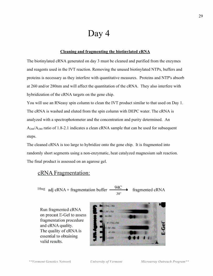
The cleaned cRNA is too large to hybridize onto the gene chip. It is fragmented into
randomly short segments using a non-enzymatic, heat catalyzed magnesium salt reaction.
The final product is assessed on an agarose gel.
**Vermont Genetics Network University of Vermont Microarray Outreach Program**
30
Day 4 Cleaning the biotinylated cRNA : ____1. Remove the Day 3 tubes from the freezer and let thaw. Transfer the entire cRNA sample to a
new1.7 ml microcentrifuge tube. Add 60 µl of DEPC water and 350 µl BME-RLT and vortex for 5 seconds.
____2. Add 250 µl 100% ethanol and vortex again.
____3. Label an RNeasy spin column and transfer the entire cRNA sample to the column. As before, be careful not to touch the tip of the pipet to the silica membrane. Place the spin column in a 2 ml collection tube, close the lid and centrifuge for 15 seconds at full speed.
____4. Remove the spin column from the collection tube. Pipet the pass through liquid (the
liquid in the collection tube) back onto the spin column and centrifuge again for 15 seconds. Discard the collection tube (with the flow through fluid) and place the spin column into a new 2 ml collection tube.
____5. Pipette 500 µl of RPE buffer onto the spin column. Close the tube and centrifuge for 15
seconds at full speed. Discard collection tube and the pass through liquid. Place the spin column into a new collection tube.
____6. Add another 500 µl RPE buffer to the spin column. Close the tube and centrifuge for
15 seconds at full speed. Discard the collection tube and pass through liquid.
____7. Transfer the spin column into a new collection tube and at full speed for 1 minute. ____8. Label a new 1.7 ml microcentrifuge tube with your identification information. Transfer the spin column to this tube. ____9. Pipet 30 µl DEPC water onto the RNeasy silica-gel membrane and wait 1 minute.
Make sure the DEPC water contacts the entire membrane. Be careful not to touch the membrane with the pipet tip. Close the tube and centrifuge for 1 minute, at full speed.
____10. Carefully transfer the 30 µl that is recovered in the 1.7 ml tube back onto the center
of the silica membrane of the same spin column. Place the column back in to the same 1.7 ml collection tube and incubate for 1 minute at room temperature. Centrifuge for 1 minute at full speed. This double extraction ensures that the maximal amount of cRNA is eluted from the membrane.
**Vermont Genetics Network University of Vermont Microarray Outreach Program**
31
____11. Label a new 1.7 microcentrifuge tube with your identifying information. The old
tube lid may have been contaminated in the last few procedures. Transfer the cRNA this tube.
____12. Determine the cRNA concentration as on Day 1. Prepare a sample for analysis by
adding 2 ul of your sample to 48 ul DEPC water. Determine the concentration and the A260/A280 ratio .
Absorbance A260of sample:_________________ A260/A280 ratio:_________________ Concentration of RNA in sample:_______________ug/ul Total yield of RNA = 30 ul x concentration = ___________________ug. Record these data on the page 5 data sheet.
**Vermont Genetics Network University of Vermont Microarray Outreach Program**
32
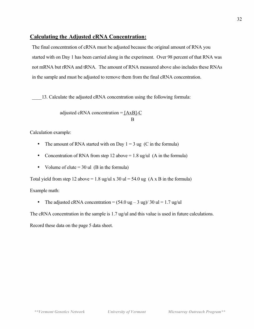
Calculating the Adjusted cRNA Concentration:
The final concentration of cRNA must be adjusted because the original amount of RNA you
started with on Day 1 has been carried along in the experiment. Over 98 percent of that RNA was
not mRNA but rRNA and tRNA. The amount of RNA measured above also includes these RNAs
in the sample and must be adjusted to remove them from the final cRNA concentration.
____13. Calculate the adjusted cRNA concentration using the following formula:
adjusted cRNA concentration = [AxB]-C
B
Calculation example:
• The amount of RNA started with on Day 1 = 3 ug (C in the formula)
• Concentration of RNA from step 12 above = 1.8 ug/ul (A in the formula)
• Volume of elute = 30 ul (B in the formula) Total yield from step 12 above = 1.8 ug/ul x 30 ul = 54.0 ug (A x B in the formula) Example math:
• The adjusted cRNA concentration = (54.0 ug – 3 ug)/ 30 ul = 1.7 ug/ul The cRNA concentration in the sample is 1.7 ug/ul and this value is used in future calculations. Record these data on the page 5 data sheet.
**Vermont Genetics Network University of Vermont Microarray Outreach Program**
33
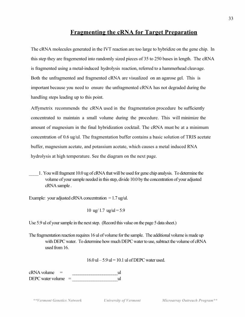
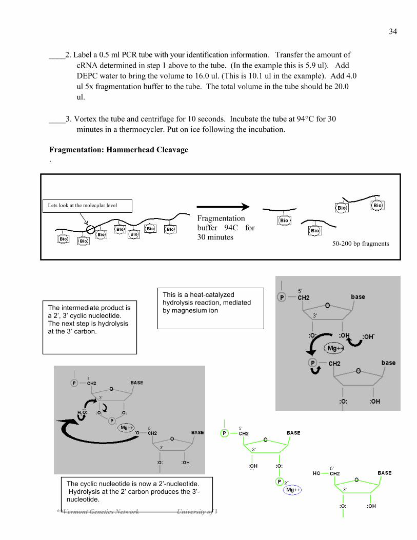
Fragmenting the cRNA for Target Preparation
The cRNA molecules generated in the IVT reaction are too large to hybridize on the gene chip. In
this step they are fragemented into randomly sized pieces of 35 to 250 bases in length. The cRNA
is fragmented using a metal-induced hydrolysis reaction, referred to a hammerhead cleavage.
Both the unfragmented and fragmented cRNA are visualized on an agarose gel. This is
important because you need to ensure the unfragmented cRNA has not degraded during the
handling steps leading up to this point.
Affymetrix recommends the cRNA used in the fragmentation procedure be sufficiently
concentrated to maintain a small volume during the procedure. This will minimize the
amount of magnesium in the final hybridization cocktail. The cRNA must be at a minimum
concentration of 0.6 ug/ul. The fragmentation buffer contains a basic solution of TRIS acetate
buffer, magnesium acetate, and potassium acetate, which causes a metal induced RNA
hydrolysis at high temperature. See the diagram on the next page.
____1. You will fragment 10.0 ug of cRNA that will be used for gene chip analysis. To determine the
volume of your sample needed in this step, divide 10.0 by the concentration of your adjusted cRNA sample .
Example: your adjusted cRNA concentration = 1.7 ug/ul. 10 ug/ 1.7 ug/ul = 5.9 Use 5.9 ul of your sample in the next step. (Record this value on the page 5 data sheet.) The fragmentation reaction requires 16 ul of volume for the sample. The additional volume is made up
with DEPC water. To determine how much DEPC water to use, subtract the volume of cRNA used from 16.
16.0 ul – 5.9 ul = 10.1 ul of DEPC water used. cRNA volume = _____________________ul DEPC water volume = _____________________ul
**Vermont Genetics Network University of Vermont Microarray Outreach Program**
34
____2. Label a 0.5 ml PCR tube with your identification information. Transfer the amount of cRNA determined in step 1 above to the tube. (In the example this is 5.9 ul). Add DEPC water to bring the volume to 16.0 ul. (This is 10.1 ul in the example). Add 4.0 ul 5x fragmentation buffer to the tube. The total volume in the tube should be 20.0 ul.
____3. Vortex the tube and centrifuge for 10 seconds. Incubate the tube at 94°C for 30
minutes in a thermocycler. Put on ice following the incubation. Fragmentation: Hammerhead Cleavage .
This is a heat-catalyzed hydrolysis reaction, mediated by magnesium ion The intermediate product is
a 2’, 3’ cyclic nucleotide. The next step is hydrolysis at the 3’ carbon.
The cyclic nucleotide is now a 2’-nucleotide. Hydrolysis at the 2’ carbon produces the 3’-nucleotide.
Fragmentation buffer 94C for 30 minutes
50-200 bp fragments
Lets look at the molecµlar level
**Vermont Genetics Network University of Vermont Microarray Outreach Program**
35
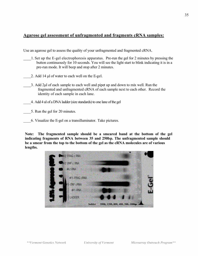
Agarose gel assessment of unfragmented and fragments cRNA samples: Use an agarose gel to assess the quality of your unfragmented and fragmented cRNA. ____1. Set up the E-gel electrophoresis apparatus. Pre-run the gel for 2 minutes by pressing the
button continuously for 10 seconds. You will see the light start to blink indicating it is in a pre-run mode. It will beep and stop after 2 minutes.
____2. Add 14 µl of water to each well on the E-gel. ____3. Add 2µl of each sample to each well and pipet up and down to mix well. Run the
fragmented and unfragmented cRNA of each sample next to each other. Record the identity of each sample in each lane.
____4. Add 4 ul of a DNA ladder (size standards) to one lane of the gel ____5. Run the gel for 20 minutes. ____6. Visualize the E-gel on a transilluminator. Take pictures. Note: The fragmented sample should be a smeared band at the bottom of the gel indicating fragments of RNA between 35 and 250bp. The unfragmented sample should be a smear from the top to the bottom of the gel as the cRNA molecules are of various lengths.
ladder 2000, 1200, 800, 400, 200, 100bp

**Vermont Genetics Network University of Vermont Microarray Outreach Program**
36
To Be Performed at the University of Vermont
These steps will be performed at the University of Vermont Microarray Core Lab and will
be shown as a powerpoint presentation during your class visit.
Preparing the Hybridization Mix
The hybridization mixture contains several internal controls that provide information
about the success of the hybridization procedure. These controls are the B2 oligo for which
there are several hundred probes along the outer edge of all expression arrays and a
checkerboard pattern in each corner. These predefined patterns provide signals for the
Affymetrix® Microarray Suite software to perform automatic grid alignment during image
analysis. They can also be used to align the grid manually. The fluorescence intensities for
control oligo B2 are not used for analyzing data. The bioB, bioC, and bioD are biotinlylated
gene fragments much like your sample which represent the biotin synthesis pathway
from the bacteria E. coli. The cre control is the recombinase gene from P1 bacteriophage.
These biological controls are prepared at standardized concentrations and signal intensities
obtained on these genes provide information on how well the hybridization, washing, and
staining procedures have performed.
____1. Prepare the following:
Fragmented cRNA [10µg] ? Control B2 Oligo 3.3 µl 20x Eukaryotic Control mix [bio B, bio C, bio D, Cre] 10 µl Herring Sperm DNA [10mg/ml] 2 µl Acetyleted BSA [50mg/ml] 2 µl 2x Hybridization Buffer 100 µl Water [QS to 200µl] ___
____2. After preparing the hybridization mixture, store the mixture at -20°C.
**Vermont Genetics Network University of Vermont Microarray Outreach Program**
37
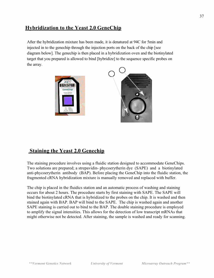
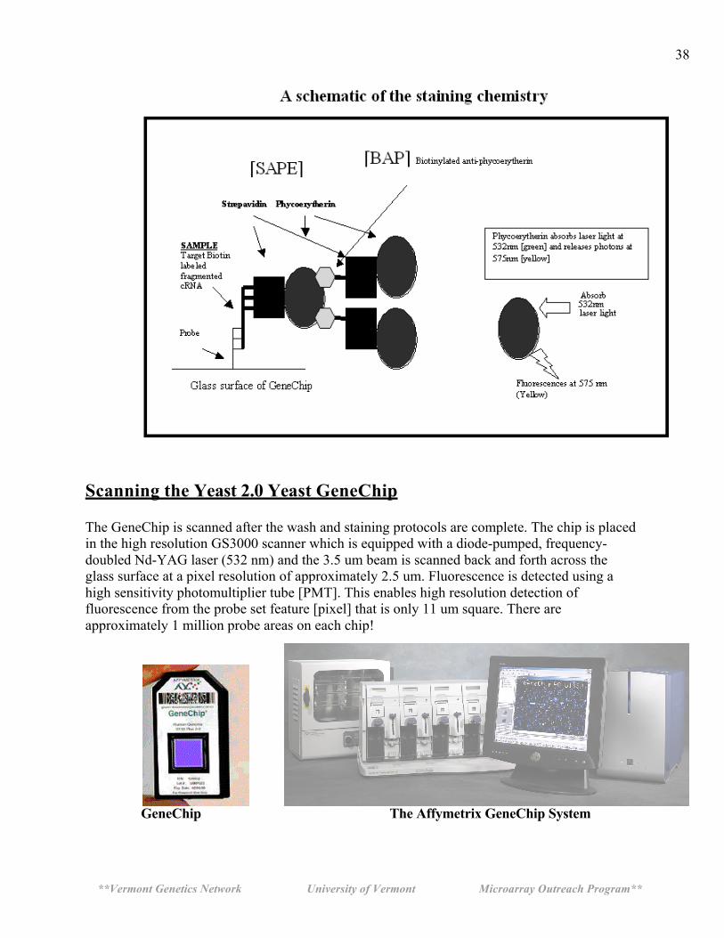
Hybridization to the Yeast 2.0 GeneChip After the hybridization mixture has been made, it is denatured at 94C for 5min and injected in to the genechip through the injection ports on the back of the chip [see diagram below]. The genechip is then placed in a hybridization oven and the biotinylated target that you prepared is allowed to bind [hybridize] to the sequence specific probes on the array.
Staining the Yeast 2.0 Genechip
The staining procedure involves using a fluidic station designed to accommodate GeneChips. Two solutions are prepared; a strepavidin- phycoerytherin dye (SAPE) and a biotinylated anti-phycoerytherin antibody (BAP). Before placing the GeneChip into the fluidic station, the fragmented cRNA hybridization mixture is manually removed and replaced with buffer. The chip is placed in the fluidics station and an automatic process of washing and staining occurs for about 2 hours. The procedure starts by first staining with SAPE. The SAPE will bind the biotinylated cRNA that is hybridized to the probes on the chip. It is washed and then stained again with BAP. BAP will bind to the SAPE. The chip is washed again and another SAPE staining is carried out to bind to the BAP. The double staining procedure is employed to amplify the signal intensities. This allows for the detection of low transcript mRNAs that might otherwise not be detected. After staining, the sample is washed and ready for scanning.
**Vermont Genetics Network University of Vermont Microarray Outreach Program**
38
Scanning the Yeast 2.0 Yeast GeneChip The GeneChip is scanned after the wash and staining protocols are complete. The chip is placed in the high resolution GS3000 scanner which is equipped with a diode-pumped, frequency-doubled Nd-YAG laser (532 nm) and the 3.5 um beam is scanned back and forth across the glass surface at a pixel resolution of approximately 2.5 um. Fluorescence is detected using a high sensitivity photomultiplier tube [PMT]. This enables high resolution detection of fluorescence from the probe set feature [pixel] that is only 11 um square. There are approximately 1 million probe areas on each chip!
GeneChip The Affymetrix GeneChip System
**Vermont Genetics Network University of Vermont Microarray Outreach Program**
39
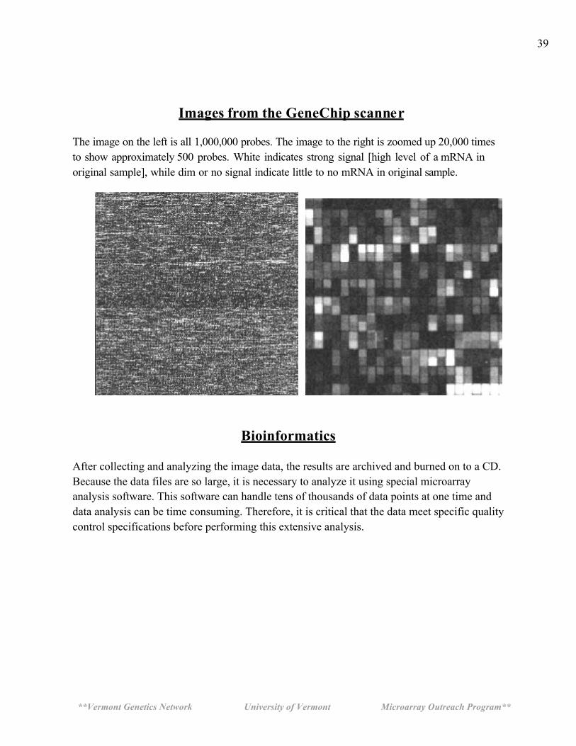
Images from the GeneChip scanner
The image on the left is all 1,000,000 probes. The image to the right is zoomed up 20,000 times to show approximately 500 probes. White indicates strong signal [high level of a mRNA in original sample], while dim or no signal indicate little to no mRNA in original sample.
Bioinformatics
After collecting and analyzing the image data, the results are archived and burned on to a CD. Because the data files are so large, it is necessary to analyze it using special microarray analysis software. This software can handle tens of thousands of data points at one time and data analysis can be time consuming. Therefore, it is critical that the data meet specific quality control specifications before performing this extensive analysis.
**Vermont Genetics Network University of Vermont Microarray Outreach Program**
40
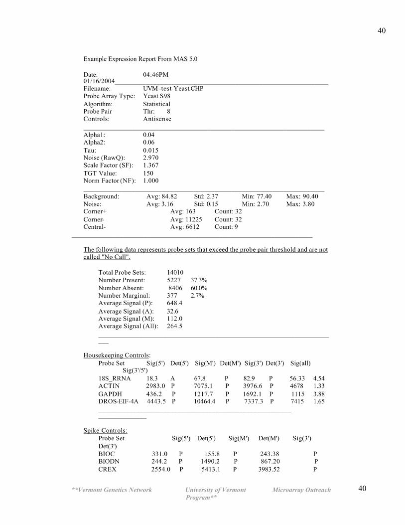
40
Example Expression Report From MAS 5.0 Date: 04:46PM 01/16/2004______________________________________________________________ Filename: UVM -test-Yeast.CHP Probe Array Type: Yeast S98 Algorithm: Statistical Probe Pair Thr: 8 Controls: Antisense ______________________________________________________________________ Alpha1: 0.04 Alpha2: 0.06 Tau: 0.015 Noise (RawQ): 2.970 Scale Factor (SF): 1.367 TGT Value: 150 Norm Factor (NF): 1.000 ______________________________________________________________________ Background: Avg: 84.82 Std: 2.37 Min: 77.40 Max: 90.40 Noise: Avg: 3.16 Std: 0.15 Min: 2.70 Max: 3.80 Corner+ Avg: 163 Count: 32 Corner- Avg: 11225 Count: 32 Central- Avg: 6612 Count: 9
______________________________________________________________________
The following data represents probe sets that exceed the probe pair threshold and are not called "No Call".
Total Probe Sets: 14010 Number Present: 5227 37.3% Number Absent: 8406 60.0% Number Marginal: 377 2.7% Average Signal (P): 648.4 Average Signal (A): 32.6 Average Signal (M): 112.0 Average Signal (All): 264.5 ______________________________________________________________________
Housekeeping Controls: Probe Set Sig(5') Det(5') Sig(M') Det(M') Sig(3') Det(3') Sig(all) Sig(3'/5') 18S_RRNA 18.3 A 67.8 P 82.9 P 56.33 4.54 ACTIN 2983.0 P 7075.1 P 3976.6 P 4678 1.33 GAPDH 436.2 P 1217.7 P 1692.1 P 1115 3.88 DROS-EIF-4A 4443.5 P 10464.4 P 7337.3 P 7415 1.65 ________________________________________________________ ______________
Spike Controls: Probe Set Sig(5') Det(5') Sig(M') Det(M') Sig(3') Det(3') BIOC 331.0 P 155.8 P 243.38 P BIODN 244.2 P 1490.2 P 867.20 P CREX 2554.0 P 5413.1 P 3983.52 P
**Vermont Genetics Network University of Vermont Microarray Outreach Program**
41
41
Appendix A: Preparing the pre-culture
1] All work must be done in a HEPA hood such as those involved with tissue culture! Always wear gloves that have been rinsed with alcohol!
2] Transfer an entire colony of yeast to a broth culture of 1/2x YPD with or without glucose. It is not important as long as we get some growth as indicated by turbidity at 48 hrs.
3] After incubation, aseptically remove a drop of the culture and examine on the microscope to ensure there is no contaminating bacteria. Remember that bacteria will be 1/5 the size of the yeast. So look for small rods or cocci beyond the yeast.
4] To inoculate the experimental culture to be used in class, aseptically transfer 0.5 ml of this culture to the 1/2x YPD-3xGlu broth 48hrs before day 1. It is advisable to use aerosol resistant pipet tips.
Alternatively:
You may streak a YPD agar plate 4 days prior and then inoculate your experimental culture with a whole colony 48 hours prior to day 1 class.