Embed Size (px)
Citation preview

Integrated Bioinformatic and Targeted Deletion Analyses of the SRSGene Superfamily Identify SRS29C as a Negative Regulator ofToxoplasma Virulence
James D. Wasmuth,a,b* Viviana Pszenny,b Simon Haile,c* Emily M. Jansen,c Alexandra T. Gast,b Alan Sher,d Jon P. Boyle,e
Martin J. Boulanger,f John Parkinson,a,g and Michael E. Griggb,c
Program in Molecular Structure and Function, Hospital for Sick Children, Toronto, Ontario, Canadaa; Molecular Parasitology Unit, Laboratory of Parasitic Diseases, NIAID,National Institutes of Health, Bethesda, Maryland, USAb; Department of Medicine, University of British Columbia, Vancouver, BC, Canadac; Immunobiology Section,Laboratory of Parasitic Diseases, NIAID, NIH, Bethesda, Maryland, USAd; Department of Biological Sciences, University of Pittsburgh, Pittsburgh, Pennsylvania, USAe;Department of Biochemistry and Microbiology, University of Victoria, Victoria, BC, Canadaf; and Departments of Biochemistry & Molecular Genetics, University of Toronto,Toronto, Ontario, Canadag
* Present address: James D. Wasmuth, Department of Ecosystem and Public Health, Faculty of Veterinary Medicine, University of Calgary, Calgary, Alberta, Canada; Simon Haile,Genome Sciences Center, BC Cancer Agency, Vancouver, BC, Canada.
J.D.W. and V.P. contributed equally to this work.
ABSTRACT The Toxoplasma gondii SRS gene superfamily is structurally related to SRS29B (formerly SAG1), a surface adhesinthat binds host cells and stimulates host immunity. Comparative genomic analyses of three Toxoplasma strains identified 182SRS genes distributed across 14 chromosomes at 57 genomic loci. Eight distinct SRS subfamilies were resolved. A core 69 func-tional gene orthologs were identified, and strain-specific expansions and pseudogenization were common. Gene expression pro-filing demonstrated differential expression of SRS genes in a developmental-stage- and strain-specific fashion and identified nineSRS genes as priority targets for gene deletion among the tissue-encysting coccidia. A �sag1 �sag2A mutant was significantlyattenuated in murine acute virulence and showed upregulated SRS29C (formerly SRS2) expression. Transgenic overexpressionof SRS29C in the virulent RH parent was similarly attenuated. Together, these findings reveal SRS29C to be an important regula-tor of acute virulence in mice and demonstrate the power of integrated genomic analysis to guide experimental investigations.
IMPORTANCE Parasitic species employ large gene families to subvert host immunity to enable pathogen colonization and causedisease. Toxoplasma gondii contains a large surface coat gene superfamily that encodes adhesins and virulence factors that facili-tate infection in susceptible hosts. We generated an integrated bioinformatic resource to predict which genes from within this182-gene superfamily of adhesin-encoding genes play an essential role in the host-pathogen interaction. Targeted gene deletionexperiments with predicted candidate surface antigens identified SRS29C as an important negative regulator of acute virulencein murine models of Toxoplasma infection. Our integrated computational and experimental approach provides a comprehensiveframework, or road map, for the assembly and discovery of additional key pathogenesis genes contained within other large sur-face coat gene superfamilies from a broad array of eukaryotic pathogens.
Received 30 August 2012 Accepted 5 September 2012 Published 13 November 2012
Citation Wasmuth JD, et al. 2012. Integrated bioinformatic and targeted deletion analyses of the SRS gene superfamily identify SRS29C as a negative regulator of Toxoplasmavirulence. mBio 3(6):e00321-12. doi:10.1128/mBio.00321-12.
Editor Louis Weiss, Albert Einstein College of Medicine
Copyright © 2012 Wasmuth et al. This is an open-access article distributed under the terms of the Creative Commons Attribution-Noncommercial-Share Alike 3.0 UnportedLicense, which permits unrestricted noncommercial use, distribution, and reproduction in any medium, provided the original author and source are credited.
Address correspondence to John Parkinson, [email protected], or Michael E. Grigg, [email protected].
Toxoplasma gondii, a highly successful protozoan parasite, in-fects nearly one-third of the world’s population. It is a member
of the subclass Coccidia, which includes Eimeria, Neospora, andSarcocystis, all genera of medical and veterinary importance.T. gondii has the ability to infect any warm-blooded animal, whileother coccidians apparently possess a more restricted host range.The coccidia are part of the phylum Apicomplexa, which in-cludes Plasmodium, Theileria, and Cryptosporidium. Theirshared mode of infection relies on physical interactions be-tween the parasite and a host cell; consequently, many apicom-plexans have evolved an arsenal of surface protein virulencegenes that facilitate parasite attachment and invasion of host
cells and evasion or modulation of host immunity (reviewed inreference 1).
The genome of T. gondii contains several distinct, coccidian-specific multicopy gene families (2), including those that encodethe SRS, ROPK, and SUSA proteins (3–5). The SRS genes encodevirulence determinants that induce lethal ileitis in a murine model(SRS29B, formerly SAG1), mediate attachment to host cells(SRS57; formerly SAG3), and establish transmissible latent infec-tions (SRS16B; formerly SRS9) (6–8). T. gondii presumably ex-presses and regulates this multigene family to successfully estab-lish chronic infections in most warm-blooded vertebrates, whereit persists within latent cysts for the life of the intermediate host.
RESEARCH ARTICLE
November/December 2012 Volume 3 Issue 6 e00321-12 ® mbio.asm.org 1
on Novem
ber 9, 2018 by guesthttp://m
bio.asm.org/
Dow
nloaded from

Unlike the serial expression of a single var or VSG gene in Plasmo-dium falciparum and Trypanosoma brucei, respectively, the SRSproteins are expressed in a developmentally regulated manner asdistinct, largely nonoverlapping sets of SRS antigens (3). An SRSgene typically contains one or two domains, each with four to sixcysteines (4-Cys or 6-Cys) that participate in disulfide bonds anda glycosylphosphatidylinositol (GPI) anchor for attachment to theparasite cell surface (9). Some SRS proteins are very closely re-lated, while others share less than 30% sequence identity. Theexact biological role of the SRS gene superfamily is poorly under-stood. It is known that SRS29B and SRS34A (formerly SAG2A) arehighly immunogenic during infection, whereas SRS29B andSRS57 function as adhesins (6, 10). An interaction with cellularligands is supported by structural evidence that SRS29B forms ahomodimer with a deep, positively charged groove capable ofdocking sulfated proteoglycans (11, 12).
With the release of the type II Me49 draft genome in 2004, 161unique SRS DNA sequences were identified (3). A combination oftargeted studies and searches of expressed sequence tag (EST) datahas revealed SRS orthologs in other coccidian genera, includingNeospora and Sarcocystis (13, 14), and an SRS-like fold in the Plas-modium 6-Cys family of parasite adhesins (15).
Exploiting the recent availability of genome sequence data forthree strains of T. gondii, here we describe a comprehensive inves-tigation exploring the evolution and expression of SRS virulencegenes. Systematic sequence searches identified eight subfamilies ofSRS domains which display distinct and nonrandom patterns ofco-occurrence, expansions, and losses across the three T. gondiistrains. Crystal structure analyses revealed a core SRS structuralscaffold with sequence divergence that presumably reflects thediversity of host ligands targeted. Integration of transcriptomicdata sets further revealed lineage- and life cycle stage-specific pro-grams of SRS gene expression that correlate with altered patternsof virulence by different parasite strains. Targeted disruption oftwo highly abundant, pancoccidial SRS proteins (SRS29B andSRS34A) resulted in a dramatic upregulation of the levels ofSRS29C (formerly SRS2), a protein that is differentially expressedby mouse-virulent and -avirulent strains of T. gondii. Transgenicoverexpression of SRS29C in a type I virulent strain significantlyattenuated virulence, and the majority of the mice infected sur-vived. Our results highlight the utility of integrating bioinformaticwith targeted genetic approaches to identify a major and unex-pected role for SRS29C in Toxoplasma acute virulence in mice.
RESULTSThere are eight subfamilies of SRS domains. To identify the fullcomplement of SRS domains in T. gondii, previously publishedSRS sequences were used to seed a hidden Markov model (HMM).New sequences were curated and added iteratively to expand thediversity of the HMM. A total of 763 SRS domains were found, ofwhich 459 had annotated ToxoDB gene models, a predicted signalpeptide, four to six invariant cysteine residues, and a predictedGPI anchor sequence consistent with an SRS protein (Table 1).SRS domains not found within a gene model were consideredpseudogenes. The low sequence identity (~15%) prevented theuse of standard methods for the construction of a robust phylog-eny. Therefore, all-against-all pairwise alignments were used togenerate a similarity score matrix, which was clustered hierarchi-cally. The domains segregated into two families and eight distinctdomain subfamilies (Fig. 1A), subfamilies 1 to 6 (which includesSRS34A) and subfamilies 7 and 8 (which includes SRS29B). Theintrasubfamily pairwise identity percentages ranged from 23% to45%, with the majority of the conservation mapping to the pro-posed �-strands in the structures (see Fig. 2, system object model[SOM] file 1).
A structure-guided alignment of each domain subfamily re-vealed that, of the six family-defining cysteines (Cys-1 to Cys-6),four are invariant (see Fig. S1 in the supplemental material). How-ever, in subfamilies 1 and 2, a subfamily-specific cysteine up-stream of Cys-5 replaces the absent Cys-3 to form the third disul-fide bond (16). Members of subfamilies 3 and 6 are also missingmiddle cysteine residues but do not have a compensatory residueto rescue the disulfide bond. In two-domain proteins, the extent towhich SRS domains vary depends upon whether the domain issolvent exposed (D1) or proximal to the membrane (D2) (SOMfile 1). The membrane-distal end of D1 is poorly conserved andlikely to be involved in mediating ligand interactions, whereas themembrane-proximal end of the D2 subunit is highly conserved toorient and tether the protein to the parasite surface (Fig. 2). Theinterface between the two domains exhibits similar patterns ofconservation, reflecting the complementary surfaces required tostabilize the interface.
SRS sequences form multigene loci that are distributedthroughout the genome. Genes encoding SRS proteins are foundon all T. gondii chromosomes (Fig. 1B). SRS genes are organizedinto 57 discrete genomic sites across the three strains sequenced
TABLE 1 SRS genes and pseudogenes by strain and number of domains
Parameter T. gondii Me49 T. gondii GT1 T. gondii VEG
No. of SRS protein-coding genes 109 90 91No. (%) of SRS protein-coding genes with:
1 domain 44 (40) 25 (27) 39 (40)2 domains 58 (53) 60 (67) 46 (50)3 domains 4 (4) 3 (3) 3 (3)�4 domains 3 (3) 2 (2) 3 (3)
No. of SRS protein domains 194 162 162No. (%) of SRS protein domains that pass filters 172 (89) 142 (88) 145 (90)No. of SRS pseudogenes 35 57 67No. of domains 48 87 110No. (%) of domains that pass filters 29 (60) 68 (78) 86 (78)No. of gene loci 52 52 56No. (%) of gene loci with �2 genes 26 (50) 26 (50) 25 (45)
Wasmuth et al.
2 ® mbio.asm.org November/December 2012 Volume 3 Issue 6 e00321-12
on Novem
ber 9, 2018 by guesthttp://m
bio.asm.org/
Dow
nloaded from

(each locus possesses either a single gene or a group of paralogousgenes that group together in a cluster). Syntenic genes and pseu-dogenes common to the three strains are described as “ortholo-gous groups.” In total, 182 SRS gene loci were identified but only122 (67%) existed as orthologous groups common to all threestrains (Fig. 3A). When pseudogenes were excluded, a stable coreof only 69 orthologous groups was identified (Fig. 3A).
The reference Me49 genome has 144 SRS gene sequences, 109with annotated gene models and 35 pseudogenes (Table 1; Fig. 1Band 3B). The SRS genes are organized into 52 genomic loci, halfexisting as multigene clusters containing more than one relatedSRS gene (designated A, B, C, etc.), accounting for 118 (81%) ofthe 144 genes. GT1 and VEG strains had fewer SRS gene modelsbut an increased number of pseudogenes, possibly reflecting lowerlevels of sequence coverage or inherent conflicts produced viascaffolding of the GT1 and VEG assemblies to the Me49 referencegenome.
We found that 60/144 (42%) Me49 SRS genes are located insubtelomeric sites and all domain subfamilies are represented(Fig. 1B). This is in contrast to the var genes in P. falciparum,
where strict partitioning of the A and B subfamilies to subtelo-meric expression sites versus partitioning of the C subfamily tocentral locations along chromosomes has occurred (17). The 69core SRS genes were distributed somewhat equally across 44 syn-tenic loci, and the majority of the singleton genes were represented(19/26; 73%). The remaining 49 genes were organized into 23 loci,and only SRS17, SRS29, SRS34, and SRS49 existed as multigeneorthologous groups that were invariant across all three strains(Fig. 1B).
SRS proteins demonstrate only a limited repertoire of poten-tial domain combinations. Most SRS gene sequences consist oftwo SRS domains (Table 1). Only 5 to 6% of the genes containthree or more domains with a single 14-domain gene model(SRS44) found in Me49 and VEG. Investigations of domain com-binations revealed nonrandom groupings. Strong associations be-tween subfamilies 1 and 5, 2 and 4, and 2 and 6 exist, and domainsfrom subfamily 7 are found only in genes with domains fromsubfamily 8 (Table 2). Domain pairing between subfamilies 7 and8 makes up approximately 30% of the T. gondii SRS genes and halfof the multidomain genes. No subfamily 7 or 4 domains were
FIG 1 SRS domain subfamily definition and distribution. (A) Four hundred thirty-seven SRS domains were clustered by using pairwise sequence similarity.Lighter green indicates higher sequence similarity. Eight subfamilies are resolved hierarchically that bifurcate into the previously defined SRS29B and SRS34Afamilies. (B) HMMs constructed by using previously published SRS genes identified 144 SRS genes in the T. gondii ME49 genome. Core conserved genes areindicated by asterisks. SRS domains containing fewer than four conserved cysteine residues were considered “degraded” (indicated by a slash). Pseudogenes(indicated by �) are SRS domains without an associated ToxoDB gene model. Domains are colored by subfamily definitions from panel A. Four SRS genes werelocated on orphan contigs DS984864, DS984866, and DS984877.
The Toxoplasma SRS Superfamily
November/December 2012 Volume 3 Issue 6 e00321-12 ® mbio.asm.org 3
on Novem
ber 9, 2018 by guesthttp://m
bio.asm.org/
Dow
nloaded from

present as single-domain genes, and domain subfamily 3 is largelyrestricted to single-domain genes.
To determine the mechanism of SRS gene expansion genomewide, a phylogenetic investigation of subfamily 7 and 8 architec-ture was performed. The consensus reconstruction placed four ofthe multigene loci into homogeneous monophyletic clades (cladesB to E) and a large heterogeneous clade (clade A), which was also
monophyletic (Fig. 4). The topology ofthe reconstruction suggested that dupli-cations of single genes and entire locihave occurred, consistent with gene con-version (SOM file 1).
Interstrain comparisons reveallineage-specific patterns of expansionsand losses. Among the three T. gondiistrains, 37 strain-specific SRS gene ex-pansions were present, 26 in multigeneloci versus 9 in bona fide genes (Fig. 3A).However, the relative abundance of pseu-dogenes should be considered prelimi-nary, as five of the multigene loci sit inregions of potential genome misassembly(J. D. Wasmuth et al., unpublished data).When considering gene loss or conver-sion of a bona fide gene into a pseudo-gene, large arrays of pseudogenes arefound most frequently within loci com-posed of four or more tandemly arrayedSRS genes (Fig. 1B). Sixty gene loci werecomposed exclusively of pseudogenesacross the three genomes analyzed andpresumably represent either a mutationevent in an ancestral T. gondii strain or anassembly error. Of the remaining 122gene loci, 31 had pseudogenes that are or-thologous with bona fide genes. Assumingno reactivation mutations, all of thechanges are likely to be relatively recent(Fig. 3). When focusing on the SRS36(formerly SAG5) locus, a variegated pat-tern of pseudogenization is observed,with only SRS36C bona fide across allthree strains (Fig. 3C). Similar patch-works of genes and pseudogenes are pres-ent at the SRS40 (Fig. 3D) and SRS53 loci(Fig. 3E).
The SRS gene family is polymorphicand differentially regulated in a strain-and developmental-stage-specific man-ner. Single-nucleotide polymorphisms inthe 69 core SRS genes were assessed (seeTable S1 in the supplemental material).In 58% of the genes, intertypic variationwas biallelic (interstrain polymorphisms,�0.4%), consistent with the tenet thatlineage types I, II, and III are recombinantprogeny from a limited number of crossesbetween two distinct ancestries (18, 19).Nineteen (28%) of the core SRS geneswere monoallelic, whereas 10 (14%) of
the genes were triallelic. Manual inspection identified problematicgene models for three triallelic genes (SRS12B, SRS18, andSRS26A). The remaining seven were essentially biallelic but hadelevated mutation rates.
To identify the evolutionary forces acting on individual SRSgenes, we estimated � (dN/dS ratio) (Fig. 5; see Table S1 in thesupplemental material). In general, SRS genes are not under
FIG 2 The SRS domain is stabilized by four invariant cysteines, and sequence diversity is greatest inligand binding regions. (A) A conservation score for individual residues was calculated for all Me49two-domain SRS proteins and mapped onto the SRS29B/SAG1 dimer in a secondary-structure formatin orthogonal orientations with the domain used for surface mapping highlighted in gray. The areaslabeled I, II, and III are membrane-distal, interface, and membrane-proximal regions, respectively. Thecolor scheme uses calculated conservation scores, with red reflecting the highest level of conservation(�0.5) and blue representing the lowest (��0.5). (B) Conservation at the apical surface and theinterdomain interface. (C) Dashed lines represent the 2-fold axis of the SRS29B dimer. Secondarystructural elements (colored green) represent the symmetry-related monomer. Analysis of the D1domain reveals that Lys33 and Lys94 are highly conserved while most of the D1 dimer interface is poorlyconserved. (D) Spatial representation of family-defining residues mapped to the D1 domain of SRS29B.Top-down and side views of the SRS29B dimer reveal the localization of the family-defining residuesshown as color-coded sticks that cluster near the D1 domain tip, distal from the dimer interface, butadjacent to the D2 domain. See File S1 in the supplemental material for additional details and Fig. S1 inthe supplemental material for sequence number correlation.
Wasmuth et al.
4 ® mbio.asm.org November/December 2012 Volume 3 Issue 6 e00321-12
on Novem
ber 9, 2018 by guesthttp://m
bio.asm.org/
Dow
nloaded from

strong selection pressure, with 38 (55%) of 69 core Me49 SRSgenes displaying an � value of �0.5 and �1. Six SRS genes areunder diversifying selection, whereas 25 SRS genes were highlyconserved and under purifying selection, where synonymous mu-tations are significantly enriched (�, �0.5). Indeed, 14 of thesegenes were 100% identical across the three strains. No skewing ofsubfamilies toward purifying or diversifying selection was ob-served. To unequivocally show that selection is operating at theseloci will, however, require more extensive population level sam-pling.
By interrogating EST and microarray data, transcript expres-sion for 95 SRS genes was confirmed (see Table S2 in the supple-mental material). The EST analyses, composed of a mixture ofcDNAs derived from different developmental stages of T. gondii,identified developmental-stage-specific expression patterns forthose SRS genes that had large numbers of ESTs. Eleven geneswere expressed primarily in tachyzoites, whereas four were appar-ently restricted to bradyzoites and only SRS28 was enriched insporozoites. Strict partitioning of SRS gene expression to a partic-
ular parasite stage appeared to be the rule,with one exception: SRS25 was abun-dantly expressed in both tachyzoites andsporozoites (Fig. 5). Analysis of VEGstrain oocyst ESTs has previously identi-fied SRS19 as sporozoite specific (20).
In the microarray experiments, para-sites were induced to differentiate fromtachyzoites into bradyzoites. Sufficienttranscript data existed for 70 genes. Four-teen genes were more abundantly ex-pressed in tachyzoites, including theSRS29 gene cluster; 31 genes possessedmore transcripts after bradyzoite induc-tion, including 10 genes whose relativeexpression was greater than 20-fold (Fig.5B). Genes within the same cluster pos-sessed similar expression profiles. Giventhe imperfect nature of the artificial in-duction of the life cycle switch, additionalexperimental validation is required in or-der to assign a relative stage expressionpattern.
Comparing SRS gene expression be-tween strains revealed 44 differentially ex-pressed SRS genes; 8 genes were down-regulated in type I, 2 genes weredownregulated and 25 genes were up-regulated in type II, and 7 genes weredownregulated and 8 genes were upregu-lated in type III (Fig. 5C). Dramatic dif-ferences in relative expression amongtachyzoite-specific SRS genes were de-tected for SRS20C (down 12-fold inVEG), SRS42 (up 9-fold in VEG), andSRS54 (up 15-fold in Me49).
Comparative and cross-species anal-yses prioritize genes for targeted dele-tion. SRS domains have also been identi-fied in Neospora caninum, N. hughesi,Sarcocystis muris, S. neurona, and Ham-
mondia hammondii. Putative orthologs of 29 genes were found inthe EST data from N. caninum and S. neurona (Fig. 5A). Despitethe use of sensitive HMMs for each subfamily, the taxonomicdistribution of the SRS superfamily was not extended beyond theSarcocystidae, tissue cyst-forming coccidian parasites that havebroad intermediate host ranges.
Integration of the bioinformatic and comparative analysesidentified nine SRS genes that were pursued for targeted genedeletion studies. SRS17A and SRS17B are nonpolymorphic, areabundantly expressed in N. caninum, and likewise show differen-tial expression among strains of T. gondii. SRS25 possesses a de-graded SRS domain containing only three cysteines, is expressedin both Neospora and Toxoplasma, and is not strictly stage specific.SRS33 is the only gene with evidence of strong expression acrossall coccidian parasites, SRS35 (SAG4) is the most abundantbradyzoite-specific SRS gene that is under positive selection and isdifferentially expressed across the archetypal strains of Toxo-plasma, and SRS54 is highly polymorphic and differentially ex-pressed among Toxoplasma strains. SRS29C is differentially ex-
Taxonomic distribution of
FIG 3 SRS orthology/paralogy among three T. gondii strains. (A) Venn diagrams showing the distri-bution of orthologous SRS genes across three Toxoplasma strains: (i) orthologous groups includingpseudogenes, (ii) orthologous groups excluding pseudogenes. (B) Syntenic relationships of the SRSgenes represented by at least one gene model in ToxoDB. Each block represents an individual chromo-some colored by strain. Links between blocks represent syntenic genes (see inset color key). (C and D)Detailed views of syntenic relationships within SRS36 and SRS40, respectively. (E) The SRS55 locus issplit and found in two locations in strain GT1, as either a lineage-specific translocation or an exampleof misassembly.
The Toxoplasma SRS Superfamily
November/December 2012 Volume 3 Issue 6 e00321-12 ® mbio.asm.org 5
on Novem
ber 9, 2018 by guesthttp://m
bio.asm.org/
Dow
nloaded from

pressed among Toxoplasma strains (21) but not at the level oftranscription (Fig. 5C), and its ortholog in Neospora is one of themost abundant proteins expressed (13). Finally, SRS29B andSRS34A are highly expressed in all T. gondii strains and are knownto induce strong immunity during infection (22).
Targeted disruption of SRS29B and SRS34A alters expres-sion of SRS29C. In this section, and throughout this paper, wehave adopted the Toxoplasma genome annotation for SAG1 andSAG2, respectively, SRS29B and SRS34A. Targeted gene deletionconstructs were engineered to disrupt SRS17A, SRS17B, SRS25,SRS29B, SRS29C, SRS33, SRS34A, and SRS35 to test their rele-vance in virulence and pathogenesis during in vivo infection in thenatural murine host. Herein we present data on a dramatic viru-lence phenotype uncovered after the targeted deletion of SRS29Band SRS34A by double-crossover homologous recombination inthe mouse-virulent type I RH strain of Toxoplasma.
Western blot analyses and flow cytometry with monoclonalantibodies (MAbs) specific for SRS29B and SRS34A confirmed theabsence of expression of SRS29B and/or SRS34A in the mutantparasite lines (Fig. 6). Western blotting for the cross-reacting de-terminant (CRD), which is exposed after a GPI-anchored proteinis cleaved by GPI-phospholipase C (PLC) (22), identified a lack ofSRS29B, SRS34A, or both proteins in the �srs29B, �srs34A, and�srs29B �srs34A double-knockout (DKO) mutant lines, respec-tively (Fig. 6A), confirming the targeted disruption of the genes inquestion. Anti-CRD immunoblotting identified no real differ-ences in the relative expression of other tachyzoite-expressed SRSproteins in the single-knockout lines but did identify an apparentincreased expression of two bands running above 35 kDa, consis-
tent with those of SRS29C (3). To confirm that SRS29C proteinlevels were increased in the DKO, the surface expression ofSRS29B, SRS34A, SRS57, SRS29A, and SRS29C was tested by flowcytometry. With fluorescence-activated cell sorter (FACS) stain-ing, no significant differences in relative SRS29B, SRS34A, SRS57,and SRS29A surface expression, apart from the obvious lack of theSRS29B and SRS34A proteins in the respective mutant parasitelines, was detected (Fig. 6D). However, a substantial increase inSRS29C protein expression was detected in the DKO mutant, con-firming the Western blotting data. TaqMan quantitative reversetranscription (qRT)-PCR results for the 18 most abundant SRSgenes confirmed the bioinformatic analyses reported in Fig. 5; therelative order and hierarchy of SRS genes from most to least abun-dant were reproducibly similar (Fig. 6C). Importantly, RNA levelswere only moderately increased for SRS29C, SRS51, and SRS52Ain the �srs29B and DKO lines, indicating that the increasedSRS29C protein expression in the DKO mutant line was likelyregulated posttranscriptionally, as has been previously shown forSRS16C (BSR4) (23).
The �srs29B �srs34A DKO mutant is less virulent in mice. Invitro replication of the �srs29B, �srs34A, and DKO strains, as wellas their ability to adhere to fibroblasts, was comparable to that ofwild-type (WT) strain RH, with the �srs29B strain exhibiting agreater invasion rate than the parental WT strain, as previouslydescribed (8). To investigate the virulence phenotype of the�srs29B, �srs34A, and DKO mutant parasites, groups of five out-bred CD-1 mice were injected intraperitoneally with ~25tachyzoites and the resulting disease was monitored. Results ofseropositive mice are shown in Fig. 6B. Mice infected with the
TABLE 2 Domain family architecture across three Toxoplasma strains
Domain architecture(s)a
No. of genes in:
Coreb
Me49 GT1 VEG
Total Minus pseudogenes Total Minus pseudogenes Total Minus pseudogenes
fam7.fam8 18 34 33 35 28 36 20fam2 8 15 9 11 6 16 8fam3 5 12 11 14 5 15 11fam6.fam2 7 8 7 11 11 9 8fam8 6 11 7 9 5 9 6fam6 5 8 5 5 3 6 4fam8.fam8 4 4 4 4 4 4 4fam1 3 13 7 9 6 13 8fam4{2}.fam2 3 4 4 9 3 7 3fam6.fam1 3 3 3 4 4 5 5fam2.fam2 2 3 3 3 3 2 2fam5.fam1 1 7 3 15 8 10 2fam2.fam1 1 1 1 1 1 1 1fam3.fam2 1 1 1 1 1 1 1(fam5.fam1){2} 1 1 1 1 1 1 1fam2.fam4{2}.fam2 1 1 1 1 1 1 1fam5 0 8 5 5 0 7 2fam4.fam2 0 3 3 3 0 4 3fam4{3}.fam2 0 2 0 0 0 2 0fam7 0 2 0 4 0 3 0fam4 0 1 0 1 0 2 0fam6.fam6 0 1 0 1 0 1 0fam2{13}.fam3 0 1 1 0 0 1 1fam4.fam4 0 0 0 0 0 1 0fam1.fam2 0 0 0 0 0 1 0All 69 144 109 147 90 158 91a In famX{N}, X is the number of the SRS domain subfamily and N is the number of consecutive instances of that domain subfamily in the architecture.b The core genes are the syntenic gene models, excluding pseudogenes, found in all three strains.
Wasmuth et al.
6 ® mbio.asm.org November/December 2012 Volume 3 Issue 6 e00321-12
on Novem
ber 9, 2018 by guesthttp://m
bio.asm.org/
Dow
nloaded from

Srs29B� and Srs34A� mutant singly deficient parasites exhibiteddelayed kinetics to death (by several days), with one survivor,relative to WT-infected mice, which all died between days 11 to 12.The DKO parasite line, however, was significantly attenuated invirulence, the majority of mice survived an inoculum of 25tachyzoites, and mice also survived infection at doses of 250 and2,500 tachyzoites (Fig. 6B).
Overexpression of SRS29C attenuates virulence. To testwhether overexpression of either SRS29C allele in the RH type IWT strain is sufficient to alter virulence in mice, we engineered aseries of SRS29C transgenic strains (designated SRS29C-I andSRS29C-II, respectively) that express either the type I or the type II
SRS29C allele at levels equivalent to thosefound on the surface of avirulent type IIstrains (Fig. 7A). The type II SRS29C allelepossesses five amino acid polymorphismsrelative to the type I allele. No differencein growth, attachment, or invasion of celllines in vitro was detected among the WT,a parallel green fluorescent protein (GFP)mutant (as a heterologous overexpressioncontrol), and the SRS29C transgenicoverexpression clones (data not shown).CD-1 outbred mice were infected withWT, GFP overexpression control,SRS29C-I, and SRS29C-II parasite clones.All mice infected with WT parasites died(Fig. 7B). Remarkably, SRS29C overex-pression attenuated the virulence of theWT strain in mice by 60 to 70% in fiveindependent experiments. Biolumines-cent imaging (BLI) to monitor in vivoparasite burdens showed that the para-sites in WT-infected mice increased expo-nentially up to day 10, when the micedied. In contrast, the parasites in mice in-fected with SRS29C-I (Fig. 7C) increasedexponentially for the first 5 days, peakedby day 8, and then progressively decreasedon days 10 to 15, coincident with resolu-tion of the acute infection phase. BLI ofday 10 SRS29C-I-infected mice showeddramatically lower photonic signals thanthat of WT-infected mice in the ex vivospleen, gastrointestinal tract, and thymus,indicating significant differences in theparasite loads in their organs (Fig. 7D).Hence, WT parasites achieve substantiallygreater parasite burdens and/or greaterparasite dissemination to peripheral or-gans than do mice infected with SRS29C-Ior -II parasites. The similar phenotypesobserved with the two alleles argues thatthe level of SRS29C expression is moreimportant to the virulence phenotypethan the type-specific allele is.
DISCUSSION
Here we present a comprehensive data set used for a systematicinvestigation of the genetic organization, evolution, regulation,and expansion of the coccidian SRS gene superfamily. The pro-teins they encode are the major constituents expressed on thesurface coats of prevalent coccidian parasites, including T. gondii,N. caninum, and S. neurona. Comparative genomic analyses iden-tified 182 SRS genes that were distributed throughout the genome,which is in contrast to surface antigens described in other proto-zoan species, where their subtelomeric location is thought to driveantigenic variation or sequential expression of a single gene on the
FIG 4 Phylogenetic relationship of the most common SRS domain architectures. The alignment andtree reconstructions were performed as described in Materials and Methods. To achieve a robustphylogeny, three tree-building methods were used, i.e., ME with DayHoff and JTT probability modelsand ML with the JTT model. The three methods returned highly similar topologies. The node supportsshown are from 1,000 bootstrap replicates. Nodes are considered resolved if they gained �50% supportin two of the three tree-building methods used. Only proteins containing nondegraded domains wereincluded in the analysis
The Toxoplasma SRS Superfamily
November/December 2012 Volume 3 Issue 6 e00321-12 ® mbio.asm.org 7
on Novem
ber 9, 2018 by guesthttp://m
bio.asm.org/
Dow
nloaded from

FIG 5 SRS gene expression patterns. The SRS gene name is used, and where appropriate, the original gene designation is included. Core genes are denoted byasterisks. Twenty core genes are not listed, as there were no expression data available for them. Column A shows the EST data for N. caninum, S. neurona, anddifferent T. gondii life cycle stages. Column B shows stage specificity calculated from microarray data. Column C depicts microarray-derived relative geneexpression differences between different T. gondii types. Column D shows evolutionary selection in core genes. Column E provides the subfamily domainarchitecture from Fig. 1.
Wasmuth et al.
8 ® mbio.asm.org November/December 2012 Volume 3 Issue 6 e00321-12
on Novem
ber 9, 2018 by guesthttp://m
bio.asm.org/
Dow
nloaded from
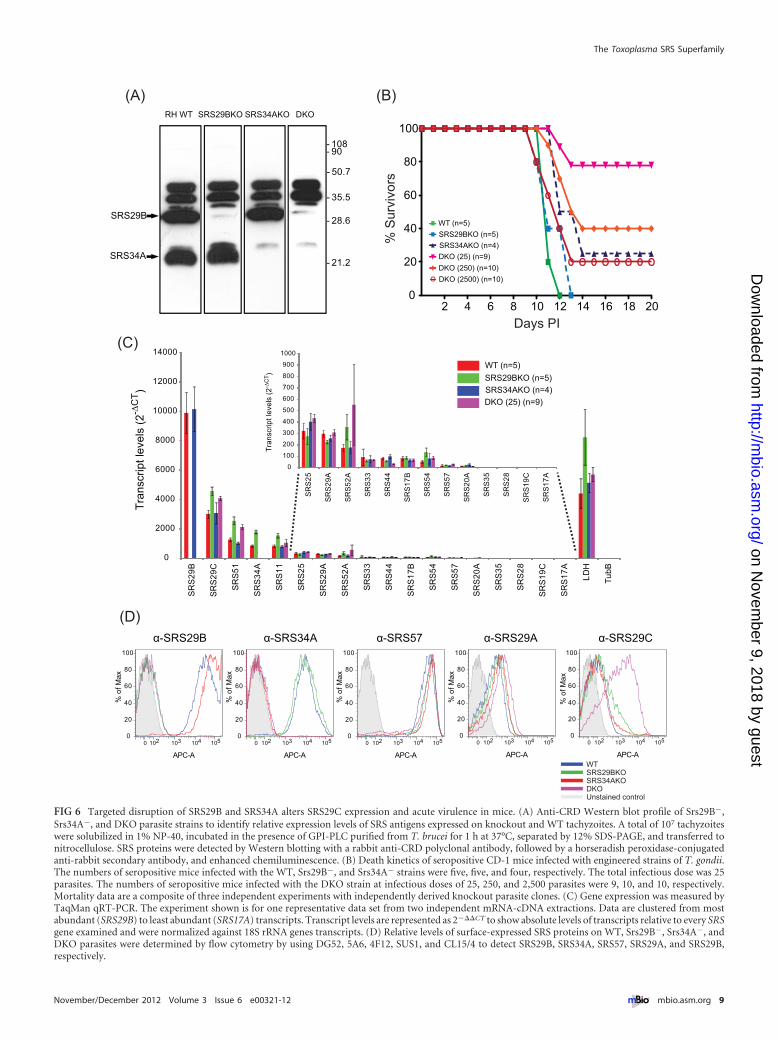
FIG 6 Targeted disruption of SRS29B and SRS34A alters SRS29C expression and acute virulence in mice. (A) Anti-CRD Western blot profile of Srs29B�,Srs34A�, and DKO parasite strains to identify relative expression levels of SRS antigens expressed on knockout and WT tachyzoites. A total of 107 tachyzoiteswere solubilized in 1% NP-40, incubated in the presence of GPI-PLC purified from T. brucei for 1 h at 37°C, separated by 12% SDS-PAGE, and transferred tonitrocellulose. SRS proteins were detected by Western blotting with a rabbit anti-CRD polyclonal antibody, followed by a horseradish peroxidase-conjugatedanti-rabbit secondary antibody, and enhanced chemiluminescence. (B) Death kinetics of seropositive CD-1 mice infected with engineered strains of T. gondii.The numbers of seropositive mice infected with the WT, Srs29B�, and Srs34A� strains were five, five, and four, respectively. The total infectious dose was 25parasites. The numbers of seropositive mice infected with the DKO strain at infectious doses of 25, 250, and 2,500 parasites were 9, 10, and 10, respectively.Mortality data are a composite of three independent experiments with independently derived knockout parasite clones. (C) Gene expression was measured byTaqMan qRT-PCR. The experiment shown is for one representative data set from two independent mRNA-cDNA extractions. Data are clustered from mostabundant (SRS29B) to least abundant (SRS17A) transcripts. Transcript levels are represented as 2���CT to show absolute levels of transcripts relative to every SRSgene examined and were normalized against 18S rRNA genes transcripts. (D) Relative levels of surface-expressed SRS proteins on WT, Srs29B�, Srs34A�, andDKO parasites were determined by flow cytometry by using DG52, 5A6, 4F12, SUS1, and CL15/4 to detect SRS29B, SRS34A, SRS57, SRS29A, and SRS29B,respectively.
The Toxoplasma SRS Superfamily
November/December 2012 Volume 3 Issue 6 e00321-12 ® mbio.asm.org 9
on Novem
ber 9, 2018 by guesthttp://m
bio.asm.org/
Dow
nloaded from

parasite surface coat (i.e., VSG genes in T. brucei) or promoterapid sequence variation via increased rates of ectopic recombina-tion (var genes in P. falciparum) (24, 25). In contrast, distinct,largely nonoverlapping sets of SRS mRNA transcripts were foundto be codominantly expressed in a strain-dependent and develop-mental life cycle stage-dependent fashion. Although no data cur-rently exist for merozoites, gametes, and developing oocysts that
represent the sexual stages of T. gondii de-velopment, it is likely that their surfacecoats will likewise be populated by a mu-tually exclusive set of SRS proteins.Hence, the genetic mechanism(s) thatregulates SRS gene expression is clearlydifferent from that of the vsg or var genes,whereby only a single locus is expressed atany one time. Analysis of the expressiondata is a first step toward understandinghow SRS genes are regulated as a system.Stage specificity data will become morerobust as new RNA sequence data setsemerge for bradyzoites sampled fromcysts in vivo, for merozoites growing infeline enterocytes, and for oocysts under-going sporulation. By moving away fromsingle-gene studies, future work will beable to look for factors that regulate thecoordinated coexpression of these life cy-cle stage-specific SRS genes, whether it beby shared transcription factor binding sitesand promoters or via epigenetic factors.
The size of the SRS superfamily, its se-quence diversity, and its limited taxo-nomic distribution point to a relativelyrecent and rapid expansion coupled to ahigh mutation rate. The tolerance of theseevents is likely due to the SRS structuralfold serving as a scaffold for the design ofnew biological properties, similar to theproposed evolution of the T. brucei trans-ferrin receptor (TfR) from the VSG gene(26). Given the plasticity of this gene fam-ily, the assignment of SRS domains wasnontrivial. The subfamily HMMs identi-fied related domains, which were curatedby match length and the presence of atleast four cysteine residues. Domains thatfailed either of the latter two criteria werelabeled as “degraded.” This led to the as-signment of an SRS domain to SRS35, theSAG4 gene. It is noteworthy that SAG4.2does not meet the HMM cutoff and con-tains too few cysteines; however, it islikely that this gene was duplicated froman SRS gene and evolved to fulfill a newfunctional niche. It is possible that a sim-ilar event led to the establishment of theSUSA gene family (5). Structural analysisand cross-species comparisons will helpreveal the role of such SRS-related se-quences.
The eight domain subfamilies present an excellent opportunityto study the SRS superfamily. An important caveat is that intrasu-bfamily relationships must remain ambiguous. Within each mul-tigene locus, member genes have similar architectures of domainfamilies and similar gene structures (number of introns) and, withthe exception of the SRS38 locus, are transcribed in the same di-rection. These strong associations between domain families and
FIG 7 Transoverexpression of SRS29C is sufficient to attenuate acute virulence in mice. (A) Flowcytometric analysis of SRS29C surface expression in transgenic clones overexpressing type II (SRS29C-II) and type I (SRS29C-I) alleles. Endogenous SRS29C expression levels for type I (RH) and type II(Me49) are shown for comparison. Unstained control refers to staining without a primary antibody. (B)Effect of SRS29C overexpression on acute virulence in mice. Infection with SRS29C-I or SRS29C-IIparasites resulted in significantly lower mortality than that caused by the WT. The number of seropos-itive mice infected with WT strain 22 was 17 for SRS29C-I and 14 for SRS29C-II. The total infectiousdose for all experiments was 50 parasites. Mortality data are a composite of six independent experimentsby using independently derived transgenic clones (two for SRS29C-I, one for SRS29C-II) and the WTparent. (C) Bioluminescent detection of parasite burdens and dissemination in vivo. CD-1 mice in-fected with 50 luciferase-positive WT or SRS29C-I parasites and imaged daily. Representative imageswith photon output intensity in photons/second/cm2/surface radiance (sr) are shown for days 3, 5, 8,and 11. The minimum number of photons was set at 3,932, and the maximum was set at 1.3 � 106. Tocontrol for background luminescence on days 8 and 11, the minimum was set at 2 � 105. (D) BLI ofluciferase-positive parasites in ex vivo organs. Spleens, gastrointestinal tracts (GIT), and thymuses frommice sacrificed on day 10 were imaged ex vivo to quantify parasite burdens.
Wasmuth et al.
10 ® mbio.asm.org November/December 2012 Volume 3 Issue 6 e00321-12
on Novem
ber 9, 2018 by guesthttp://m
bio.asm.org/
Dow
nloaded from

the small number of observed domain architectures (only 9 of apotential 36 nondirectional two-domain combinations) implythat exon switching is not a significant mechanism for increasingprotein divergence. Taken together, these data suggest that theseloci arose by tandem gene duplication. Whether the expansion isdue to duplications of single genes or whole loci is unclear. Con-sidering a subset of two-domain proteins (families 7 and 8), itappears that a mixture of the two is most likely (Fig. 4). The situ-ation is further complicated by convergent evolution. A recentanalysis of the expanded SRS gene family in N. caninum has re-vealed intralocus genetic exchange as an important mechanismhomogenizing SRS genes (27).
The spectrum of biological functions for known SRS proteinshas led to the hypothesis that the SRS family has evolved distinctsubsets that either function as adhesins (e.g., SRS57) or immunedecoys (e.g., SRS34A) or possess some form of dual activity (e.g.,SRS29B). In fact, the tachyzoite-specific SRS29B, SRS34A, andSRS29C proteins elicit high-titer antibodies during the acute in-fection phase (28). Such immune dominance has been postulatedto focus immunity against the disseminating tachyzoite stage tolimit their proliferation and away from the transmissible tissue-encysting bradyzoite form (which no longer expresses SRS29B,SRS34A, and SRS29C). Alternatively, immunity to these domi-nant antigens might function, at least in part, to provide a differ-entiation signal to switch tachyzoites into bradyzoites. This couldrepresent a novel type of surface coat variation that is coupled tothe developmental program of the parasite. These two mecha-nisms are not necessarily mutually exclusive, and the latter possi-bility is further supported by previous studies that implicate com-ponents of host immunity as differentiation signals (29). At least29 orthologs of the SRS superfamily are present in other tissue-dwelling coccidians. In contrast, no SRS orthologs were identifiedin other apicomplexan parasites (Eimeria, Plasmodium, or Cryp-tosporidium) on the basis of primary sequence similarity searches.Recent studies, however, identified an SRS-like fold in the 10-member Pfs230-related 6-Cys family of gamete surface proteins,potentially extending the role of SRS domains as fertility factors ingamete-gamete recognition and attachment during fertilization(15, 30). It is unclear whether this represents the convergent evo-lution of unrelated proteins or is the result of divergent evolutionof genes that arose from the same ancestral gene, possibly a pre-cursor of the 6-Cys proteins that are a feature of the apicomplexangenomes (1).
Toxoplasma presumably regulates the expression of specificSRS antigens to successfully initiate infection and regulate the de-velopment of host immunity in order to establish patent, trans-missible infections. Differential expression of certain alleles of theROPK genes ROP5, ROP16, and ROP18 has previously beenshown to facilitate parasite infection by actively alteringimmunity-related GTPases (IRGs) and cytokine signaling to pro-mote parasite infection (4, 31–33). Integration of the SRS expres-sion and polymorphism data identified numerous parasitelineage-specific differences and prioritized nine SRS genes forfunctional investigation to determine their role in parasite viru-lence. Targeted disruption of the SRS29B and SRS34A genes in thevirulent type I RH strain did not affect parasite replication in vitrobut did have a dramatic effect on mouse virulence. These DKOparasites showed a strong and specific upregulation in theirSRS29C surface expression to a level found on the surface ofmouse avirulent type II and III parasites. Upregulation was not at
the level of transcription (Fig. 6C) but more likely to be the resultof increased mRNA stability or to be regulated posttranscription-ally, since similar levels of SRS29C message were detected in type Istrains, which express low levels of SRS29C protein, and the DKOparasites and type II and III strains, which are high expressers.Importantly, the SRS29C-I and SRS29C-II strains that transgeni-cally overexpress the two SRS29C alleles in the virulent RH strainpossessed similar lethal dose kinetics in mice, suggesting that theSRS29C expression level, rather than polymorphism, is the morerelevant factor modulating the acute-virulence phenotype. Thedecreased virulence seen is most likely the function of an alteredhost immune response because no intrinsic growth defect wasobserved in the transgenic parasites and they were not impaired inthe ability to invade host cells. Our preliminary results measuringsystemic gamma interferon (IFN-�) levels at the peak of infectionshowed that parasites expressing high levels of SRS29C proteinhad substantially reduced levels of IFN-� during in vivo infection(V. Pszenny et al., unpublished data). Hence, the absolute levels ofIFN-� produced in mice infected with the SRS29C-I or SRS29C-IIparasites relative to the WT RH parent strain are controlledthroughout infection. Murine infections with type I strains typi-cally produce dysregulated levels of interleukin-12 and IFN-� andinduce severe immunopathology and death (34, 35). It is possiblethat antigens such as SRS29C have evolved to fine-tune and/ornegatively regulate the proinflammatory capacity of other immu-nodominant antigens (SRS29B) or polymorphic effector mole-cules that directly alter cytokine induction and/or signaling(ROP5, ROP16, ROP18, ROP38, GRA15) in a parasite strain- andhost genetics-dependent fashion, resulting in dysregulated immu-nity and gross pathology (4, 8, 31, 32, 36). Ultimately, these twoconceivably opposing mechanisms might ensure the persistenceof the bradyzoite form of the parasite by actively subvertingand/or fine-tuning the potent proinflammatory immune re-sponse that tachyzoite-specific antigens such as SRS29B, ROP16,ROP18, and GRA15 induce. This notion is strongly reinforced bya recent study implicating SRS genes that regulate the persistenceof parasite cysts (7). This incredibly successful Toxoplasma para-site might thus have evolved such an intricate mechanism to allowfor early parasite proliferation and dissemination to sites ofchronic infection in the host (e.g., brain, heart) before a potenthost response is mounted against the tachyzoite form to limit theparasite burden and the pathology of infection.
It is conceivable that Toxoplasma’s complex virulence traits aremultifactorial, with a variety of different positive and/or negativemodulatory factors that cooperatively function to confer variousdegrees of virulence among parasite strains. Ultimately, thiswould allow the efficient expansion of the range of hosts and eco-logical niches this successful parasite occupies. Attempts to alterthe expression levels and/or engineer a targeted deletion ofSRS29C in the avirulent type II Prugniaud strain are ongoing.Genetic disruption of the endogenous gene and/or subsequenttransgenic manipulation to allow allele-specific and regulated ex-pression of SRS29C will be required to elucidate the mechanisticbasis governing the role of SRS29C in the acute-virulence pheno-type across a variety of parasite genotypes.
In conclusion, this report highlights the importance of inte-grating comparative and functional genomic data sets to generatetestable scientific hypotheses that are assumptionless in their ap-proach. The identification of SRS29C as an important regulator ofvirulence capable of altering parasite pathogenesis and the devel-
The Toxoplasma SRS Superfamily
November/December 2012 Volume 3 Issue 6 e00321-12 ® mbio.asm.org 11
on Novem
ber 9, 2018 by guesthttp://m
bio.asm.org/
Dow
nloaded from

opment of protective immunity underpins the critical role api-complexan parasite surface-expressed antigens play among eu-karyotic pathogens that interact with and subvert host immuneresponses in order to establish patent, transmissible infections.
MATERIALS AND METHODSSequences, SRS domain family searches, phylogeny, and selection. Thegenome sequences, protein sequences, and coding sequences were down-loaded from ToxoDB (version 5.3 [Tg]), and the ESTs were downloadedfrom dbEST. HMMer (v3.0.3b) was used to build HMMs to performdomain searches against Toxoplasma protein models, as well as six-frametranslations (transeq) of the genome (http://hmmer.janelia.org/). Pair-wise distances were used to cluster the domains (metric, Kendall’s tau;method, complete linkage). The seed model for Pfam domain PF04092 (v.23) was searched against the T. gondii protein sequences. The gatheringscore (GA) cutoff was used. The CD-Hit program was used to build arepresentative set of domain sequences (95% identity). All-against-allglobal alignments were produced by using the Needleman-Wunsch algo-rithm, and the bit score was used to hierarchically cluster the domains(metric, Kendall’s tau; method, complete linkage). The eight SRS subfam-ilies were aligned by using PROMALS. The alignment was seeded withSRS29B (1KZQ), SRS28 (2X28), and SRS16C (2JKS) structures. Threerules were used in domain identification; a sequence was accepted as anSRS domain if it aligned with one of the domain models with an E value of�e�5, was at least 90 amino acids in length, and contained at least fourcysteine residues. Aligned regions with fewer than four cysteines weredesignated “degraded” domains. Assignment of orthologous groups wasdone manually and guided by MultiParanoid. The phylogenetic trees weregenerated by using the minimum-evolution (ME) and maximum-likelihood (ML) methods. To estimate � (dN/dS ratio), the Slr programwas used.
Pseudogenes were defined as SRS domains that met the search criteriabut were not part of a ToxoDB gene model. Two domains separated by lessthan 1,000 bp were considered multidomain pseudogenes separated by anintron. Stop codons in SRS domains were present in 11% of the pseudo-genes; for the remaining 89%, the absence of a gene model was likely dueto a stop codon elsewhere in the sequence but outside the SRS domain.
Expression profiling. ESTs were mapped onto SRS gene models byusing BLASTX. Microarray data were downloaded from ToxoDB. To de-tect stage-specific expression, an arbitrary cutoff of �2-fold divergencewas used.
Polymorphism analyses. Strain type-specific single-nucleotide poly-morphisms were downloaded for each SRS gene model, and the ratio ofsynonymous-to-nonsynonymous changes was calculated. Protein-basedmultiple-sequence alignments were generated for orthologous groupsfound in all three strains. Coding sequences were mapped onto the align-ment, and pairwise distances were calculated by using DNADIST. Toestimate � (dN/dS ratio), the Slr program was used; an � of �1 is positiveselection, and an � of �0.5 is purifying.
Plasmid constructs and parasite strains. Single-knockout parasiteswere engineered by replacing the SRS17A/B, SRS25, SRS29B/C, SRS33,SRS34A, and SRS35 genes with the drug-selectable hxgprt marker in theRH �hxgprt “WT” strain. To construct the SRS29B/SRS34A DKO, thehxgprt gene inserted in the SRS29B locus was negatively selected by using6-thioxanthine to remove the hxgprt gene and yield RH �srs29B �hxgprt.SRS34A was then disrupted by using the SRS34A single-knockout con-struct. A 3.2-kb fragment encompassing the SRS29C gene was amplifiedfrom type I RH and type II ME49 and cloned into pTOPO. Heterologousoverexpression of SRS29C in the parental RH (WT) strain was selected byFACS using MAb CL15/4, which recognizes SRS29C.
Flow cytometry. Parasites were fixed and stained with anti-SRS29BMAb DG52, anti-SRS34A MAb 5A6, anti-SRS57 MAb 4F12, anti-SRS29Apolyclonal antibody SUS1, and anti-SRS29C MAb CL15/4. Stained para-sites were analyzed by flow cytometry (FACS) with a FACSCalibur (BDBiosciences).
Mouse infection. Seven- to 10-week-old female CD-1 outbred micewere infected by intraperitoneal injection with a total of 25, 50, 250, or2,500 tachyzoite parasites diluted in 400 �l of phosphate-buffered saline.Mice were imaged daily to detect firefly luciferase activity by using an IVISBLI system from Xenogen to monitor parasite burdens. Mice were in-jected with 200 �l (3 mg) of d-luciferin substrate and imaged for 5 min todetect the photons emitted.
qRT-PCR. Total RNA (2 �g) isolated from tachyzoites with an RNeasyminikit (Qiagen) was reverse transcribed by using random primers andSuperScript II (Invitrogen). Gene expression was measured by TaqManqRT-PCR by using an Applied Biosystems 7900HT real-time PCR system.For the primer and probe sets used, see Table S3 in the supplementalmaterial. The cycling program included 2 min at 50°C and 10 min ofincubation at 95°C, followed by 40 cycles of 95°C for 15 s and 60°C for1 min. The Toxoplasma 18S rRNA gene was used as a reference to normal-ize the quantity of transcripts. Transcript levels were represented as2���CT to show absolute levels of transcripts relative to every SRS geneexamined.
SUPPLEMENTAL MATERIALSupplemental material for this article may be found at http://mbio.asm.org/lookup/suppl/doi:10.1128/mBio.00321-12/-/DCSupplemental.
File S1, DOCX file, 0.1 MB.Figure S1, PDF file, 0.3 MB.Table S1, XLS file, 0.1 MB.Table S2, XLS file, 0.1 MB.Table S3, XLSX file, 0.1 MB.
ACKNOWLEDGMENTS
This study was financially supported by the CIHR (MOP no. 84556 to J.P.and M.E.G.), the Intramural Research Program of the NIH and NIAID(M.E.G.), and the Ontario MRI (J.P.). M.J.B. was supported by the CIHR(MOP no. 82915).
We acknowledge EuPathDB (http://eupathdb.org/) for providing apublicly available repository for all Toxoplasma genomic data sources andannotations. M.E.G. is a scholar of the Canadian Institute for AdvancedResearch Integrated Microbial Biodiversity Program. We thank JonathanWastling, Adam Reid, and Arnab Pain for critical reading of the manu-script.
REFERENCES1. Templeton TJ. 2007. Whole-genome natural histories of apicomplexan
surface proteins. Trends Parasitol. 23:205–212.2. Wasmuth J, Daub J, Peregrín-Alvarez JM, Finney CA, Parkinson J.
2009. The origins of apicomplexan sequence innovation. Genome Res.19:1202–1213.
3. Jung C, Lee CY, Grigg ME. 2004. The SRS superfamily of Toxoplasmasurface proteins. Int. J. Parasitol. 34:285–296.
4. Peixoto L, et al. 2010. Integrative genomic approaches highlight a familyof parasite-specific kinases that regulate host responses. Cell Host Microbe8:208 –218.
5. Pollard AM, Onatolu KN, Hiller L, Haldar K, Knoll LJ. 2008. Highlypolymorphic family of glycosylphosphatidylinositol-anchored surface an-tigens with evidence of developmental regulation in Toxoplasma gondii.Infect. Immun. 76:103–110.
6. Dzierszinski F, Mortuaire M, Cesbron-Delauw MF, Tomavo S. 2000.Targeted disruption of the glycosylphosphatidylinositol-anchored surfaceantigen SAG3 gene in Toxoplasma gondii decreases host cell adhesion anddrastically reduces virulence in mice. Mol. Microbiol. 37:574 –582.
7. Kim SK, Karasov A, Boothroyd JC. 2007. Bradyzoite-specific surfaceantigen SRS9 plays a role in maintaining Toxoplasma gondii persistence inthe brain and in host control of parasite replication in the intestine. Infect.Immun. 75:1626 –1634.
8. Rachinel N, et al. 2004. The induction of acute ileitis by a single microbialantigen of Toxoplasma gondii. J. Immunol. 173:2725–2735.
9. Manger ID, Hehl AB, Boothroyd JC. 1998. The surface of Toxoplasmatachyzoites is dominated by a family of glycosylphosphatidylinositol-anchored antigens related to SAG1. Infect. Immun. 66:2237–2244.
Wasmuth et al.
12 ® mbio.asm.org November/December 2012 Volume 3 Issue 6 e00321-12
on Novem
ber 9, 2018 by guesthttp://m
bio.asm.org/
Dow
nloaded from

10. Mineo JR, et al. 1993. Antibodies to Toxoplasma gondii major surfaceprotein (SAG-1, P30) inhibit infection of host cells and are produced inmurine intestine after peroral infection. J. Immunol. 150:3951–3964.
11. Boulanger MJ, Tonkin ML, Crawford J. 2010. Apicomplexan parasiteadhesins: novel strategies for targeting host cell carbohydrates. Curr.Opin. Struct. Biol. 20:551–559.
12. He XL, Grigg ME, Boothroyd JC, Garcia KC. 2002. Structure of theimmunodominant surface antigen from the Toxoplasma gondii SRS su-perfamily. Nat. Struct. Biol. 9:606 – 611.
13. Howe DK, Crawford AC, Lindsay D, Sibley LD. 1998. The p29 and p35immunodominant antigens of Neospora caninum tachyzoites are homol-ogous to the family of surface antigens of Toxoplasma gondii. Infect. Im-mun. 66:5322–5328.
14. Howe DK, et al. 2008. Strains of Sarcocystis neurona exhibit differences intheir surface antigens, including the absence of the major surface antigenSnSAG1. Int. J. Parasitol. 38:623– 631.
15. Gerloff DL, Creasey A, Maslau S, Carter R. 2005. Structural models forthe protein family characterized by gamete surface protein Pfs230 of Plas-modium falciparum. Proc. Natl. Acad. Sci. U. S. A. 102:13598 –13603.
16. Crawford J, et al. 2010. Structural and functional characterization ofSporoSAG: a SAG2-related surface antigen from Toxoplasma gondii. J.Biol. Chem. 285:12063–12070.
17. Kraemer SM, et al. 2007. Patterns of gene recombination shape var generepertoires in Plasmodium falciparum: comparisons of geographically di-verse isolates. BMC Genomics 8:45.
18. Boyle JP, et al. 2006. Just one cross appears capable of dramaticallyaltering the population biology of a eukaryotic pathogen like Toxoplasmagondii. Proc. Natl. Acad. Sci. U. S. A. 103:10514 –10519.
19. Grigg ME, Bonnefoy S, Hehl AB, Suzuki Y, Boothroyd JC. 2001. Successand virulence in Toxoplasma as the result of sexual recombination be-tween two distinct ancestries. Science 294:161–165.
20. Li L, et al. 2003. Gene discovery in the Apicomplexa as revealed by ESTsequencing and assembly of a comparative gene database. Genome Res.13:443– 454.
21. Manger ID, et al. 1998. Expressed sequence tag analysis of the bradyzoitestage of Toxoplasma gondii: identification of developmentally regulatedgenes. Infect. Immun. 66:1632–1637.
22. Boothroyd JC, Hehl A, Knoll LJ, Manger ID. 1998. The surface ofToxoplasma: more and less. Int. J. Parasitol. 28:3–9.
23. Knoll LJ, Boothroyd JC. 1998. Isolation of developmentally regulatedgenes from Toxoplasma gondii by a gene trap with the positive and negative
selectable marker hypoxanthine-xanthine-guanine phosphoribosyltrans-ferase. Mol. Cell. Biol. 18:807– 814.
24. Cross GA, Wirtz LE, Navarro M. 1998. Regulation of vsg expression sitetranscription and switching in Trypanosoma brucei. Mol. Biochem. Para-sitol. 91:77–91.
25. Scherf A, et al. 1998. Antigenic variation in malaria: in situ switching,relaxed and mutually exclusive transcription of var genes during intra-erythrocytic development in Plasmodium falciparum. EMBO J. 17:5418 –5426.
26. Salmon D, et al. 1997. Characterization of the ligand-binding site of thetransferrin receptor in Trypanosoma brucei demonstrates a structural re-lationship with the N-terminal domain of the variant surface glycoprotein.EMBO J. 16:7272–7278.
27. Reid AJ, et al. 2012. Comparative genomics of the apicomplexan parasitesToxoplasma gondii and Neorospora caninum: coccidia differing in hostrange and transmission strategy. PLoS Pathog. 8:e1002567.
28. Lekutis C, Ferguson DJ, Grigg ME, Camps M, Boothroyd JC. 2001.Surface antigens of Toxoplasma gondii: variations on a theme. Int. J. Para-sitol. 31:1285–1292.
29. Bohne W, Heesemann J, Gross U. 1993. Induction of bradyzoite-specificToxoplasma gondii antigens in gamma interferon-treated mouse macro-phages. Infect. Immun. 61:1141–1145.
30. van Dijk MR, et al. 2010. Three members of the 6-cys protein family ofPlasmodium play a role in gamete fertility. PLoS Pathog. 6:e1000853.
31. Saeij JP, et al. 2006. Polymorphic secreted kinases are key virulencefactors in toxoplasmosis. Science 314:1780 –1783.
32. Saeij JP, et al. 2007. Toxoplasma co-opts host gene expression by injec-tion of a polymorphic kinase homologue. Nature 445:324 –327.
33. Taylor S, et al. 2006. A secreted serine-threonine kinase determines vir-ulence in the eukaryotic pathogen Toxoplasma gondii. Science 314:1776 –1780.
34. Gavrilescu LC, Denkers EY. 2001. IFN-gamma overproduction and highlevel apoptosis are associated with high but not low virulence Toxoplasmagondii infection. J. Immunol. 167:902–909.
35. Mordue DG, Monroy F, La Regina M, Dinarello CA, Sibley LD. 2001.Acute toxoplasmosis leads to lethal overproduction of Th1 cytokines. J.Immunol. 167:4574 – 4584.
36. Reese ML, et al. 2011. Polymorphic family of injected pseudokinases isparamount in Toxoplasma virulence. Proc. Natl. Acad. Sci. U. S. A. 108:10568 –10573.
The Toxoplasma SRS Superfamily
November/December 2012 Volume 3 Issue 6 e00321-12 ® mbio.asm.org 13
on Novem
ber 9, 2018 by guesthttp://m
bio.asm.org/
Dow
nloaded from