Embed Size (px)
Citation preview

Integrating Incremental Learning and Episodic Memory Models of theHippocampal Region
M. MeeterVrije Universiteit Amsterdam
C. E. Myers and M. A. GluckRutgers University
By integrating previous computational models of corticohippocampal function, the authors develop andtest a unified theory of the neural substrates of familiarity, recollection, and classical conditioning. Thisapproach integrates models from 2 traditions of hippocampal modeling, those of episodic memory andincremental learning, by drawing on an earlier mathematical model of conditioning, SOP (A. Wagner,1981). The model describes how a familiarity signal may arise from parahippocampal cortices, giving anovel explanation for the finding that the neural response to a stimulus in these regions decreases withincreasing stimulus familiarity. Recollection is ascribed to the hippocampus proper. It is shown how theproperties of episodic representations in the neocortex, parahippocampal gyrus, and hippocampus propermay explain phenomena in classical conditioning. The model reproduces the effects of hippocampal,septal, and broad hippocampal region lesions on contextual modulation of classical conditioning,blocking, learned irrelevance, and latent inhibition.
Keywords: memory, classical conditioning, hippocampus, medial septum, cerebellum
Many recent theories of hippocampal functioning are variants ofthe idea that the hippocampus, or more broadly the hippocampalregion (hippocampus proper; dentate gyrus; subiculum; and ento-rhinal, perirhinal, and parahippocampal cortices), stores episodicmemories (Eichenbaum, 1992; Hasselmo & Wyble, 1997; Marr,1971; McClelland & Goddard, 1996; Meeter & Murre, in press;Meeter, Talamini, & Murre, 2004; Norman & O’Reilly, 2003;Talamini, Meeter, Murre, Elvevåg, & Goldberg, in press). In itsstrictest definitions, episodic refers to the memories for an unstruc-tured array of things that were present or events that occurred atone specific location and moment in time. Evidence that thehippocampus plays a role in storing such memories comes fromelectrophysiological recordings (Eichenbaum, 2000; Ferbintineau& Shapiro, 2003; O’Keefe, 1979), functional imaging (Cabeza &Nyberg, 2000), patient data (Reed & Squire, 1998; Rempel-Clower, Zola, Squire, & Amaral, 1996; Scoville & Milner, 1957),and lesion studies with experimental animals (Gilbert, Kesner, &Lee, 2001; Jarrard, 1995).
However, lesion data have also implicated the hippocampalregion in tasks that do not conform very well to the episodic label.These findings, discussed below, suggest that the hippocampus hasa role in what is usually referred to as associative learning (Pearce& Bouton, 2001) or incremental learning (Gluck, Meeter, & My-ers, 2003). Both names are apt, as this domain involves tasks inwhich the learner is required to associate a set of stimuli or a state
of the environment with a behavioral output. Moreover, the outputusually develops over tens or hundreds of trials. This is a far cryfrom episodic memory tasks, which often require retrieval afterjust one presentation of a certain material.
Findings from the incremental learning literature have led to asecond strain of theories of the hippocampal region. The hip-pocampal region is theorized to aid the slow development of thecorrect representations to support behavioral performance (Gluck& Myers, 1993; Schmajuk & DiCarlo, 1992). In classical condi-tioning, for example, this may take the form of developing repre-sentations that distinguish conditions predictive of an uncondi-tioned stimulus and conditions that predict no such stimulus willoccur. The hippocampal region may thus slowly alter and adaptrepresentations in response to environmental feedback (i.e., re-wards, punishments, unconditioned stimuli).
The two groups of theories, those concerned with episodicmemory and those addressing incremental learning, seem diamet-rically opposed in the demands they pose to hippocampal func-tioning. Incremental learning is sensitive to behavioral outcome,whereas episodic learning is thought of as unsupervised, automaticcoding of whatever is present. Episodic learning is fast (often onetrial), whereas incremental learning is slow. It is not immediatelyobvious how a model could reconcile episodic and incrementallearning in a parsimonious way. Here, we present a model thatattempts to do just that—reconcile episodic memory with incre-mental learning. The conceptual glue that binds these two ap-proaches is an earlier mathematical model of conditioning, thesometimes opponent processing, or SOP, model (Wagner, 1981).The model is able to account for the basic phenomena of episodicmemory as well as for classic findings in the conditioning litera-ture. Moreover, it offers a novel explanation for a puzzling findingfrom neurophysiology, namely that familiarity decreases (ratherthan increases) neural responses to familiar stimuli (Li, Miller, &Desimone, 1993; Xiang & Brown, 1998).
M. Meeter, Department of Cognitive Psychology, Vrije UniversiteitAmsterdam, Amsterdam, the Netherlands; C. E. Myers, Department ofPsychology, Rutgers University; M. A. Gluck, Center for Molecular andBehavioral Neuroscience, Rutgers University.
Correspondence concerning this article should be addressed to M.Meeter, Department of Cognitive Psychology, Vrije Universiteit Amster-dam, Vd Boechorststraat 1, 1081 BT Amsterdam, the Netherlands. E-mail:[email protected]
Psychological Review Copyright 2005 by the American Psychological Association2005, Vol. 112, No. 3, 560–585 0033-295X/05/$12.00 DOI: 10.1037/0033-295X.112.3.560
560

In the remainder of this article, we first outline the two strainsof models and some of the evidence for the roles of the hippocam-pal region in different kinds of memory. Then we present ourmodel, both in its theoretical basis and in its applications toconcrete data. Although it is applicable to a wide range of memorytasks, we focus in this article on what may be seen as twoextremes: explicit episodic memory and classical conditioning.The article ends with a discussion of the merits and limitations ofthe model, a comparison with other models, and a list of untestedpredictions.
Role of Hippocampal Region in Episodic Memory
The role of the hippocampus in memory has been the focus ofmany qualitative theories and computational models (Eichenbaum,1992; Hasselmo & Wyble, 1997; Marr, 1971; McClelland &Goddard, 1996; Meeter, Murre, & Talamini, 2002; Meeter et al.,2004; Norman & O’Reilly, 2003; Talamini et al., in press). Thesemodels have assumed that the hippocampal region simply storeswhatever pattern is presented to it by the neocortex. Input from theneocortex is modeled as arbitrary vectors to be stored. The hip-pocampal region forms a compact code that is bidirectionallylinked to such neocortical representations. If a partial cue can laterreactivate this compact code, it can retrieve the neocortical repre-sentation in its entirety.
The hippocampus is not the only structure involved in episodicmemory. Parahippocampal cortices adjacent to the hippocampushave also been implicated in memory, especially in the processingof stimulus familiarity (Aggleton & Brown, 1999; Davachi, Mitch-ell, & Wagner, 2003; Ranganath et al., 2004; Zhu, McCabe,Aggleton, & Brown, 1997). Areas of the neocortex, down tounimodal primary cortices, play a role too: retrieval of visualmemories will activate not only the medial temporal lobe but alsovisual areas down to primary visual area V1 (Cabeza & Nyberg,2000; Reber & Squire, 1998). In line with these findings, lesionsto visual neocortical areas in the occipital lobe can cause amnesiafor visual details (Ogden, 1993).
Damage to the basal forebrain can also cause a dense amnesia,suggesting a role in memory (Tranel, Damasio, & Damasio, 2000).Within this set of structures, the importance of the medial septumfor hippocampal functioning has often been stressed (Hasselmo &Bower, 1993; Hasselmo & Wyble, 1997; Meeter et al., 2004;Myers et al., 1996; Rokers, Mercado, Allen, Myers, & Gluck,2002), as it is the main source of acetylcholine in the hippocampus(Alonso, Sang, & Amaral, 1996). Because acetylcholine facilitateslearning (Hasselmo, 1999), the medial septum may thus control thelearning process within the hippocampus.
From these findings rises a view of memories as a set of rich,modality-specific representations stored in the neocortex, boundtogether by high-level compound representations stored in thehippocampus (Murre, 1996; Teyler & DiScenna, 1986). This dis-tinction between representations of different complexity need notbe viewed as a dichotomy; in fact, representations of intermediatecomplexity may exist in areas such as the parahippocampal gyrus.A natural view of episodic memories would thus be a hierarchy ofrepresentations all bound together, with low-level features storedin the neocortex and integrated by higher level representations inthe hippocampal region (Talamini et al., in press). The septum may
fit in this story as the controller of the learning rate in thehippocampus.
Role of the Hippocampal Region in Incremental Learning
In classical conditioning, subjects learn that a previously neutralstimulus (the conditioned stimulus [CS]) precedes a response-evoking stimulus (the unconditioned stimulus [US]). With time,they learn to give an anticipatory or preparatory response (theconditioned response [CR]) to the CS. Such conditioning can beanalyzed rather successfully as the learning of relations betweenstimuli and motor outputs. Where stimuli and outputs are con-nected in the brain has remained a difficult question, but of somebrain regions, it is now clear that they are important in condition-ing. For classical conditioning, the cerebellum plays a major role(Steinmetz, 1998; Thompson, 1990). The basal ganglia are knownto be involved in operant conditioning and to code for rewards(Lauwereyns, Watanabe, Coe, & Hikosaka, 2002; Pagnoni, Zink,Montague, & Berns, 2002; Peoples, Uzwiak, Gee, & West, 1997).In fear conditioning, the amygdala has been identified as a centralstructure (LeDoux, 1996).
These structures are not the only important ones. Conditioningcan occur to very complex stimuli as well as to simple ones, suchas an entire environment in contextual fear conditioning(Fanselow, 2000). It is unlikely that the structures enumeratedabove do the necessary stimulus processing, as they are smallrelative to the size of the whole brain. More likely, they takeadvantage of processing elsewhere in the brain, using processed,high-level representations as their input.
Indeed, there are several lines of evidence that point to theimportance of neocortical processing areas for conditioning. Pri-mary sensory cortices have, for example, been found to reorganizethemselves under influence of contingencies in conditioning tasks(Recanzone, Schreiner, & Merzenich, 1993). In addition, the hip-pocampal region seems to have a role in incremental learning, asis suggested by electrophysiological recordings during condition-ing tasks: During eyeblink conditioning, hippocampal neural ac-tivity develops in response to a CS, and its shape and durationmimic the CR (Berger & Thompson, 1978a).
Hippocampal involvement in incremental learning is also evi-dent from the effects of hippocampal lesions on conditioningparadigms. Although, for example, classical conditioning itself isnot slowed by hippocampal damage (Schmaltz & Theios, 1972;Shohamy, Allen, & Gluck, 2000), animals with lesions in thehippocampal region will in many variations of the basic paradigmperform differently than normal animals. Animals with lesions tothe hippocampus and overlaying cortex do not show the decreasedrates of responding typically seen in intact ones when conditionedstimuli are presented in a different context than the one in whichconditioning took place (Penick & Solomon, 1991). They also donot show the temporary dip in responding that occurs in intactanimals when a CS predictive of a US is suddenly accompanied byanother stimulus (Allen, Padilla, Myers, & Gluck, 2002).
The broader hippocampal region (including adjacent parahip-pocampal cortices and the subiculum) seems to be important forlatent inhibition and learned irrelevance. In latent inhibition, theCS is presented many times to the animal in the environment inwhich training will take place, prior to training. When trainingstarts and the CS is followed by a US, the animal is severely
561INCREMENTAL LEARNING AND EPISODIC MEMORY

slowed down acquiring the CS–US association (Lubow & Moore,1959). In learned irrelevance, the animal is first subjected to manyexplicitly uncorrelated presentations of the stimulus and the US.When in the training phase the CS is reliably followed by the US,learning the CS–US association is slowed relative to a controlcondition in which animals do not receive the uncorrelated preex-posure (Mackintosh, 1973). Both phenomena, which tap complexlearning about environmental contingencies, are abolished afterhippocampal region ablations (Ackil, Mellgren, Halgren, & From-mer, 1969; Allen, Chelius, & Gluck, 2002; Han, Gallagher, &Holland, 1995).
Gluck and Myers’s (1993) Model of Incremental Learning
One theory of incremental learning, Gluck and Myers’s (1993)model, has accounted for these effects of hippocampal lesions bymodeling the broader hippocampal region as a predictive autoen-coder, which interacts with a simpler module representing thecerebellum (see Figure 1). The task of this autoencoder is topredict its environment, including the presence or absence of allstimuli, including CSs, contextual stimuli, and a US, if any. Thisleads to the formation of representations that the autoencoder canuse to predict an upcoming US. With the predictive encoder intact,the model reproduces standard phenomena in the conditioningliterature. With the autoencoder lesioned, it reproduces the above-mentioned effects of hippocampal lesions on conditioning.
Several features of autoencoders help to make Gluck and My-ers’s (1993) model a good model of the hippocampal role inclassical and operant conditioning. Autoencoders will compressfeatures that consistently occur together (i.e., have the same pre-
dictive value). Features that do not occur together will, conversely,be differentiated. This explains the model’s performance in para-digms in which contextual discrimination plays a role: A stimulusA may be rewarding in context X, but not in context Y, whereas itis the other way round for a stimulus B. A simple neural networksuch as a perceptron cannot solve this nonlinear problem, becauseits response to A and X together is by necessity the sum of itsresponses to A and X separately, and neither on its own will predictthe reward. The model solves this problem by developing a sep-arate representation for the compound of A and X, and this com-pound can then predict occurrence of the reward (Myers & Gluck,1994).
The tendency of autoencoders to compress features that occurtogether also explains paradigms such as latent inhibition andlearned irrelevance. In these paradigms stimulus and context be-come bound into a single representation during exposure, in whichneither predicts anything other than itself. This works againstsubsequent learning to respond in the presence of the CS but notthe context alone.
Another feature of autoencoders is that the amount of learningtaking place is a function of prediction error. If the error is large,as it will be at the onset of learning, learning takes big steps. If,after training, the error is small (i.e., the autoencoder can predict itsenvironment and the US), learning will be slow. This explainsblocking, a paradigm in which a CS is paired with a US at amoment that the US is already well predicted. In blocking, ananimal first goes through many trials in which a stimulus A ispaired with a US. When the animal reliably makes a CR to thestimulus, stimulus A is presented in combination with a novelstimulus B, and that compound is still followed by the US. When,after many such trials stimulus B is presented on its own, animalstypically emit CRs only at very low rates (Kamin, 1969). Nostimulus B–CR link has been made, even though stimulus B waspredictive of a US on all trials in which it was presented. In Gluckand Myers’s (1993) model, this is explained by the fact that theanimal makes no errors during training on the compound, as italready emits CRs to stimulus A. Without errors to be corrected, nolearning occurs in the cerebellar network in which CSs and CRsare connected (this explanation essentially follows from theRescorla–Wagner rule discussed in a later section).
Limitations of the Model
The hippocampal role in many incremental learning paradigmscan thus be captured by the mathematics of error-correction learn-ing that underlies predictive autoencoders. Three features standout: that the model can form representations at the right level ofcomplexity (i.e., it forms compound or differentiated representa-tions as needed), that its hippocampal network learns swiftly onlywhen a stimulus has not already been incorporated into the context,and that it learns new CS–CR associations only when a US is notyet well predicted.
Nevertheless, there are clear grounds for improvement on themodel. One is that the model does not clearly map to brainanatomy. For example, the model has one module for the hip-pocampal region and can thus not differentiate the role of thehippocampus proper from that of the surrounding areas. Another isthat the model does not provide any account of episodic memoryin the hippocampus. A third is that error-correction learning may
Figure 1. The corticohippocampal model of Gluck and Myers (1993).The model receives inputs representing conditioned stimuli, such as lightsand tones, as well as contextual information. One network, representingprocessing that is dependent on the hippocampal region, learns to recon-struct these inputs and also to predict arrival of the unconditioned stimulus(US), such as a corneal airpuff. As the hippocampal-region network doesthis, it forms new stimulus representations in its internal layer that com-press redundant information and differentiate predictive information. Asecond network, assumed to represent long-term memory sites in cerebraland cerebellar cortices, adopts the internal representation provided by thehippocampal-region network and then maps from this to an output thatrepresents the strength or probability of a conditioned response (CR), suchas a protective eyeblink. Adapted from “Hippocampal Mediation of Stim-ulus Representation: A Computational Theory,” by M. A. Gluck and C. E.Myers, 1993, Hippocampus, 3, p. 495. Copyright 1993 by Wiley.
562 MEETER, MYERS, AND GLUCK

indeed occur in the cerebellum, but it has not been shown to takeplace in the hippocampus. The necessary prediction error signalhas never been identified there, and neither has a response com-mensurate with error reduction. Below, we present a model thattakes over many features of Gluck and Myers’s (1993) model butseeks to remedy the limitations just noted.
Incremental and Episodic in One Model
At the heart of our model is a welding of two theories: Gluckand Myers’s (1993) model and a generic version of an episodicmemory model. From Gluck and Myers’s (1993) model, our modeltakes an architecture in which a hippocampal memory systeminteracts with more output-oriented brain structures. But the au-toencoder hippocampal submodel is replaced by a multilayermodel capable of forming episodic memories (Meeter et al., 2002;Talamini et al., in press). This simple model of episodic memory,as shown in Figure 2, contains three layers in which stimuli and,more broadly, the environment of the organism are represented.An input layer, modeling the neocortex, codes for stimuli andcontext features. A second layer stands for the parahippocampalregion: the perirhinal, entorhinal, and postrhinal–parahippocampalcortices. This layer has integrated representations, with some partscoding mostly for context features and some mostly for stimuli, butwith all parts also getting input of the other kind. The third layerstands for the hippocampus proper, with nodes representing den-tate granule cells and/or pyramidal cells in Ammon’s horn. Thishippocampal layer forms a compact code for the whole situation inwhich the organism finds itself, for which we use the term ensem-
ble (Murnane, Phelps, & Malmberg, 1999). Such representationsform the basis of episodic memory.
To simulate incremental learning tasks, the model has to containmodules coding for the outputs of memory. For classical condi-tioning, the cerebellum is most relevant, but for other tasks ofincremental learning, one would have to include output regions forrewards and operant behaviors (basal ganglia) and for fear re-sponses (amygdala). The cerebellar circuit, the only output struc-ture implemented here, is a simplified version of the cerebellarnetwork in Gluck and Myers’s (1993) model. All three represen-tational layers project to the output modules. These connectionsallow the output modules to attach behavioral significance tosimple and complex representations of the same set of stimuli,thereby allowing stimulus configurations to have different associ-ations than the constituent stimuli on their own.
Multilevel Representations
This may explain one feature of Gluck and Myers’s (1993)model, the formation of compound and single representations.Instead of representations at the right level of complexity beingslowly formed through error-correction learning, they may beformed automatically in episodic memory on the first presentationof the stimuli. Conditioning may then proceed by associatingevents such as the occurrence of a US with episodic representa-tions at the right level of complexity.
Consider the following example: A simple stimulus such as atone is coded for in the neocortical layer of the model. This sametone, mixed in with contextual elements, is coded for in theparahippocampal layer. The hippocampal layer has one compoundrepresentation that stands for the ensemble of all available cues—that is, the situation in which the animal finds itself. An outcomecan now be connected to the neocortical representation, as insimple conditioning to a tone independent of the context in whichthe tone occurs (connection labeled “a.” in Figure 2). It can also beconnected to a parahippocampal representation, which in thismodel would be equivalent to conditioning to a tone in a context(connection labeled “b.” in Figure 2). Finally, it can be connectedto a hippocampal representation, which would be equivalent toconditioning to a whole situation (connection labeled “c.” in Fig-ure 2). In most cases all three associations may develop at the sametime. It may also be, however, that over the course of manylearning trials, low-level features are associated with other out-comes than compounds of the same stimuli. This would be the casein contextually modulated conditioning.
Effects of Outcome Predictability
In addition to its ability to form representations of the rightcomplexity, the second feature mentioned above of Gluck andMyers’s (1993) model is its ability to learn at the right time.Speeded acquisition of a CR or an operant response occurs onlywhen an unpredicted outcome is presented together with a novelstimulus. When a stimulus loses its novelty before it is combinedwith an unpredicted outcome, both the model and experimentalanimals suffer from learned irrelevance or latent inhibition. Whenthe novel stimulus occurs after the outcome has lost its unpredict-ability, blocking will impede learning.
Figure 2. Brain regions that play a role in incremental learning and thatare part of the model. Representations are assumed to be formed in ahierarchy of regions. All stimuli have a basic neocortical representation.They are also represented in the parahippocampal gyrus (parahippocam-pus), but here stimulus representations are loaded with context. In thehippocampus, stimuli and context are bound together in holistic ensembles.These representations form under the influence of acetylcholine (ACh). Allthree representational layers are assumed to project to output regions.These are the cerebellum for classical conditioning, the basal ganglia foroperant conditioning, and the amygdala for fear conditioning. Of these,only the cerebellum is implemented in the model. a, b, and c: Differentkinds of information that can form the basis for classical conditioning; seethe main text for further explanation.
563INCREMENTAL LEARNING AND EPISODIC MEMORY

The effects of outcome predictability on learning are capturedvery well by the Rescorla–Wagner rule of associative learning(Rescorla & Wagner, 1972). This rule assumes that the amount oflearning taking place at any one trial is a function of how well theoutcome is, on that trial, predicted by all cues taken together (seethe Appendix for a mathematical formulation). When an outcomepaired with a CS is already predicted by other cues, no learning tothe CS will take place, and blocking ensues. The circuitry in thecerebellum has been shown to indeed implement the Rescorla–Wagner rule (Kim, Krupa, & Thompson, 1998; Thompson &Gluck, 1991), and it is part of both Gluck and Myers’s (1993)model and the cerebellar network of our current model. Thecurrent model thus explains blocking in the same way as Gluckand Myers’s (1993) model does.
In classical conditioning, learning is thus explicitly dependenton prediction failure. Although not directly relevant to the currentwork, it is important to note that a dependence of learning onoutcome predictability is also plausible for kinds of incrementallearning involving different brain regions. In the basal ganglia,long-term potentiation (LTP), a candidate mechanism for long-term memory, is enhanced by dopamine (Thomas & Malenka,2003), a neuromodulator. Electrophysiological data suggest thatdopamine release reflects the unpredictability of current rewards(Schultz, 1998, 2002). These two facts together imply that learningin the basal ganglia is fast when unpredicted rewards occur andslow once a reward is well predicted. Operant conditioning,thought to rely at least partly on the basal ganglia (Lauwereyns etal., 2002; Pagnoni et al., 2002; Peoples et al., 1997), may thus begoverned by the same rules as classical conditioning. In fearconditioning, Fanselow (1998) has argued that the interplay be-tween opioid receptors and fear elicitors in the amygdala may beequivalent to the Rescorla–Wagner rule. In particular, if an animalhas learned a fear response to a CS, this CS may elicit the releaseof endogenous opioids, which dampens the effect of the negativereinforcer. In this way, the reinforcing effect of a fear elicitor maydecrease during learning, as it does in the Rescorla–Wagner rule.
Stimulus Novelty, SOP, and Familiarity
As described above, stimulus novelty also affects incrementallearning. These effects have been formalized in other theories ofassociative learning (Mackintosh, 1975; Pearce & Hall, 1980), oneof which is sometimes opponent processes, or SOP (Wagner,1981). Although SOP has many subtleties, the mechanism bywhich novelty affects incremental learning is fairly straightforward(see also Donegan, Gluck, & Thompson, 1989). The theory as-sumes that stimulus representations can be in one of two states(three, if one includes absolute quiescence). When a stimulus isnovel to the animal, it will on presentation bring its representationinto a very active state, A1. Later, when the stimulus has beenpresented often in a certain context, the animal will come to expectthe stimulus in that context. In this case, when the stimulus ispresented, its representation enters in a lower state of activity, A2.SOP assumes that a stimulus representation can be associated witha US only when it is in State A1, not in A2. In latent inhibition, thestimulus is first presented alone for a number of trials. At the endof this preexposure, the stimulus will elicit only A2 activity.Because learning does not take place in A2, associating the stim-ulus with the US will be retarded, relative to a nonexposed control
condition in which the stimulus is novel and hence in A1. SOP alsopredicts that latent inhibition is reduced or eliminated by a changein context between preexposure and training phases: The newcontext will not be associated with the stimulus and will thereforenot bring it into State A2 on presentation. This release from latentinhibition following context shift has indeed been found (Mack-intosh, Kaye, & Bennett, 1991; Symonds & Hall, 1995).
SOP can be contrasted with a development in the episodicmemory field. Since the 1970s, many researchers in the field ofepisodic recognition memory have argued that recognition judg-ments need not be based on retrieval of the item to be recognizedbut can also be based on a more fuzzy feeling that the item matchesold memories (e.g., Atkinson & Juola, 1974; Humphreys, Bain, &Pike, 1989; Jacoby, 1991; Mandler, 1980; Yonelinas, 2002). Thesetwo kinds of memory are often referred to as recollection andfamiliarity. Recollection is the retrieval of a particular memory, aswhen an item must be reproduced in recall tasks. Familiarity is asignal computed through a comparison of a memory probe with allmemories in the memory store. During a recognition task, theprobe is thought of as eliciting a strong familiarity signal if itresembles some or many stored memories. Formalized in compu-tational models of recognition, the contribution of the familiaritysignal can explain many dissociations between recognition andrecall (Humphreys et al., 1989; Norman & O’Reilly, 2003; Raaij-makers & Shiffrin, 1992).
Lesion studies (Aggleton & Brown, 1999), electrophysiologicalevidence (Zhu et al., 1997), and imaging data (Ranganath et al.,2004) have all implicated the perirhinal cortex in familiarity. In themodels, familiarity is modeled as an increase in a scalar signalcoming out of the memory system. When neural responses in theperirhinal cortex to novel and familiar stimuli are directly com-pared, however, results seem to contradict this idea. Instead ofbeing larger for familiar stimuli, neural responses to a stimulusdecrease in the perirhinal cortex with increasing familiarity (Li etal., 1993; Xiang & Brown, 1998). Although these results (see, e.g.,Figure 3) contradict many theories of familiarity, they are exactlywhat one would expect from SOP: a state of high activity at thefirst presentations, with less activity being elicited by an oft-repeated stimulus. Here, we suggest that the familiarity effect, thedecreased parahippocampal response to familiar stimuli, is whatcauses the effects of stimulus novelty on the speed of conditioning.This leaves open how this familiarity effect is caused.
A Theory of Familiarity
The decrease in firing for familiar stimuli effects may resultfrom the interaction between a few uncontroversial neural mech-anisms. The basic idea is that stimulus representations may alwayspartly reflect the latest context in which they were seen. This willmake them responsive to that context on itself, which will in turnlead to less responsiveness to the stimulus if presented in the samecontext because of adaptation and accommodation.
Figure 4 shows how a parahippocampal node may come torepresent context as well as its preferred stimulus. The input to theparahippocampal gyrus during an experiment consists of a phasicinput when a stimulus is briefly presented and of more or lesscontinuous stimuli that together represent context (e.g., relativelyconstant features of the physical environment). We assume thateach parahippocampal node receives one strong connection from
564 MEETER, MYERS, AND GLUCK

the input it codes for and weak connections from other inputs.Although this is a gross simplification, the underlying idea thatparahippocampal cells have preferred stimuli is well supported(see below).
In Figure 4a, connections for one parahippocampal node codingfor a stimulus are drawn in. This node receives a strong connectionfrom a lower layer node coding for its preferred stimulus, but otherinputs, together forming the context, also reach it. If the preferredstimulus is presented, the node will fire strongly, and the weakconnections from contextual inputs will be strengthened throughLTP. These connections may now be strong enough to make thenode fire at a low rate when its preferred stimulus is absent (see
Figure 4b). This in turn will lead to adaptation in the node. Whenthe preferred stimulus is presented again in that context, the nodewill be in a less responsive state because of the built-up adaptation(see Figure 4c). It will therefore respond at a lower rate than whenthe stimulus was first presented. Exactly this pattern has beenfound in perirhinal and entorhinal neurons (Li et al., 1993; Xiang& Brown, 1998).
On a side note, firing decrements have not yet been observed inthe parahippocampal cortex. This seems at odds with our inclusionof the parahippocampal cortex in the substrate of our parahip-pocampal layer. The parahippocampal cortex relays spatial layoutinformation, among other things, to the hippocampus (Bohbot,Allen, & Nadel, 2000; Vann, Brown, Erichsen, & Aggleton, 2000),and we felt it was important to include this source of hippocampalinputs in our model. To our knowledge, it has not been investi-gated whether parahippocampal neurons show a familiarity firingdecrement for preferred stimuli (this is the case in our model, butit plays no role in the current simulations).
Summary of the Model
In summary, we propose a new model of brain regions involvedin learning and memory. With it we hope to elucidate phenomenafrom the literature of episodic memory and classical conditioning.The model contains three layers that form representations and thatstand for the neocortex, the parahippocampal gyrus, and the hip-pocampus proper (see Figure 2). Of the output regions in Figure 2,only the cerebellum is implemented for this article. The basicassumptions of the theory behind our model, as presented above,are listed below. A comparison of these assumptions with those ofother models is addressed in the General Discussion.
1. The neocortex, parahippocampal gyrus, and hippocam-pus form a hierarchy in which episodic representations ofincreasing complexity are formed.
2. Stimulus representations at different levels of complexitymay acquire different or even opposite associations.
3. Learning in the cerebellum is governed by the Rescorla–Wagner rule. This explains why incremental learning ismodulated by outcome predictability.
4. Incremental learning is modulated by stimulus novelty,
Figure 3. Neuronal responses of a familiarity neuron, in this case located in area TE, to a visual stimulus eitherpresented for the first time (left) or repeated in a different session (first presentation in that session; right).Reprinted from Neuropharmacology, 37, J. Z. Xiang and M. W. Brown, “Differential Neuronal Encoding ofNovelty, Familiarity, and Recency in Regions of the Anterior Temporal Lobe,” p. 665, Copyright 1998, withpermission from Elsevier.
Figure 4. Theory of the familiarity effect. Some cortical nodes code forfeatures that are more or less permanent in the current environment(labeled “context”), and others code for phasic cues (labeled “stimulus”).Each cortical node has a strong connection to one parahippocampal node(the solid arrow). In addition, the cortical nodes send weak connections toother parahippocampal nodes (connections to only one parahippocampalnode are drawn; the dashed arrows). a: When a phasic cue is presentedtogether with a set of more permanent context features, cortical nodescoding for the context features are active synchronously with parahip-pocampal nodes activated by the phasic cue. This will allow long-termpotentiation to occur in the connections from the cortical nodes to theparahippocampal nodes. b: The strengthened connections now allow thepermanent context features to weakly activate the parahippocampal nodeon their own. c: The resulting adaptation in the parahippocampal node willmake it less responsive to its preferred input, the phasic cue, whichproduces the familiarity effect.
565INCREMENTAL LEARNING AND EPISODIC MEMORY

as suggested by SOP, because firing to stimuli in theparahippocampal gyrus decreases with increasingfamiliarity.
5. Firing to familiar stimuli decreases in the parahippocam-pal gyrus because stimulus representations become re-sponsive to the context in which stimuli are presented.
Steps 1–4 do not depend on Step 5 being correct. They requireonly that there is a difference between the parahippocampal activ-ity elicited by novel and familiar stimuli, and this has already beenobserved, as described above. Step 5 is one plausible way in whichthis could occur (other theories of the familiarity firing decrementare reviewed in the General Discussion).
Model Implementation
The five assumptions above have been implemented as a com-putational model, which we describe here at a conceptual level.Formulas and technical aspects of the implementation are rele-gated to the Appendix.
The three representational modules are implemented as threelayers of linear input integrators with a continuous firing rate. Thearchitecture is loosely based on a model of episodic memory(Meeter et al., 2002; Talamini et al., in press); feedback connec-tions existing in that model are omitted here, and a more explicitrepresentation of neuronal activity is chosen. The cerebellar net-work is taken from previous work (Gluck, Allen, Myers, &Thompson, 2001; Gluck & Myers, 1993); in its basis, it is a simpleperceptron in which an input layer codes for stimuli and an outputlayer codes for a CR. Weights between these layers are slowlymodified with the Rescorla–Wagner rule, so that the output of thecerebellum comes to predict a target output (equivalent to the US).
Firing rate of the representational nodes is a thresholded, linearfunction of cell activity, which is itself equal to summed inputsmultiplied by a factor accounting for adaptation. Adaptation is afunction of previous node firing rate, whereas inputs to a nodeconsist of node-specific excitation and undifferentiated feedfor-ward inhibition. Feedforward inhibition is a linear function of thetotal activity in the layer below, and excitation is a weighted sumof the outputs of its nodes. Weights on the excitatory connectionscan change through learning, governed by a variant of Hebb’s ruleoften used in competitive learning, the Oja rule (Oja, 1982). Thisrule models both LTP and heterosynaptic long-term depression(LTD): Weights are strengthened when pre- and postsynapticnodes are both active and weakened when the postsynaptic node isactive while the presynaptic node is not. Time is discrete, withiterations standing for 0.5 s.
Connections from the cortical to the parahippocampal layer arenot uniform. In the brain, distinct pathways carry distinct kinds ofinputs to the hippocampus, but these streams become more andmore intertwined as they make their way through the parahip-pocampal gyrus (Witter, Wouterlood, Naber, & Van Haeften,2000). In the model, this is simplified so that each parahippocam-pal node receives a strong, topological connection from a corre-sponding neocortical node and weak, distributed connections fromother neocortical nodes. Functionally, this scheme implies that aparahippocampal node has a preferred stimulus it reacts strongly tobut also becomes weakly active in response to other inputs, mean-
ing that context can influence representations in the parahippocam-pal layer. Indeed, parahippocampal primary neurons are known tobe stimulus selective in their responses but also to be contextuallymodulated (Dusek & Eichenbaum, 1997; Suzuki, Miller, & Desi-mone, 1997).
Connections from the parahippocampal layer to the hippocam-pal layer are dense and fanning. They model the perforant pathprojections from entorhinal cortex to dentate gyrus and hippocam-pal field CA3, which have such characteristics (Witter et al.,2000). This arrangement allows the hippocampal layer to formensemble representations, coding for the combination of all stimuliin the parahippocampal layer. Compared with connections fromthe cortical to the parahippocampal layer, initial weights are largerin this connection, but this is balanced by the absence of the strong,topological connections that are present in the lower connections.Feedforward inhibition is proportional to initial weight strengthand thus also is higher in the parahippocampal-to-hippocampalconnections than in the cortex-to-parahippocampal connections.
The three representational layers send projections to each onethird of the input layer of the cerebellar network. Connections fromnodes in the representational layers to the cerebellar input layer areall one to one. Auditory cortical areas are known to project to thecerebellar cortex via the basilar pontine nuclei (Aitkin & Boyd,1978). There is less evidence with regard to mesocortical afferents.Anatomically, no direct projections from the hippocampal regionto the cerebellum have been found. However, indirect, polysynap-tic connections have been suggested to exist (Berger, Weikart,Bassett, & Orr, 1986). For example, a retrograde tracing study inwhich viruses were injected in the cerebellum found that both thehippocampus and the entorhinal cortex projected indirectly to thecerebellum (Kaufman, Mustari, Miselis, & Perachio, 1996). More-over, stimulation in field CA1 of the dorsal hippocampus leads toa response in the cerebellum (Yu, Wang, & Chen, 1989). Oneindirect pathway may be from the subiculum to the ventromedialhypothalamus (Kohler, 1990), a region that itself projects to thecerebellum (Haines, May, & Dietrichs, 1990). Another possibilityis that projections go via entorhinal and other cortical areas. (Ashippocampal outputs are processed in entorhinal deep layers whilefeedforward processing takes place in the superficial layers of thatstructure, the assumption of independence of parahippocampal andhippocampal projections to the cerebellum would still be tenable.)Nevertheless, until the existence of such indirect connections hasreceived stronger anatomical or neurophysiological support, itremains a critical assumption of the model.
The effects of septal cholinergic innervation of the hippocampusare modeled by including an extra learning phase whenever one oftwo situations applies. First, following several models of episodicmemory (Hasselmo & Wyble, 1997; Meeter et al., 2004), septalactivity is assumed whenever parahippocampal inputs fail to elicitactivity in the hippocampus. Such lack of activity indicates a novelsituation. The mechanism assumed by these models, disinhibitionof the septum if firing in the hippocampus is low, is consistent withdata indicating an inhibitory effect of hippocampus on the septum(Dragoi, Carpi, Recce, Csicsvari, & Buzsaki, 1999; McLennan &Miller, 1974). The other situation in which we assume septalactivity, following Rokers et al. (2002), is in the presence of anunpredicted US. Indeed, it has been found that a US tends toactivate the medial septum at the beginning of training, whereasthis activity tapers off when conditioned responses start appearing
566 MEETER, MYERS, AND GLUCK

(Berger & Thompson, 1978b). In both situations eliciting septalactivity, a subset of nodes with the largest inputs undergoes LTPand LTD with a larger learning parameter, modeling one of thephysiological effects of acetylcholine (Hasselmo, 1999).
Reported results are all averages from 10 replications.
Results: Episodic Memory
The primary goal of the model is to show how the hippocampalregion can have a role in both episodic memory and incrementallearning. First, we discuss the ways in which the present modeladdresses findings in episodic memory.
Above, a distinction commonly made by theories of episodicmemory was described, namely that between familiarity and rec-ollection. Norman and O’Reilly (2003) showed that many charac-teristics of the two processes can be explained by assuming thatthey are differently responsive to input overlap; see Yonelinas(2001) for a presentation of a similar idea in a more formalframework. Norman and O’Reilly equated familiarity with a signalthat varies continuously with the overlap of probes with previouslystored memories and recollection with a process that yields anoutput only when the probe closely resembles a stored memory.The left panel of Figure 5 shows how the strength of the twosignals depends on the overlap between the probe and previouslystored items. Familiarity follows input overlap nearly linearly,whereas recollection only delivers a strong signal at large inputoverlaps. The right panel of Figure 5 shows how this may explainthe relative usefulness of familiarity and recollection with differentkinds of lures. In recognition, targets (studied items) have to bediscriminated from unstudied lures. These lures can be similar ordissimilar to targets, with similar lures overlapping in more fea-tures with targets than dissimilar ones. The right panel of Figure 5shows how the two signals differ for targets and similar anddissimilar lures. As can be seen, targets can be separated quiteeasily from similar lures with both a familiarity and a recollectionsignal, but only in recollection is the difference between a targetand a similar lure large. (On a side note, Norman and O’Reilly,2003, showed that very similar lures lead to retrieval errors, or
“false memories,” making recollection unreliable when such luresare used.)
Here, we first show how our model produces both signals. Wethen show how signal strength in our model depends on inputoverlap in the same way as in Norman and O’Reilly’s (2003)model.
Familiarity
To test the theory of familiarity presented in the introduction, wepresented a stimulus A twice to the parahippocampal layer in onecontext. We then compared the response to the second presentationof A with the response to a second, novel stimulus B. Eachstimulus was represented by 1 cortical node, active for 10 itera-tions (i.e., 5 s), and was preceded by 20 iterations of only contextpresentation. As in all further simulations, 15 active cortical nodesrepresented context, active cortical nodes had an output of 0.5, andinactive ones had an output of 0.
Figure 6a plots the response of the parahippocampal node max-imally responsive to the first presentation of A. It shows that theresponse to a repetition of A is indeed lower than that to a novelstimulus B, replicating the basic finding (Li et al., 1993; Xiang &Brown, 1998). Note that at the second presentation, the parahip-pocampal node has started responding to context, which drives upits adaptation and thus lowers its response to the stimulus.
The response decrement for repetitions occurs under a widerange of parameter sets, but both its strength and the time courseof its induction are determined by parameter values. Three impor-tant factors are weight change during one presentation (less changewill lead to a slower induction of the familiarity effect), adaptationconstants (stronger adaptation will lead to a stronger effect), andthe weight distribution at the onset of the simulation. Such factorsmay partly explain the variability in neuronal responses to famil-iarity reported by Xiang and Brown (1998). These authors ana-lyzed responses as a function of two variables: repetition withinsessions and repetition in different sessions. The analysis led tofour types of neurons: Novelty neurons were those that reducedtheir responses to both within-session and between-session repe-
Figure 5. Recollection and familiarity in Norman and O’Reilly’s (2003) model. Left: The familiarity (famil.)and recollection (recoll.) signals to a probe decrease with decreasing overlap between the probe and previouslystored inputs. Right: Strength of both signals to studied items (targets), similar lures (simil. lures) that overlapin 80% of features with targets, and dissimilar lures (dissim. lures) that overlap in 20% of features with targets.The left panel was plotted with data from Norman and O’Reilly (2003).
567INCREMENTAL LEARNING AND EPISODIC MEMORY

tition. They fired strongly to novel patterns, but not to any repe-tition. Familiarity neurons were those that were influenced bybetween-session repetitions but showed no significant effect ofwithin-session repetition. These neurons seemed to gradually de-crease their firing with continued repetition. Recency neurons wereinfluenced by within-session repetition but not by between-sessionrepetition. They decreased their firing when a stimulus had beenpresented recently but not when it had been presented in earliersessions. Finally, visually responsive neurons were not influencedby repetition of any kind.
The results shown in Figure 6a represent a novelty neuron, onethat fires differently on the first presentation of a stimulus ascompared with all later presentations. It was produced using a highparahippocampal learning rate (0.1). In the data of Xiang andBrown (1998), responses to familiar patterns were only around30%–50% of that to the original presentation. With a learning rateof 0.1, the ratio of responses to novel and familiar stimuli wasaround 50% (this drops to below 25% with even higher learningrates).
Figure 6b shows the results with the low learning rate used inthe remaining simulations (0.01). Here, the effect takes shapeover a number of trials. This would make the node akin toXiang and Brown’s (1998) familiarity neurons, neurons whosefiring to familiar patterns (i.e., ones seen many times) is low-ered as compared with that to unfamiliar stimuli, but not nec-essarily showing a decrement in firing on the first repetition ofa pattern.
Simulating Xiang and Brown’s (1998) recency neuronswould necessitate the assumption of weight decay mechanisms,which are not present in the model. If weights decayed betweensessions, neurons would decrement firing in response to repe-titions within sessions but not across sessions. Xiang andBrown’s (1998) novelty neurons also show stronger firing dec-
rements when a repetition is within a session than when it is inanother session, suggesting that such decay may exist for allrecorded neurons but is parametrically stronger in recencyneurons.
The fourth kind of neuron, visually responsive neuronswhose responses were no different for repetitions than for firstpresentations, was actually the most common in the data set ofXiang and Brown (1998). Very low learning rates could pro-duce such neurons, but they may also be thought of as theneurons coding that receive no or too few inputs from afferentcells coding for contextual elements to be influenced by con-text. In summary, variations in wiring, weight decay, and learn-ing rate may produce the four kinds of neurons found by Xiangand Brown (1998).
One further thing to note about Figure 6a is the transient natureof much of the firing elicited by the stimulus. The strong firing inthe first time step that the stimulus is present quickly returns tolower levels through adaptation. This is highly realistic: Even insituations in which there is a sustained neural response to astimulus, the sustained response is usually preceded by a strongertransient signal at stimulus onset (e.g., Lamme, Rodriguez-Rodriguez, & Spekreijse, 1999; Tsujimoto & Sawaguchi, 2004;Xiang & Brown, 1998). As a consequence, processing in ourmodel is biased toward stimulus onsets; even though contextrepresentations are always larger than stimulus representations,responses elicited by stimuli at onset are in the same range orstronger than the summated response to all context elements. Thisis important in our simulations of classical conditioning. As onlynovel, phasically present stimuli elicit a large response in theparahippocampal layer, the system will have a built-in, automaticbias to associate a US with such stimuli instead of with constantcontext elements.
Figure 6. Parahippocampal (parahip) coding of stimulus familiarity. a: Response of the parahippocampal nodeto its preferred stimulus, with rapid learning from the cortical layer to the parahippocampal layer (� � 0.1). Theleft panel shows the node’s response to a stimulus presented for the first time in a context (black bar along thex-axis). The right panel shows a comparison of stimuli presented after this stimulus in the same context. If anovel stimulus is presented (“new”), the response of the parahippocampal node coding for this stimulus is of thesame magnitude. If the first stimulus is repeated (“old”), the response is attenuated. b: Response of aparahippocampal node to its preferred stimulus repeated 100 times in the same context, with slow learning fromthe cortical layer to the parahippocampal layer (� � 0.01). The response slowly habituates with repeatedpresentations.
568 MEETER, MYERS, AND GLUCK

Recollection
A second way in which to query episodic memory is recollec-tion or retrieval. This is usually modeled as pattern completion,occurring after a pattern has been strengthened by Hebbian learn-ing (Hasselmo & Wyble, 1997; McClelland & Goddard, 1996;Meeter et al., 2004; Norman & O’Reilly, 2003; Talamini et al., inpress). To test whether our model would show similar behavior,we performed a simulation in which a pattern was presented forfive time steps in a context (again represented by 15 corticalnodes). Later in the simulation, either a degraded version of thepattern (with 40% of features set to 0) or an entirely novel patternwas presented in the same context. To allow for partial patterns,we designed the patterns in our recollection simulations to consistof 5 cortical nodes, whereas they consist of 1 cortical node in othersimulations. This does not change the results, as a pattern of 1cortical node also elicits the creation of a new hippocampalpattern.
Figure 7a plots parahippocampal and hippocampal activity inthe model, averaged over the whole layers as representations formover time. In the first time steps, a hippocampal representation isformed of the context. In the very first time step, no hippocampalactivation results from the large input; the random, initial weightsare not strong enough for hippocampal nodes to overcome feed-forward inhibition (weaker feedforward inhibition and strong inputconnections from the cortical layer to the parahippocampal layerpreclude such silencing by feedforward inhibition in the parahip-pocampal layer). In vivo electrophysiological recordings haveindeed shown a period of several hundreds of milliseconds ofseverely dampened firing in CA3 and CA1 following presentation
of a novel stimulus (Vinogradova, Kitchigina, & Zenchenko,1998). The silence in the hippocampal layer triggers septal acti-vation and the formation of a hippocampal pattern to represent itsinput. In the next time step, a hippocampal pattern has been formedand becomes active. Thereafter, both parahippocampal and hip-pocampal activation decrease because of adaptation, until a plateauis reached at around the 10th time step. At Time Step 16, thepattern is presented (the black bar along the x-axis in left panel ofFigure 7a), leading to a strong response in the parahippocampallayer and a second interruption of firing in the hippocampal layer.Again, the random weights are not strong enough for hippocampalnodes to overcome the feedforward inhibition resulting from thenew inputs. The interruption once more triggers septal activation,and a hippocampal pattern is now formed to represent the item inits context.
When the pattern is presented again in degraded form (see theright panel in Figure 7a), it immediately activates its hippocampalrepresentation. A novel pattern does not activate it; instead, itinterrupts firing in the hippocampal layer for a third time. In thenext time step, this new pattern has itself been stored underinfluence of septal activation. In the first time step in which apattern is presented, hippocampal activity thus distinguishessharply between old patterns, which elicit activity even in de-graded form, and novel patterns, which do not. This contrast canform the basis for recollection-based recognition in ways exploredby other models of episodic memory.
As a measure of pattern retrieval, we use the correlation over allhippocampal nodes between the hippocampal representations ofthe first pattern and either the degraded or the novel pattern. As can
Figure 7. a: Response of the parahippocampal (parahip) and hippocampal (hip.) layers (average response of allnodes in each layer) to novel and old patterns. The left panel shows responses to the first presentation of a patternat Time Step 16 (black bar along the x-axis). The right panel shows the responses to either a novel (“new”)pattern or a degraded (“degr.”) version of the first pattern (black bar along the x-axis). The degraded old patternimmediately elicits activity. Novel patterns cause a pause in firing in the hippocampus in the first time step theyare presented. Acetylcholine-induced learning subsequently creates a pattern, which is active in later time steps.The same is true for the first presentation of the context, at Time Step 1 (black arrow in the left panel). b:Correlation of hippocampal activity elicited by the first pattern with that elicited by either the degraded versionof the first or the novel pattern. The response to the degraded pattern resembles that to the first pattern, whereasthe novel pattern generates an entirely new pattern of hippocampal activity, negatively correlated with the firstpattern.
569INCREMENTAL LEARNING AND EPISODIC MEMORY

be seen in Figure 7b, this correlation was very large for thedegraded pattern but negative for the novel pattern. The fullhippocampal pattern was produced with partial cues, which con-stitutes pattern completion in the hippocampus. Strengthened con-nections between nodes in the parahippocampal and hippocampalpatterns underlie this pattern completion.
Comparison of Recollection and Familiarity
To investigate the relation between input overlap and parahip-pocampal and hippocampal activity, we performed a simulation inwhich the overlap between a first and a second stimulus wassystematically varied. A stimulus represented by five neocorticalnodes was presented once for five time steps in a standard context.After a delay, a second five-node stimulus was presented thatoverlapped in zero, one, two, three, four, or five nodes with thefirst stimulus. The zero-overlap condition was equivalent to thenovel pattern condition in the previous simulation.
Figure 7 shows that parahippocampal and hippocampal re-sponses to stimulus repetition are opposite: Whereas parahip-pocampal nodes fire more strongly to a novel pattern, the hip-pocampal layer responds more strongly to an old pattern. The samepattern emerges when pattern overlap is systematically varied.With decreasing overlap, and thus increasing pattern novelty,parahippocampal responses increase while hippocampal responsesdecrease (see Figure 8a). To ease comparison, we rescaled bothsignals so that both were maximal at 100% overlap (i.e., straightrepetition of Pattern 1).1 As the inset in Figure 8a shows, ourmodel reproduces, though in an exaggerated way, the responses offamiliarity and recollection in Norman and O’Reilly’s (2003)model to input overlap (see Figure 5a). Parahippocampal responseto the second pattern varies linearly with input overlap, whereashippocampal response depended in a very nonlinear way on inputoverlap. As our model reproduces the central distinction betweenfamiliarity and recollection in Norman and O’Reilly’s (2003)
model, it also reproduces their results on similar and dissimilarlures (see Figure 8b).
The magnification of small differences between inputs to largedifferences between hippocampal patterns also explains anotherfeature of the model. Although context alone and context plusstimulus overlap in most features (namely, in all context features),the addition of the stimulus nevertheless causes a whole newpattern to be formed (see Figure 7). This is the case even when astimulus consists of a single node (because of adaptation to con-text, at first presentation the stimulus will produce a large para-hippocampal activity compared with the context; see Figure 6).
Discussion
Although our simulations of episodic memory are not veryelaborate, they can be seen as a proof of concept: They show thatour architecture is sufficient to model basic episodic memorytasks, on the basis of either recall–recollection or familiarity. In thecase of recollection this is not surprising, as the model conforms inmany ways to the common denominator of computational modelsof episodic memory. The idea that parahippocampal areas computea familiarity signal is also widespread (Aggleton & Brown, 1999;Bogacz, Brown, & Giraud-Carrier, 2001; Norman & O’Reilly,2003), but this signal is implemented here in a novel way: as adecrease of parahippocampal activity caused by adaptation to thecurrent context. Several predictions that follow from this novelimplementation are given in the General Discussion.
Familiarity in our proposal is context-specific—to a lesser ex-tent, this is also the case in other implementations of familiarity
1 The transformation for the parahippocampal response was (r100 �rx)/(r100 � r0), where rx is the response to a particular level of overlap, r100
is the response to 100% overlap (i.e., to straight repetition), and r0 is theresponse to 0% overlap (i.e., to a wholly new pattern). For the hippocampalresponse it was (rx � r100)/(r0 � r100).
Figure 8. a: Responses of the parahippocampal (parahip) and hippocampal (hip) layer to a second pattern withvarying overlap with an already stored pattern. Simulation structure is as in Figure 7. The average node outputin each layer on the first time step that the second pattern is presented divided by the average node output tocontext alone is plotted. The inset shows layer activities replotted as familiarity and recollection signals, as inFigure 5. b: Strength of both signals to studied items (targets), similar lures (simil. lures) that overlap in 80%of features with targets, and dissimilar lures (dissim. lures) that overlap in 20% of features with targets.
570 MEETER, MYERS, AND GLUCK

(see the General Discussion). In some theories, however, familiar-ity is thought of as explicitly context free (e.g., Mandler, 1980).The “butcher on the bus” phenomenon, the familiar face that wecannot place because we see it outside the normal context, suggestsfamiliarity is context independent. However, there may also bebutchers on the bus who we miss completely because they evokeno familiarity outside of their usual context.
In controlled studies, simple context manipulations sometimesinfluence human recognition memory and sometimes do not. Arecent meta-analysis concluded, however, that a global contextshift (e.g., a room change) has as much effect on recognition as ithas on recall scores (Smith & Vela, 2001). This does not imply thatthe two hypothetical underlying processes, familiarity and recol-lection, are both context dependent. The two most pertinent studiesdiffer radically in their conclusion with regard to this question.Macken (2002) concluded on the basis of the remember–knowparadigm that all context effects in recognition derived from thecontributions of recollection estimates and that familiarity wascontext insensitive. McKenzie and Tiberghien (2004) found theexact opposite result with a process dissociation procedure: smallcontext effects in recollection and large ones in familiarity esti-mates. A factor that may play a role in these conflicting findingsis the timing of context presentations. In our model, context worksby slightly activating associated stimuli in the interval before theyare presented. When context and item information are attended toat the same time, the effect of context should thus be attenuated oreliminated. Macken (2002), who found familiarity to be unaffectedby context, presented context and item information at the sametime, which suggests they were also attended to at the same time.In McKenzie and Tiberghien’s (2004) study, added context ele-ments were presented before the item was, and opposite conclu-sions were reached.
Although stimulus timing may thus be a factor, it is clear that thequestion of the context specificity of familiarity is still open. Thequestion of whether all familiarity relies on the parahippocampalcontext is also not wholly solved. It would be odd if, for example,for faces specialized face processing systems (Farah, Wilson,Drain, & Tanaka, 1998; Kendrick, da Costa, Leigh, Hinton, &Peirce, 2001) do not also play a role in computing familiarity.
One aspect of context effects in recognition is that a contextchange tends to result not only in a lower hit rate but also in alower false alarm rate (e.g., Macken, 2002). At first sight, this isparadoxical, as it implies that a context shift lowers familiarity forlures never presented in that context. It is, however, uncontrover-sial if we think of items as represented by multiple features. If luresoverlap in some features with targets (studied items), neuronscoding for these features will show a familiarity firing decrementin the old context but not in a new one. Therefore, firing would belowered for lures sharing features with targets in the old context,but not in new ones. This implies that context effects shouldinteract with target–lure similarity, in that a context change shouldaffect false alarm rates more for similar lures than for nonsimilarlures. This stands as a prediction of the model.
Results: Classical Conditioning
The previous section has shown how the model implements bothrecall–recollection and familiarity. Recall–recollection is based onthe reactivation of compound representations formed at acquisi-
tion, and familiarity is based on the decreased responding tofamiliar stimuli. The episodic memory simulations pave the wayfor our work on classical conditioning. The familiarity effectmodeled in the parahippocampal layer implements the central ideaof SOP, described in the introduction, that only novel stimuli elicita state suitable for fast acquisition of responses to them. Theformation of episodic memories in the hippocampal layer allowsfor conditioning to occur to complex, compound representations.
We first describe our simulations of basic conditioning. The roleof different representations in generating simple conditioned re-sponses is investigated both through inspection of individual layercontributions and through lesion studies. We then investigate sev-eral paradigms from the classical conditioning literature. Our aimin these simulations is twofold: to show that the intact model canreproduce findings in the literature and to show that lesions in themodel have the same consequences for performance as they havein experimental animals.
Basic Conditioning
In our simulations of classical conditioning, no attempt wasmade to quantitatively fit animal data. Instead, we assumed ageneric setup in which an animal is placed in a context, is allowedto familiarize itself with it, and is then subjected to 99 trials ofconditioning to a CS followed by a US. Context consisted again of15 continuously active cortical nodes, with both the stimulus andthe US being represented by 1 active cortical node. A CS had aduration of one time step (0.5 s) and was immediately followed bya US also of one time step. Between each presentation of thestimulus were 20 iterations (10 s) in which only the context wasactive. As our measure of conditioning we took the output of thecerebellar network, which we assume to be monotonically relatedto the likelihood of a CR.
The left panel of Figure 9 shows how the output of the cerebel-lum in the time step after a CS rises with increasing numbers ofpresentations of the CS. It also shows the decomposition of theoutput into contributions from inputs from the three layers. Thesewere computed by summing the contribution to cerebellar outputof cerebellar nodes receiving input from a particular layer. As canbe seen, the hippocampal and parahippocampal layers contributemost to the generation of cerebellar output, with the corticalcontribution rising slowly throughout training. The fact that para-hippocampal and hippocampal contributions rise faster than thoseof the cortical layer is the result of strong responses to novelstimuli: Whereas in the cortex the CS is just one of many activeelements, it generates a much larger signal in the parahippocampallayer because it is novel and only phasically active. Becausehippocampal output is a function of parahippocampal input, the CSelicits strong activity also in this layer. The CS thus generates astronger signal in parahippocampal and hippocampal layers, mak-ing it easier for the cerebellum to form a CS–US connection usinginputs from these layers. Later in learning, parahippocampal re-sponses to the more and more familiar CS decrease. The advantageof the parahippocampal CS representation over its cortical prede-cessor thus disappears, which results in that later in learning, thecortical CS representation accrues more strength as a CR elicitor inthe cerebellum (see later portion of learning curve in the left panelof Figure 9).
571INCREMENTAL LEARNING AND EPISODIC MEMORY

The hippocampal contribution does not come right at the onsetof training. The right panel of Figure 9 shows performance on thefirst 10 trials. The hippocampus starts contributing to cerebellaroutput only after a few trials. In those trials, a hippocampalrepresentation of the CS-context combination has to be formed andthen strengthened under influence of the septum.
Figure 10 shows cerebellar output to the context alone.Throughout the whole training episode, the model correctly gen-erates no CRs to context alone. Underneath this nonresponse liesa revealing pattern: Through the training, hippocampal and to alesser degree parahippocampal inputs come to drive the cerebel-lum to incorrectly emit a CR to context alone, a tendency that isinhibited only through negative contributions from the corticallayer. This reflects pattern completion: The context alone is part of
the pattern of context and stimulus and will therefore weaklyactivate the hippocampal representation of the context–stimulusensemble even in the absence of the stimulus. Moreover, it willweakly activate parahippocampal stimulus representations. Thecontext alone thus comes to prefigure the stimuli that regularlyappear in it and therefore activate CS–US associations that wereformed within it.2 Although that may sound like an intuitivefinding, it leads to the counterintuitive prediction that individualelements making up the context come to have negative predictivevalue during training. They should thus function as mild inhibitorswhen presented in a different context. This could be tested in anexperiment in which animals are classically conditioned in acontext X. Salient elements of that context could be removed andlater placed back. Removal of the elements should lead to in-creased responding, and returning them to the context should leadto a drop in responding.
Effects of Lesions
To investigate the roles of different layers, we repeated theabove simulation with one or more layers lesioned. Three lesionswere investigated. In one, the hippocampal layer was removed,modeling a selective hippocampal lesion in animals. In the second,both the hippocampal and the parahippocampal layers were re-moved, modeling a broader hippocampal region lesion. Finally, weinvestigated selective lesions of the medial septum by not applyingthe extra learning phase under influence of acetylcholine. In thissimulation, only base-rate learning occurred in the hippocampus.We made the assumption that after a lesion of a representationallayer, remaining inputs become stronger through the compensatory
2 Activation of the context-CS ensemble in the context-alone situationwill also weaken its association with the US, as no US follows context-alone presentations. This counteracts further learning of the CS–US asso-ciation, which is another reason for the stagnant parahippocampal andhippocampal contributions to the CR.
Figure 9. Acquisition of cerebellar response over 100 trials in which a conditioned stimulus is combined withan unconditioned stimulus. Both the total cerebellar response (thick black line) and the contributions of the threesources of cerebellar inputs (hippocampal layer [hip], parahippocampal layer [parahip], and cortical layer [ctx])to this response are shown. The right panel shows the first 10 trials, highlighting that the hippocampalcontribution starts only after a few trials.
Figure 10. Cerebellar response to context alone, measured on the itera-tion before presentation of a stimulus. Although the total response isstable at 0, the different inputs acquire different values: Hippocampal (hip)inputs provide a drive toward a conditioned response, requiring the otherinputs to actively inhibit a conditioned response. ctx � cortical; parahip �parahippocampal.
572 MEETER, MYERS, AND GLUCK

processes generally seen when an area or neuron is partly deaffer-entiated: Synapses from remaining connections grow larger andmore numerous (Robertson & Murre, 1999). This was modeled bymultiplying remaining inputs to the cerebellum with a factorrepresenting the proportion of inputs lost (i.e., if only the hip-pocampal layer was lesioned, remaining inputs were multiplied by1.5; if both the hippocampal and parahippocampal layers werelesioned, remaining inputs were multiplied by 3).
Under this assumption, CS–CR associations were formed at thesame speed after lesions of the hippocampal and/or parahippocam-pal layers as in the intact model (see Figure 11a). Acquisition ofthe CR was slowed substantially only after a septal lesion. Gluckand Myers’s (1993) model makes the same predictions, for thesame reason. With removal of the hippocampal region, corticalinputs still support the formation of CRs to simple stimuli. After aseptal lesion, no new representations are formed in the hippocam-pal layer, but hippocampal outputs are still projected to the cere-bellum. In both Gluck and Myers’s (1993) model and in the currentone, no hippocampal input is better than a dysfunctional hip-pocampal region input. This pattern is consistent with findingsfrom rabbit eyeblink conditioning (see Figure 11b): Acquisition ofan eyeblink response to a simple cue is not impaired after hip-pocampal region lesions (Schmaltz & Theios, 1972) or selectivelesions of the hippocampus (Allen, Padilla, Myers, & Gluck, 2002)but is slowed after medial septal lesions (Allen, Padilla, & Gluck,2002).
In our model, the results depend critically on the assumption ofsynaptic compensation. Although the weight multiplication factorcan be somewhat below the level of total compensation, if it weredramatically lower or if compensation did not occur in the cere-bellum, lesions to the hippocampal or parahippocampal layerwould affect the speed of conditioning.
The lesions discussed above were made before training. Lesionscan also be made during or after training. What the model wouldpredict in such cases can be gleaned from Figure 9, by comparing
the full cerebellar response to what it would be if one or morecomponents were missing. The hippocampal and parahippocampallayers play a large role in generating cerebellar output early intraining, with the cortical layer contributing substantially only laterin training. If the hippocampus is lesioned early in training, per-formance will thus suffer to a large extent, with the effect becom-ing gradually smaller if the lesion is made later in training. In asimilar vein, Figure 9 shows that a lesion encompassing parahip-pocampal regions as well as the hippocampus proper will affectperformance in all stages of training. Our model thus predicts noeffects of both types of lesions when they are made before trainingbut effects of varying strength when they are made during or aftertraining. Although we know of no data showing these effects forclassical conditioning, they have been found in fear conditioning,in which lesions sufficient to cause retrograde amnesia (i.e., loss oftrained responses) hardly affect the acquisition of new responses(Anagnostaras, Gale, & Fanselow, 2001).
Sensitivity to Context in the Intact and Lesioned Model
If after conditioning in one context, the response is tested inanother, a decrement in performance is often found (Penick &Solomon, 1991). It is as if a change in context removes part of thecues for the CR. This effect is not universally found, however.Reviewing many experiments, Myers and Gluck (1994) suggestedthat the amount of training is an important determinant, withresponse decrements likely if the context is changed early intraining but less likely if the context is changed after extensivetraining.
Figure 12a shows how cerebellar output changes in the modelwhen context is changed after a certain number of trials. The effectof context change is relatively large after 30 trials but becomessmaller later in training, as was argued to be the case in experi-mental animals (Myers & Gluck, 1994). Gluck and Myers’s (1993)model explained this pattern by a tuning of representations. Early
Figure 11. a: Conditioned stimulus–unconditioned stimulus learning in rabbit eyeblink classical conditioning:Only medial septal (MS) lesions reliably slow acquisition (control and MS data are from Allen, Padilla, & Gluck,2002; hippocampal-region [H�] lesion data are from Allen, Chelius, & Gluck, 2002; selective hippocampal [H]lesion data are from Allen, Padilla, Myers, & Gluck, 2002; all studies used same stimulus parameters andprocedure). b: Results of simulations with classical conditioning under different lesions. As in the data, only MSlesions slow acquisition. CRs � conditioned responses.
573INCREMENTAL LEARNING AND EPISODIC MEMORY

in training, the CS and context are part of a single representation;later in training, representations for the rewarded CS and theunrewarded context become more and more separate. Our modelproduces the same pattern for a similar reason: Early in training,the hippocampal layer plays a larger role in generating cerebellaroutput than it does later in training (see Figure 9). As the hip-pocampal layer codes for stimulus–context ensembles, CRs thusdepend on representations that are highly context laden early intraining but less so later in training. This leads to gradually smallercontext effects with more training. Both the current model andGluck and Myers’s (1993) model predict that overtraining shouldeventually abolish context effects.
Figure 12b shows the effects of a context change after 30 trialsfor lesioned models. A decrement is only seen in the intact model.There was no context shift decrement after a hippocampal regionlesion. Penick and Solomon (1991) showed that context changeeffects were indeed abolished following hippocampal lesions (seeFigure 12c). In the model, responses are even strengthened by acontext change after such a lesion. This results from the fact thatfamiliarity decreases responses in a context-dependent way. Achange of context will therefore increase the parahippocampalresponse to the CS. As cerebellar output is a linear function of its
inputs, this increase in parahippocampal response will translateinto a larger cerebellar output, resulting in more, not less, respond-ing in the new context. In several data sets, responses are indeedlarger in the novel context than in the old one after hippocampallesions. This has been found in rabbit eyeblink conditioning (seeFigure 12c) and also in rat operant conditioning (Honey & Good,1993). It is unclear whether this effect was reliable in these datasets; so, it stands as a prediction of the model. The model furtherpredicts disruption of context effects by septal lesions, which alsoremains to be tested empirically.
Blocking
Blocking refers to the phenomenon that an animal will not learna connection between a stimulus and an outcome (say, a US) thatis already well predicted. As in that of Gluck and Myers (1993),the model’s explanation for blocking relies on the presence of aprediction error term in the Rescorla–Wagner rule describinglearning in the cerebellum (see the Appendix). A well-predictedUS does not elicit learning in the cerebellum, because there is noprediction error to drive learning. As this effect depends on thecerebellum, lesions in the hippocampal region should not affect it.
Figure 12. Effects of context change on cerebellar output. a: In the intact model, output is smaller after acontext switch. This effect is large early in training and disappears with more trials. b: Lesions including thoseeither in the hippocampus or in the medial septum abolish the context change effect. c: In rabbit eyeblinkconditioning, control rabbits show decrement in learning the conditioned response (CR) when the conditionedstimulus is presented in a new context. This decrement is abolished following ablation of the dorsal hippocampus(data are from Penick & Solomon, 1991). H � hippocampal; H� � hippocampal region; MS � medial septal.
574 MEETER, MYERS, AND GLUCK

This is indeed what was found in the model. After 99 trials inwhich stimulus A was paired with a US, 99 trials were given inwhich stimulus A was joined by another stimulus B. In the lasttrial, only stimulus B was presented. A strong blocking effect wasfound in all conditions, consisting of no responding to B alone. Nolesion abolished the blocking effect (see Figure 13a). This repli-cates experimental findings (see Figure 13b) in that neither hip-pocampal lesions (Allen, Padilla, Myers, & Gluck, 2002) normedial septum lesions (Baxter, Gallagher, & Holland, 1999) abol-ish blocking.
Hippocampal lesions do have a more subtle effect, however. Inintact animals, introduction of the novel cue causes a temporarydrop in the rate of responding (Allen, Padilla, Myers, & Gluck,2002; Rokers et al., 2002). This novel cue effect (see Figure 13d)is abolished after hippocampal lesions (Allen, Padilla, Myers, &Gluck, 2002). Our model reproduces these findings, as the novelcue causes a new hippocampal representation to be formed, dis-rupting older representations that underlie part of the cerebellaroutput. Figure 13c compares output on the last trial before theintroduction of the new stimulus with output on the 10 trials afterintroduction. Consistent with the rabbit data, a decrement in re-
sponse was found in the intact model but not in the model withhippocampal lesions. The predictions for the broader hippocampalregion and medial septal lesions remain to be tested empirically.
Learned Irrelevance and Latent Inhibition
Learned irrelevance and latent inhibition are produced, in themodel, through the effects of familiarity. A novel cue elicits strongfiring in the parahippocampal region, which makes it easy to attachsignificance to the cue. Familiar stimuli elicit much weaker firing,rendering conditioning to such stimuli more difficult. This thenresults in learned irrelevance or latent inhibition.
In our latent inhibition simulation, stimulus A was first pre-sented for 99 trials on its own. Subsequently, A was paired with aUS for another 99 trials. In such situations, responses are acquiredmore slowly than in a control condition with no stimulus preex-posure. The model reproduced this finding: In the latent inhibitioncondition, a response developed more slowly than in a controlcondition in which the model was given only context exposureprior to the 99 CS–US pairings (see Figure 14). At this point, theCS in the control condition was novel and thus elicited large
Figure 13. a: Blocking in the intact model and all lesion conditions, measured as the decrease in cerebellarresponse to a stimulus B alone as compared with the response to a compound, AB. b: Blocking in rabbit eyeblinkconditioning is intact in animals with selective hippocampal lesion, as measured by the decrease in respondingto B in a blocking condition and in a control condition in which only B has been paired with the unconditionedstimulus. c: The novel cue effect: The decrement in cerebellar response on the first trial that AB is presented,as compared with the last trial on which A alone is presented. d: Novel cue effect in rabbit eyeblink conditioning:Decrement in first AB block, for both sham-lesioned controls and rabbits with selective hippocampal lesions(data in b and d are from Allen, Padilla, Myers, & Gluck, 2002). H � hippocampal; H� � hippocampal region;MS � medial septal; CR � conditioned response.
575INCREMENTAL LEARNING AND EPISODIC MEMORY

responses in the parahippocampal nodes. This made it easy for thecerebellar layer to attach predictive value to the CS. In the latentinhibition condition, the CS was familiar during the CS–US pair-ings and thus did not elicit strong parahippocampal responsesanymore.
In the learned irrelevance condition, a CS and US were bothpresented 99 times during a first training phase, but in an unpairedfashion (each US was presented somewhere in the 20 time-stepinterval between two CS presentations). In a second phase, the CSwas paired with the US for 99 trials. This produced slower learningthan in the control condition (same as in the latent inhibition case;see Figure 14). Again, predictive value is more easily attached tonovel stimuli than to stimuli with which the animal has becomefamiliar.
Although latent inhibition is a robust phenomenon, it is gener-ally eliminated by a change in context between the two learningphases (Mackintosh et al., 1991; Symonds & Hall, 1995). As wasalready discussed, SOP predicts this finding, and as the currentmodel is in a sense an implementation of SOP, it is no surprise thatthe model also reproduces the finding. In our simulation, wepresented a stimulus for 99 trials in Context 1 and then paired it for99 trials with the US in Context 2. Learning now proceeded at thesame speed as in the nonexposed control condition (see Figure 14).A context change restores parahippocampal firing to the level of anovel stimulus and therefore allows conditioning to the stimulus tooccur at normal speed.
In additional simulations, we investigated the sensitivity tolesions of latent inhibition and also of the effect of context change.The results were unambiguous: Because in our model latent inhi-bition is a result of the lower parahippocampal firing in responseto familiar stimuli, only lesions involving the parahippocampallayer interfere with latent inhibition (see Figure 15). This wasindeed found in rabbit eyeblink conditioning: Parahippocampal(specifically, entorhinal cortex) lesions did interfere with latentinhibition, whereas lesions restricted to the hippocampus properdid not (Shohamy et al., 2000). Although septal lesions have not
yet been investigated in this paradigm, learned irrelevance was stillpresent with systemic injections with scopolamine, which disruptseptohippocampal cholinergic projections (Moore, Goodell, & So-lomon, 1976). The sparing of latent inhibition by medial septallesions stands as a prediction of the model.
General Discussion
Although at first blush episodic memory and incremental learn-ing seem to pose contradictory demands, the computational modelpresented in this article shows how the two may in fact be com-plementary. The properties of episodic representations in theneocortex, parahippocampal gyrus, and hippocampus proper mayexplain phenomena in classical conditioning. The model repro-duces the effects of hippocampal, septal, and broad hippocampal-region lesions on classical conditioning and its contextual modu-lation. This is done while respecting gross brain anatomy andtaking into account many findings from neurophysiology andneuropharmacology.
What the Model Accounts For or Can Account For
Many theories of episodic memory distinguish between twooutputs of memory: familiarity and recollection. We have de-scribed how each output may arise from different parts of thehippocampal region, as has been argued previously by others(Aggleton & Brown, 1999; Norman & O’Reilly, 2003; Yonelinas,2002). A familiarity signal may arise from parahippocampal cor-tices, and a recollection–recall output may be provided by thehippocampus proper. Our model diverges from others only in howthe familiarity signal arises: as a decrease in signal due to increas-ing contextual sensitivity.
Our simulations of episodic memory are essentially a proof ofprinciple. We showed that familiarity and recollection have thesame characteristics in our model as they have in other models.Familiarity is sensitive to the overlap between the memory probeand stored memories. Recollection is sensitive to strong overlapwith a single memory and will generate complete memories todegraded cues. These characteristics help to simulate a large num-ber of findings from episodic memory, as has been shown byothers (Humphreys et al., 1989; Norman & O’Reilly, 2003; Ta-lamini et al., in press; Yonelinas, 2001). Our current model wouldhave to be scaled up to entertain such simulations, however, as itis currently too small to support storage of lists of items.
The characteristics of familiarity and recollection were used tosimulate several paradigms of incremental learning. Ensemblerepresentations formed in the hippocampal layer could explain thedrop in CRs when a CS is presented in a new context. It could alsoexplain the novel cue effect in blocking. The drop in parahip-pocampal response to stimuli with increasing familiarity repro-duced learned irrelevance and latent inhibition. The fact that para-hippocampal representations include contextual informationproduces a release from learned irrelevance and latent inhibitionwith context change.
We did not attempt to quantitatively fit precise experiments. Wedid this for several reasons. One was that because of the simplicityof the cerebellar model, its output is not directly comparable to thenumber of CRs emitted by the animal. Direct fitting would haverequired a mapping function, which because of a lack of data
Figure 14. Acquisition of a cerebellar response in a control conditionwith extended context exposure, in a latent inhibition (inh.) condition inwhich conditioned stimulus–unconditioned stimulus (CS–US) contingencyis preceded by preexposure trials to the CS, in a condition in which thecontext is changed between preexposure and learning (latent inh. ctxt), andin a learned irrelevance (irrel.) condition in which both the CS and US arepresented before learning but in an uncorrelated fashion.
576 MEETER, MYERS, AND GLUCK
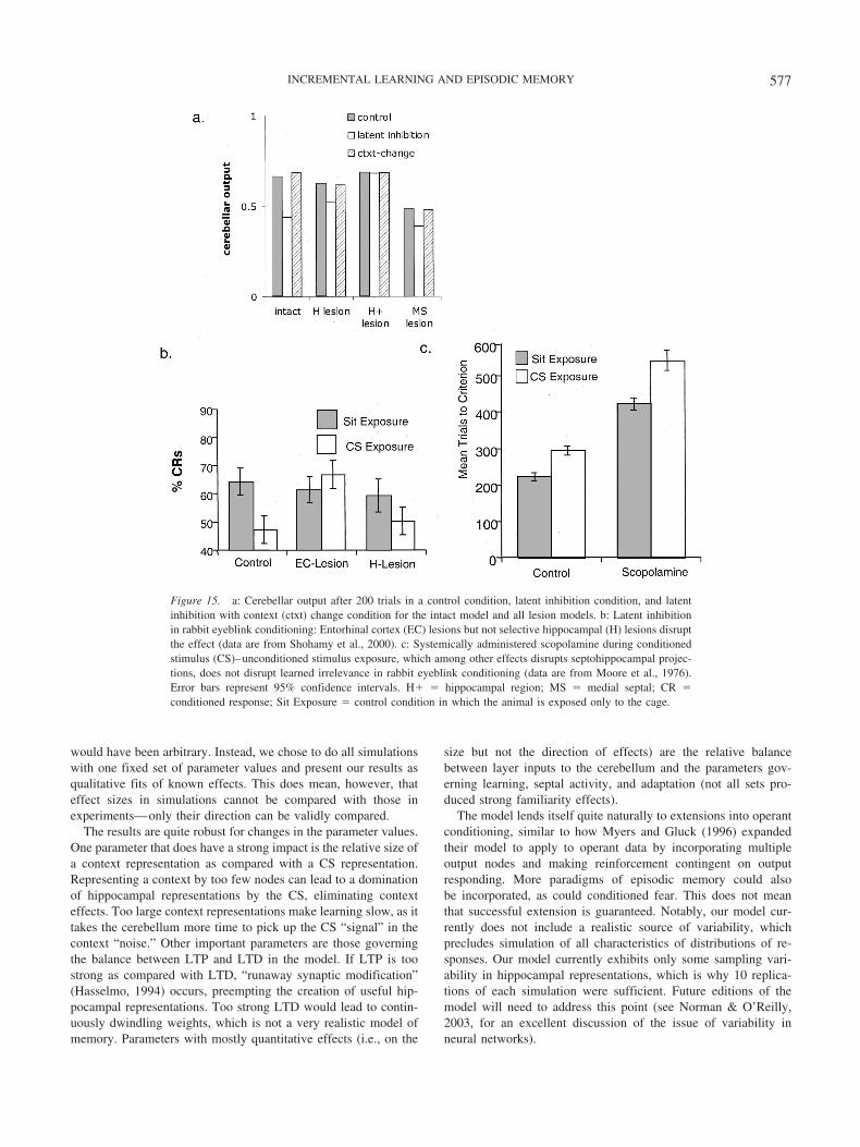
would have been arbitrary. Instead, we chose to do all simulationswith one fixed set of parameter values and present our results asqualitative fits of known effects. This does mean, however, thateffect sizes in simulations cannot be compared with those inexperiments—only their direction can be validly compared.
The results are quite robust for changes in the parameter values.One parameter that does have a strong impact is the relative size ofa context representation as compared with a CS representation.Representing a context by too few nodes can lead to a dominationof hippocampal representations by the CS, eliminating contexteffects. Too large context representations make learning slow, as ittakes the cerebellum more time to pick up the CS “signal” in thecontext “noise.” Other important parameters are those governingthe balance between LTP and LTD in the model. If LTP is toostrong as compared with LTD, “runaway synaptic modification”(Hasselmo, 1994) occurs, preempting the creation of useful hip-pocampal representations. Too strong LTD would lead to contin-uously dwindling weights, which is not a very realistic model ofmemory. Parameters with mostly quantitative effects (i.e., on the
size but not the direction of effects) are the relative balancebetween layer inputs to the cerebellum and the parameters gov-erning learning, septal activity, and adaptation (not all sets pro-duced strong familiarity effects).
The model lends itself quite naturally to extensions into operantconditioning, similar to how Myers and Gluck (1996) expandedtheir model to apply to operant data by incorporating multipleoutput nodes and making reinforcement contingent on outputresponding. More paradigms of episodic memory could alsobe incorporated, as could conditioned fear. This does not meanthat successful extension is guaranteed. Notably, our model cur-rently does not include a realistic source of variability, whichprecludes simulation of all characteristics of distributions of re-sponses. Our model currently exhibits only some sampling vari-ability in hippocampal representations, which is why 10 replica-tions of each simulation were sufficient. Future editions of themodel will need to address this point (see Norman & O’Reilly,2003, for an excellent discussion of the issue of variability inneural networks).
Figure 15. a: Cerebellar output after 200 trials in a control condition, latent inhibition condition, and latentinhibition with context (ctxt) change condition for the intact model and all lesion models. b: Latent inhibitionin rabbit eyeblink conditioning: Entorhinal cortex (EC) lesions but not selective hippocampal (H) lesions disruptthe effect (data are from Shohamy et al., 2000). c: Systemically administered scopolamine during conditionedstimulus (CS)–unconditioned stimulus exposure, which among other effects disrupts septohippocampal projec-tions, does not disrupt learned irrelevance in rabbit eyeblink conditioning (data are from Moore et al., 1976).Error bars represent 95% confidence intervals. H� � hippocampal region; MS � medial septal; CR �conditioned response; Sit Exposure � control condition in which the animal is exposed only to the cage.
577INCREMENTAL LEARNING AND EPISODIC MEMORY

What the Model Does Not Account For
There are also data that the model does not account for andaspects of the model that are clear simplifications. The rich liter-ature on extinction is a case in point. Many complex phenomena inthis literature implicate the hippocampus (Frohardt, Guarraci, &Bouton, 2000), and our model seems ideally placed to make acontribution to this literature. However, preliminary simulationsshowed that it already fails to provide an adequate explanation ofextinction at a low level. It often takes much longer to extinguisha CR to a stimulus than it takes to learn it, as if the system errs onthe side of caution. However, preliminary simulation showed thatour model extinguishes a response at approximately the speed atwhich it acquires them, as do other associative models of condi-tioning. This is because the Rescorla–Wagner rule at the core ofour cerebellar model decreases the CS–CR association after anomission (CR with no US) in the same way as it increases thatassociation after a commission (no CR before the US). However,recent research has suggested that the cerebellar mechanisms be-hind acquisition and extinction of CRs are different (Krupa &Thompson, 2003).
Another area in which improvements could be made is the timecourse at which hippocampal responses occur during conditioning(Berger & Thompson, 1978a). It takes more trials to developstrong responses in the rat than it takes here, and they start off asresponses to the US, which here they do not. It would be possibleto fit these data, as we have incorporated the US in the input to thehippocampus. In the current setup that would not serve any pur-pose; a model on a more fine time scale might be needed to givemeaningful interpretations to the data. Although this leaves some-thing to be explained, it does not challenge the model as it stands.
The model does not make a contribution to a long-standingdiscussion in the episodic memory literature, namely whetherepisodic memories are consolidated in the neocortex (for review,see Meeter & Murre, 2004). Both the view that they are consoli-dated in the neocortex (McClelland, McNaughton, & O’Reilly,1995; Meeter & Murre, in press; Squire & Alvarez, 1995; Squire,Cohen, & Nadel, 1984) and the view that they are not (Nadel &Moscovitch, 1997; Nadel, Samsonovitch, Ryan, & Moscovitch,2000) could be reconciled with the current model.
An area to which the model also does not make a contributionis the timing of responses and the influence of timing of condi-tioned and unconditioned stimuli on the acquisition of responses.Here, we chose for a CS lasting one time step and a US thatimmediately follows the CS, which makes the model not dissimilarto trial-based models with added context-only trials (e.g., Gluck &Myers, 1993). In contrast with trial-based models, however, theexplicit representation of time makes our model essentially capa-ble of being extended to other timing regimes. Two additionswould be necessary. Delay conditioning could be simulated bymaking the elicited CR part of the input of the cerebellar layer, ashas been shown previously (Gluck et al., 2001). The secondaddition would be a loop between the parahippocampal and hip-pocampal layers, as has been implemented in models of episodicmemory (e.g., Talamini et al., in press). This would allow repre-sentations to remain active in both regions through reverberations,as has indeed been observed between the hippocampus and ento-rhinal cortex (Iijima et al., 1996). Reverberatory representationscould be used to form associations in trace conditioning tasks, in
which CS and US are not coactive. Indeed, some evidence haspointed to the hippocampus as being necessary in delay condition-ing (Clark, Manns, & Squire, 2001; Clark & Squire, 1999).
Relation to Previous Models
The model is a variant of associative memory theories, and assuch it has many of the strengths and weaknesses of these theories(e.g., in the field of extinction). It has borrowed heavily from twoassociative theories in particular, that of Rescorla and Wagner(1972) and SOP (Wagner, 1981), bringing the latter from anabstract level to a neurobiologically testable level (see alsoDonegan et al., 1989). Central elements of both theories areimplemented in the current model.
The current model replaces and builds on Gluck and Myers’s(1993) model of corticohippocampal function in classical andoperant conditioning (Gluck & Myers, 1993, 2001; Myers &Gluck, 1994, 1996; Myers, Gluck, & Granger, 1995) and itsextensions to septohippocampal interactions (Myers et al., 1996;Rokers et al., 2002) and cerebellar function in eyeblink condition-ing (Gluck et al., 2001; Gluck & Thompson, 1987). The imple-mentation of the cerebellum and septohippocampal system wereadapted from these models. The implementation of the hippocam-pal region was changed—there as an autoassociator and here as ahierarchy of representational layers. This allowed us to circumventthree limitations of the previous model: that it was anatomicallyimprecise (remedied only to a degree in the current model), that itplaced unobserved error-correcting learning in the hippocampus,and that it did not create room for episodic memories in thatstructure. It also means that at a mechanistic level, the two modelsdiffer substantially. An example is the speed at which representa-tions are formed. Whereas in the previous model representationswere slowly tuned under influence of behavioral contingencies,here we take the episodic memory perspective in which the bulk ofhippocampal learning occurs at the first confrontation with a novelsituation or stimulus and in an unsupervised fashion.
At a more functional level, however, many explanations of thecurrent model are very similar to those of the earlier framework.As an example, we consider the proposed mechanisms behindlatent inhibition. In Gluck and Myers’s (1993) model, this phe-nomenon is explained through a hippocampal region-mediatedcompression of the CS with the context during the first phase oftraining, in which the CS and US are presented in an uncoupledfashion. This makes the task of learning a CS–US association inthe second phase of training hard, as the model cannot differentiatebetween context alone and context plus CS. In the current model,compression of CS and context into one representation occurs inthe hippocampus, but it is not necessary for latent inhibition.During the first phase of learning, the CS stops eliciting a strongresponse in its habitual context (the familiarity effect), and this isassumed to underlie latent inhibition. Although the details of bothexplanations differ, they bear family resemblance: Both assumethat the CS has more or less “faded” into the background contextduring the first phase of learning. At a more abstract level, the twomodels thus deliver similar explanations for the findings in theconditioning literature. They also share many predictions, such asa declining influence of context on classical conditioning.
As was already mentioned, the model is also similar to manyneural network models of episodic memory. The structure of its
578 MEETER, MYERS, AND GLUCK

representational layers was loosely based on a model of episodicmemory (Meeter et al., 2002; Talamini et al., in press), and itsbehavior in the episodic memory simulations is similar to that ofother episodic memory models (Hasselmo & Wyble, 1997; Mc-Clelland & Goddard, 1996; Meeter et al., 2004; Norman &O’Reilly, 2003). Our assumptions about septal activity are alsowidely shared by episodic memory models (Hasselmo & Wyble,1997; Meeter & Murre, in press; Meeter et al., 2004).
In many respects, the current model thus merely pulls togetherthreads that were evident in earlier models of the hippocampus. Inat least two aspects it is different from those other models:
1. In its explicit modeling of the interaction between thecerebellum and episodic representations stored in thehippocampal region.
2. In how sensitivity to context in parahippocampal repre-sentations leads to lower responses to familiar stimuli.
Comparison With Theories of Familiarity
The proposed explanation of the familiarity effect is not the onlyone, however. It has been suggested that a few perirhinal neuronscoding strongly for the stimulus show increased firing rates, whichin turn inhibit other neurons (Norman & O’Reilly, 2003). Becausethere would be more neurons with decreased responses, thesewould be the ones observed in studies of familiarity neurons. Theobserved decreases may also reflect neurons that lose out in theself-organization phase in which a new representation is formedfor a novel stimulus (Sohal & Hasselmo, 2000). Both proposalssuggest that whereas firing is decreased in many neurons, it isincreased in “winners,” those neurons that really code for thestimulus. However, although Xiang and Brown (1998) recordedfrom hundreds of neurons, none showed an increase in firing rate.
One model suggests one reason why such increases might not,in fact, occur. In the model of Bogacz et al. (2001), winners inhibitthemselves via feedback loops. Although firing is increased in thefirst milliseconds after a stimulus appears, it is quickly extin-guished, leading to an overall decrease in firing during repetitionseven in these winners. With some parameter settings, Norman andO’Reilly’s (2003) model could behave in a similar way. The initialincrease in firing frequency was not found by Xiang and Brown(1998); they noted that from the first spike on, firing rates arelower for familiar stimuli. However, it is possible that noise inresponse times causes the initial increase to be invisible, as thegraphs of Xiang and Brown (1998) are averaged over many trials.
Bogacz and Brown (2002, 2003) also proposed another model,in which familiarity neurons activated strongly by a novel stimulusundergo LTD instead of LTP. This so-called anti-Hebbian modelprovides an elegant explanation for the reduced firing to familiarstimuli, because winners firing strongly at the first presentation ofa stimulus will, through LTD, limit their firing during repetitions.The model does not depend on inhibition for the decrease and doesnot predict firing increases in any group of neurons. However, itpresupposes that LTD occurs between highly active pre- andpostsynaptic neurons, which is counter to the standard situations inwhich LTD occurs and has yet not been observed in neurons(Bogacz & Brown, 2003).
Another advantage of the anti-Hebbian model is its large theo-retical capacity: With the same number of neurons, a model based
on anti-Hebbian learning could store many more patterns thansome models based on Hebbian learning (Bogacz & Brown, 2003).Models based on Hebbian learning increase their capacity, how-ever, by including homosynaptic LTD—LTD observed when apresynaptic neuron is active but the postsynaptic neuron is not(Bogacz & Brown, 2002). This lowers the correlation betweenneurons not involved in coding for the same pattern, and a math-ematical analysis has shown that an on-average positive correla-tion between firing in different perirhinal nodes is the main lim-iting factor in the capacity of a perirhinal memory store (Bogacz &Brown, 2003). Such LTD was not needed in the current model, butit could be included without changes to the simulations.
In summary, all published models of the familiarity effect aswell as the current proposal leave something to be proven. Theproposals based on inhibition or reorganization would seem torequire either an increase in the firing frequency of some famil-iarity neurons, either continuously or immediately after a stimulushas been presented. This has not been observed, but it is possiblethat averaging over trials obscures the increase. The current pro-posal and anti-Hebbian learning may be easier to reconcile withexisting data, but both make strong claims that have so far not beentested. The current proposal suggests that the familiarity effectshould diminish with context change and even disappear with atotal context change. The anti-Hebbian model suggests that LTDshould dominate LTP in the perirhinal cortex.
As already stated in the introduction, the current implementationof familiarity could be replaced by other theories without endan-gering most simulation results. There is one caveat, however. Oneeffect, latent inhibition, does depend on an aspect of the theory,namely on the context dependence of familiarity. As discussed inthe introduction, SOP explains the context dependence of latentinhibition by assuming that the context of learning brings repre-sentations in a state of low activity. The equation of familiaritywith this state in SOP is thus only possible if familiarity is contextdependent. Familiarity is indeed context dependent in our imple-mentation, and it also would be in the theory of Norman andO’Reilly (2003), in the anti-Hebbian learning model, and in thetheory of Sohal and Hasselmo (2000). In all three, context featureswould presumably be part of the input to parahippocampal–medialtemporal lobe cortex–perirhinal modules and thus be partly re-sponsible for the match between study items and test probes.
Comparison With Complementary Learning Systems
Norman and O’Reilly’s (2003) model of episodic memory is aninstantiation of a broader framework, dubbed ComplementaryLearning Systems (McClelland et al., 1995; O’Reilly & Rudy,2000). Within the same framework, O’Reilly and Rudy (2001)presented simulations both of tasks that could be seen as episodicand of paradigms from incremental learning. Their focus in thelatter is on operant and fear conditioning. They simulated four setsof findings: nonlinear discrimination problems, contextual fearconditioning, transitivity problems, and context-sensitive respond-ing. In the latter three sets, the simulations rely on pattern com-pletion in their hippocampal system as the causal mechanism. Inparticular, O’Reilly and Rudy showed how the hippocampus couldbuild up a compound representation of context, which could be-come associated for example with a shock in the contextual fearparadigm. Because pattern completion is also essential to the
579INCREMENTAL LEARNING AND EPISODIC MEMORY

functioning of our hippocampal layer, our model would simulatethese sets in the same way as their model (although it wouldrequire a theory of the formation of context representations thatwould take into account the role of stimulus novelty in our model).In the last case, that of context-sensitive responding, this is obvi-ous from our simulations of the same phenomenon in classicalconditioning. The first set, of nonlinear discrimination problems,would be treated fundamentally differently in our model than intheir framework.
Nonlinear discrimination problems are those in which patternsof stimuli that are not linearly separable must be discriminated bythe animal. An example is negative patterning, a paradigm inwhich two stimuli, A and B, predict a reward, but their combina-tion, AB, does not. In most experiments, experimental animals willtake a long time to correctly respond to A and B, but not to AB.Such tasks require a separate representation for the combinationAB, which can then be associated with different consequences thanthe individual stimuli A and B. Such compound representationswould naturally be formed in our hippocampal layer, and indeedmany models of hippocampal functioning would predict that thehippocampus is necessary for negative patterning and similarnonlinear discrimination problems (Sutherland & Rudy, 1989).However, experiments have shown that animals with hippocampallesions can still perform on such tasks (Alvarado & Rudy, 1995;Davidson, McKernan, & Jarrard, 1993), although they are oftenslowed compared with normal animals (Alvarado & Rudy, 1995;Rudy & Sutherland, 1995). This seems to require compoundrepresentations (i.e., for the compound AB) outside of the hip-pocampus. In O’Reilly and Rudy’s (2001) model, the cortex canform these representations through error-correction learning. Inour model, such representations would automatically be formed,because stimuli occurring simultaneously form each other’s con-text. Parahippocampal nodes coding for A would thus become“loaded” with B just as they become loaded with context. If somesuch nodes received inputs coding for B and others did not, theirdifferential responses to the AB compound could form the basis fornonlinear discriminations such as negative patterning.
The different ways in which the two models deal with negativepatterning point to a major difference, namely in learning rules.O’Reilly and Rudy’s (2001) model assumes that two forms oflearning, Hebbian and error-correction learning, are present in boththe cortical and the hippocampal memory systems. Hebbian learn-ing underlies performance in episodic tasks, whereas error correc-tion is important for the incremental learning simulations. Thisseems unparsimonious, but a justification could be that the differ-ence reflects task demands: Error signals are important in incre-mental learning tasks but not in episodic memory tasks. Our mainargument not to follow their assumption is that error signals anderror-driven learning have not been observed in the hippocampusor in the cortex (although see O’Reilly, 1996, for a defense oferror-driven learning in cortical areas). This contrasts with thecerebellum, where error-driven learning has been convincinglydemonstrated (Kim et al., 1998; Thompson & Gluck, 1991), andwith the basal ganglia and amygdala, which could operate undersimilar regimes (see the introduction). The result of this differenceis that in O’Reilly and Rudy’s model, representations are tuned byerror correction, whereas in our model representations are strictlyepisodic and tuning occurs in the connections from the represen-tational layers to output systems such as the cerebellum.
There are, however, also many features in which the Comple-mentary Learning Systems framework overlaps with the one laidout in this article. For example, the hippocampus in both frame-works automatically forms rapid compound representations of thesituation, and these representations explain context dependence inincremental learning tasks. At a more fundamental level, the twoagree in that the same memory representations may underlie verydifferent forms of learning in animals (as expressed by classical,operant, and fear conditioning) and in that there is a fundamentalcontinuity between human and animal memory tasks. They alsooverlap in what is a fundamentally associative view of incrementallearning: In both models, representations of stimuli are associatedwith outputs that drive responses.
Comparison With Comparator Theories
The strongest contrast is probably between our model, firmly inthe associative tradition, and comparator theories (Gallistel, 1990;Gallistel & Gibbon, 2000; Gibbon & Balsam, 1981; Miller &Matzel, 1988). The latter theories claim that conditioning is not amatter of forming associations between stimuli and responses butthat it involves estimations as to when an emitted response will berewarded. Gallistel and Gibbon (2000) have made a convincingclaim that current associative theories are incomplete and have notyet resolved certain internal inconsistencies within the associativeframework. We agree that there are phenomena, especially in thetiming domain, for which associative theories do not fully account.We hope to be able to extend our model in that direction.
Nevertheless, there are also areas in which associative theoriesoffer better explanations than timing theories, notably in describ-ing context effects. For example, it is not obvious within a frame-work such as rate estimation theory (Gallistel, 1990; Gallistel &Gibbon, 2000) why context change would eliminate latent inhibi-tion, as the expectations generated by stimuli are not affected bycontext in these models. The biggest drawback of comparatortheories, however, is that most are entirely functional, with fewguidelines as to how they might be implemented in the brain.Associative theories have an advantage here, as shown perhapsmost clearly by findings on the cerebellum and the Rescorla–Wagner rule (Kim et al., 1998; Thompson, 1990). Perhaps whencomparator theories are made biologically more plausible andassociative theories are brought to bear on timing issues, they willconverge onto a common framework.
Untested Predictions
Many predictions follow from the model as it stands. As a favorto our future falsifiers, we end with a simple enumeration of these.We start with those that follow exclusively from our theory offamiliarity and from its dependence on context:
1. If there is a change in context between presentations of astimulus, no firing decrement should be observed inparahippocampal areas.
2. Familiarity estimates should show context dependence.
3. Familiarity should depend on context being present be-fore the stimulus is presented.
580 MEETER, MYERS, AND GLUCK

4. A context change should affect false alarm rates more forsimilar lures than for nonsimilar lures.
5. Decrements in firing to familiar stimuli in parahippocam-pal areas should be preceded by increases in backgroundfiring rate in the same cell, reflecting on stronger sensi-tivity to context alone.
Other predictions do not depend on our theory of familiaritybeing correct, although some rely on the fact that familiar stimulielicit smaller parahippocampal responses (as has been found).Predictions 6, 7, and 8 are shared with Gluck and Myers’s (1993)model; the others are not.
6. A lesion to the hippocampal region after training willaffect even simple conditioned responses that can belearned normally if the lesion is made before training.
7. The effect of a lesion to the hippocampal region on aconditioned response will be larger during early phasesof training (when hippocampal representations play arelatively large role; see Figure 9) than later in training.
8. Overtraining will abolish or severely limit contextchange effects in classical conditioning.
9. Medial septal lesions will abolish context change effectsin simple conditioning.
10. Context effects in simple conditioning will be in theopposite direction in animals with lesions restricted tothe hippocampus proper: They will show an incrementin responding after context change.
11. Individual elements making up the context come to havenegative predictive value during training. They shouldthus function as inhibitors when presented in a differentcontext.
Although vigorous testing of these and other predictions of ourmodel will almost certainly prove it wrong in details (and perhapswrong in its entirety), we believe that the model highlights anadvantage of computational modeling: that knowledge from dif-ferent task domains and brain areas can be integrated into onecoherent framework and lead to testable predictions. With moreand more knowledge being generated about the brain, we believethat such an integrating function will become more and moreessential.
References
Ackil, J., Mellgren, R., Halgren, C., & Frommer, G. (1969). Effects of CSpreexposures on avoidance learning in rats with hippocampal lesions.Journal of Comparative and Physiological Psychology, 69, 739–747.
Aggleton, J. P., & Brown, M. W. (1999). Episodic memory, amnesia, andthe hippocampal-anterior thalamic axis. Behavioral and Brain Sciences,22, 425–489.
Aitkin, L., & Boyd, J. C. (1978). Accoustic input to the lateral pontinenuclei. Hearing Research, 1, 17–77.
Allen, M. T., Chelius, L., & Gluck, M. A. (2002). Selective entorhinal andnonselective cortical-hippocampal region lesions, but not selective hip-
pocampal lesions, disrupt learned irrelevance in rabbit eyeblink condi-tioning. Cognitive, Affective, and Behavioral Neuroscience, 2, 214–226.
Allen, M. T., Padilla, Y., & Gluck, M. A. (2002). Ibotenic acid lesions ofthe medial septum retard delay eyeblink conditioning in rabbits (Oryc-tolagus cuniculus). Behavioral Neuroscience, 116, 733–738.
Allen, M. T., Padilla, Y., Myers, C. E., & Gluck, M. A. (2002). Selectivehippocampal lesions disrupt a novel cue effect but fail to eliminateblocking in rabbit eyeblink conditioning. Cognitive, Affective, and Be-havioral Neuroscience, 2, 318–328.
Alonso, J., Sang, U. H., & Amaral, D. (1996). Cholinergic innervation ofthe primate hippocampal formation: II. Effects of fimbria/fornix tran-section. Journal of Comparative Neurology, 375, 527–551.
Alvarado, M., & Rudy, J. (1995). A comparison of kainic acid pluscolchicine and ibotenic acid-induced hippocampal formation damage onfour configural tasks in rats. Behavioral Neuroscience, 109, 1052–1062.
Anagnostaras, S. G., Gale, G. D., & Fanselow, M. S. (2001). Hippocampusand contextual fear conditioning: Recent controversies and advances.Hippocampus, 11, 8–17.
Atkinson, R. C., & Juola, J. F. (1974). Search and decision processes inrecognition memory. In D. H. Krantz, R. C. Atkinson, R. D. Luce, & P.Suppes (Eds.), Contemporary developments in mathematical psychology(pp. 243–260). San Francisco: Freeman.
Baxter, M., Gallagher, M., & Holland, P. (1999). Blocking can occurwithout losses in attention in rats with selective removal of hippocampalcholinergic input. Behavioral Neuroscience, 113, 881–890.
Berger, T. W., & Thompson, R. (1978a). Neuronal plasticity in the limbicsystem during classical conditioning of the rabbit nictitating membraneresponse. I. The hippocampus. Brain Research, 145, 323–346.
Berger, T. W., & Thompson, R. (1978b). Neuronal plasticity in the limbicsystem during classical conditioning of the rabbit nictitating membraneresponse. II: Septum and mammillary bodies. Brain Research, 156,293–314.
Berger, T. W., Weikart, C. L., Bassett, J. L., & Orr, W. B. (1986). Lesionsof the retrosplenial cortex produce deficits in reversal learning of therabbit nictitating membrane response: Implications for potential interac-tions between hippocampal and cerebellar brain systems. BehavioralNeuroscience, 100, 802–809.
Bogacz, R., & Brown, M. W. (2002). The restricted influence of sparsenessof coding on the capacity of familiarity discrimination networks. Net-work: Computations in Neural Systems, 13, 457–485.
Bogacz, R., & Brown, M. W. (2003). Comparison of computational modelsof familiarity discrimination in the perirhinal cortex. Hippocampus, 13,494–524.
Bogacz, R., Brown, M. W., & Giraud-Carrier, C. (2001). Model of famil-iarity discrimination in the perirhinal cortex. Journal of ComputationalNeuroscience, 10, 5–23.
Bohbot, V. D., Allen, J. J., & Nadel, L. (2000). Memory deficits charac-terized by patterns of lesions to the hippocampus and parahippocampalcortex. In H. E. Scharfman, M. P. Witter, & R. Schwarcz (Eds.), Annalsof the New York Academy of Sciences: Vol. 911. The parahippocampalregion: Implications for neurological and psychiatric diseases (pp.355–368). New York: New York Academy of Sciences.
Cabeza, R., & Nyberg, L. (2000). Imaging cognition II: An empiricalreview of 275 PET and fMRI studies. Journal of Cognitive Neuro-science, 12, 1–47.
Clark, R., Manns, J., & Squire, L. (2001). Trace and delay eyeblinkconditioning: Contrasting phenomena of declarative and nondeclarativememory. Psychological Science, 12, 304–308.
Clark, R., & Squire, L. (1999). Human eyeblink classical conditioning:Effects of manipulating awareness of the stimulus contingencies. Psy-chological Science, 10, 14–18.
Davachi, L., Mitchell, J. P., & Wagner, A. D. (2003). Multiple routes tomemory: Distinct medial temporal lobe processes build item and source
581INCREMENTAL LEARNING AND EPISODIC MEMORY

memories. Proceedings of the National Academy of Sciences, USA, 100,2157–2162.
Davidson, T., McKernan, M., & Jarrard, L. (1993). Hippocampal lesionsdo not impair negative patterning: A challenge to configural associationtheory. Behavioral Neuroscience, 107, 227–234.
Donegan, N. H., Gluck, M. A., & Thompson, R. F. (1989). Integratingbehavioral and biological models of conditioning. In R. D. Hawkins &G. H. Bower (Eds.), Psychology of learning and motivation (Vol. 3, pp.109–156). New York: Academic Press.
Dragoi, G., Carpi, M., Recce, M., Csicsvari, J., & Buzsaki, G. (1999).Interactions between hippocampus and medial septum during sharpwave and theta oscillation in the behaving rat. Journal of Neuroscience,19, 6191–6199.
Dusek, J. A., & Eichenbaum, H. (1997). The hippocampus and memory fororderly stimulus relations. Proceedings of the National Academy ofSciences, USA, 94, 7109–7114.
Eichenbaum, H. (1992). The hippocampal system and declarative memoryin animals. Journal of Cognitive Neuroscience, 4, 217–231.
Eichenbaum, H. (2000). Hippocampus: Mapping or memory? CurrentBiology, 10, R785–R787.
Fanselow, M. S. (1998). Pavlovian conditioning, negative feedback andblocking: Mechanisms that regulate association formation. Neuron, 20,625–627.
Fanselow, M. S. (2000). Contextual fear, gestalt memories, and the hip-pocampus. Behavioural Brain Research, 110, 73–81.
Farah, M. J., Wilson, K., Drain, M., & Tanaka, J. (1998). What is “special”about face perception? Psychological Review, 105, 482–498.
Ferbintineau, J., & Shapiro, M. L. (2003). Prospective and retrospectivememory coding in the hippocampus. Neuron, 40, 1227–1239.
Fleidervish, I. A., Friedman, A., & Gutnick, M. J. (1996). Slow inactivationof Na� current and slow cumulative spike adaptation in mouse andguinea-pig neurocortical neurones in slices. Journal of Physiology (Lon-don), 493, 83–97.
Frohardt, R., Guarraci, F., & Bouton, M. (2000). The effects of neurotoxichippocampal lesions on two effects of context following fear extinction.Behavioral Neuroscience, 114, 227–240.
Gallistel, C. R. (1990). The organization of learning. Cambridge, MA:Bradford Books/MIT Press.
Gallistel, C. R., & Gibbon, J. (2000). Time, rate, and conditioning. Psy-chological Review, 107, 289–344.
Gibbon, J., & Balsam, P. (1981). Spreading association in time. In C. M.Locurto, H. S. Terrace, & J. Gibbon (Eds.), Autoshaping and condition-ing theory (pp. 219–253). New York: Academic Press.
Gilbert, P., Kesner, R., & Lee, I. (2001). Dissociating hippocampal sub-regions: A double dissociation between dentate gyrus and CA1. Hip-pocampus, 11, 626–636.
Gluck, M. A., Allen, M., Myers, C., & Thompson, R. (2001). Cerebellarsubstrates for error correction in motor conditioning. Neurobiology ofLearning and Memory, 76, 314–341.
Gluck, M. A., Meeter, M., & Myers, C. E. (2003). Computational modelsof the hippocampal region: Linking incremental learning and episodicmemory. Trends in Cognitive Sciences, 7, 269–276.
Gluck, M. A., & Myers, C. E. (1993). Hippocampal mediation of stimulusrepresentation: A computational theory. Hippocampus, 3, 491–516.
Gluck, M. A., & Myers, C. E. (2001). Gateway to memory: An introductionto neural network modeling of the hippocampus in learning and memory.Cambridge, MA: MIT Press.
Gluck, M. A., Myers, C. E., & Thompson, R. (1994). A computationalmodel of the cerebellum and motor-reflex conditioning. In S. Zornetzer,J. Davis, C. Lau, & T. McKenna (Eds.), An introduction to neural andelectronic networks (pp. 91–98). New York: Academic Press.
Gluck, M. A., & Thompson, R. F. (1987). Modeling the neuron substrateof associative learning and memory: A computational approach. Psy-chological Review, 94, 176–191.
Haines, D. E., May, P. J., & Dietrichs, E. (1990). Neuronal connectionsbetween the cerebellar nuclei and hypothalamus in Macaca fascicularis:Cerebello-visceral circuits. Journal of Comparative Neurology, 299,106–122.
Han, J. S., Gallagher, M., & Holland, P. (1995). Hippocampal lesionsdisrupt decrements but not increments in conditioned stimulus process-ing. Journal of Neuroscience, 15, 7323–7329.
Hasselmo, M. E. (1994). Runaway synaptic modification in models ofcortex: Implications for Alzheimer’s disease. Neural Networks, 7, 13–40.
Hasselmo, M. E. (1999). Neuromodulation: Acetylcholine and memoryconsolidation. Trends in Cognitive Sciences, 3, 351–359.
Hasselmo, M. E., & Bower, J. M. (1993). Acetylcholine and memory.Trends in Neuroscience, 16, 218–222.
Hasselmo, M. E., & Wyble, B. P. (1997). Free recall and recognition in anetwork model of the hippocampus: Simulating effects of scopolamineon human memory function. Behavioural Brain Research, 89, 1–34.
Honey, R., & Good, M. (1993). Selective hippocampal lesions abolish thecontextual specificity of latent inhibition and conditioning. BehavioralNeuroscience, 107, 23–33.
Humphreys, M. S., Bain, J. D., & Pike, R. (1989). Different ways to cue acoherent system: A theory for episodic, semantic and procedural tasks.Psychological Review, 96, 208–233.
Iijima, T., Witter, M. P., Ichikawa, M., Tominaga, T., Kajiwara, R., &Matsumoto, G. (1996, May 24). Entorhinal-hippocampal interactionsrevealed by real-time imaging. Science, 272, 1176–1179.
Jacoby, L. L. (1991). A process dissociation framework: Separating auto-matic from intentional use of memory. Journal of Memory and Lan-guage, 30, 513–541.
Jarrard, L. (1995). What does the hippocampus really do? Behavioral BrainResearch, 71, 1–10.
Kamin, L. (1969). Predictability, surprise, attention and conditioning. InB. Campbell & R. Church (Ed.), Punishment and aversive behavior (pp.279–296). New York: Appleton-Century-Crofts.
Kaufman, G. D., Mustari, M. J., Miselis, R. R., & Perachio, A. A. (1996).Transneuronal pathways to the vestibulocerebellum. Journal of Com-parative Neurology, 370, 501–523.
Kendrick, K. M., da Costa, A. P., Leigh, A. E., Hinton, M. R., & Peirce,J. W. (2001, November 8). Sheep don’t forget a face. Nature, 414,165–166.
Kim, J., Krupa, D., & Thompson, R. (1998, January 23). Inhibitorycerebello-olivary projections and blocking effect in classical condition-ing. Science, 279, 570–573.
Kohler, C. (1990). Subicular projections to the hypothalamus and brain-stem: Some novel aspects revealed in the rat by the anterograde Phaseo-lus vulgaris leukoagglutinin (PHA-L) tracing method. Progress in BrainResearch, 83, 59–69.
Krupa, D. J., & Thompson, R. F. (2003). Inhibiting the expression of aclassically conditioned behavior prevents its extinction. Journal of Neu-roscience, 23, 10577–10584.
Lamme, V. A. F., Rodriguez-Rodriguez, V., & Spekreijse, H. (1999).Separate processing dynamics for texture elements, boundaries andsurfaces in primary visual cortex of the maquaque monkey. CerebralCortex, 9, 406–413.
Lauwereyns, J., Watanabe, K., Coe, B., & Hikosaka, O. (2002, July 25). Aneural correlate of response bias in monkey caudate nucleus. Nature,418, 413–417.
LeDoux, J. (1996). The emotional brain. New York: Simon & Schuster.Levy, W. B., Colbert, C. M., & Desmond, N. L. (1990). Elemental adaptive
processes in neurons and synapses: A statistical/computational perspec-tive. In M. A. Gluck & D. E. Rumelhart (Eds.), Neuroscience andconnectionist theory (pp. 187–235). Hillsdale, NJ: Erlbaum.
Li, L., Miller, E. K., & Desimone, R. (1993). The representation of
582 MEETER, MYERS, AND GLUCK

stimulus familiarity in anterior inferior temporal cortex. Journal ofNeurophysiology, 13, 1918–1929.
Lubow, R., & Moore, A. (1959). Latent inhibition: The effect of non-reinforced pre-exposure to the conditional stimulus. Journal of Compar-ative and Physiological Psychology, 52, 415–519.
Macken, W. J. (2002). Environmental context and recognition: The role ofrecollection and familiarity. Journal of Experimental Psychology:Learning, Memory, and Cognition, 28, 153–161.
Mackintosh, N. J. (1973). Stimulus selection: Learning to ignore stimulithat predict no change in reinforcement. In R. Hinde & J. Stevenson-Hinde (Ed.), Constraints on learning: Limitations and predispositions(pp. 75–96). New York: Academic Press.
Mackintosh, N. J. (1975). A theory of attention: Variations in the associa-bility of stimuli with reinforcement. Psychological Review, 82, 276–298.
Mackintosh, N. J., Kaye, H., & Bennett, C. H. (1991). Perceptual learningin flavour aversion conditioning. Quarterly Journal of ExperimentalPsychology: Comparative and Physiological Psychology, 43(B), 297–322.
Mandler, G. (1980). Recognizing: The judgment of previous occurrence.Psychological Review, 87, 252–271.
Marr, D. (1971). Simple memory: A theory for archicortex. PhilosophicalTransactions of the Royal Society of London, Series B, 262, 23–81.
McClelland, J. L., & Goddard, G. V. (1996). Considerations arising froma complementary learning systems perspective on hippocampus andneocortex. Hippocampus, 6, 654–665.
McClelland, J. L., McNaughton, B. L., & O’Reilly, R. C. (1995). Whythere are complementary learning systems in the hippocampus andneocortex: Insights from the successes and failures of connectionistmodels of learning and memory. Psychological Review, 102, 419–457.
McKenzie, W. A., & Tiberghien, G. (2004). Context effects in recognitionmemory: The role of familiarity and recollection. Consciousness andCognition, 13, 20–38.
McLennan, H., & Miller, J. J. (1974). The hippocampal control of neuronaldischarges in the septum of the rat. Journal of Physiology, 237, 607–624.
Meeter, M., & Murre, J. M. J. (2004). Consolidation of long-term memo-ries: Evidence and alternatives. Psychological Bulletin, 130, 843–857.
Meeter, M., & Murre, J. M. J. (in press). TraceLink: A connectionist modelof consolidation and amnesia. Cognitive Neuropsychology.
Meeter, M., Murre, J. M. J., & Talamini, L. M. (2002). A computationalapproach to memory deficits in schizophrenia. Neurocomputing, 44,929–936.
Meeter, M., Talamini, L. M., & Murre, J. M. J. (2004). Mode shiftingbetween storage and recall based on novelty detection in oscillatinghippocampal circuits. Hippocampus, 14, 722–741.
Miller, R. R., & Matzel, L. D. (1988). The comparator hypothesis: Aresponse rule for the expression of associations. In G. H. Bower (Ed.),The psychology of learning and motivation (Vol. 22, pp. 51–92). NewYork: Academic Press.
Moore, J., Goodell, N., & Solomon, P. (1976). Central cholinergic block-ade by scopolamine and habituation, classical conditioning, and latentinhibition of the rabbit’s nictitating membrane response. PhysiologicalPsychology, 4, 395–399.
Murnane, K., Phelps, M. P., & Malmberg, K. (1999). Context-dependentrecognition memory: The ICE theory. Journal of Experimental Psychol-ogy: General, 128, 403–415.
Murre, J. M. J. (1996). TraceLink: A model of amnesia and consolidationof memory. Hippocampus, 6, 675–684.
Myers, C. E., Ermita, B., Harris, K., Hasselmo, M., Solomon, P., & Gluck,M. (1996). A computational model of the effects of septohippocampaldisruption on classical eyeblink conditioning. Neurobiology of Learningand Memory, 66, 51–66.
Myers, C. E., & Gluck, M. A. (1994). Context, conditioning and hippocam-pal re-representation. Behavioral Neuroscience, 108, 835–847.
Myers, C. E., & Gluck, M. (1996). Cortico-hippocampal representations insimultaneous odor discrimination learning: A computational interpreta-tion of Eichenbaum, Mathews, and Cohen (1989). Behavioral Neuro-science, 110, 685–706.
Myers, C. E., Gluck, M. A., & Granger, R. (1995). Dissociation ofhippocampal and entorhinal function in associative learning: A compu-tational approach. Psychobiology, 28, 116–138.
Nadel, L., & Moscovitch, M. (1997). Memory consolidation, retrogradeamnesia and the hippocampal complex. Current Opinion in Neurobiol-ogy, 7, 217–227.
Nadel, L., Samsonovitch, A., Ryan, L., & Moscovitch, M. (2000). Multipletrace theory of human memory: Computational, neuroimaging and neu-ropsychological results. Hippocampus, 10, 352–368.
Norman, K. A., & O’Reilly, R. C. (2003). Modeling hippocampal andneocortical contributions to recognition memory: A complementarylearning systems approach. Psychological Review, 110, 611–646.
Ogden, J. A. (1993). Visual object agnosia, prosopagnosia, achromatopsia,loss of visual imagery, and autobiographical amnesia following recoveryfrom cortical blindness: Case M.H. Neuropsychologia, 31, 571–589.
Oja, E. (1982). A simplified neuron model as a principal componentanalyzer. Journal of Mathematical Biology, 15, 267–273.
O’Keefe, J. (1979). A review of the hippocampal place cells. Progress inNeurobiology, 13, 419–439.
O’Reilly, R. C. (1996). Biologically plausible error-driven learning usinglocal activation differences: The generalized recirculation algorithm.Neural Computation, 8, 895–938.
O’Reilly, R. C., & Rudy, J. W. (2000). Computational principles oflearning in the neocortex and hippocampus. Hippocampus, 10, 389–397.
O’Reilly, R. C., & Rudy, J. W. (2001). Conjunctive representations inlearning and memory: Principles of cortical and hippocampal function.Psychological Review, 108, 311–345.
Pagnoni, G., Zink, C. F., Montague, P. R., & Berns, G. S. (2002). Activityin human ventral striatum locked to errors of reward prediction. NatureNeuroscience, 5, 97–98.
Pearce, J. M., & Bouton, M. E. (2001). Theories of associative learning inanimals. Annual Review of Psychology, 52, 111–139.
Pearce, J. M., & Hall, G. (1980). A model for Pavlovian learning: Varia-tions in the effectiveness of conditioned but not of unconditioned stim-uli. Psychological Review, 87, 532–552.
Penick, S., & Solomon, R. (1991). Hippocampus, context and conditioning.Behavioral Neuroscience, 105, 611–617.
Peoples, L. L., Uzwiak, A. J., Gee, F., & West, M. O. (1997). Operantbehavior is necessary and sufficient of rat accumbens neurons duringsessions of intravenous cocaine infusions. Brain Research, 757, 280–284.
Raaijmakers, J. G. W., & Shiffrin, R. M. (1992). Models for recall andrecognition. Annual Review of Psychology, 43, 205–234.
Ranganath, C., Yonelinas, A. P., Cohen, M. X., Dy, C. J., Tom, S. M., &D’Esposito, M. (2004). Dissociable correlates of recollection and famil-iarity within the medial temporal lobes. Neuropsychologia, 42, 2–13.
Reber, P. J., & Squire, L. R. (1998). Contrasting cortical activity associatedwith category memory and recognition memory. Learning & Memory, 5,420–428.
Recanzone, G. H., Schreiner, C. E., & Merzenich, M. M. (1993). Plasticityin the frequency representation of primary auditory cortex followingdiscrimination training in adult owl monkeys. Journal of Neuroscience,13, 87–103.
Reed, J. M., & Squire, L. R. (1998). Retrograde amnesia for facts andevents: Findings from four new cases. Journal of Neuroscience, 18,3943–3954.
Rempel-Clower, N. A., Zola, S. M., Squire, L. R., & Amaral, D. G. (1996).Three cases of enduring memory impairment after bilateral damage
583INCREMENTAL LEARNING AND EPISODIC MEMORY

limited to the hippocampal formation. Journal of Neuroscience, 16,5233–5255.
Rescorla, R., & Wagner, A. (1972). A theory of Pavlovian conditioning:Variations in the effectiveness of reinforcement and non-reinforcement.In A. Black & W. Prokasy (Ed.), Classical conditioning II: Currentresearch and theory (pp. 64–99). New York: Appleton-Century-Crofts.
Robertson, I. H., & Murre, J. M. J. (1999). Why do damaged brainsrecover? A neuropsychological analysis in a connectionist framework.Psychological Bulletin, 125, 544–575.
Rokers, B., Mercado, E., Allen, M. T., Myers, C. E., & Gluck, M. A.(2002). A connectionist model of septohippocampal dynamics duringconditioning: Closing the loop. Behavioral Neuroscience, 116, 48–62.
Rudy, J. W., & Sutherland, R. (1995). Configural association theory andthe hippocampal formation: An appraisal and reconfiguration. Hip-pocampus, 5, 375–398.
Sah, P., & Clements, J. D. (1999). Photolytic manipulation of [CA2�]reveals slow kinetics of potassium channels underlying the afterhyper-polarization in hippocampal pyramidal neurons. Journal of Neuro-science, 19, 3657–3664.
Schmajuk, N., & DiCarlo, J. (1992). Stimulus configuration, classicalconditioning, and hippocampal function. Psychological Review, 99,268–305.
Schmaltz, L., & Theios, J. (1972). Acquisition and extinction of a classi-cally conditioned response in hippocampectomized rabbits (Oryctolaguscuniculus). Journal of Comparative and Physiological Psychology, 79,328–333.
Schultz, W. (1998). Predictive reward signal of dopamine neurons. Journalof Neurophysiology, 80, 1–27.
Schultz, W. (2002). Getting formal with dopamine and reward. Neuron, 36,241–263.
Scoville, W. B., & Milner, B. (1957). Loss of recent memories afterbilateral hippocampal lesions. Journal of Neurology, Neurosurgery andPsychiatry, 20, 11–21.
Shohamy, D., Allen, M., & Gluck, M. (2000). Dissociating entorhinal andhippocampal involvement in latent inhibition. Behavioral Neuroscience,114, 867–874.
Smith, S. M., & Vela, E. (2001). Environmental context-dependent mem-ory: A review and meta-analysis. Psychonomic Bulletin & Review, 8,2003–2220.
Sohal, V. S., & Hasselmo, M. E. (2000). A model for experience-dependent changes in the responses of enfero-temporal neurons. Net-work: Computations in Neural Systems, 11, 169–190.
Squire, L. R., & Alvarez, P. (1995). Retrograde amnesia and memoryconsolidation: A neurobiological perspective. Current Opinion in Neu-robiology, 5, 169–175.
Squire, L. R., Cohen, N. J., & Nadel, L. (1984). The medial temporalregion and memory consolidation: A new hypothesis. In H. Weingarter& E. Parker (Eds.), Memory consolidation (pp. 185–210). Hillsdale, NJ:Erlbaum.
Steinmetz, J. (1998). The localization of a simple type of learning andmemory: The cerebellum and classical eyeblink conditioning. CurrentDirections in Psychological Science, 7, 72–77.
Sutherland, R., & Rudy, J. (1989). Configural association theory: The roleof the hippocampal formation in learning, memory and amnesia. Psy-chobiology, 17, 129–144.
Suzuki, W. A., Miller, E. K., & Desimone, R. (1997). Object and placememory in the macaque entorhinal cortex. Journal of Neurophysiology,78, 1062–1081.
Symonds, M., & Hall, G. (1995). Perceptual learning in flavor aversionconditioning: Roles of stimulus comparison and latent inhibition ofcommon stimulus elements. Learning and Motivation, 26, 203–219.
Talamini, L. M., Meeter, M., Murre, J. M. J., Elvevåg, B., & Goldberg,T. E. (in press). Integration of parallel input streams in parahippocampalmodel circuits; implications for schizophrenia. Archives of GeneralPsychiatry.
Teyler, T. J., & DiScenna, P. (1986). The hippocampal memory indexingtheory. Behavioral Neuroscience, 100, 147–154.
Thomas, M. J., & Malenka, R. C. (2003). Synaptic plasticity in themesolimbic dopamine system. Philosophical Transactions of the RoyalSociety of London, Series B, 358, 815–819.
Thompson, R. (1990). Neural mechanisms of classical conditioning. Phil-osophical Transactions of the Royal Society of London, Series B, 329,161–170.
Thompson, R., & Gluck, M. (1991). Brain substrates of basic associativelearning. In H. Weingartner & R. Lister (Ed.), Cognitive neuroscience(pp. 24–45). New York: Oxford University Press.
Tranel, D., Damasio, H., & Damasio, A. R. (2000). Amnesia caused byherpes simplex encephalitis, infarctions in basal forebrain, and anoxia/ischemia. In F. Boller & J. Grafman (Eds.), Handbook of neuropsychol-ogy (2nd ed., Vol. 2, pp. 85–110). Amsterdam: Elsevier Science.
Tsujimoto, S., & Sawaguchi, T. (2004). Properties of delay-period neuro-nal activity in the primate prefrontal cortex during memory- andsensory-guided saccade tasks. European Journal of Neuroscience, 19,447–457.
Vann, S. D., Brown, M. W., Erichsen, J. T., & Aggleton, J. P. (2000). Fosimaging reveals differential patterns of hippocampal and parahippocam-pal subfield activation in rats in response to different spatial memorytests. The Journal of Neuroscience, 20, 2711–2718.
Vinogradova, O. S., Kitchigina, V. F., & Zenchenko, C. I. (1998). Pace-maker neurons of the forebrain medial septal area and theta rhythm ofthe hippocampus. Membrane and Cell Biology, 11, 715–725.
Wagner, A. (1981). SOP: A model of automatic memory processing inanimal behavior. In N. Spear & R. Miller (Ed.), Information processingin animals: Memory mechanisms (pp. 5–47). Hillsdale, NJ: Erlbaum.
Witter, M. P., Wouterlood, F. G., Naber, P. A., & Van Haeften, T. (2000).Anatomical organization of the parahippocampal-hippocampal network.In H. E. Scharfman, M. P. Witter, & R. Schwarcz (Eds.), Annals of theNew York Academy of Sciences: Vol. 911. The parahippocampal region:Implications for neurological and psychiatric diseases (pp. 1–24). NewYork: Annals of the New York Academy of Sciences.
Xiang, J. Z., & Brown, M. W. (1998). Differential neuronal encoding ofnovelty, familiarity, and recency in regions of the anterior temporal lobe.Neuropharmacology, 37, 657–676.
Yonelinas, A. P. (2001). Components of episodic memory: The contribu-tion of recollection and familiarity. Philosophical Transactions of theRoyal Society of London, Series B, 356, 1363–1374.
Yonelinas, A. P. (2002). The nature of recollection and familiarity: Areview of 30 years of research. Journal of Memory and Language, 46,441–517.
Yu, Q. X., Wang, J. J., & Chen, J. (1989). Hippocampus-cerebellarcortex-cerebellar nuclei projection in the rat: Electrophysiological andHRP studies. Shen Li Xue Bao, 41, 231–240.
Zhu, X. O., McCabe, B. J., Aggleton, J. P., & Brown, M. W. (1997).Differential activation of the rat hippocampus and perirhinal cortex bynovel visual stimuli and a novel environment. Neuroscience Letters, 229,141–143.
584 MEETER, MYERS, AND GLUCK

Appendix
Representational Layers
Both the cortical and the parahippocampal layers consist of 33 nodes,enough to code for 2 contexts of 15 nodes, 2 stimuli of 1 node, and a US.The hippocampal layer consists of 40 nodes. Weights between the corticaland parahippocampal layers are of two kinds. Each cortical node has animmutable connection with weight 1.8 with 1 parahippocampal node. Withall other parahippocampal nodes, cortical nodes have connections subjectto learning that are initialized at values taken from a uniform distributionwith mean .05 and a 25% spread. Parahippocampal nodes are in turnconnected to 85% of hippocampal nodes with connections subject tolearning; weights on these connections are initialized at values taken froma uniform distribution with mean .3 and a 10% spread.
The parahippocampal and hippocampal layers consist of simple firingrate nodes that compute an output from linearly summated inputs. Input tothe cells, gi, consists of excitation, Ei, and inhibition, Ii, both originatingfrom the layer below. Excitation, unique to each node, is a weighted sumof the output of the layer below; the inhibitory component, modelingundifferentiated feedforward inhibition, is a fraction of the summed output,the same for all nodes:
gi � Ei � Ii � ��wijoj� � ���oj�,
where oj is the output of presynaptic node j and wij is the weight on itsconnection to node i. The inhibition fraction � is set to a value 1.2 timesthe average weight at the outset of the simulation (0.06 from the cortical tothe parahippocampal layer, and 0.36 from the parahippocampal to thehippocampal layer), so as to strongly inhibit nodes that are not part of acued memory. A node’s output is a function of its input gi, and (throughadaptation) of its own previous firing record. With t as the time index,
oi�t� � gi�t� � �1 � �Ai�t�� � H �gi�t��
and
Ai�t� � ��Ai �t � 1� � oi�t � 1�.
H is the Heavyside step function that maps negative values and zero to 0and positive values to 1, and 1 � �A(t) is an adaptation function. Theaccretion parameter of adaptation is set to 0.5, its gain parameter � to 0.9,and its decay constant � to 0.1. This implies a slow recovery fromadaptation (seconds instead of milliseconds), as is indeed present in at leastsome subtypes of neocortical (Fleidervish, Friedman, & Gutnick, 1996)and hippocampal (Sah & Clements, 1999) neurons.
Learning is implemented with Oja’s rule:
wij � ���oj oi��1 � wij� � ���1 � oj�oiwij.
This variant of Hebb’s rule seems to describe LTP well (Levy, Colbert, &Desmond, 1990) and allows learning to asymptotically approach a plateauwhose level is determined by the strength of the input oj and the balancebetween �� and ��. A modified output level o’i is used in the learning
rule, which is equal to oi � 0.4, truncated at zero. Only strongly firingnodes are thus allowed to learn. This models data showing that thethreshold for LTP is usually higher than the firing threshold.
The positive learning rate �� is set at 0.01 for the connections from thecortical layer to the parahippocampal layer (except in the first familiaritysimulation, where it was set at 0.1). For the connections from the parahip-pocampal layer to the hippocampal layer, it is set at 0.05. The negative rate�� was set to 0.5 � �� in all connections.
Septal Activity
Learning under the influence of acetylcholine is implemented in theconnection of the parahippocampus to the hippocampus. This was not donein great detail. Learning under the influence of acetylcholine is triggeredwhenever fewer than three hippocampal nodes have an output of more than0.05 (i.e., when patterns were new) or whenever prediction error in thecerebellum is higher than 0.5. In these cases, the 6 nodes with the highestinputs are allowed to strengthen the weights on their connections from theparahippocampal layers according to the following rule:
wij � �ojoi��1 � wij� � 0.5�1 � oj�wij.
Here oi is the output of the ith hippocampal node, oj is the output of the jthparahippocampal node, and � is the cholinergic input [10 in case of a novelpattern, 1.5 � (error � 0.5) in case of septal activation through error].Weight change is bound to the interval [�0.25, 0.3].
Cerebellum
The cerebellar network is the same as that in earlier work (Gluck, Myers,& Thompson, 1994), consisting of a simple perceptron trained with theRescorla–Wagner rule:
woi � �c�oo � to�oi,
where (oo � to) is the error of the output node (its output minus its target)and oi is the output of the ith input node. The target of the output node isthe presence of a US on the next iteration (1 if present, 0 if absent). Thereare 99 input nodes, with 33 getting input from each 1 context node(transmitted via weights of strength 1), 33 more getting input from each 1parahippocampal node (transmitted via weights of strength 1), and the last33 getting connections with strength 0.5 from each of a random 15% ofhippocampal nodes. Weights between the input layer and output node areinitialized at values taken from a uniform distribution between �.05 and.05. The learning rate �c is set at 0.05 in all simulations.
Received May 13, 2004Revision received December 8, 2004
Accepted January 13, 2005 �
585INCREMENTAL LEARNING AND EPISODIC MEMORY


![[DL輪読会]Neural Episodic Control/Model-Free Episodic Control](https://img.pdfslide.net/doc/110x75/5a64790d7f8b9a57568b463b/dlneural-episodic-controlmodel-free-episodic-control.jpg)