Embed Size (px)
Citation preview
Integrating Molecular Findings into the Diagnosis and
Prognosis of MDS
Rafael Bejar MD, PhD
Biological and Clinical Advances in MDS
San Diego, CA
December 2nd, 2016
Disclosures
Celgene: consultant, steering committee, honoraria, DSMB Genoptix: consultant, honoraria, licensed IP Foundation Medicine: honoraria, ad-hoc advisory board Alexion: ad-hoc advisory board
Overview
• Genetic Heterogeneity in MDS
• Somatic Mutations as Diagnostic Markers
• Germline Predisposition Mutations
• Somatic Mutations as Prognostic Markers
MDS Genetics
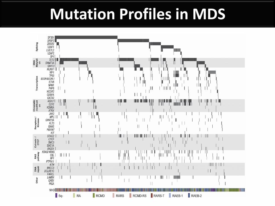
Mutation Profiles in MDS
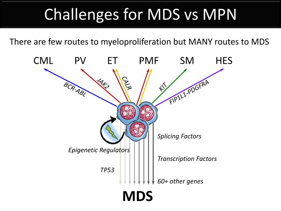
Challenges for MDS vs MPN
CML PV ET PMF SM HES
MDS
Splicing Factors
Epigenetic Regulators Transcription Factors
TP53
60+ other genes
There are few routes to myeloproliferation but MANY routes to MDS
Diagnostic Mutations (?)
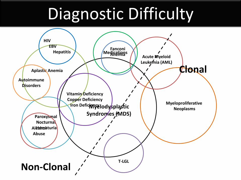
Myelodysplastic Syndromes (MDS)
Aplastic Anemia
Acute Myeloid Leukemia (AML)
Paroxysmal Nocturnal Hematuria
T-LGL
Fanconi Anemia
Myeloproliferative Neoplasms
Diagnostic Difficulty
Vitamin Deficiency Copper Deficiency
Iron Deficiency
HIV EBV Hepatitis
Alcohol Abuse
Medications
Autoimmune Disorders
Non-Clonal
Clonal
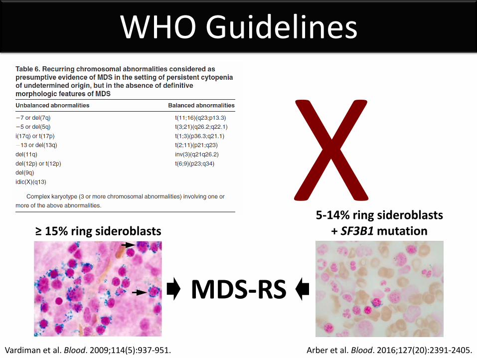
Vardiman et al. Blood. 2009;114(5):937-951.
WHO Guidelines
≥ 15% ring sideroblasts 5-14% ring sideroblasts
+ SF3B1 mutation
Arber et al. Blood. 2016;127(20):2391-2405.
MDS-RS
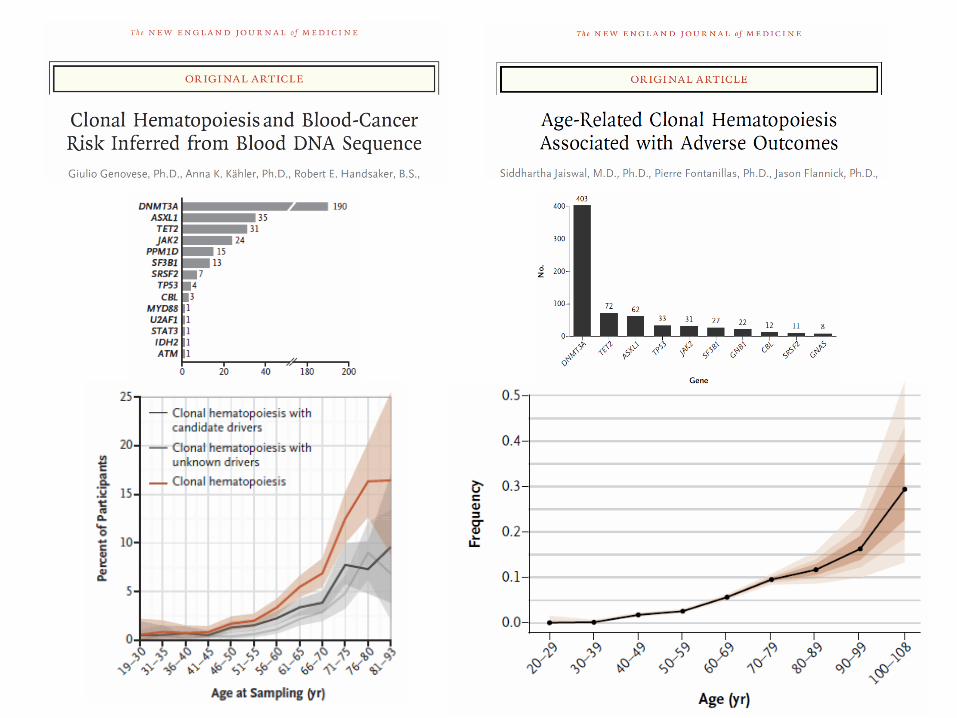
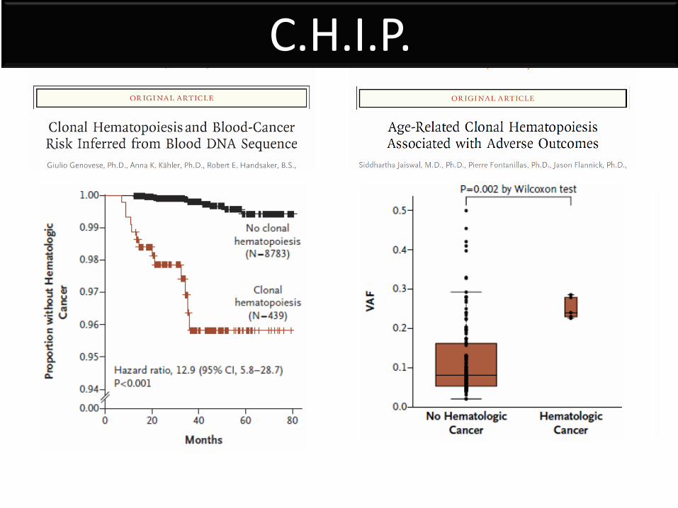
C.H.I.P.
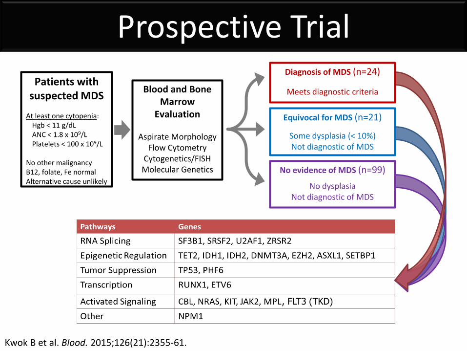
Prospective Trial
Kwok B et al. Blood. 2015;126(21):2355-61.
Patients with suspected MDS
At least one cytopenia: Hgb < 11 g/dL ANC < 1.8 x 10⁹/L Platelets < 100 x 109/L
No other malignancy B12, folate, Fe normal Alternative cause unlikely
Blood and Bone Marrow
Evaluation Aspirate Morphology
Flow Cytometry Cytogenetics/FISH
Molecular Genetics
Diagnosis of MDS (n=24)
Meets diagnostic criteria
No evidence of MDS (n=99)
No dysplasia Not diagnostic of MDS
Equivocal for MDS (n=21)
Some dysplasia (< 10%) Not diagnostic of MDS
, FLT3 (TKD)
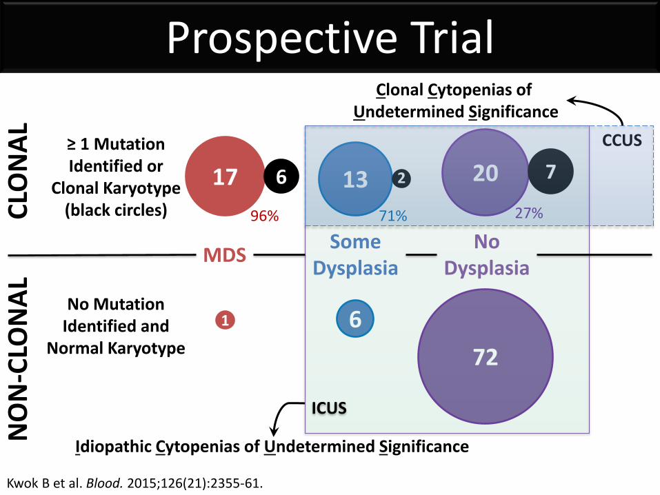
Prospective Trial
Kwok B et al. Blood. 2015;126(21):2355-61.
17
1
13
6
20
72
≥ 1 Mutation Identified or
Clonal Karyotype (black circles)
No Mutation Identified and
Normal Karyotype
96% 71% 27%
7 6
Idiopathic Cytopenias of Undetermined Significance
Clonal Cytopenias of Undetermined Significance
CCUS
2
ICUS
MDS Some
Dysplasia No
Dysplasia
CLO
NA
L N
ON
-CLO
NA
L
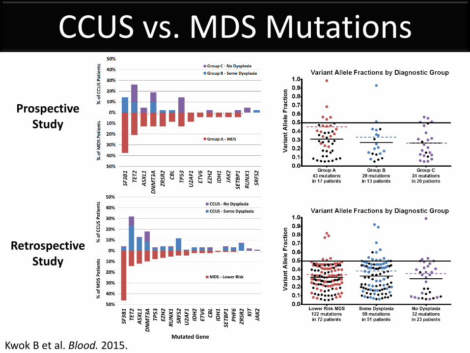
CCUS vs. MDS Mutations
Kwok B et al. Blood. 2015.
Prospective Study
Retrospective Study
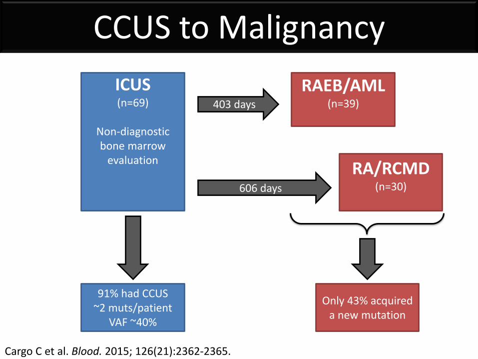
CCUS to Malignancy
Cargo C et al. Blood. 2015; 126(21):2362-2365.
ICUS (n=69)
Non-diagnostic bone marrow
evaluation
RAEB/AML (n=39)
403 days
606 days
91% had CCUS ~2 muts/patient
VAF ~40%
Only 43% acquired a new mutation
RA/RCMD (n=30)
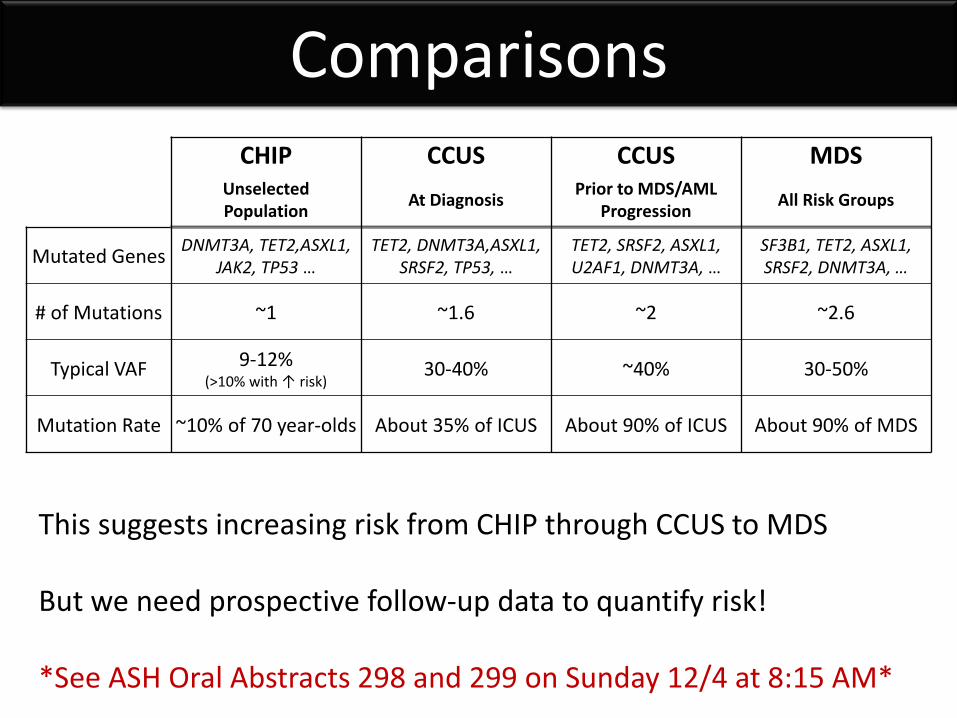
Comparisons CHIP CCUS CCUS MDS
Unselected Population
At Diagnosis Prior to MDS/AML
Progression All Risk Groups
Mutated Genes DNMT3A, TET2,ASXL1,
JAK2, TP53 … TET2, DNMT3A,ASXL1,
SRSF2, TP53, … TET2, SRSF2, ASXL1, U2AF1, DNMT3A, …
SF3B1, TET2, ASXL1, SRSF2, DNMT3A, …
# of Mutations ~1 ~1.6 ~2 ~2.6
Typical VAF 9-12% (>10% with ↑ risk)
30-40% ~40% 30-50%
Mutation Rate ~10% of 70 year-olds About 35% of ICUS About 90% of ICUS About 90% of MDS
This suggests increasing risk from CHIP through CCUS to MDS But we need prospective follow-up data to quantify risk! *See ASH Oral Abstracts 298 and 299 on Sunday 12/4 at 8:15 AM*
Germline Mutations
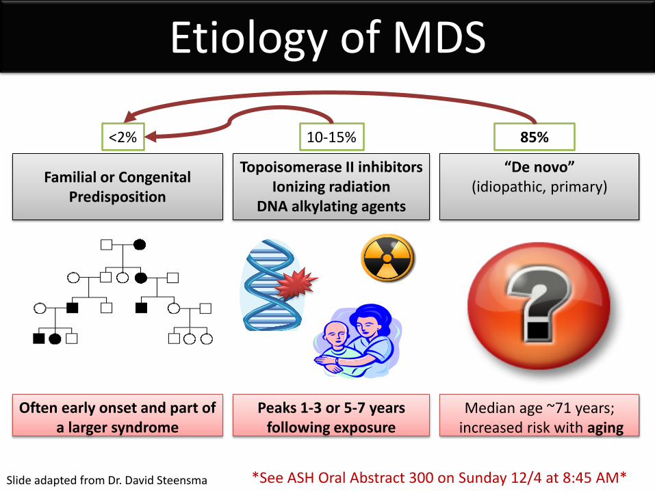
“De novo” (idiopathic, primary)
Median age ~71 years; increased risk with aging
85%
Familial or Congenital Predisposition
<2%
Topoisomerase II inhibitors Ionizing radiation
DNA alkylating agents
10-15%
Peaks 1-3 or 5-7 years following exposure
Etiology of MDS
Slide adapted from Dr. David Steensma
Often early onset and part of a larger syndrome
*See ASH Oral Abstract 300 on Sunday 12/4 at 8:45 AM*
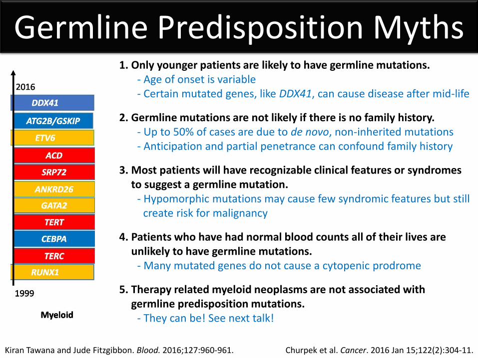
Germline Predisposition Myths
Kiran Tawana and Jude Fitzgibbon. Blood. 2016;127:960-961.
1. Only younger patients are likely to have germline mutations. - Age of onset is variable - Certain mutated genes, like DDX41, can cause disease after mid-life
2. Germline mutations are not likely if there is no family history. - Up to 50% of cases are due to de novo, non-inherited mutations - Anticipation and partial penetrance can confound family history
3. Most patients will have recognizable clinical features or syndromes to suggest a germline mutation.
- Hypomorphic mutations may cause few syndromic features but still create risk for malignancy
4. Patients who have had normal blood counts all of their lives are unlikely to have germline mutations.
- Many mutated genes do not cause a cytopenic prodrome
5. Therapy related myeloid neoplasms are not associated with germline predisposition mutations.
- They can be! See next talk!
Churpek et al. Cancer. 2016 Jan 15;122(2):304-11.
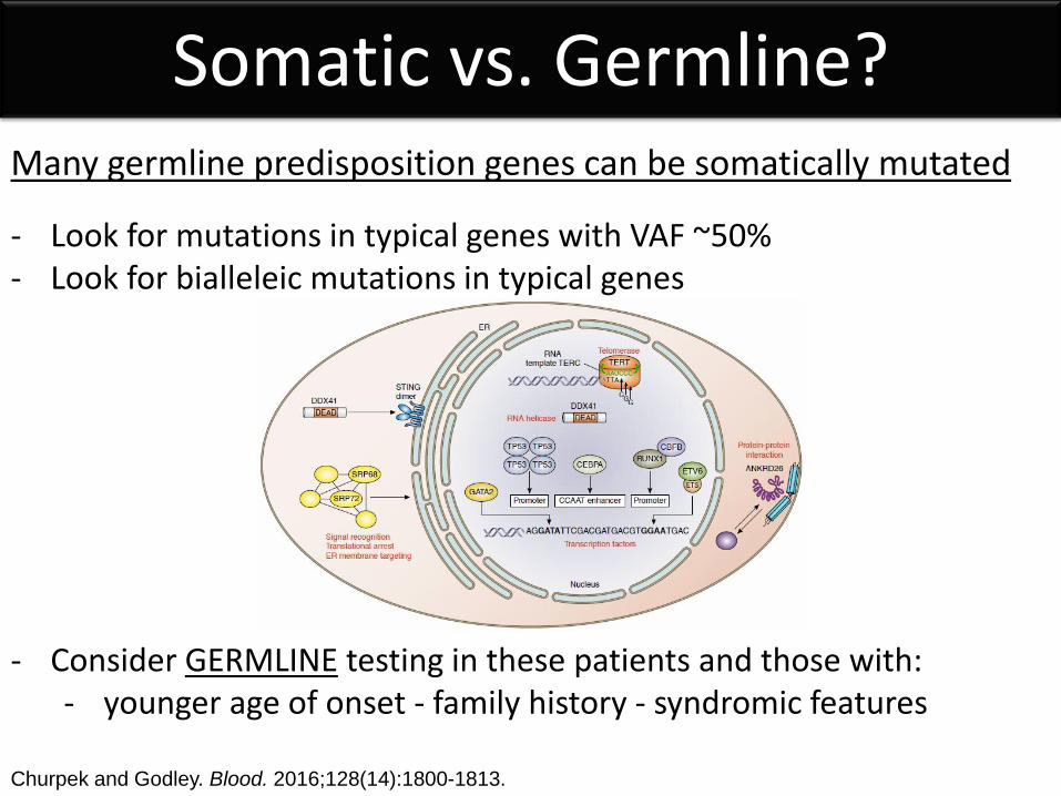
Somatic vs. Germline?
Churpek and Godley. Blood. 2016;128(14):1800-1813.
Many germline predisposition genes can be somatically mutated
- Look for mutations in typical genes with VAF ~50% - Look for bialleleic mutations in typical genes
- Consider GERMLINE testing in these patients and those with: - younger age of onset - family history - syndromic features

Classification and Prognosis
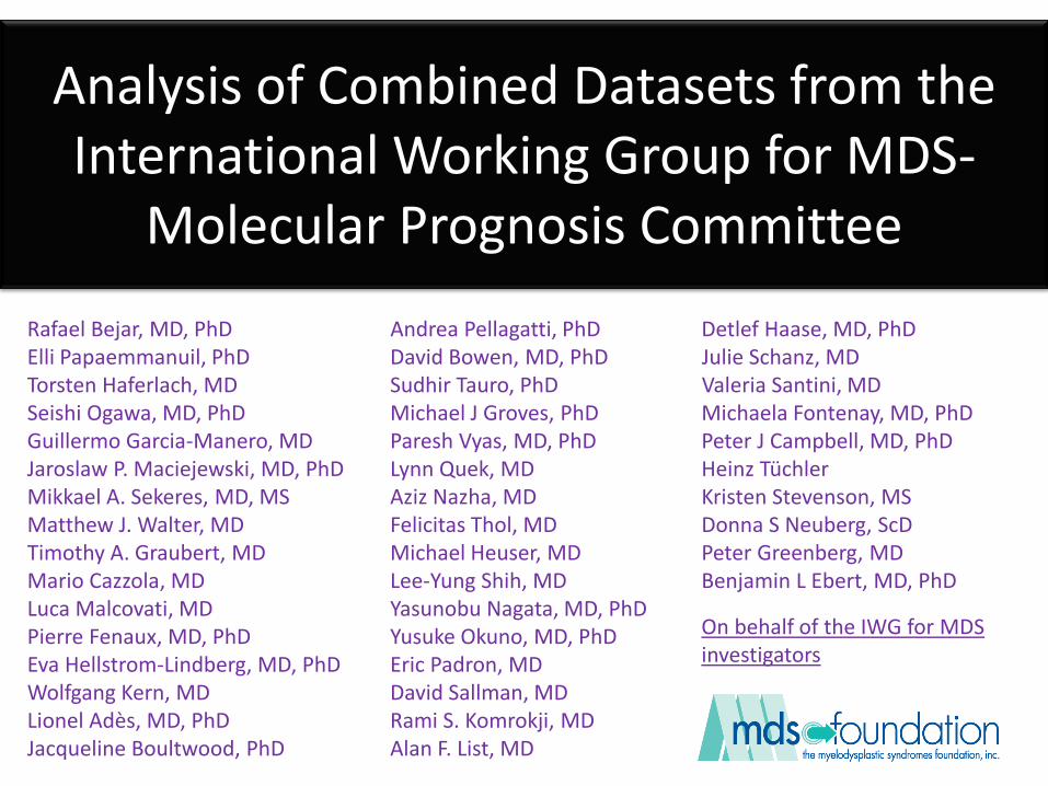
Analysis of Combined Datasets from the International Working Group for MDS-
Molecular Prognosis Committee
Rafael Bejar, MD, PhD Elli Papaemmanuil, PhD Torsten Haferlach, MD Seishi Ogawa, MD, PhD Guillermo Garcia-Manero, MD Jaroslaw P. Maciejewski, MD, PhD Mikkael A. Sekeres, MD, MS Matthew J. Walter, MD Timothy A. Graubert, MD Mario Cazzola, MD Luca Malcovati, MD Pierre Fenaux, MD, PhD Eva Hellstrom-Lindberg, MD, PhD Wolfgang Kern, MD Lionel Adès, MD, PhD Jacqueline Boultwood, PhD
Andrea Pellagatti, PhD David Bowen, MD, PhD Sudhir Tauro, PhD Michael J Groves, PhD Paresh Vyas, MD, PhD Lynn Quek, MD Aziz Nazha, MD Felicitas Thol, MD Michael Heuser, MD Lee-Yung Shih, MD Yasunobu Nagata, MD, PhD Yusuke Okuno, MD, PhD Eric Padron, MD David Sallman, MD Rami S. Komrokji, MD Alan F. List, MD
Detlef Haase, MD, PhD Julie Schanz, MD Valeria Santini, MD Michaela Fontenay, MD, PhD Peter J Campbell, MD, PhD Heinz Tüchler Kristen Stevenson, MS Donna S Neuberg, ScD Peter Greenberg, MD Benjamin L Ebert, MD, PhD
On behalf of the IWG for MDS investigators
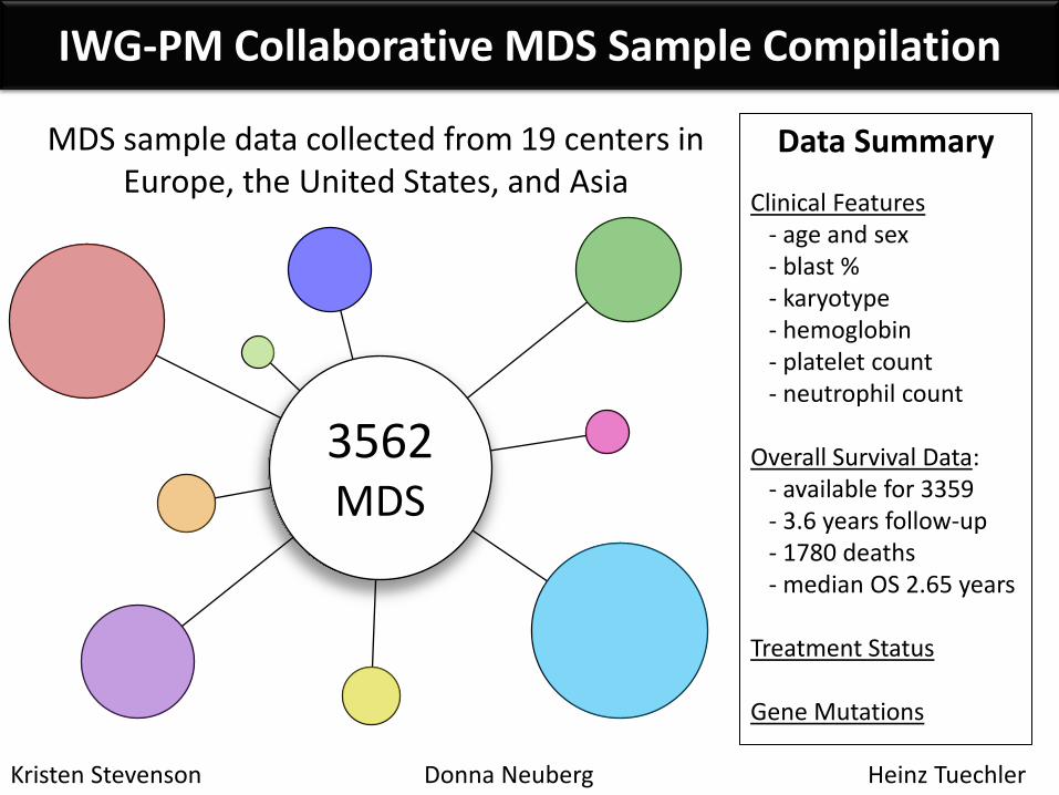
IWG-PM Collaborative MDS Sample Compilation
Kristen Stevenson Donna Neuberg Heinz Tuechler
Data Summary
Clinical Features - age and sex - blast % - karyotype - hemoglobin - platelet count - neutrophil count Overall Survival Data: - available for 3359 - 3.6 years follow-up - 1780 deaths - median OS 2.65 years Treatment Status Gene Mutations
3562 MDS
MDS sample data collected from 19 centers in Europe, the United States, and Asia
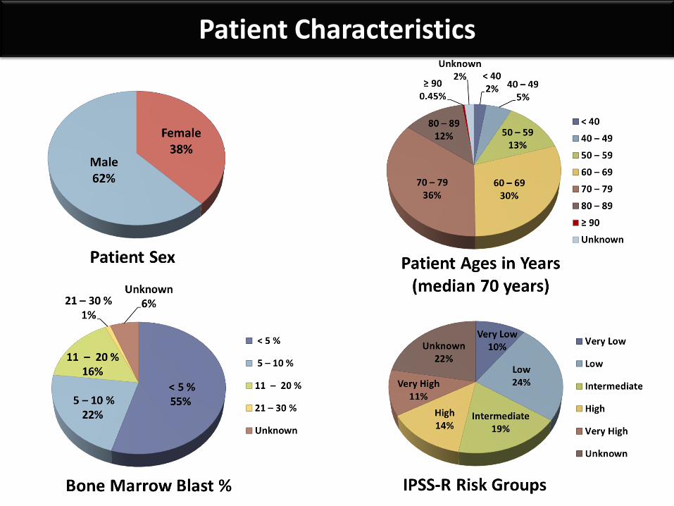
Patient Characteristics
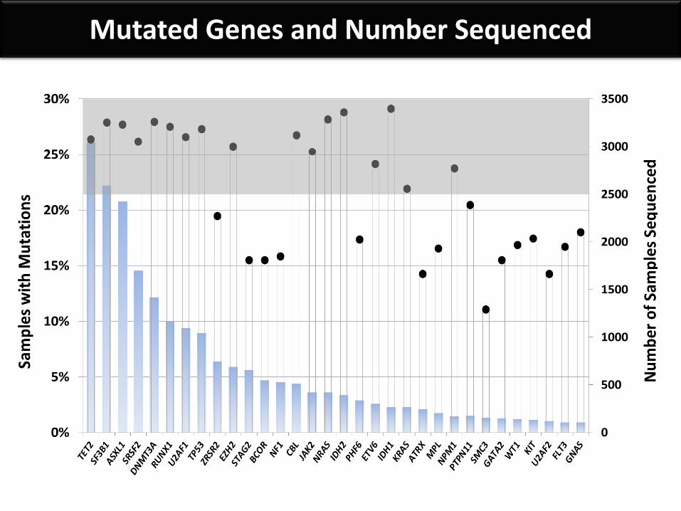
Mutated Genes and Number Sequenced
Y e a r s
Ov
era
ll S
urv
iva
l (%
)
0 2 4 6 8 1 0 1 2 1 4
0
1 0
2 0
3 0
4 0
5 0
6 0
7 0
8 0
9 0
1 0 0
0 (n = 3 7 7 )
1 (n = 5 9 5 )
2 (n = 4 6 0 )
3 (n = 2 1 0 )
4 (n = 1 2 5 )
5 /6 /7 (n = 2 2 )
S F 3 B 1 o n ly (n = 2 0 7 )
N u m b e r o f M u ta te d G e n e s
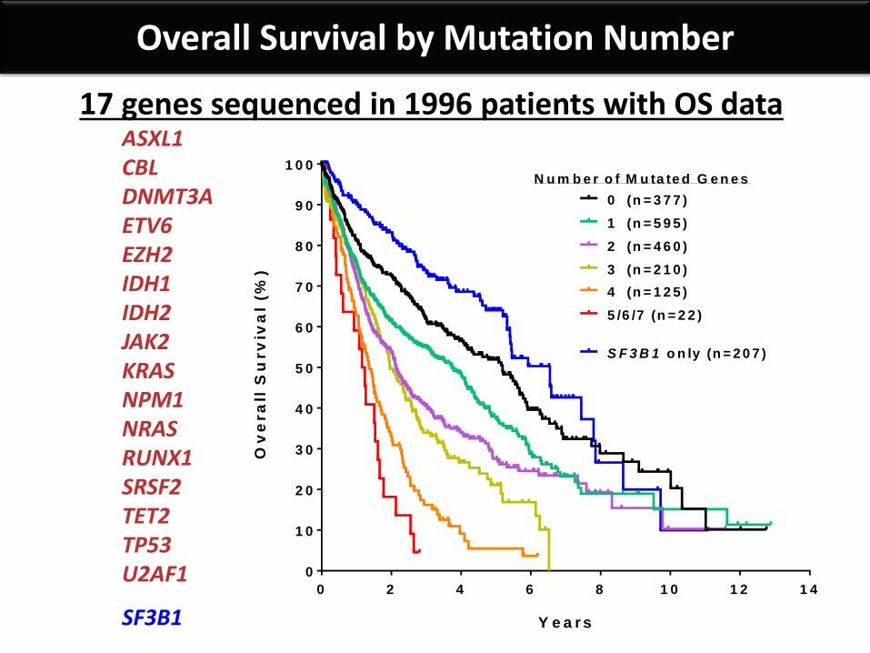
Overall Survival by Mutation Number
17 genes sequenced in 1996 patients with OS data ASXL1 CBL DNMT3A ETV6 EZH2 IDH1 IDH2 JAK2 KRAS NPM1 NRAS RUNX1 SRSF2 TET2 TP53 U2AF1
SF3B1
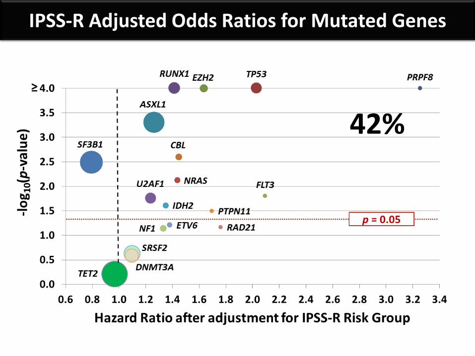
IPSS-R Adjusted Odds Ratios for Mutated Genes
42%
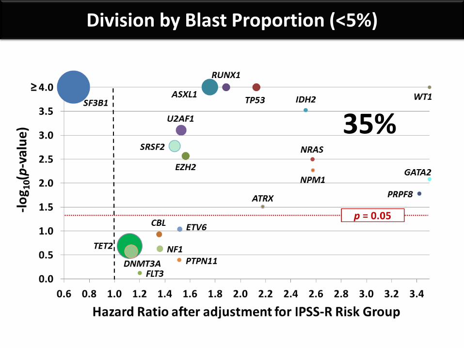
Division by Blast Proportion (<5%)
35%
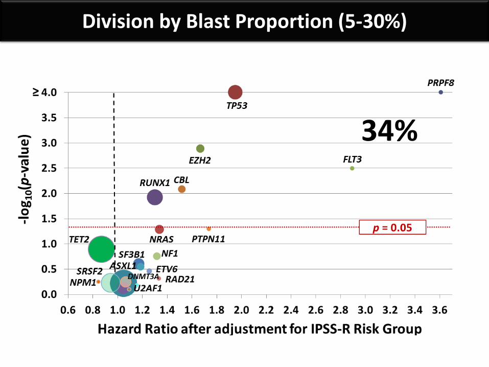
Division by Blast Proportion (5-30%)
34%
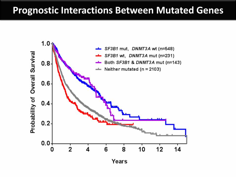
Prognostic Interactions Between Mutated Genes
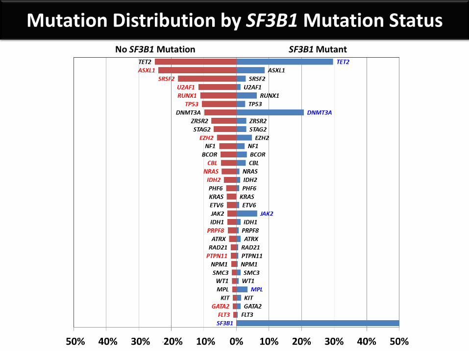
Mutation Distribution by SF3B1 Mutation Status
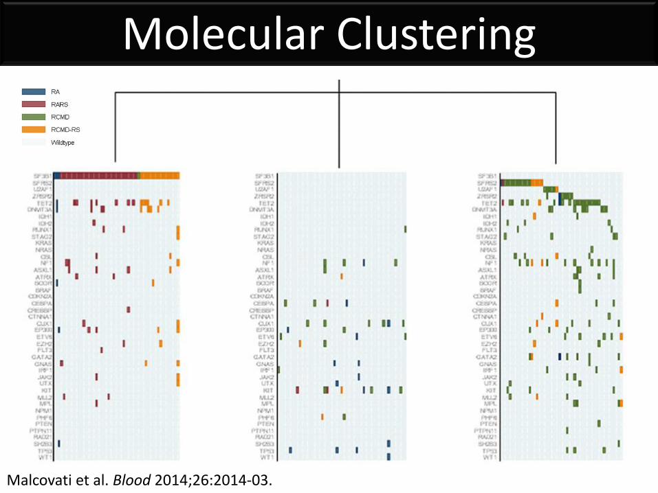
Molecular Clustering
Malcovati et al. Blood 2014;26:2014-03.
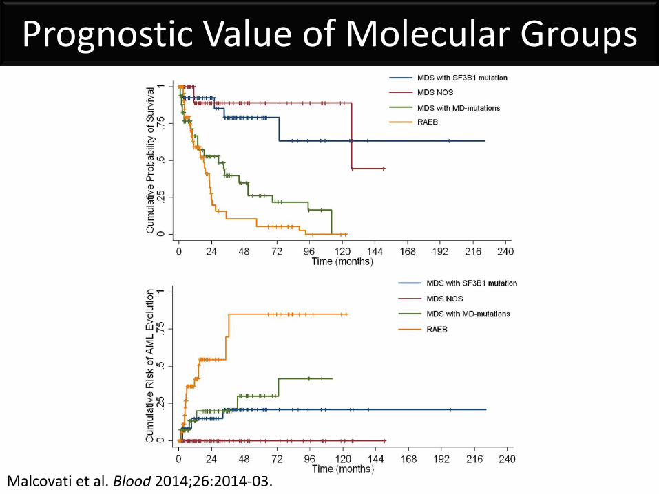
Prognostic Value of Molecular Groups
Malcovati et al. Blood 2014;26:2014-03.
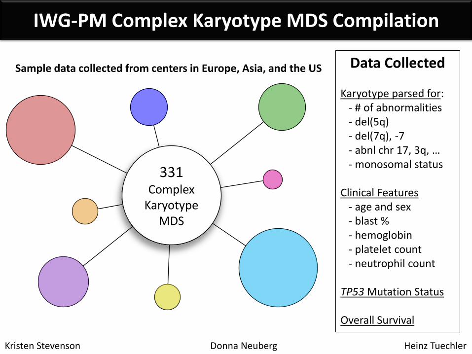
IWG-PM Complex Karyotype MDS Compilation
331 Complex
Karyotype MDS
Kristen Stevenson Donna Neuberg Heinz Tuechler
Data Collected Karyotype parsed for: - # of abnormalities - del(5q) - del(7q), -7 - abnl chr 17, 3q, … - monosomal status Clinical Features - age and sex - blast % - hemoglobin - platelet count - neutrophil count TP53 Mutation Status Overall Survival
Sample data collected from centers in Europe, Asia, and the US

TP53 Mutations and Other Genetic Features
TP53 Mutations and Most Frequent Somatic Mutations **
***p < 0.001, **p = 0.001 , *p = 0.014 for association with NO TP53 mutation
*** *
TP53 Mutations, Monosomal Karyotype, and # of Abnormalities
3 4 5+** 3 4 5+**
**
** p < 0.001 for association with TP53 mutation
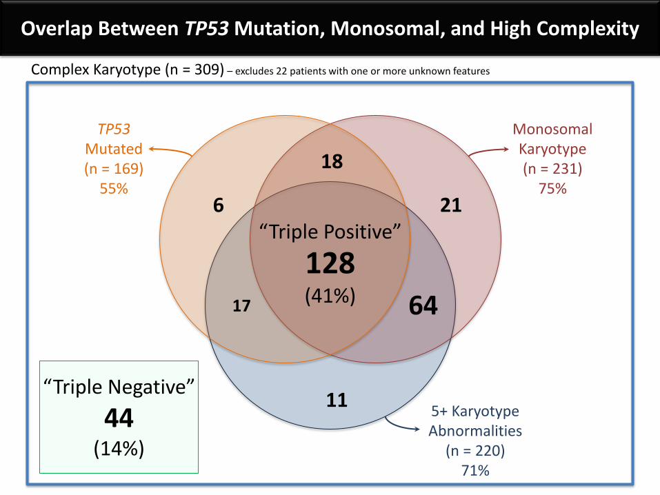
Complex Karyotype (n = 309) – excludes 22 patients with one or more unknown features
Monosomal Karyotype (n = 231)
75%
TP53 Mutated (n = 169)
55%
5+ Karyotype Abnormalities
(n = 220) 71%
“Triple Negative”
44 (14%)
128 (41%)
18
64 17
11
21 6
Overlap Between TP53 Mutation, Monosomal, and High Complexity
“Triple Positive”
Y e a rs
Pe
rce
nt
su
rviv
al
0 1 2 3 4 5 6 7
0
2 0
4 0
6 0
8 0
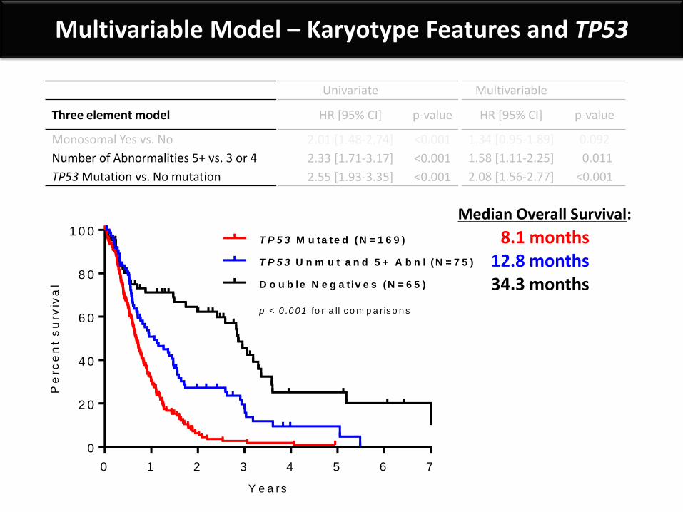
1 0 0T P 5 3 M u ta te d (N = 1 6 9 )
p < 0 .0 0 1 fo r a ll c o m p a r is o n s
T P 5 3 U n m u t a n d 5 + A b n l (N = 7 5 )
D o u b le N e g a t iv e s (N = 6 5 )
Multivariable Model – Karyotype Features and TP53
Univariate Multivariable
Three element model HR [95% CI] p-value HR [95% CI] p-value
Monosomal Yes vs. No 2.01 [1.48-2.74] <0.001 1.34 [0.95-1.89] 0.092
Number of Abnormalities 5+ vs. 3 or 4 2.33 [1.71-3.17] <0.001 1.58 [1.11-2.25] 0.011
TP53 Mutation vs. No mutation 2.55 [1.93-3.35] <0.001 2.08 [1.56-2.77] <0.001
Median Overall Survival:
8.1 months 12.8 months 34.3 months
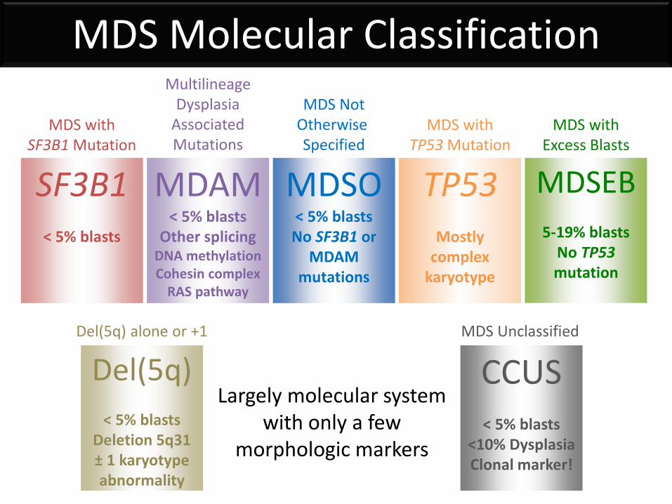
MDS with Excess Blasts
MDSEB
5-19% blasts No TP53 mutation
MDS with TP53 Mutation
TP53
Mostly complex
karyotype
MDS Molecular Classification
MDSO < 5% blasts No SF3B1 or
MDAM mutations
SF3B1
< 5% blasts
MDS Not Otherwise Specified
MDS with SF3B1 Mutation
Multilineage Dysplasia
Associated Mutations
MDAM < 5% blasts
Other splicing DNA methylation Cohesin complex
RAS pathway
CCUS
< 5% blasts <10% Dysplasia Clonal marker!
MDS Unclassified
Del(5q)
< 5% blasts Deletion 5q31 ± 1 karyotype abnormality
Del(5q) alone or +1
Largely molecular system with only a few
morphologic markers
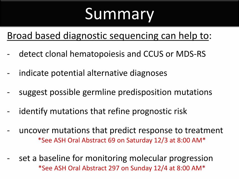
Summary Broad based diagnostic sequencing can help to:
- detect clonal hematopoiesis and CCUS or MDS-RS
- indicate potential alternative diagnoses
- suggest possible germline predisposition mutations
- identify mutations that refine prognostic risk
- uncover mutations that predict response to treatment *See ASH Oral Abstract 69 on Saturday 12/3 at 8:00 AM*
- set a baseline for monitoring molecular progression *See ASH Oral Abstract 297 on Sunday 12/4 at 8:00 AM*
Bejar Lab Albert Perez Sigrid Katz
Tiffany Tanaka Brian Reilly
Emily Wheeler Armon Azizi
Fiona Gowen-Huang
Acknowledgements MDS Center of Excellence at UCSD Elizabeth Broome Huanyou Wang - Hematopathology
Edward Ball Peter Curtin - BMT Group
Matthew Wieduwilt Dimitrios Tzachanis
Carolyn Mulroney Caitlin Costello
Januario Castro Dan S. Kaufman
Sandford Shattil John Adamson - Hematology Group
Catriona Jamieson Michael Choi
Erin Reid Tom Kipps
Natalie Galanina Annette Von Drygalski
Our amazing CLINIC and INFUSION CENTER nurses and staff
And most of all – our incredible patients and families!
Evans Foundation for MDS


![PRINTED: 10/01/2009 · 2012. 1. 6. · Data Set [MDS] assessment for Resident #4. The findings include: Resident #4 was admitted to the facility May 6, 2009. An admission MDS was](https://img.pdfslide.net/doc/110x75/6040d20cc21ed067c50e1ff1/printed-10012009-2012-1-6-data-set-mds-assessment-for-resident-4-the.jpg)