Embed Size (px)
Citation preview

Joe Pistack MS/ED
Integumentary System and Body Temperature-Chapter 7

INTEGUMENTARY SYSTEM
Integumentary system includes: The skin Accessory structures:- sweat glands
-oil glands
- hair
- nails

FUNCTIONS OF SKIN
The skin performs the following functions:
Keeps harmful substances out of the body and helps retain water and electrolytes.
Protects the internal structures and organs from injuries due to blows, cuts, harsh chemicals, sunlight burns, and pathogenic microorganisms.
Performs an excretory function. Secretes water and small amounts of urea.

FUNCTIONS OF THE SKIN
Acts as a gland by synthesizing vitamin D. Vitamin D is necessary for absorption of calcium from the digestive tract.
Performs a sensory role by housing the sensory receptors for touch, pressure, pain, and temperature.
Plays an important role in the regulation of body temperature.

STRUCTURE OF THE SKIN
Skin: Considered an organ Also called integument or cutaneous membrane
Skin has 2 layers: Epidermis-outer layer Dermis-inner layer
Dermatology-the study of skin and skin disorders.

LAYERS OF SKIN
Epidermis-thin outer layer of skin.
Composed of stratified squamous epithelium.
Has no blood supply of it’s own, avascular.
Oxygen and nutrients diffuse into the epidermis from blood supply from the dermis.

LAYERS OF SKIN
The epidermis can be divided into 5 layers the two of interest here are the deeper stratum germinativum and the more superficial stratum corneum
1.Stratum germinativum-
-lies on top of the dermis.
-has access to a rich supply of blood.
-cells of this layer constantly divide, push old cells to the surface.

LAYERS OF SKIN
Changes take place as cells move away from surface: 1. cells begin to die
2. keratinization takes place
Keratinization-process whereby tough protein
called keratin is deposited within the cell, keratin hardens and flattens the cells as they move toward surface. This makes the skin water-resistant.
LAYERS OF THE SKIN
Stratum Corneum: Surface layer of the epidermis. Composed of about 30 layers of dead cells. Dead cells are continuously sloughed off. Sloughed cells are called dander, dandruff when
clumped by oil on the skull.

LEVELS OF SKIN
Insensible perspiration-500ml/day of perspiration that is lost through the skin.
Sensible perspiration-due to activity of the sweat glands.
If the epidermis is damaged, the rate of insensible perspiration increases. E.g. burns
LEVELS OF SKIN
Dermis: Located under the epidermis. Largest portion of the skin Composed of dense, fibrous, connective tissue. Contains collagen and elastin fibers that make the skin
strong and stretchable. E.g. Pregnancy

LAYERS OF SKIN
Subcutaneous layer or hypodermis: Not considered part of the skin, Lies under the skin. Composed primarily of loose connective and adipose
tissue.

LAYERS OF SKIN
Subcutaneous tissue performs two main roles:
1. Helps to insulate the body from extreme temperature changes in the external environment.
2. Anchors the skin to the underlying structures. Several areas of the body have no subcutaneous layer and
are anchored directly to bone Drugs are administered (SubQ) because hypodermis has a
rich supply of blood vessels.

LAYERS OF SKIN

SQ INJECTIONS
22 to20 ga. 5/8 to 3/4 long

SKIN COLOR
Skin color is determined by: Genetic factors Physiological factors Disease
Melanocytes-skin cells within the epidermal layer. Melanin-darkening pigment, stains the
surrounding cells causing them to darken.

SKIN COLOR
The more melanin, the darker the skin.
Amount of melanin secreted determines the skin color.
Exposure to ultraviolet sunlight increases the secretion of melanin=suntan.

MALFUNCTIONING MELANOCYTE Conditions involving malfunctioning melanocyte: Albinism:
- melanocytes fail to secrete melanin.
- skin, hair, and iris (colored part of eye) are white. Vitiligo:
-loss of pigment in certain areas of skin.
-creates patches of white skin. Freckles and Moles:
-Areas in the skin where melanin is concentrated Malignant melanoma
-A mole that has changed in character and has become cancerous

SKIN CONDITIONS
Carotene-yellowish pigment to skin.
Cyanosis-blue look to skin, result of poorly oxygenated blood.
Blushing-dilation of the blood vessels.
Pallor-constriction of blood vessels, decrease in oxygenated blood.
ACCESSORY STRUCTURES
Accessory structures include:
- hair
- nails
- glands
Hairless body parts: palms of hands, soles of feet, lips, nipples, and parts of the external reproductive organs.

PARTS OF HAIR
Chief parts: Shaft-part above the surface of the skin.
Root-part that extends from the dermis to the surface.
Hair follicle-formed by downward extension of epithelial cells.

FUNCTIONS OF HAIR
Functions: Eyelashes and eyebrows-protect the eyes from
dust and perspiration. Nasal hairs trap dust and prevent it from entering
the lungs. Hair of the scalp keeps us warm.
FUNCTION OF HAIR
Hair growth-influenced by sex hormones.
Puberty-growth of hair in axillary and pubic areas in male and females.
Hirsutism-excessive hair growth in females, caused by too much testosterone.

HAIR FOLLICLE
Epidermal cells –receive blood supply from the dermal blood vessels.
Keratinization of cells- cells die as they move away from their source of nourishment.
Hair that we brush, blow dry, and curl is dead.

HAIR COLOR
Hair color: Genetically controlled by the amount of melanin. Abundance of melanin-dark hair. Less melanin-blond hair. Absence of melanin-white hair.

SHAPE OF HAIR
Shape of the hair shaft: Determines the appearance of hair. Round shaft produces straight hair. Oval shaft produces wavy hair. Flat hair shafts produce curly and kinky hair.

HAIR FOLLICLE

HAIR FOLLICLE
Arrector Pili muscle- attached to the hair follicle.
Bundle of smooth muscle fibers, when these muscles contract, hair stands on end.
Contract when cold or frightened.
Also called goose bumps.

HAIR STANDING ON END

ALOPECIA
Alopecia-loss of hair.
Male-pattern baldness most common type. Characterized by a gradual loss of hair.
Drug toxicity second most common type. Eg. Chemo, radiation.

HAIR LOSS FROM RADIATION

NAILS
Nails: Thin plates of stratified squamous epithelial cells. Contain a hard form of keratin. Found on the distal end of the fingers and toes. Protect structures from injury.

NAIL STRUCTURE
Structure: Free edge Nail body (finger nail) Nail root

NAIL STRUCTURE
Nail growth-determined by half-moon shaped lunula located at the base of the nail.
As nail grows, it slides over the nailbed.
Underlying dermal layer contains blood vessels which give pink color to nail.
Cuticle-fold of stratum corneum-grows onto proximal portion of the nail body.

NAIL STRUCTURE

NAILS

NAILS

ASSESSMENT
Assessment of the nails should include:
-shape
-dorsal curvature
-adhesion to the nail bed
-color
-thickness

NAIL CONDITIONS
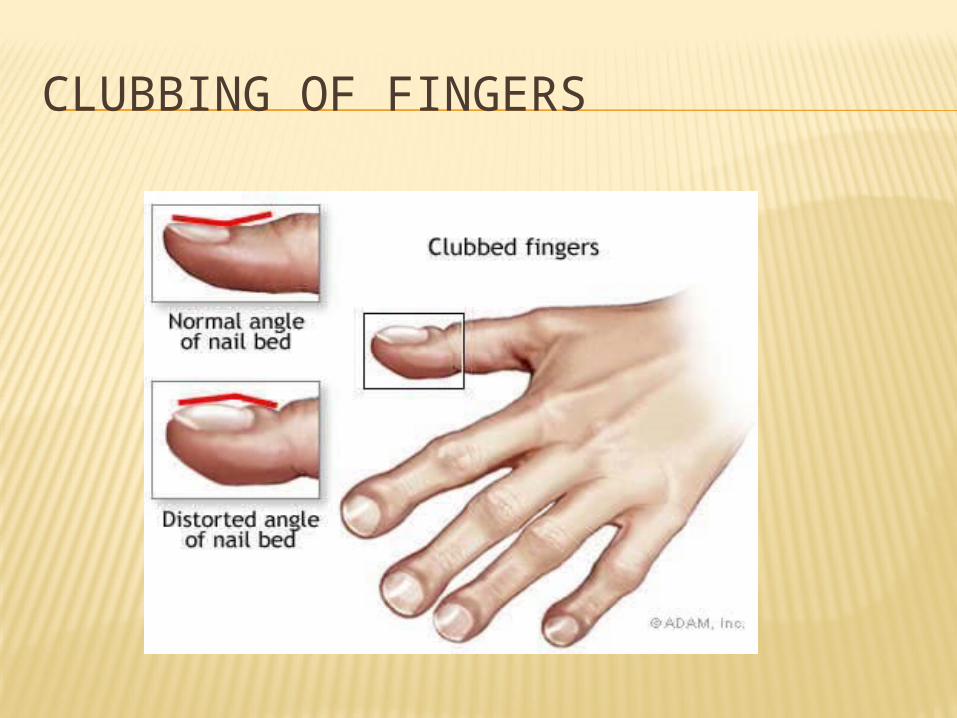
clubbing-condition that indicates fingertips have received an insufficient supply of oxygenated blood over a period of time.
Fingertips become large, nails become think, hard, shiny and curved at the free end.
Causes-chronic heart and lung disease.
CLUBBING OF FINGERS

CYANOSIS
Cyanosis-poor oxygenation makes the blood appear bluish, this in turn makes the nails appear bluish.
Nail abuse-trauma to the nail that causes the nail to thicken and hypertrophy.
Brittle- generally due to poor oxygenation or poor nutrition, or anemias.

CYANOSIS

GLANDS
Two major glands: Sebaceous glands Sweat glands
Sebaceous glands or oil glands-associated with the hair follicles, found in all body areas that have hair.
Sebum-oily substance that flows into hair follicle or onto surface of skin.

GLANDS
Function: Sebum lubricates and helps waterproof skin and
hair. Inhibits bacteria on the surface of the skin. Production decreases with aging, results in dry
skin and brittle hair. Vernix caseosa-cream cheese covering that babies
are born with, secreted by sebaceous glands.
GLANDS
Glands can become blocked by accumulating sebum and debris. A blackhead forms when sebum is exposed to air and
dries out
A pimple forms when the blocked sebum becomes infected with staphylococci-it becomes a pustule

SEBACEOUS GLANDS

SWEAT GLANDS
Sweat glands or sudoriferous glands: Located in the dermis. Secrete sweat.
Sweat is secreted into a duct that opens onto the skin as a pore.
We have approximately three million sweat glands.

SWEAT GLANDS
Two types of sweat glands:
1) Apocrine glands-usually associated with the hair follicles, found in the axillary and genital areas. Respond to emotional stress and become activated
when a person is frightened, upset, in pain or sexually excited.
Become activated during puberty.

SWEAT GLANDS
Body odor- occurs when the substances in sweat are degraded by bacteria into chemicals with a strong unpleasant odor.
2) Eccrine glands-more numerous and widely distirubuted throughout the body. Especially numerous on the forehead, neck, back, upper lip, palms, and soles.

GLANDS
Eccrine glands: Not associated with hair follicles. Sweat that is secreted plays an important role
in temperature regulation. As sweat evaporates on the skin, heat is lost. Sensible perspiration-secreted by the eccrine
glands, can secrete a gallon of sweat per hour.

GLANDS
Modified sweat glands: Ceruminous –found in the external auditory
canal, secrete cerumen. Cerumen- yellow, sticky, wax-like secretion that
repels insects and traps foreign materials. Mammary glands-located in the breasts, secrete
milk.

BODY TEMPERATURE
Normal body temperature is 98.6 degrees F . Body temp. differs from one part of the body to
another.
Core temperature-reflects the temperature of the inner parts of the body, (cranial, thoracic, and abdominal cavities).
Shell temperature-reflects the temperature of the skin and mouth.
BODY TEMPERATURE
Thermoregulation-the mechanism whereby the body balances heat production and heat loss.
Failure to regulate body temperature causes the body temperature to fluctuate.
Hypothermia-excessive decrease in body temperature.
Hyperthermia-excessive increase in body temperature.
Extreme changes in body temperature may be fatal.

HEAT LOSS
80% of heat loss occurs through the skin.
20% is lost through the respiratory system and excretory products.
Heat loss occurs by four means: Radiation Conduction Convection Evaporation

HEAT LOSS
Radiation-heat is lost from a warm object (the body) to the cooler air surrounding the warm object. Eg. Person loosing heat in a cold room.
Conduction-loss of heat from a warm body to a cooler object in contact with the warm body.
Eg. Warm person becomes cold when sitting on a block of ice.
Eg. Cooling blanket for hyperthermia-warm object (feverish patient) looses heat to the cooler object, the cooling blanket.

HEAT LOSS
Convection-loss of heat by air currents moving over the surface of the skin. E.g. Fan moving across the surface of the skin.
Evaporation-heat may be lost through changing a liquid (sweat) to a gas.
E.g. during strenuous exercise, sweat on the surface of the skin evaporates and cools the body.

BODY TEMPERATURE
Normal body temperature is regulated by several mechanisms:
- Hypothalamus-thermostat of the body, located in the brain.
-senses changes in body temperature and sends information to the skin. (blood vessels, sweat glands and skeletal muscle).

BODY TEMPERATURE
Exercise
Temperature elevates
Blood vessels dilate
Increased blood flow to the skin

BODY TEMPERATURE
Heat is transferred to deeper tissue surfaces
Sweat glands activate
Heat is lost as sweat evaporates
Body temperature lowers

BODY TEMPERATURE REGULATION
RESPONSE TO DECREASING TEMP.
Decreased temperature: Blood vessels constrict. Traps blood and heat in the deeper tissues
(prevents heat loss) Sweat glands become less active, preventing heat
loss.

RESPONSE TO DECREASING TEMP.
Skeletal muscles contract vigorously and involuntarily causing shivering and an increase
in the production of heat.
Contraction of the arrector pili muscles causes goose bumps indicating a decline in body temp.
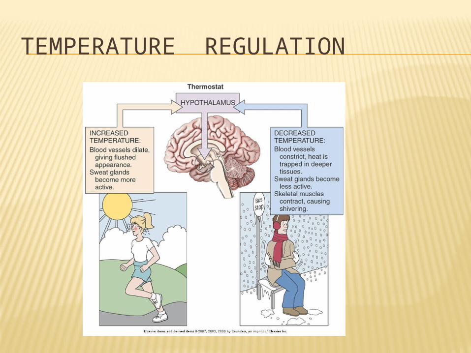
TEMPERATURE REGULATION

BURNS
Classified according to depth.
Classified as either partial-thickness burns or full-thickness burns.
Partial thickness are divided into first-degree and second-degree burns.

FIRST DEGREE BURNS
First degree burns: Red Painful Slightly edematous (swollen) Only epidermis involved
E.g. sunburn

SECOND DEGREE BURN
Second degree burns: Redness Pain Edema Blister formation
May appear red tan or white

THIRD DEGREE BURNS
Third degree burns:
(full thickness burns) Both epidermis and
dermis are destroyed Painless-sensory
receptors destroyed May appear white, tan,
brown, black or cherry red

BURNS
Rule of nines:
System used to measure the extent of burns.
Total body surface is divided into regions.
The assigned percentages are related to the number 9.

BURNS

BURNS Severe burns are associated with eschar
formation. Eschar is dead, burned tissue that forms a thick,
inflexible scab-like layer over the surface. Can act as a tourniquet and cut off blood supply to
extremity, or if the burn is in the trunk area it cal limit the ability to breath.
Though initially sterile it can become a breeding ground for bacteria, and their toxic secretions can easily enter the blood.

AGING SKIN
As we age: Epidermis becomes thinner. Skin is more translucent. Melanocyte decreases. Dermis becomes thinner, Decreased amount of collagen and elastin fibers. Increased wrinkles. Skin heals slower.

AGING SKIN
AS WE AGE!