Embed Size (px)
Citation preview
Intemal morphology and taphonomic history of the Neoproterozoic vase-shaped microfossils from the Visingso Group, Sweden
MONICA MARTf MUS & MALGORZATA MOCZYDLOWSKA
Marti Mus, M. & Moczydlowska, M. Intemal morphology and taphonomic history of the Neoproterozoic vase-shaped microfossils from the Visingsii Group, Sweden. Norsk Geologisk Tidsskrift, Vol. 80, pp. 2 13-228. Oslo 2000. ISSN 0029- 196X.
The morphology and mineral composition of the vase-shaped microfossils (V SMs) from phosphatic nodules of the upper Visingsii Group in the Lake Vattem area have been studied using confocal and transmitted light microscopy along with the EDX analytical technique. The taphonomic history of the microfossils and the internal anatomy of the original organism were reconstructed. An early diagenetic precipitation of phosphate accounts for the formation of phosphatic surfaces replicating the extemal and internal morphology of the vase-shaped organism. The vase-shaped vesicle had an internal compartrnent containing a reproductive cell or cyst. The presence of these structures indicates that the vesicle does not in itself represent an encystrnent stage of a protist as previously suggested by some authors. Hypotheses on the biological affinities of the VSMs are reviewed. The VSMs global record is considered in the palaeogeographic and palaeoenvironmetal contexts, suggesting that the vase-shaped microfossils were produced by stenothermal planktonic organisms, thriving in shallow marine seas in tropical to temperate climatic zones.
M. Martf-Mus & M. Moczydlowska, Department of Earth Sciences, Historical Geology and Palaeontology, Uppsala University, Norbyviigen 22, SE-752 36, Uppsala, Sweden
Introduction
Vase-shaped microfossils (VSMs) are a morphologically distinct group of microfossils known from Neoproterozoic marine sediments world-wide. They have a characteristic vase shape, being rounded at the aboral pole and showing a short, tapering, truncated neck with a narrow terminal opening at the oral pole. The vesicle has a smooth wall and lacks processes. VSMs were first recorded by Ewetz ( 1933), though at the time un-named and simply called 'single-celled organism with shell', from phosphatic nodules of the Visingso Group, southern Sweden. Since then, several occurrences of VSMs have been reported from Neoproterozoic (850-650 Ma) sediments of many parts of the world (Table 1 ). The organically preserved specimens from the shales of the K wagunt Formation (Bloeser et al. 1977; Bloeser 1 985) are the best-known morphologically and the only ones formally described under the generic name Melanocyrillium Bloeser, 1 985. Differences in preservation between these exceptionally preserved specimens and specimens from other localities have hindered detailed morphological comparisons and have generally prevented the identification of the latter ones as Melanocyrillium. Even so, the different microfossils summarized in Tab le l share distinct morphological similarities and have preservational features in common (perhaps associated with the chemistry of the wall; see below) that suggest close biological affinities .
VSMs occur in rocks of various lithologies: shales, carbonates, cherts and phosphatic nodules, encompassing a wide array of shallow marine depositional settings. Both
their world-wide distribution and their presence in a variety of lithologic facies supports the interpretation of VSMs as representing marine planktonic organisms (Bloeser et al. 1977; Knoll & Vidal 1980; Bloeser 1 985; Knoll et al. 1 989; Fairchild et al. 1 99 1 ). These intriguing microfossils thus constitute the earliest record of planktonic organisms showing morphological polarity (Bloeser 1 985) which, together with the morphological complexity of the oral pole and the herein described internat structures, places them among the most complex Neoproterozoic microfossils recovered to date.
It has been observed that the mode of preservation of VSMs changes according to the lithology, but little is known about the details of their preservation or the taphonomic processes which they have undergone in different depositional environments. In only a few occurrences the VSMs are preserved as three-dimensional organic-walled vesicles that can be extracted from the sediment by standard palynological techniques (Bloeser et al. 1 977; Vidal l 979; Vidal & Ford 1 985; Bloeser 1985). In most of the localities VSMs are preserved as moulds and casts of entire, undeformed vesicles, and the organic matter seems to be partially or comp1etely degraded. Knoll ( 1 992, p. 57) pointed out that 'Marked taphonomic differences between these fossils and acritarchs suggest original differences in wall chernistry' (see below).
The VSMs from phosphatic nodules of the Visingso Group were first described and figured by Ewetz (1933) and subsequently studied in more detail by Knoll & Vidal ( 1 980). Ewetz ( 1933) observed that the VSMs possessed 'shells' with rough internat surfaces and that phosphate
214 M. Marti Mus & M. Moczydlowska
Table l. Global record of VSMs.
Region
North America
Greenland
Sweden
Saudi Arabia Svalbard
Tasmania
Stratigraphic unit
KwaguntFm
Pahrump Group Limestone-Dolomite 'Series' (Eleonore Bay Group) Visingso Group
Jabal Rokharn Carbonates Rysso Fm
Draken Fm Backlundtoppen Fm Elbobreen Fm Smithton Dolomite
had entered the fossil vesicles through their 'mouths'. He also observed an internal, round, brownish structure, which he interpreted as a resting spore, and concluded that the Visingso VSMs could represent fossil rhizopods. Knoll & Vidal ( 1980) described the morphology and dimensional variation of a greater population of VSMs and compared them structurally and ecologically with modem tintinnids.
In this study, newly observed internal structures preserved in the VSMs are reconstructed in the light of the taphonomic history of the microfossils, and their biological significance is assessed.
Geological setting and palaeoenvironments
The Visingso Group is a sedimentary succes si on consisting of terrigenous rocks with minor carbonates of early Neoproterozoic age that extend in a restricted area of Lake Vattern in south-central Sweden (Fig. lA, B). The sediments are preserved in a graben within the crystalline basement presently occupied by the lake. The succes si on is unmetamorphosed and exposed in the coastal cliffs of the lake and on the Vising Island (Visingso), and is known also from a number of boreholes in the vicinity of the lake (Brotzen 1941 ; Collini 1951; Vidal 1974, 1976, 1982, 1985; Fig. lB) . Three informal lithostratigraphic units (lower, middle and upper) which are in the rank of formations (Vidal 1985), are distinguished in the Visingso Group, and together they exceed 1 000 m in thickness (Collini 1951) .
The lower unit of the Visingso Group consists of crossbedded, fluvial quartz sandstone with minor conglomerate and was deposited in a delta plain environment. The middle unit is characterized by alternating feldspathic arenite, conglomerate and siltstone and was accumulated in a shallow marine prodelta environment (Vidal 1 974, 1 976, 1982, 1 985; Larsen & Nørgaard-Pedersen 1988). The upper unit of the Visingso Group is of major interest in this study because of the occurrence of V SMs and consists
Age
Late Proterozoic
Late Proterozoic Late Proterozoic
Late Riphean
Late Precarnbrian Late Riphean
Late Riphean Late Riphean Late Proterozoic Late Precambrian
NORSK GEOLOGISK TIDSSKRIFT 80 (2000)
References
Bloeser et al. 1977 Bloeser 1 985 Vida! & Ford 1985 Horodyski 1987, 1993 Porter & Knoll (in press) Horodyski 1987, 1993 Vida! 1979 Green et al. 1988 Ewetz 1933 Knoll & Vidal 1980 This study Binda & Bokhari 1980 Knoll l 982 Knoll & Calder 1983 Knoll et al. 1991 Knoll et al. 198 9 Knoll 1990, 1992 Saito et al. 1988
of alternating micaceous shale and siltstone, with coarse sandstone at the bottom and interbeds of dolomitic limestone with stromatolites at the top (Vidal 1 972, 1976, 1985). The total thickness of the unit was estimated as being more than 580 m (Collini 1951). The observed sedimentary structures and the presence of abundant marine planktonic microfossils in the upper Visingso Group conform with deposition in a shallow marine environment with oscillating water depth, including shelf and subtidal to intertidal carbonate mud flats (Vidal 1 972, 1976, 1985; Samuelsson & Strauss 1 999). The cosmopolitan acritarch species dominating the assemblage also attest to the existence of a connection between the Vattern Basin and the global ocean (Vidal 1 985) maintaining the free dispersal of microplankton. The total organic carbon (TOC) content in the whole-rock upper Visingso samples has an average value of 3.56 mg C/g for shales (Strauss & Moore 1992; Samuelsson & Strauss 1999). This value is higher than the average value for late Proterozoic shales (2.30 mg C/g; Strauss et al. 1992) suggesting that not only high burial of organic matter, but also high organic productivity must have prevailed in the Visingso Basin. This, together with the presence of phosphate nodules in the upper Visingso Group is compatible with the Visingso basin being influenced by upwelling of nutrient-rich waters, as suggested by Knoll & Vidal ( 1 980).
The climatic conditions prevailing during the deposition of the Visingso Group, related to the palaeogeographic position of the host craton, have been constrained by several independent lines of evidence; i.e. palaeobiology, diagenetic mineralogy and palaeomagnetic data. The presence of stromatolites in the upper Visingso Group, characteristic of tropical and warm temperature beits, is a direct indication of the climatic conditions (Vidal 1 972, 1976). The abundance of detrital biotite suggests dry climatic conditions (Vidal 1985). The early diagenetic mineral berthierine, present inside the VSMs preserved in phosphate nodules and occurring also in the host shale of the upper Visingso Group, has been interpreted to
NORSK GEOLOGISK TIDSSKRIFr 80 (2000)
A B D
N
1 o '-------'
1 Km
m
6
5
4
3
2
o
Neoproterozoic microfossils, Sweden
sh
E---- V72G14
sit sslt d
§ Dark-grey micaceous shale
[-:-j Grey siltstone
CJ Grey sandy siltstone
� Grey laminated algal dolomite
215
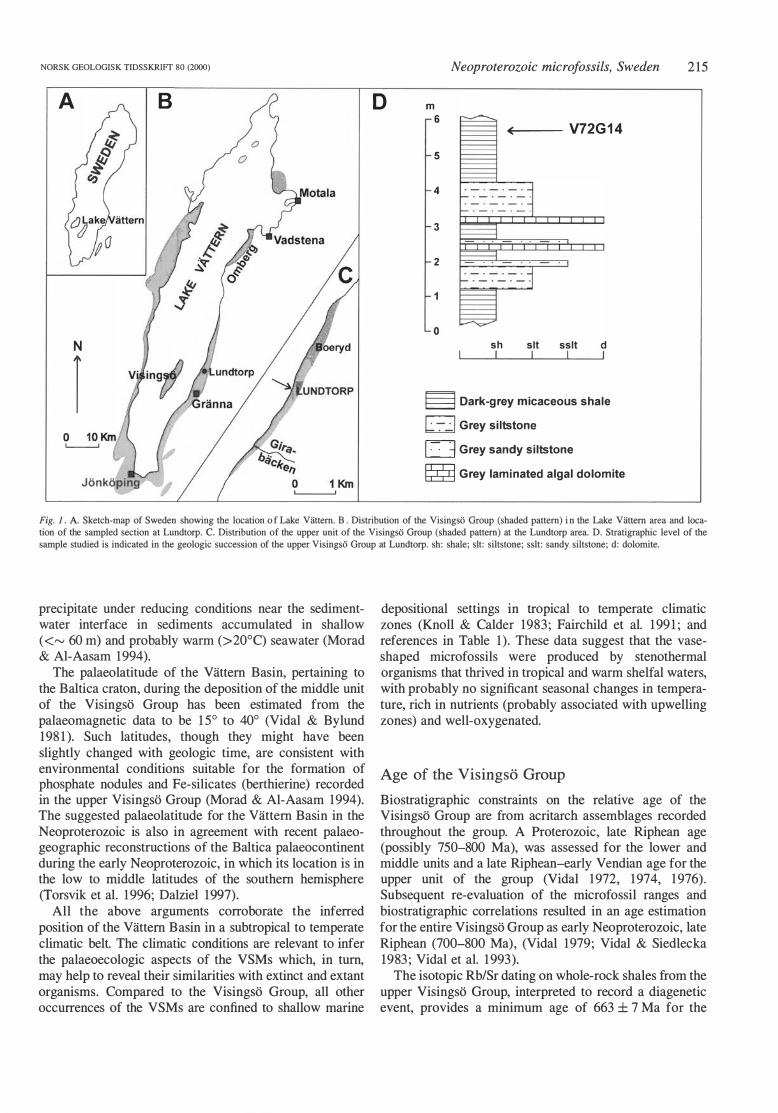
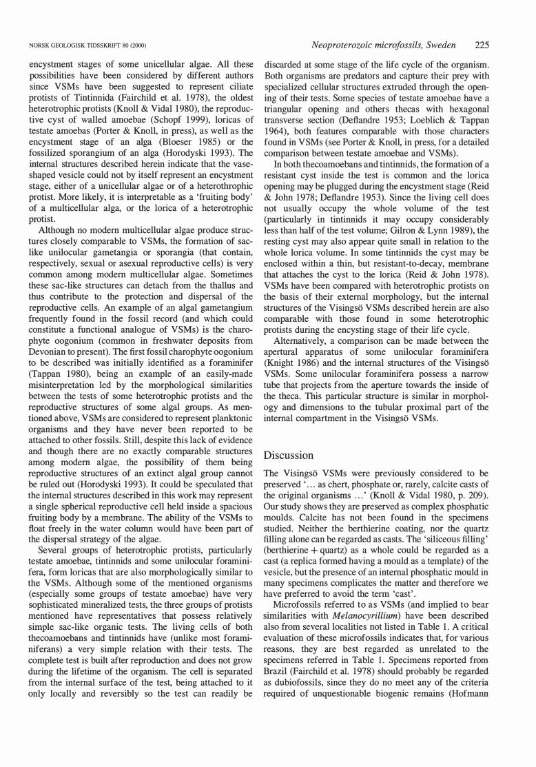
Fig. l . A. Sketch-map of Sweden showing the location o f Lake Viittern. B . Distribution of the Visingsti Group (shaded pattern) i n the Lake Viittern area and location of the sampled section at Lundtorp. C. Distribution of the upper unit of the Visingsti Group (shaded pattern) at the Lundtorp area. D. Stratigraphic levet of the sample studied is indicated in the geologic succession of the upper Visingsti Group at Lundtorp. sh: shale; sit: siltstone; sslt: sandy siltstone; d: dolomite.
precipitate under reducing conditions near the sedimentwater interface in sediments accumulated in shallow ( <"' 60 m) and probably warm (>20°C) seawater (Morad & Al-Aasam 1 994).
The palaeolatitude of the Vattem Basin, pertaining to the Bal ti ca craton, during the deposition of the rniddle unit of the Visingso Group has been estimated from the palaeomagnetic data to be 1 5° to 40° (Vidal & By1und 198 1 ). Such latitudes, though they rnight have been slightly changed with geologic time, are consistent with environmental conditions suitable for the formation of phosphate nodules and Fe-silicates (berthierine) recorded in the upper Visingso Group (Morad & Al-Aasam 1994). The suggested palaeolatitude for the Vattem Basin in the Neoproterozoic is also in agreement with recent palaeogeographic reconstructions of the Baltica palaeocontinent during the early Neoproterozoic, in which its location is in the low to rniddle latitudes of the southem hernisphere (Torsvik et al. 1 996; Dalziel 1997).
All the above arguments corroborate the inferred position of the Vattem Basin in a subtropical to temperate climatic belt. The climatic conditions are relevant to infer the palaeoecologic aspects of the VSMs which, in turn, may help to reveal their sirnilarities with extinct and ex tant organisms. Compared to the Visingso Group, all other occurrences of the VSMs are confined to shallow marine
depositional settings in tropical to temperate climatic zones (Knoll & Calder 1983; Fairchild et al. 1 99 1 ; and references in Table 1 ). These data suggest that the vaseshaped microfossils were produced by stenothermal organisms that thrived in tropical and warm shelfal waters, with probably no significant seasonal changes in temperature, rich in nutrients (probably associated with upwelling zones) and well-oxygenated.
Age of the Visingso Group
Biostratigraphic constraints on the relative age of the Visingso Group are from acritarch assemblages recorded throughout the group. A Proterozoic, late Riphean age (possibly 750-800 Ma), was assessed for the lower and middle units and a late Riphean-early Vendian age for the upper unit of the group (Vidal 1 972, 1 974, 1 976) . Subsequent re-evaluation of the microfossil ranges and biostratigraphic correlations resulted in an age estimation for the entire Visingso Group as earl y Neoproterozoic, late Riphean (700-800 Ma), (Vidal 1979; Vidal & Siedlecka 1983; Vidal et al. 1 993).
The isotopi c Rb/Sr dating on whole-rock shales from the upper Visingso Group, interpreted to record a diagenetic event, provides a minimum age of 663 ± 7 Ma for the
216 M . Marti Mus & M. Moczydlowska NORSK GEOLOGISK TIDSSKR!Ff 80 (2000)
NORSK GEOLOGISK TIDSSKR!Ff 80 (2000)
group (Bonhomme & W elin 1984 ), confonning in general with the biochronologic assessment.
Material and methods
Microfossils have been studied in thin-sections from phosphate nodules occurring in dark-grey and organicrich shale of the upper Visingso Group. The sampled succession is at Lundtorp, on the eastern shore of Lake Vattern, northeast of Granna and between the Girabacken Valley and Boeryd (Vidal l976, and unpublished data; Fig. lB , C). Strata belonging to the upper portion of the upper Visingso Group are exposed along the ca. 4 km cliff-coast but their thickness is only a few metres. They are folded but unmetamorphosed. In this exposure, the basal and topmost beds of the upper Visingso Group are missing. The measured succession consists of shale alternating with siltstone and thin layers of laminated algal dolomite (Vidal 1976; Fig. 1 0) . The phosphate nodules are lenticular in shape and randomly scattered within the shale. Detailed petrologic and geochemical analyses of phosphate nodules and host shales are provided by Morad & Al-Aasam ( 1994). The TOC content in shales in the Lundtorp area is high and ranges between 0.39 and 5.38 mg C/g (Strauss & Moore 1 992), and it is even higher (up to 1 1 .6 mg C/g) in the nearby locality at Boeryd (Samuelsson & Strauss 1999). The thermal alteration index (TAl) of the kerogens preserved at Boeryd (Fig. l C) indicates a catagenesis stage of thermal alteration and burial temperatures around 1 50°C (Samuelsson & Strauss 1 999). A similar stage of kerogen maturation is most likely in Lundtorp because of the uniform thermal alteration of the organic matter in the upper Visingso Group, estimated previously to be around 1 00°C (Vidal 1 976). The stratigraphic position of the sampled level with the occurrence of VSMs studied is shown in the lithologic log in Fig. 10.
Five thin-sections with the collection reference numbers PMU-V72Gl4 - l, 2, 3, 4, 5, have been exarnined. The VSMs are very abundant in some of the thin-sections, numbering up to several hundred specimens . The thinsections were studied using transmitted light microscopy.
Neoproterozoic microfossils, Sweden 217
Selected specimens were further studied with confocal microscopy (Leica® TCS 40) equipped with an ArgonKrypton laser. The autofluorescent properties of phosphate (probably francolite, Morad & Al-Aasam 1 994) were used for image acquisition, and the excitation wavelength was 488 nm. Among all minerals present in the Visingso nodules, only phosphate showed autofluorescent properties when illuminated with light in the visible spectrum. It was thus possible to obtain images showing only the morphology of phosphatised parts of the fossils. This was especially valuable since the taphonomic study of the Visingso VSMs indicates that only the phosphatised parts of the fossils contain relevant biological information (see below). Most of the final images were obtained processing an 'image series' with the 3D software available in the Leica ® system. Bach 'image series' consisted of 90 optical sections scanned at successive planes of focus covering the whole volume of a specimen.
Bnergy dispersive X-ray analysis (BDX), together with the appearance of the different minerals under optical, confocal and scanning electron (equipped with a backscattered electrons detector) microscopes, was used to infer the mineral composition of different parts of the microfossils. For EDX analyses the thin-sections were coated with carbon.
Bach figured specimen is named with the last digit of the thin section reference number ( 1 -5) and a code indicating its location in Bngland-Finder co-ordinates.
The tenninology established by Bloeser ( 1 985) to describe the external morphology of the VSMs from the Kwagunt Formation is followed in this paper. Some additional terms are introduced for the newly described internal structures and all terms are shown in Fig . 8 .
Description of the Visingso VSMs
VSMs have not been recovered from acid-resistant residues of the Visingso nodules, nor are they preserved in the host shale (Knoll & Vidal 1980). They are accumulated on the bedding planes, with their major axis oriented more or less horizontally. Their azimuthal
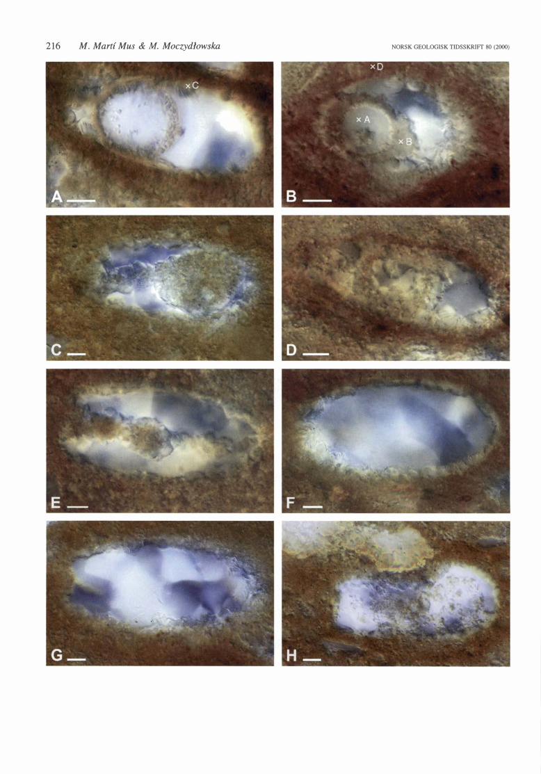
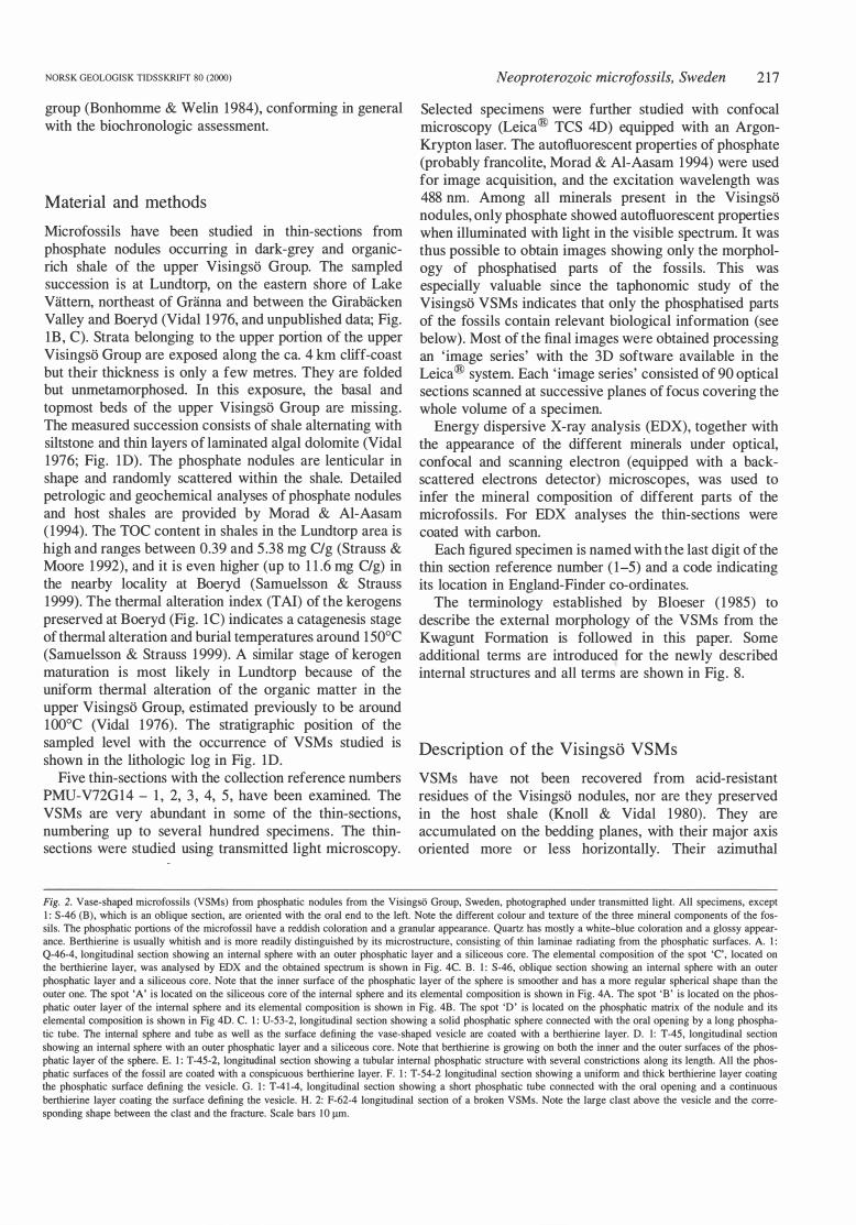
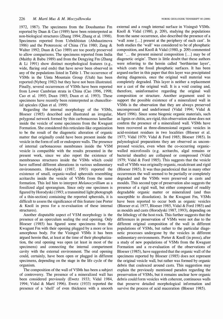
Fig. 2. Vase-shaped microfossils (VSMs) from phosphatic nodules from the Visingsii Group, Sweden, photographed under transmitted light. All specimens, except 1: S-46 (B), which is an oblique section, are oriented with the oral end to the left. Note the different colour and texture of the three mineral components of the fossils. The phosphatic portions of the microfossil have a reddish coloration and a granular appearance. Quartz has mostly a white-blue coloration and a glossy appearance. Berthierine is usually whitish and is more readily distinguished by its microstructure, consisting of thin laminae radiating from the phosphatic surfaces. A. l: Q-46-4, longitudinal section showing an internal sphere with an outer phosphatic layer and a siliceous core. The elementa! composition of the spot 'C', located on the berthierine layer, was analysed by EDX and the obtained spectrum is shown in Fig. 4C. B. l: S-46, oblique section showing an in terna! sphere with an outer phosphatic layer and a siliceous core. Note that the inner surface of the phosphatic layer of the sphere is smoother and has a more regular spherical shape than the outer one. The spot 'A' is located on the siliceous core of the internal sphere and its elementa! composition is shown in Fig. 4A. The spot 'B' is located on the phosphatic outer layer of the internal sphere and its elementa! composition is shown in Fig. 4B. The spot 'D' is located on the phosphatic matrix of the nodule and its elementa! composition is shown in Fig 40. C. 1: U-53-2, longitudinal section showing a solid phosphatic sphere connected with the oral opening by a long phosphatic tube. The internal sphere and tube as well as the surface defining the vase-shaped vesicle are coated with a berthierine layer. D. l: T -45, longitudinal section showing an internal sphere with an outer phosphatic layer and a siliceous core. Note that berthierine is growing on both the inner and the outer surfaces of the phosphatic layer of the sphere. E. l: T-45-2, longitudinal section showing a tubular in terna! phosphatic structure with several constrictions along its length. All the phosphatic surfaces of the fossil are coated with a conspicuous berthierine layer. F. 1: T-54-2 longitudinal section showing a uniform and thick berthierine layer coating the phosphatic surface defining the vesicle. G. 1: T-41-4, longitudinal section showing a short phosphatic tube connected with the oral opening and a continuous berthierine layer coating the surface defining the vesicle. H. 2: F-62-4 longitudinal section of a broken VSMs. Note the large clast above the vesicle and the corresponding shape between the clast and the fracture. Scale bars 10 !!ID.
218 M. Mart{ Mus & M. Moczydlowska NORSK GEOLOGISK TIDSSKR1Ff 80 (2000)
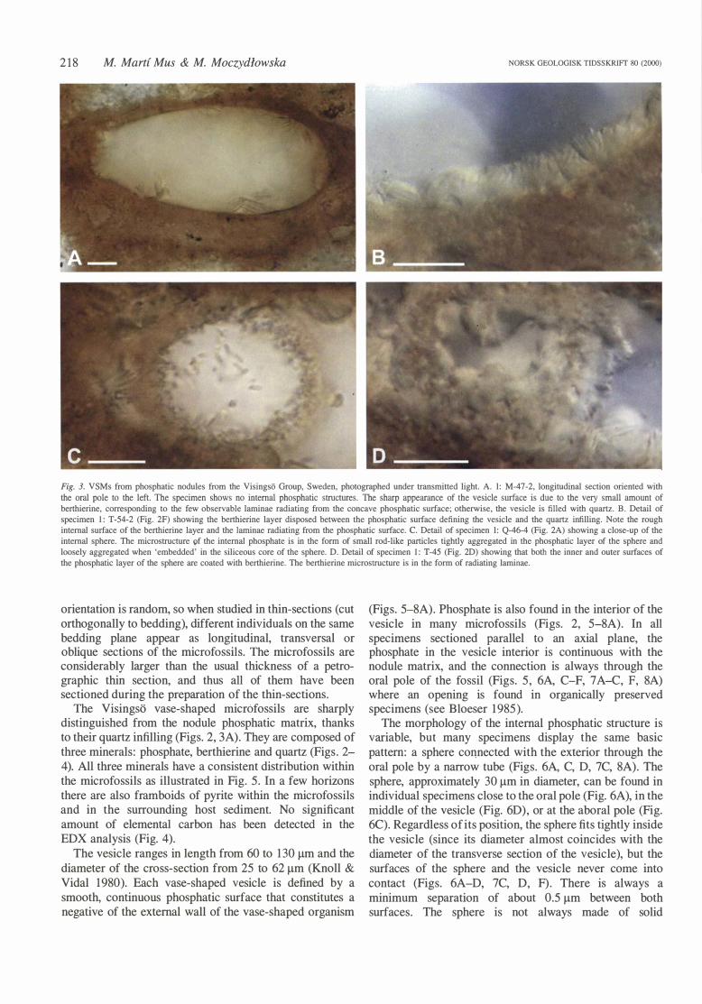
Fig. 3. VSMs from phosphatic nodules from the Visingso Group, Sweden, photographed under transmitted light. A. l: M-47-2, longitudinal section oriented with the oral pole to the left. The specimen shows no internal phosphatic structures. The sharp appearance of the vesicle surface is due to the very small amount of berthierine, corresponding to the few observable laminae radiating from the concave phosphatic surface; otherwise, the vesicle is filled with quartz. B. Detail of specimen 1: T-54-2 (Fig. 2F) showing the berthierine layer disposed between the phosphatic surface defining the vesicle and the quartz infilling. Note the rough internat surface of the berthierine layer and the laminae radiating from the phosphatic surface. C. Detail of specimen l: Q-46-4 (Fig. 2A) showing a close-up of the internat sphere. The microstructure of the internat phosphate is in the form of small rod-like particles tightly aggregated in the phosphatic layer of the sphere and loosely aggregated when 'embedded' in the siliceous core of the sphere. D. Detail of specimen 1: T-45 (Fig. 2D) showing that both the ioner and outer surfaces of the phosphatic layer of the sphere are coated with berthierine. The berthierine microstructure is in the form of radiating Iaminae.
orientation is random, so when studied in thin-sections (cut orthogonally to bedding), different individuals on the same bedding plane appear as longitudinal, transversal or oblique sections of the microfossils. The microfossils are considerably larger than the usual thickness of a petrographic thin section, and thus all of them have been sectioned during the preparation of the thin-sections.
The Visingso vase-shaped rnicrOfossils are sharply distinguished from the nodule phosphatic matrix, thanks to their quartz infilling (Figs. 2, 3A). They are composed of three minerals: phosphate, berthierine and quartz (Figs . 2-4). All three minerals have a consistent distribution within the microfossils as illustrated in Fig. 5. In a few horizons there are also framboids of pyrite within the microfossils and in the surrounding host sediment. No significant amount of elementa} carbon has been detected in the EDX analysis (Fig. 4).
The vesicle ranges in length from 60 to 130 J..lm and the diameter of the cross-section from 25 to 62 J..lm (Knoll & Vidal 1980). Each vase-shaped vesicle is defined by a smooth, continuous phosphatic surface that constitutes a negative of the extemal wall of the vase-shaped organism
(Figs. 5-8A). Phosphate is also found in the interior of the vesicle in many microfossils (Figs. 2, 5-8A). In all specimens sectioned parallel to an axial plane, the phosphate in the vesicle interior is continuous with the nodule matrix, and the connection is always through the oral pole of the fossil (Figs. 5, 6A, C-F, 7 A-C, F, 8A) where an opening is found in organically preserved specimens (see Bloeser 1 985).
The morphology of the intemal phosphatic structure is variable, but many specimens display the same basic pattem: a sphere co11nected with the exterior through the oral pole by a narrow tube (Figs. 6A, C, D, 7C, 8A). The sphere, approximately 30 J..lm in diameter, can be found in individual specimens dose to the oral pole (Fig. 6A), in the middle of the vesicle (Fig. 60), or at the aboral pole (Fig. 6C). Regardless of its position, the sphere fits tightly inside the vesicle (since its diameter almost coincides with the diameter of the transverse section of the vesicle), but the surfaces of the sphere and the vesicle never come into contact (Figs. 6A-D, 7C, D, F) . There is always a minimum separation of about 0.5 J..lm between both surfaces. The sphere is not always made of solid
NORSK GEOLOGISK TIDSSKRIFr 80 (2000)
Si
o
c
Si o
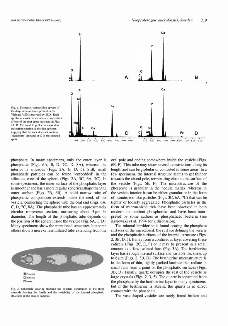
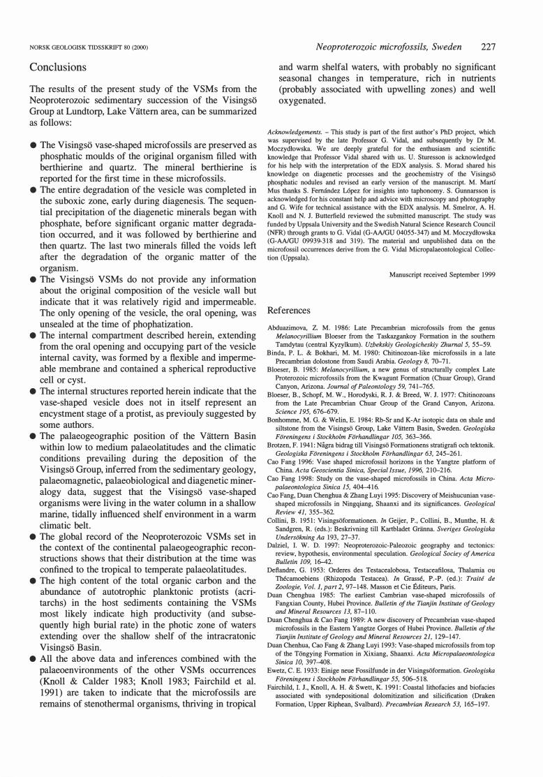
Fig. 4. Elementa! composition spectra of the diagenetic minerals present in the Visingso VSMs analysed by EDX. Each
spectrum shows the elementa! composition of one of the four spots indicated in Figs. 2A, B. The small C peaks correspond to the carbon coating of the thin-sections, implying that the rock does not contain 'significant' amounts of C in the selected spots.
1.00 2.00 3.00 4.00 5.00
phosphate. In many specimens, only the outer layer is phosphatic (Figs. 6A, B, D, 7C, D, 8A), whereas the interior is siliceous (Figs. 2A, B, D, 5). Still, small phosphatic particles can be found 'embedded' in the siliceous core of the sphere (Figs. 2A, 3C, 6A, 7C). In some specimens, the inner surface of the phosphatic layer is smoother and has a more regular spherical shape than the outer surface (Figs. 2B, 6B). A solid narrow tube of phosphatic composition extends inside the neck of the vesicle, connecting the sphere with the oral end (Figs. 6A, C, D, 7C, 8A). The phosphatic tube has an approximately circular transverse section, measuring about 5 J.lm in diameter. The length of the phosphatic tube depends on the position of the sphere inside the vesicle (Fig. 6A, C, D). Many specimens show the mentioned structures, but some others show a more or less inftated tube extending from the
.......... 1m Bethlerine
[i]QuoriZ
Fig. 5. Schematic drawing showing the constant distribution of the three
minerals forming the fossils and the variability of the internal phosphatic structures in the studied samples.
Neoproterozoic microfossils, Sweden 219
Ca
p
A B
l Ca
Fe p
o
c o
6.00 7.00 8.00 9.00 1.00 2.00 3.00 4.00 5.00 6.00 7.00 8.00 9.00
oral pole and ending somewhere inside the vesicle (Figs. 6E, F). This tube may show several constrictions along its length and can be g1obu1ar or contorted in some areas. In a few specimens, the internal structure seems to get thinner towards the aboral pole, terminating close to the surface of the vesicle (Figs. 6E, F). The rnicrostructure of the phosphate is granu1ar in the nodule matrix, whereas in the vesicle interior it can be either granular or in the form of rninute, rod-like particles (Figs. 3C, 6A, 7C) that can be tightly or loosely aggregated. Phosphatic particles in the form of rnicron-sized rods have been observed in both modem and ancient phosphorites and have been interpreted by some authors as phosphatized bacteria (see Krajewski et al. 1994 for a discussion).
The mineral berthierine is found coating the phosphate surfaces of the rnicrofossil: the surface defining the vesicle and the phosphatic surfaces of the internal structure (Figs. 2, 3B, D, 5). It may form a continuous layer covering these entirely (Figs. 2C, E, F) or it may be present in a small amount as a few iso1ated fans (Fig. 3A). The berthierine 1ayer has a rough internal surface and variable thickness up to 6 J.lm (Figs. 2, 3B , D). The berthierine rnicrostructure is in the form of thin, tightly packed larninae that radiate in small fans from a point on the phosphatic surfaces (Figs. 3B , D). Finally, quartz occupies the rest of the vesicle as large crystals (Figs. 2, 3, 5). The quartz is separated from the phosphate by the berthierine layer in many specimens, but if the berthierine is absent, the quartz is in direct contact with the phosphate.
The vase-shaped vesicles are rarely found broken and
220 M. Mart{ Mus & M. Moczydlowska NORSK GEOLOGISK TIDSSKRIFT 80 (2000)
NORSK GEOLOGISK TIDSSKRIFT 80 (2000)
are never collapsed or deformed. In a few broken specimens, the phosphatic surface defining the vesicle shows a brittle fracture with the original curvature of the broken pieces retained (Fig. 2H, 7E, F). The fractured fragments are always directed towards the vesicle interior and, in most cases, there is a large clast in the host sediment just above or below the fractured area. The shape and size of the clast seem to correspond to the shape and size of the fracture. In specimen 2: F-62-4 (Figs. 2H, 7F) the fracture determines the shape of the intemal phosphatic structure that seems accomrnodated to the shape of the broken vesicle. The internal structure is compressed against the side of the vesicle opposite to the breakage and shows a constriction coinciding with the most inwardly protruding broken fragment. Despite this compression, the separation distance between the intemal structure and the vesicle surface is kept.
Taphonomic history of the Visingso VSMs
The position and distribution of the VSMs in the sediment are compatible with them being suspended in the water column before deposition. The microstructure and distribution of the minerals as well as the morphology of the different mineralized parts of the microfossil allow the reconstruction of the diagenetic history of the microfossils (Fig. 9). The fact that the phosphate replicates the external morphology of the vase-shaped vesicles (Figs. 6, 7) so accurately suggests that precipitation of phosphate began in the sediment when the external wall of the organism was still intact and formed its external mould (Figs. 8B, 9C). The continuity of the phosphate between the nodule matrix and the vesicle interior through the oral pole indicates that the oral opening was unsealed, allowing pore waters to enter the vesicle. The consistent and precise morphology of the intemal phosphatized area, as well as the fact that phosphate never fills the whole vesicle cavity, indicates that phosphate precipitated inside an intemal compartment and formed its internal mould. Several features observable in the broken specimens (i.e. inwardly directed fractured fragments, presence of a clast in close vicinity to the broken area, the corresponding shape and size of the clast and the broken area; Figs. 2H, 7E, F) indicate that the fracture was probably formed during sediment compaction by the clast pressing against the vesicle. This implies that the fracturing occurred befare the nodule was formed and the sediment was still susceptible to compression. It is possible, however, that phosphate precipitation bad started
Neoproterozoic microfossils, Sweden 221
locally. In specimen 2: F-62-4 (Figs. 2H, 7F), the mould of the internal compartment reflects a p las tie deformation that occurred as a response to the pressure created by the inwardly directed fracture. This deformation must have happened before phosphate precipitated in the internal compartment, since a phosphatic body is not susceptible to plastic deformation. This, along with the variability in shape and position of the internal mould in different specimens indicates that the intemal compartment was defined by a flexible membrane that could be deformed and displaced inside the vesicle. The vesicle cavity must have been occupied either by fluid or was empty since the internal compartment could be displaced within it. The cavity may have escaped phosphatization by being sealed off from the pore waters by the vesicle wall and the compartment membrane (Figs. 8C, 9C). The distance between the internal and external moulds (Figs. 6A-D, 7C, D, F) is interpreted to represent the space that was occupied, when phosphatization took place, by the vesicle wall plus the membrane of the intemal compartment (Fig. 9C). The existence of this space supports the suggestion that the wall was intact when phosphate precipitation took place (see above). The internal compartment membrane appears to have been attached to the inner surface of the vesicle wall at the oral pole around the opening (Figs . 8C, 9C). In some specimens, the posterior end of the internal mould is dose to the vesicle surface at the aboral pole (Figs. 6E, F), which may indicate that the compartment membrane was locally attached to the vesicle wall also at that point (Fig. 8C). As mentioned above, in many specimens only the outer layer of the internal sphere is of phosphatic composition (Figs. 6A, B, D, 7C, D, 8A) and, at least in some specimens, the inner surface of this phosphatic layer is smoother and has a more regular shape than the outer one (Fig. 6B). This suggests that, at the time of phosphatization, the compartment was occupied by a spherical body. The inn er surface of the phosphatic la y er of the internal sphere would, thus, constitute the external mould of the spherical body (Figs. 8B, C, 9C, D). This observations also suggest that the spherical body had a smooth, rather finn surface (more so than the membrane of the compartment). Considering its location, shape and inferred surface features, this intemal body is here interpreted as a reproductive cell or cyst.
The vesicle wall of the vase-shaped organism was more rigid than the membrane of the intemal compartment because its external mould does not refiect any sort of plastic deformation. Rarely, the external mould shows brittle fractures . These fractures occurred early in the
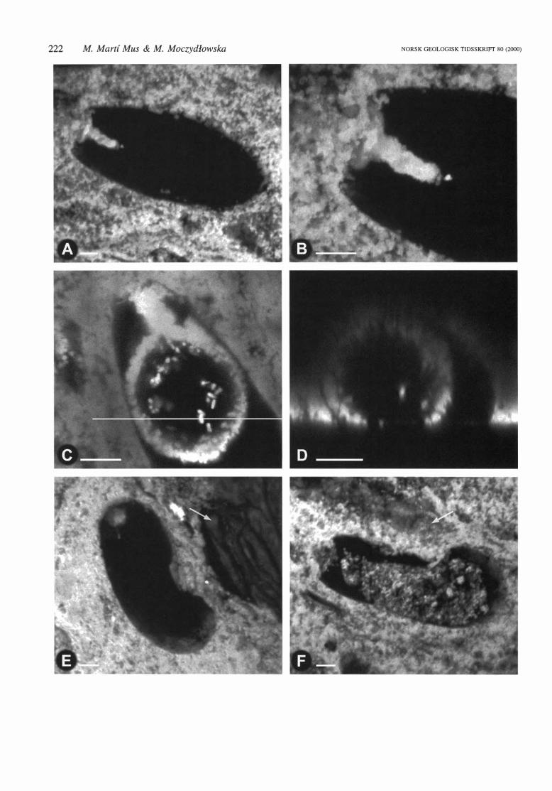
Fig. 6. Confocal microscope fluorescence irnages of VSMs from phosphatic nodules from !he Visingso Group, Sweden. Each image assembled from 90 horizontal scans, covering !he whole volume of !he specimen. Only !he phosphatic structures are seen. All specimens, except l: S-46 (B), which is an oblique section. are
oriented wiih !he oral end to !he left. A. 1: Q-46-4, longitudinal section of a VSMs showing !he morphology of !he intemal phosphatic structure. Note !he separation distance between the empty intemal sphere and !he vesicle surface, despite !he light fit of !he sphere inside !he vesicle. B. l: S-46, oblique section of a vesicle wiih
an empty intemal sphere, having a smooih and regular inner surface. The separation distance between !he outer surface of the sphere and the vesicle surface is observable. C. l: U-53-2, longitudinal section wiih a solid sphere connected wiih !he oral opening by a long tube. D. 1: T-45, longitudinal section showing an empty
in tema! sphere tightly fitted inside !he vesicle. The separation between !he sphere and !he vesicle surface is evident. E. l : T -45-2, longitudinal section wiih a tubular intemal structure wiih constrictions along its length and ending as a narrow thread in the aboral pole of !he vesicle. F. 1: T-54-2 longitudinal section showing a con
torted tu bul ar intern al structure ending as a narrow thread el ose to !he a bora! pole of !he vesicle.
222 M. Mart{ Mus & M. Moczydlowska NORSK GEOLOGISK TIDSSKRIFT 80 (2000)
NORSK GEOLOGISK TIDSSKRIFf 80 (2000)
taphonomic history of the microfossil (befare the phosphatic nodule was formed and befare phosphate precipitated inside the vesicle) at a time when the vesicle wall was still intact. It is thus possible that the brittle fractures were present in the original wall and therefore reflect the consistency of the original wall. Still, it is possible that phosphate precipitation had started locally, coating an unbroken vesicle wall with a thin, rigid phosphatic layer that was fractured under pressure deforming the original wall underneath. This second scenario is supported by the observation that, in the broken specimens, phosphate did not precipitate inside the vesicle through the broken area (as it could have done if the vesicle wall had an open fracture ). In the broken specimens, internal phosphatization is also restricted to the internal compartment (Fig. 7F), indicating that the only connection between the exterior and the interior of the vesicle was through the oral opening.
The pattern of crystal growth and the distribution of berthierine (Fig. 2, 3B, D, 5) indicate that it started to precipitate directly on the phosphatic surfaces. Because the phosphatic surfaces are moulds that formed having as a template biological structures, berthierine could have precipitated only when both the vesicle wall and the internal compartment membrane were degraded (Figs. 9D, E). The growth pattern of the berthierine, with thin larninae radiating irregularly towards the interior of the vesicle (Figs. 3B, D) indicates that the mineral growth was not restricted in this direction, and that berthierine growth preceded quartz precipitation. Quartz was the last mineral to precipitate inside the microfossil, filling the rest of the vesicle (Figs. 2, 3, 5, 9F) .
The taphonomic history of the Visingso VSMs is summarized in Fig 9. An early diagenetic precipitation of phosphate, befare significant degradation of organic matter, formed a mould of the parts of the organism that were exposed or connected to the exterior, and only the morphology of the phosphatized parts of the fossil contains relevant biological information. Both berthierine and quartz fill the void that was left after the complete degradation of the vase-shaped organism.
Morad & Al-Aasam ( 1994) studied the diagenetic history of the Visingso phosphatic nodules from a geochernical point of view. They inferred the sequence of authigenic mineral precipitation that began with francolite (the cryptocrystalline phosphate) was followed by berthierine (Fe-rich phyllosilicate) and quartz, and the burial depth at which the phosphate nodules grew to be
Neoproterozoic microfossils, Sweden 223
within several decimetres to metres below the sea floor. The taphonomic history of the vase-shaped microfossils proposed in the present work (Fig. 9) is in agreement with the reconstruction of the progressive burial-diagenetic evolution of the nodules. Taking the geochemical interpretation and the present reconstruction of the vase-shaped organisms' fossilization into account, it can be concluded that the complete degradation of the most resistant materials forming the vase-shaped organism (presumably the vesicle wall) must have taken place very early during the diagenetic history, in the suboxic diagenetic zone.
Palaeobiological reconstruction
Several conclusions about the morphology (Fig. 8C) and the properties of the original material of the vase-shaped organism can be drawn from the study of the Visingso V SMs, despite the fact that all the original material of the organism is gone. The vesicle wall was smooth and less than 0.5 jlm thick ( considerably thinner than the 5 to 7 jlm thick wall of the specimens described by Bloeser 1985 ; see below). It was relatively rigid and impermeable, at least to some ions. The only opening of the vesicle wall was at the oral pole. The oral opening was about 5 jlm in diameter. Inside the vase-shaped vesicle there was an internal compartment connected to the exterior through the oral opening and defined by a flexible membrane. This membrane was also impermeable to some ions. The internal compartment had an elaborate morphology, being tubular and narrow at the oral pole and extending into a more spacious spherical part that contained a reproductive cell or a cyst. The contained cell bad a smooth and regular spherical surface and a relatively finn wall. The membrane of the compartment may have been locally attached to the internal surface of the vesicle wall towards the aboral pole. The vesicle cavity (the space extending between the wall and the internal compartment) was empty or filled with fluid.
Biological affinities
The biological affinities of the VSMs remain uncertain, though several ideas have been put forward since the discovery of these microfossils. The morphological resemblance to the problematic group Chitinozoa was observed
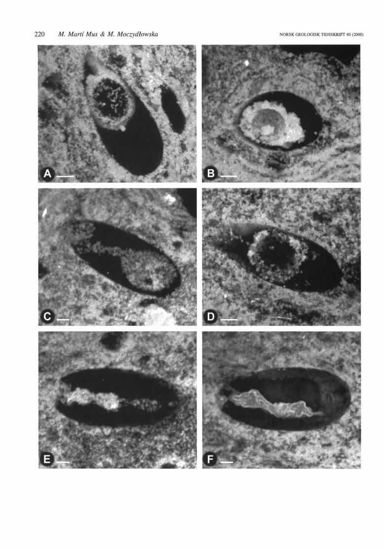
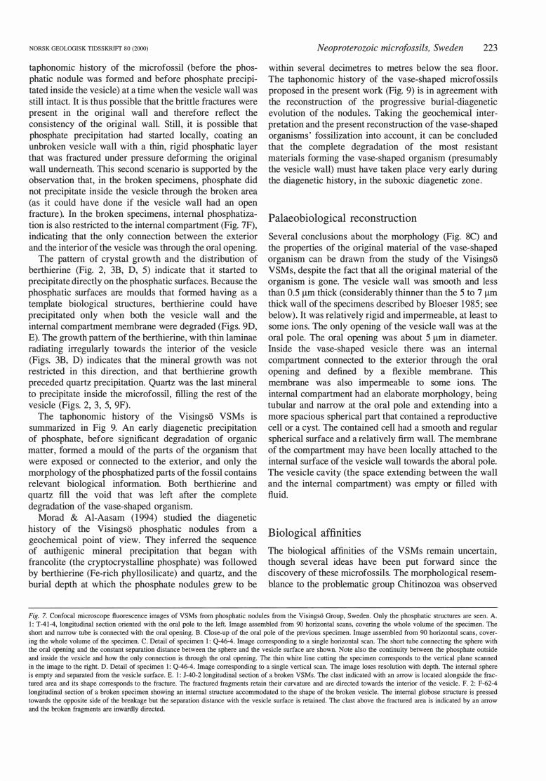
Fig. 7. Confocal microscope fluorescence images of VSMs from phosphatic nodules from the Visingsti Group, Sweden. Only the phosphatic structures are seen. A. l: T -41-4, longitudinal section oriented with the oral pole to the left. Image assembled from 90 horizontal scans, covering the whole volume of the specimen. The short and narrow tube is connected with the oral opening. B. Close-up of the oral pole of the previous specimen. Image assembled from 90 horizontal scans, covering the whole volume of the specimen. C. Detail of specimen l: Q-46-4. Image corresponding to a single horizontal scan. The short tube connecting the sphere with the oral opening and the constant separation distance between the sphere and the vesicle surface are shown. Note also the continuity between the phosphate outside and inside the vesicle and bow the only connection is through the oral opening. The thin white line cutting the specimen corresponds to the vertical plane scanned in the image to the right. D. Detail of specimen l: Q-46-4. Image corresponding to a single vertical scan. The image loses resolution with depth. The intemal sphere is empty and separated from the vesicle surface. E. l: J-40-2 longitudinal section of a broken VSMs. The clast indicated with an arrow is located alongside the fractured area and its shape corresponds to the fracture. The fractured fragments retain their curvature and are directed towards the interior of the vesicle. F. 2: F-62-4 longitudinal section of a broken specimen showing an intemal structure accommodated to the shape of the broken vesicle. The intemal globose structure is pressed towards the opposite side of the breakage but the separation distance with the vesicle surface is retained. The clast above the fractured area is indicated by an arrow and the broken fragments are inwardly directed.
224
A
M. Martf Mus & M. Moczydlowska
surface defining the vase-shaped vesicle
tube + phosphatic layer of the sphere
(internal phosphatic structure)
external mould of spherical cell
external mould of vesicle wall
internal mould of internal compartment
8
lumen of internal compartment
vesicle wall
oral opening ,.---.
internal compartment membrane
c
spherical cell (reproductive cell/ cyst)
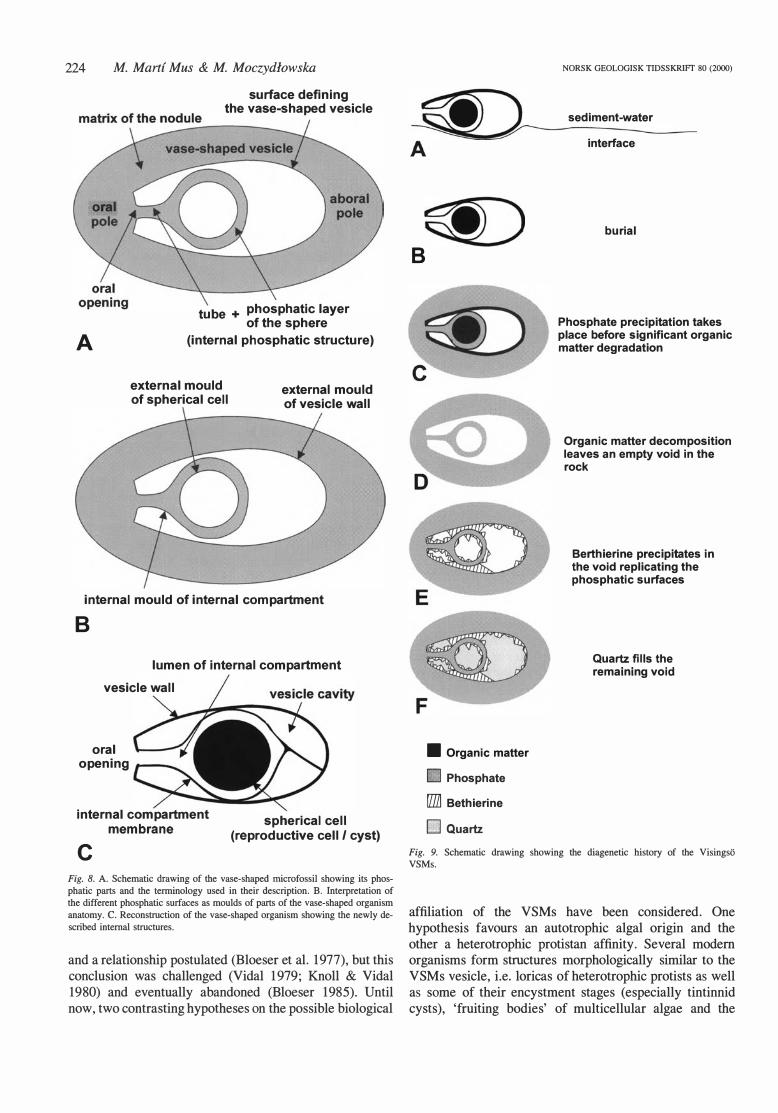
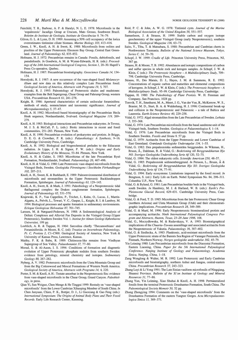
Fig. 8. A. Schematic drawing of the vase-shaped microfossil showing its phosphatic parts and the terminology used in their description. B. lnterpretation of the different phosphatic surfaces as moulds of parts of the vase-shaped organism anatomy. C. Reconstruction of the vase-shaped organism showing the newly described intemal structures.
and a relationship postulated (Bloeser et al. 1977), but this conclusion was challenged (Vidal 1 979; Knoll & Vidal 1980) and eventually abandoned (Bloeser 1985). Until now, two contrasting hypotheses on the possible biological
~ B
• Organic matter
11 Phosphate
rriJJ Bethierine
E5J Quartz
NORSK GEOLOGISK TIDSSKR!Ff 80 (2000)
sediment-water
interface
bu ria l
Phosphate precipitation takes place before significant organic matter degradation
Organic matter decomposition leaves an empty vold in the rock
Berthierine precipitates in the vold replicating the phosphatic surfaces
Quartz fills the remaining vold
Fig. 9. Schematic drawing showing the diagenetic history of the Visingso V SMs.
affiliation of the VSMs have been considered. One hypothesis favours an autotrophic algal origin and the other a heterotrophic protistan affinity. Several modem organisms form structures morphologically similar to the V SMs vesicle, i.e. loricas of heterotrophic protists as well as some of their encystment stages (especially tintinnid cysts), 'fruiting bodies' of multicellular algae and the
NORSK GEOLOGISK TIDSSKRIFT 80 (2000)
encystment stages of some unicellular algae. All these possibilities have been considered by different authors since VSMs have been suggested to represent ciliate protists of Tintinnida (Fairchild et al. 1 978), the oldest heterotrophic protists (Knoll & Vidal 1980), the reproductive cyst of walled amoebae (Schopf 1 999), loricas of testate amoebas (Porter & Knoll, in press), as well as the encystment stage of an alga (Bloeser 1985) or the fossilized sporangium of an alga (Horodyski 1993). The internat structures described herein indicate that the vaseshaped vesicle could not by itself represent an encystment stage, either of a unicellular algae or of a heterothrophic protist. More likely, it is interpretable as a 'fruiting body' of a multicellular alga, or the lorica of a heterotrophic protist.
Although no modem multicellular algae produce structures closely comparable to VSMs, the formation of saclike unilocular gametangia or sporangia (that contain, respectively, sexual or asexual reproductive cells) is very cornmon among modem multicellular algae. Sometimes these sac-like structures can detach from the thallus and thus contribute to the protection and dispersal of the reproductive cells. An example of an algal gametangium frequently found in the fossil record (and which could constitute a functional analogue of VSMs) is the charophyte oogonium ( cornmon in freshwater deposits from Devonian to present). The first fossil charophyte oogonium to be described was initially identified as a forarninifer (Tappan 1980), being an example of an easily-made misinterpretation led by the morphological similarities between the tests of some heterotrophic protists and the reproductive structures of some algal groups. As mentioned above, V SMs are considered to represent planktonic organisms and they have never been reported to be attached to other fossils. Still, despite this lack of evidence and though there are no exactly comparabte structures among modem atgae, the possibitity of them being reproductive structures of an extinct algat group cannot be ruted out (Horodyski 1993). It coutd be specutated that the internat structures described in this work may represent a single spherical reproductive cell hetd inside a spacious fruiting body by a membrane. The ability of the VSMs to float freety in the water column would have been part of the dispersal strategy of the algae.
Severat groups of heterotrophic protists, particutarly testate amoebae, tintinnids and some unilocutar foraminifera, form toricas that are also morphologically similar to the VSMs. Although some of the mentioned organisms (especialty some groups of testate amoebae) have very sophisticated mineralized tests, the three groups of protists mentioned have representatives that possess retativety simple sac-like organic tests. The living cells of both thecoamoebans and tintinnids have (unlike most foraminiferans) a very simple relation with their tests. The complete test is built after reproduction and does not grow during the lifetime of the organism. The cell is separated from the internal surface of the test, being attached to it only locally and reversibly so the test can readily be
Neoproterozoic microfossils, Sweden 225
discarded at some stage of the tife cycle of the organism. Both organisms are predators and capture their prey with specialized cellular structures extruded through the opening of their tests. Some species of testate amoebae have a triangular opening and others thecas with hexagonal transverse section (Deflandre 1 953; Loeblich & Tappan 1 964), both features comparabte with those characters found in V SMs (see Porter & Knoll, in press, for a detaited comparison between testate amoebae and VSMs).
In both thecoamoebans and tintinnids, the formation of a resistant cyst inside the test is cornmon and the lorica opening rna y be plugged during the encystment stage (Reid & John 1 978; Deflandre 1953). Since the living cell does not usually occupy the whote volume of the test (particularly in tintinnids it may occupy considerably less than half of the test volume; Gilron & Lynn 1 989), the resting cyst may also appear quite small in retation to the whote lorica votume. In some tintinnids the cyst may be enclosed within a thin, but resistant-to-decay, membrane that attaches the cyst to the torica (Reid & John 1 978). VSMs have been compared with heterotrophic protists on the basis of their external morphology, but the internat structures of the Visingso V SMs described herein are atso comparable with those found in some heterotrophic protists during the encysting stage of their tife cycle.
Alternatively, a comparison can be made between the aperturat apparatus of some unilocular forarninifera (Knight 1 986) and the internal structures of the Visingso VSMs. Some unilocutar forarninifera possess a narrow tube that projects from the aperture towards the inside of the theca. This particutar structure is sirnilar in morphology and dimensions to the tubular proximal part of the internat compartment in the Visingso VSMs.
Discussion
The Visingso VSMs were previousty considered to be preserved ' ... as chert, phosphate or, rarely, calcite casts of the original organisms ... ' (Knoll & Vidal 1980, p. 209). Our study shows they are preserved as complex phosphatic moulds. Calcite has not been found in the specimens studied. Neither the berthierine coating, nor the quartz filting alone can be regarded as casts. The 'siliceous filling' (berthierine + quartz) as a whole could be regarded as a east (a rep ti ca form ed ha ving a mould as a temp late) of the vesicle, but the presence of an internat phosphatic moutd in many specimens complicates the matter and therefore we have preferred to avoid the term 'east'.
Microfossils referred to as VSMs (and implied to bear similarities with Melanocyrillium) have been described also from several localities not listed in Table l. A critical evaluation of these microfossils indicates that, for various reasons, they are best regarded as unrelated to the specimens referred in Table l. Specimens reported from Brazil (Fairchild et al. 1978) should probabty be regarded as dubiofossils, since they do no meet any of the criteria required of unquestionable biogenic remains (Hofmann
226 M. Marti Mus & M. Moczydlowska
1972, 1 987). The specimens from the Doushantuo Fm reported by Duan & Cao (1989) have been reinterpreted as non-biological structures (Zhang 1994; Zhang et al. 1998). Specimens reported from Uzbekhistan (Abduazimova 1986) and the Proterozoic of China (Yin 1980; Zang & Walter 1992; Duan & Cao 1989) are too poorly preserved to allow comparisons. The specimens reported from India (Maithy & Babu 1989) and from the Dengying Fm (Zhang & Li 199 1 ) show distinct morphological features (e.g., wide, flaring oral ends) that have never been observed in an y of the populations listed in Tab le l. The occurrence of VSMs in the Uinta Mountain Group (Utah) has been reported (Nyberg 1982) but they have not been illustrated. Finally, several occurrences of VSMs have been reported from Lower Cambrian strata in China (Cao 1996, 1 998; Cao et al. 1995; Duan 1985; Duan et al. 1 993). These specimens have recently been reinterpreted as chancelloriid spicules (Qian et al. 1999)
Regarding the internal morphology of the VSMs, Bloeser ( 1 985) described and illustrated an irregular, polygonal network formed by thin carbonaceous lamellae inside the vesicle of some specimens from the K wagunt Formation. She considered this reticulate-like organization to be the result of the diagenetic alteration of organic matter that originally could have been present inside the vesicle in the form of cell or endospore walls . The presence of internal carbonaceous membranes inside the VSM vesicle is compatible with the results presented in the present work, since we also report the existence of membranous structures inside the VSMs which could have suffered different alterations in various sedimentary environments. Horodyski ( 1987, 1993) reported the existence of small, organic-walled spheroids resembling acritarchs inside the vesicle of VSMs from the same formation. This led him to interpret Melanocyrillium as a fossilized algal sporangium. Since only one specimen is figured by Horodyski ( 1993; a transmitted light photograph of a thin-section) containing the reported spheroids, it is difficult to assess the significance of this feature (see Porter & Knoll in press for a re-evaluation of these internal structures) .
Another disputable aspect of VSM morphology is the presence of an operculum sealing the oral opening. Only Bloeser ( 1985) has figured some specimens from the Kwagunt Fm with their opening plugged by a more or less amorphous body. For the Visingso VSMs it has been argued herein that, at least at the time of their phosphatization, the oral opening was open (at }east in most of the specimens) and connecting the internal compartment cavity with the external environment. The oral opening could, certainly, have been open or plugged in different specimens, depending on the stage in the life cycle of the organism.
The composition of the wall of V SMs has been a subject of controversy. The presence of a mineralized wall has been considered previously (Ewetz 1933; Vidal 1 984, 1994; Vidal & Martf 1 996) . Ewetz ( 1933) reported the presence of a 'shell' of even thickness with a smooth
NORSK GEOLOGISK TIDSSKRIFT 80 (2000)
external and a rough internat surface in Visingso VSMs. Knoll & Vidal ( 1 980, p. 209), studying the populations from the same occurrenee, also deseribed the presenee of a 'wall zone [ . . . ] present at the periphery of eaeh east' . In both studies the 'wall' was eonsidered to be of phosphatic composition, and Knoll & Vidal ( 1980, p. 209) commented that ' . . . the present mineral eomposition [ . . . ] may be of diagenetie origin' . There is little doubt that these authors were referring to the herein ealled 'berthierine la y er' , whieh eoats the fossil phosphatie surfaees. It has been argued earlier in this paper that this layer was precipitated during diagenesis, once the original wall material was completely degraded. This layer is neither a replacement nor a east of the original wall. It is a void coating and, therefore, uninformative regarding the original wall composition or structure. Another argument used to support the possible existence of a mineralized wall in VSMs is the observation that they are always preserved uncompressed and undeformed (Vidal 1 994; Vidal & Marti 1996) . Since some biogenic organic materials, such as lignin or chitin, are rigid, this observation alone does not confirm the presence of a mineralized wall. VSMs have been recovered as three-dimensional organic vesicles in acid-resistant residues in two localities (Bloeser et al. 1 977; Vidal 1 979; Vidal & Ford 1985; Bloeser 1985). In palynologieal preparations they are observed as uncompressed vesicles, even when the co-occurring organicwalled microfossils (e.g. acritarchs, algal remains or bacterial sheaths) are eollapsed or compressed (Vidal 1979; Vidal & Ford 1 985). This suggests that the vesicle wall of V SMs was originally organic, recalcitrant and rigid (Bloeser 1985). It was thus paradoxical that in many other oceurrenees the wall seemed to be partially or completely degraded and the VSMs were preserved as casts and moulds. This second type of preservation also suggests the presence of a rigid wall, but either composed of readily degradable organic matter or mineralized (and thus susceptible to dissolution) . In the Kwagunt Fm, VSMs have been reported to occur both as organic vesicles (Bloeser et al. 1977, Bloeser 1985, Vidal & Ford 1 985) and as moulds and easts (Horodyski 1987, 1 993), depending on the lithology of the host rock. This further suggests that the differenees in preservation of VSMs were not due to the different original composition of the wall in different populations of VSMs, but rather to the partieular diagenetic processes undergone by the vesicles in different sedimentary environments. Porter & Knoll (in press), after a study of new populations of VSMs from the Kwagunt Formation and a re-evaluation of the observations of Bloeser ( 1 985), have suggested that the organic wall of the specimens reported by Bloeser ( 1985) does not represent the original vesicle wall, but rather was formed by organic debris that eoaleseed around casts. This suggestion may explain the previously mentioned paradox regarding the preservation of V SMs, but it remains unclear bow organic debris could form vesicles with coherent, continuous walls that preserve detailed morphological information and survive the proeess of acid maceration (Bloeser 1985).
NORSK GEOLOGISK TIDSSKR!Ff 80 (2000)
Conclusions
The results of the present study of the VSMs from the Neoproterozoic sedimentary succession of the Visingso Group at Lundtorp, Lake Vattem area, can be summarized as follows:
• The Visingso vase-shaped microfossils are preserved as phosphatic moulds of the original organism filled with berthierine and quartz. The mineral berthierine is reported for the first time in these microfossils.
e The entire degradation of the vesicle was completed in the suboxic zone, early during diagenesis. The sequential precipitation of the diagenetic minerals began with phosphate, before significant organic matter degradation occurred, and it was followed by berthierine and then quartz. The last two minerals filled the voids left after the degradation of the organic matter of the organism.
• The Visingso VSMs do not provide any information about the original composition of the vesicle wall but indicate that it was relatively rigid and impermeable. The only opening of the vesicle, the oral opening, was unsealed at the time of phophatization.
e The internat compartment described herein, extending from the oral opening and occupying part of the vesicle internat cavity, was formed by a ftexible and impermeable membrane and contained a spherical reproductive cell or cyst.
• The internat structures reported herein indicate that the vase-shaped vesicle does not in itself represent an encystment stage of a protist, as previouly suggested by some authors.
• The palaeogeographic position of the Vattern Basin within low to medium palaeolatitudes and the climatic conditions prevailing during the deposition of the Visingso Group, inferred from the sedimentary geology, palaeomagnetic, palaeobiological and diagenetic mineralogy data, suggest that the Visingso vase-shaped organisms were living in the water column in a shallow marine, tidally inftuenced shelf environment in a warm climatic belt.
• The global record of the Neoproterozoic VSMs set in the context of the continental palaeogeographic reconstructions shows that their distribution at the time was confined to the tropical to temperate palaeolatitudes.
e The high content of the total organic carbon and the abundance of autotrophic planktonic protists (acritarchs) in the host sediments containing the VSMs most likely indicate high productivity (and subsequently high burial rate) in the photic zone of waters extending over the shallow shelf of the intracratonic Visingso Basin.
• All the above data and inferences combined with the palaeoenvironments of the other VSMs occurrences (Knoll & Calder 1983; Knoll 1 983; Fairchild et al. 199 1 ) are taken to indicate that the microfossils are remains of stenothermal organisms, thriving in tropical
Neoproterozoic microfossils, Sweden 227
and warm shelfal waters, with probably no significant seasonal changes in temperature, rich in nutrients (probably associated with upwelling zones) and well oxygenated.
Acknowledgements. - This study is part of the first author' s PhD project, which was supervised by the late Professor G. Vida!, and subsequently by Dr M. Moczydlowska. We are deeply grateful for the enthusiasm and scientific knowledge that Professor Vida! shared with us. U. Sturesson is acknowledged for his help with the interpretation of the EDX analysis. S. Morad shared his knowledge on diagenetic processes and the geochemistry of the Visingso phosphatic nodules and revised an early version of the manuscript. M. Martf Mus thanks S. Fermindez L6pez for insights into taphonomy. S. Gunnarsson is acknowledged for his constant help and advice with microscopy and photography and G. Wife for technical assistance with the EDX analysis. M. Smelror, A. H. Knoll and N. J. Butterfield reviewed the submitted manuscript. The study was funded by Uppsala University and the Swedish Natura! Science Research Council (NFR) through grants to G. Vida! (G-AA/GU 04055-347) and M. Moczydlowska (G-AA/GU 09939-318 and 319) . The material and unpublished data on the microfossil occurrences derive from the G. Vida! Micropalaeontological Collection (Uppsala).
Manuscript received September 1999
References
Abduazimova, Z. M. 1986: Late Precambrian microfossils from the genus Melanocyrillium Bloeser from the Taskazgankoy Forrnation in the southern Tamdytau (central Kyzylkum). Uzbekskiy Geologicheskiy Zhumal 5, 55-59.
Binda, P. L. & Bokhari, M. M. 1980: Chitinozoan-like microfossils in a late Precambrian dolostone from Saudi Arabia. Geology 8, 70-71.
Bloeser, B. 1985: Melanocyrillium, a new genus of structurally complex Late Proterozoic microfossils from the Kwagunt Formation (Chuar Group), Grand Canyon, Arizona. Journal of Paleontology 59, 741-765.
Bloeser, B., Schopf, M. W., Horodyski, R. J. & Breed, W. J. 1977: Chitinozoans from the Late Precambrian Chuar Group of the Grand Canyon, Arizona. Science 195, 676-679.
Bonhomme, M. G. & Welin, E. 1984: Rb-Sr and K-Ar isotopic data on shale and siltstone from the Visingso Group, Lake Vattern Basin, Sweden. Geologiska
Foreningens i Stockholm Forhandlingar 105, 363-366. Brotzen, F. 1941: Några bidrag till Visingso Formationens stratigrafi och tektonik.
Geologiska Foreningens i Stockholm Forhandlingar 63, 245-261. Cao Fang 1996: Vase shaped microfossil horizons in the Yangtze p1atform of
China. Acta Geoscientia Sinica, Special Issue, 1996, 210-216. Cao Fang 1998: Study on the vase-shaped microfossils in China. Acta Micro
palaeontologica Sinica 15, 404-416. Cao Fang, Duan Chenghua & Zhang Luyi 1995: Discovery of Meishucunian vase
shaped microfossils in Ningqiang, Shaanxi and its significances. Geological
Review 41, 355-362. Collini, B. 1951: Visingsoformationen. In Geijer, P., Collini, B., Munthe, H. &
Sandgren, R. (eds.): Beskrivning till Kartbladet Granna. Sveriges Geologiska Undersokning Aa 193, 27-37.
Dalziel, l. W. D. 1997: Neoproterozoic-Paleozoic geography and tectonics: review, hypothesis, environmental speculation. Geological Sociey of America Bulletin 109, 16-42.
Deflandre, G. 1953: Orderes des Testacealobosa, Testaceafilosa, Thalarnia ou Thecamoebiens (Rhizopoda Testacea). In Grasse, P.-P. (ed.): Traite de Zoologie, Vol. l, part 2, 97-148. Masson et Cie Editeurs, Paris.
Duan Chenghua 1985: The earliest Cambrian vase-shaped microfossils of Fangxian County, Hubei Province. Bulletin of the Tianjin Institute ofGeology and Mineral Resources 13, 87-110.
Duan Chenghua & Cao Fang 1989: A new discovery of Precambrian vase-shaped microfossils in the Eastern Y angtze Gorges of Hubei Province. Bulletin of the
Tianjin Institute ofGeology and Mineral Resources 21, 129--147. Duan Chenhua, Cao Fang & Zhang Luyi 1993: Vase-shaped microfossils from top
of the Tongying Formation in Xixiang, Shaanxi. Acta Micropalaeontologica
Sinica JO, 397-408. Ewetz, C. E. 1933: Einige neue Fossilfunde in der Visingsoformation. Geologiska
Foreningens i Stockholm Forhandlingar 55, 506--518. Fairchild, l. J., Knoll, A. H. & Swett, K. 1991: Coastal lithofacies and biofacies
associated with syndepositional dolomitization and silicification (Draken Formation, Upper Riphean, Svalbard). Precambrian Research 53, 165-197.
228 M. Mart{ Mus & M. Moczydlowska
Fairchild, T. R., Barbour, A. P. & Haralyi, N. L. E. 1978: Microfossils in the 'eopaleozoic' Jacadigo Group at Urucum, Mato Grosso, Southwest Brazil. Boletim do lnstituto de Geologia, lnstituto de Geociencias 9, 74-79.
Gilron, G. L. & Lynn, D. H. 1989: Assurning a 50% cell occupancy of the lorica overestimates tintinnine ciliate biomass. Marine Biology 103, 413-416.
Green, J. W., Knoll, A. H. & Swett, K. 1988: Microfossils from oolites and pisolites of the Upper Proterozoic Eleonore Bay Group, Central East Greenland. Journal of Paleontology 62, 835-852 .
Hofmann, H. J. 1972: Precambrian remains in Canada: Fossils, dubiofossils, and pseudofossils. In Goodwin, A. M. & Wynne-Edwards, H. R. (eds.): Proceed
ings of the 24th International Geological Congress, Section l, 20-30. Hapell's Press Co-operative, Quebec.
Hofmann, H. J. 1987: Precambrian biostratigraphy. Geoscience Canada 14, 134-154.
Horodyski, R. J. 1987: A new occurrence of the vase-shaped fossil Melanocyr
illium and new data on this relatively complex Late Precambrian fossil. Geological Society of America, Abstracts with Programs 19, 5, 707.
Horodyski, R. J. 1993: Paleontology of Proterozoic shales and mudstones: examp1es from the Belt Supergroup, Chuar Group and Pahrump Group, western USA. Precambrian Research 61, 241-278.
Knight, R. 1986: Apertural characteristics of certain unilocular forarninifera: methods of study, nomenc1ature and taxonornic significance. Journal of
Micropalaeontology 5, 37-47. Knoll, A. H. 1982: Microfossil-based biostratigraphy of the Precambrian Hecla
Hoek sequence, Nordaustlandet, Svalvard. Geological Magazine 119, 269-279.
Knoll, A. H. 1983: Biological interactions and Precambrian eukaryotes.ln Tevesz, M. J. S. & McCall, P. L. (eds.): Biotic interactions in recent and fossil communities, 251-283. Plenum, New York.
Knoll, A. H. 1990: Precambrian evolution of prokaryotes and protists. In Briggs, D. E. G. & Crowther, P. R. (eds.): Palaeobiology: A Synthesis, 9-16. Cambridge University Press, Cambridge.
Knoll, A. H. 1992: Biological and biogeochernical pre1udes to the Ediacaran radiation. In Lipps, J. H. & Signor, P. W. (eds.): Origins and Early
Evolutionary His tory of the Metazoa, 53-84. Plenum, New Y ork. Knoll, A. H. & Calder, S. 1983: Microbiotas of the late Precambrian Rysii
Forrnation, Nordaustlandet, Svalbard. Palaeontology 26, 467-496. Knoll, A. H. & Vida!, G. 1980: Late Proterozoic vase-shaped rnicrofossils from the
Visingso Beds, Sweden. Geologiska Foreningens i Stockholm Forhand/ingar
102, 207-211. Knoll, A. H., Sweet, K. & Burkhardt, E. 1989: Paleoenvironmental distribution of
rnicrofossils and stromatolites in the Upper Proterozoic Backlundtoppen Formation, Spitsbergen. Journal of Paleontology 63, 129-145.
Knoll, A. H., Swett, K. & Mark, J. 1991: Paleobiology of a Neoproterozoic tida! fiatllagoonal complex: the Draken conglomerate formation, Spitsbergen. Journal of Paleontology 65, 531-570.
Krajewski, K. P., Van Cappellen, P., Trichet, J., Kuhn, 0., Lucas, J., MartfnAlgarra, A., Prevot, L., Tewari, V. C., Gaspar, L., Knight, R. l. & Lamboy, M. 1994: Biological processes and apatite formation in sedirnentary environments. Eclogae Geologicae Helveatiae 87, 701-745 .
Larssen, M. & Nørgaard-Pedersen, N. 1988: A Sedimentological Analysis of Deltaic Complexes and Alluvial Fan Deposits in the Visingsii Group (Upper Proterozoic) , Southem Sweden Vol. l. Institut for Almen Geologi Københavns Universitet, 199 pp.
Loeblich, A. R. & Tappan, H. 1964: Sarcodina chiefiy 'Thecamoebians' and Foraminiferida. In Moore, R. C. (ed): Treatise on lnvertebrate Paleontology, Pt. C, Protista 2, C1- C900. Geological Society of America, New York & University of Kansas Press, Lawrence, Kansas.
Maithy, P. K. & Babu, R. 1989: Chitinozoa-like remains from Vindhyan Supergroup of Son Valley. Palaeobotanist 37, 77-80.
Morad, S. & Al-Aasm, I. S. 1994: Conditions of formation and diagenetic evolution of Upper Proterozoic phosphate nodules from southern Sweden: evidence from petrology, mineral chemistry and isotopes. Sedimentary
Geology 88, 267-282. Nyberg, A. V. 1982: Proterozoic microfossils from the Uinta Mountain Group and
from the Big Cottonwood and Mescal Formations of Western North America. Geological Society of America, Abstracts with Programs 14, 4, 220.
Porter, S. M. & Knoll, A. H.: Testate amoebae in the Neoproterozoic Era: evidence from vase-shaped microfossils in the Chuar Group, Grand Canyon. Paleobiology, in press.
Qian Yi, Sun Weiguo, Chen Menge & He Tinggui 1999: Restudy on 'vase-shaped rnicrofossils' from the Lower Cambrian Xihaoping Member of South China. In Chen Junyuan, Chien, P. K., Bottjer, D. J., Li Guoxiang & Gao Feng (eds.): International Symposium: The Origins of Animal Body Plans and Their Fossil
Records. Early Life Research Center, Kunming.
NORSK GEOLOGISK TIDSSKRIFT 80 (2000)
Reid, P. C. & John, A. W. G. 1978: Tintinnid cysts. Journal of the Marine
Biological Association of the United Kingdom 58, 551-557. Samuelsson, J. & Strauss, H. 1999: Stable carbon and oxygen isotope
geochemistry of the upper Visingsii Group (earl y Neoproterozoic ), southem Sweden. Geological Magazine 13 6, 63-73.
Saito, Y., Tiba, T. & Matsubara, S. 1988: Precambrian and Cambrian cherts in Northwestem Tasmania. Bulletin of the National Science Museum, Tokyo,
Series C. 14, 59-70. Schopf, J. W. 1999: Cradle of Life. Princeton University Press, Princeton, NJ,
367 pp. Strauss, H. & Moore, T. B. 1992: Abundances and isotopic compositions of carbon
and sulfur species in whole rock and kerogen samples. In Schopf, J. W. & Klein, C (eds.): The Proterozoic biosphere- A Multidisciplinary Study, 709-798. Cambridge University Press, Cambridge.
Strauss, H., Des Marais, D. J., Hayes, J. M. & Summons, R. E. 1992: Concentrations of organic carbon and maturities and elementa! compositions of kerogens. In Schopf, J. W. & Klein, C (eds.): The Proterozoic biosphere- A
Multidisciplinary Study, 95-99. Cambridge University Press, Cambridge. Tappan, H. 1980: The Paleobiology of Plant Protists. W. H. Freeman and
Company, San Francisco, 1028 pp. Torsvik, T.H., Smethurst, M. A., Meert, J. G., Van der Voo, R., McKerrow, W. S.,
Brasier, M. D., Sturt, B. A. & Walderhaug, H. J. 1996: Continental break-up and collision in the Neoproterozoic and Palaeozoic - a tale of Baltica and Laurentia. Earth-Science Reviews 40, 229-258 .
Vida!, G. 1972: Algal stromatolites from the Late Precambrian of Sweden. Lethaia
5, 353-368 . Vi dal, G. 197 4: Late Precambrian microfossils from the basal sandstone unit of the
Visingso beds, Southern Sweden. Geologica et Palaeontologica 8, 1-14. Vida!, G. 1976: Late Precambrian rnicrofossils from the Visingso Beds in
Southern Sweden. F ossils and Strata 9, 57 pp. Vida!, G. 1979: Acritarchs from the Upper Proterozoic and Lower Cambrian of
East Greenland. Grønlands Geologiske Undersøgelse 134, 1-55. Vida!, G. 1982: Den prepaleozoiska sedirnentiira berggrunden. In Wikman, H.,
Bruun, Å., Dah1man, B. & Vida!, G.: Beskrivning till Berggrundskartan Hjo
NO. Sveriges Geologiska Undersokning Serie Af 120 , 52-75. Vida!, G. 1984: The oldest eukaryotic cells. Scientific American 250, 48-57. Vida!, G. 1985: Prepaleozoisk sedimentberggrund. In Persson, L., Bruun, Å &
Vida!, G.: Beskrivning till Berggrundskartan Hjo SO. Sveriges Geologiska
Undersokning Serie Af 134, 77-91. Vida!, G. 1994: Early ecosystems: Lirnitations imposed by the fossil record. In
Bengtson, S. (ed.): Early Life on Earth. Nobel Symposium No. 84 , 298-311. Columbia U.P., New York.
Vida!, G. & Bylund, G. 1981: Late Precambrian boulder beds in the Visingso beds, south Sweden. In Hambrey, M. J. & Harland, W. B. (eds.): Earth's Pre
Pleistocene Glacial Record, 629-631. Cambridge University Press, Cambridge.
Vida!, G. & Ford, T.D. 1985: Microbiotas from the late Proterozoic Chuar Group (northem Arizona) and Uinta Mountain Group (Utah) and their chronostratigraphic irnplications. Precambrian Research 28, 349-389.
Vida!, G. & Martf, M. 1996: Early Neoproterozoic biornineralized protists and accompanying acritarchs. Ninth International Palynological Congress Program and Abstracts, Huston, Texas, 23-28 lune 1996, 168.
Vida!, G., Moczydlowska, M. & Rudavskaya, V. A. 1993: Biostratigraphical implications of the Chuaria-Tawuia assemblage and associated acritarchs from the Neoproterozoic of Yakutia. Palaeontology 36, 387-402.
Vida!, G. & Sied1ecka, A. 1983: Planktonic, acid-resistant microfossils from the Upper Proterozoic strata of the Barents Sea Region of Varanger Peninsula, East Finmark, Northem Norway. Norges geologiske undersøkelse 382, 45-79.
Yin Leirning 1980: Late Precambrian rnicrofossils from the Diaoyutai Formation, Eastern Liaoning, China. Paper for the 5th International Palynological
Conference. Nanjing Institute of Geology and Palaeontology. Academia
Sinica, Nanjing, China, 1-18. Zang Wenglong & Walter, M. R. 1992: Late Proterozoic and Early Cambrian
microfossils and biostratigraphy, northem Anhui and Jiangsu, central-eastern China. Precambrian Research 57, 243-323.
Zhang Luyi & Li Y ong 1991: The Late Sinian vasiform rnicrofossils of Ningqiang, Shaanxi Province. Bulletin of the Xi'an Institute of Geology and Mineral
Resources 31, 77-86. Zhang Yun, Yin Leirning, Xiao Shuhai & Knoll, A. H. 1998: Perrnineralized
fossils from the terminal Proteroroic Doushantuo Formation, South China. The
Paleontological Society Memoir 50, 52 pp. Zhang Zhongying 1994: Comments on the 'vase-shaped microfossils' from the
Doushantou Formation of the eastem Y angtze Gorges. Acta Micropalaeonto
logica Sinica Il, 369-371.