Embed Size (px)
Citation preview

Biochem. J. (2011) 439, 505–516 (Printed in Great Britain) doi:10.1042/BJ20101650 505
Interaction between the SH3 domain of Src family kinases and theproline-rich motif of HTLV-1 p13: a novel mechanism underlying deliveryof Src family kinases to mitochondriaElena TIBALDI*, Andrea VENERANDO*†, Francesca ZONTA*, Carlo BIDOIA‡, Elisa MAGRIN*, Oriano MARIN*,Antonio TONINELLO*, Luciana BORDIN*, Veronica MARTINI†, Mario Angelo PAGANO*1 and Anna Maria BRUNATI*1,2
*Department of Biological Chemistry, University of Padua, Viale G. Colombo 3, 35131 Padua, Italy, †Venetian Institute of Molecular Medicine (VIMM), Via G. Orus, 2, 35129 Padua,Italy, and ‡Centre for Research in Infectious Diseases, School of Medicine and Medical Science, University College Dublin, Belfield, Dublin 4, Ireland
The association of the SH3 (Src homology 3) domain of SFKs(Src family kinases) with protein partners bearing proline-richmotifs has been implicated in the regulation of SFK activity, andhas been described as a possible mechanism of relocalization ofSFKs to subcellular compartments. We demonstrate in the presentstudy for the first time that p13, an accessory protein encodedby the HTLV-1 (human T-cell leukaemia virus type 1), bindsthe SH3 domain of SFKs via its C-terminal proline-rich motif,forming a stable heterodimer that translocates to mitochondriaby virtue of its N-terminal mitochondrial localization signal. Asa result, the activity of SFKs is dramatically enhanced, with asubsequent increase in mitochondrial tyrosine phosphorylation,and the recognized ability of p13 to insert itself into the
inner mitochondrial membrane and to perturb the mitochondrialmembrane potential is abolished. Overall, the present study,in addition to confirming that the catalytic activity of SFKsis modulated by interactors of their SH3 domain, leads us tohypothesize a general mechanism by which proteins bearing aproline-rich motif and a mitochondrial localization signal at thesame time may act as carriers of SFKs into mitochondria, thuscontributing to the regulation of mitochondrial functions undervarious pathophysiological conditions.
Key words: human T-cell leukaemia virus type 1 (HTLV-1) p13,mitochondrion, Src family kinase (SFK), SH3 domain.
INTRODUCTION
SFKs (Src family kinases) are non-receptor tyrosine kinasesacting as molecular switches that are generally described asassociated with the inner face of the plasma membrane, integratingdiverse signals generated by the cell surface receptors in responseto a large number of extracellular cues, and regulate a varietyof cellular events, such as cell growth and proliferation, celladhesion and migration, differentiation, survival and death [1–3]. In the last decade, SFKs have been reported to target othersubcellular compartments, such as endosomes, secretory granulesor phagosomes, the endoplasmic reticulum, the Golgi apparatusand mitochondria [4–9]. Although distinctive localizations of SFKmembers have been implicated in their specific functions, themechanism of localization and their function exerted in theseorganelles still remain to be determined. SFKs comprise eightmembers classed into two groups on the basis of the phylogenetictree inferred from the analysis of the complete sequence of eachkinase [10], namely Src-related (Src, Yes, Fyn and Fgr) andLyn-related (Lyn, Hck, Lck and Blk). SFKs possess a commonmodular structure consisting of: (i) a SH4 (Src homology 4)domain, a unique N-terminal sequence for myristoylation and/orpalmitoylation; (ii) a SH3 domain, which associates with specificproline-rich sequences; (iii) a SH2 domain, which recognizesp-Tyr (phosphotyrosine) motifs; and (iv) a kinase domain, alsoreferred to as a SH1 domain, followed by a C-terminal tail playing
a negative regulatory role when phosphorylated by the inhibitoryCSK (c-Src tyrosine kinase) [11].
SFKs are ordinarily kept in a closed inactive conformationthrough two major intramolecular inhibitory interactions, namelythe binding of the C-terminal CSK-phosphorylated tyrosine (i.e.Tyr527 of c-Src) to the SH2 domain, and the interaction of a PPII(polyproline type II) helical motif (containing the core consensussequence Pro-X-X-Pro; where X is any amino acid) within theSH2-kinase linker with the SH3 domain [12,13]. Conformationaltransition from the inactive to the active form of SFKs is inducedby an array of factors, including physiological and pathologicalstimuli triggering dephosphorylation of the C-terminal tail byseveral tyrosine phosphatases, and displacement of the SH2 orSH3 domains by specific ligands that lead to the disruption ofthe inhibitory intramolecular interactions; these events can occursingly, sequentially or synergistically [14–20]. Importantly, theopen active form of SFKs is in turn susceptible to recognitionby interacting proteins capable of engaging the non-catalyticdomains, resulting in modulation of SFK activity [3,11].
The complex regulation of SFK activity is well exemplifiedby the specific binding of the SH3 domain to ligands displayinga proline-rich motif, which, on the basis of the activation stateof SFKs and the type of interactors, can be a mechanism ofactivation or inhibition of SFKs themselves [3,11]. In fact,although the HIV accessory protein Nef was the first moleculeshown to elicit the SFK activity through this mode of interaction
Abbreviations used: AIF, apoptosis-inducing factor; CSK, c-Src tyrosine kinase; GST, glutathione transferase; HRP, horseradish peroxidase; HTLV-1,human T-cell leukaemia virus type 1; LDH, lactate dehydrogenase; MLS, mitochondrial localization sequence; PP2, 4-amino-5-(4-chlorophenyl)-7-(t-butyl)pyrazolo[3,4-d]pyrimidine; PPII motif, polyproline type II motif; p-Tyr, phosphotyrosine; RLM, rat liver mitochondria; ROS, reactive oxygen species;SFK, Src family kinase; SH domain, Src homology domain; TBS, Tris-buffered saline; TPP+ , tetraphenylphosphonium; TRITC, tetramethylrhodamineβ-isothiocyanate.
1 These authors contributed equally to this work.2 To whom correspondence should be addressed (email [email protected]).
c© The Authors Journal compilation c© 2011 Biochemical Society

506 E. Tibaldi and others
[18], followed by other viral [21–24] and cellular [25–29]proteins, which all bear a proline-rich motif, p66shc (Src homologyand collagen homology, 66 kDa isoform) has recently beendescribed to interact with the active form of SFKs and initiateinhibition through a proline-rich motif/SH3 interaction [30].Emerging evidence indicates that, in addition to the prevailingview of a regulatory role of the kinase activity, this type ofinteraction can influence the turnover of these kinases [31] and/ortheir localization in various subcellular compartments [27,32].Recently, we have demonstrated that Lyn is not only resident inthe intermembrane space of mitochondria [33], but can also betranslocated from the plasma membrane to mitochondria in theearly phases of liver regeneration, taking part in a multiproteincomplex, ultimately preserving mitochondrial integrity.
The aim of the present study was to assess whether SFKs cantraffic to mitochondria through assembly with proteins known tobe imported into mitochondria and containing a proline-rich motifputatively capable of binding SH3 domains. In this regard, theHTLV-1 (human T-cell leukaemia virus type 1) accessory proteinp13 fulfilled the above criteria, being targeted to mitochondriaand possessing a well-defined C-terminal proline-rich motif [34].In the present study we demonstrate that the interaction betweenp13 and distinct SFKs dramatically activates SFK activity andresults in the import of SFKs into mitochondria. Intriguingly, therecognized capability of p13 to trigger an inward K+ current,leading to swelling, depolarization and increased respiratorychain activity in mitochondria [34], is strongly impaired bythe SH3-dependent assembly with SFKs, supporting the notionof a scaffolding function of this family of protein kinases anddisclosing novel insights into the action exerted by p13 in HTLV-1-infected cells.
EXPERIMENTAL
Materials
All analytical grade reagents, cell culture media and phosphataseinhibitor cocktails 2 and 3 were from Sigma–Aldrich. [γ -33P]ATP (3000 Ci/mmol) was purchased from PerkinElmer.Anti-Lyn (sc-15), anti-Src (sc-19), anti-Fyn (sc-434), anti-Fgr (sc-130) and anti-TOM20 rabbit polyclonal FL-145 (sc-11415) (anti-TOM20 R) and F-10 mouse monoclonal (sc-17764) (anti-TOM20 M) antibodies were from Santa CruzBiotechnology. The monoclonal anti-p-Tyr antibody (clonePY-20) was from BioSource International. Anti-aconitase(ab102803), anti-AIF (apoptosis-inducing factor) (ab1998) andanti-LDH (lactate dehydrogenase) (ab52488) antibodies werefrom Abcam. Complete protease inhibitor cocktail tablets andanti-c-Myc antibody (clone 9E10) were from Roche Diagnostics.Anti-phospho-SFK (anti-Src pY416) antibody was from CellSignaling Technology. PP2 {4-amino-5-(4-chlorophenyl)-7-(t-butyl)pyrazolo[3,4-d]pyrimidine} as well as HRP (horseradishperoxidase)-conjugated antibodies were purchased from Calbio-chem. The ECL (enhanced chemiluminescence) detection systemwas from GE Healthcare.
Peptide synthesis, production and purification of anti-p13 antibody
The full-length form of wild-type p13, the p13-derived peptidecovering the N-terminal region (p139–41), and the p13-derivedpeptide covering the C-terminal proline-rich motif (p1361–87)were synthesized by solid-phase peptide synthesis as describedpreviously [35]. The anti-p13 antibody was raised againstp13 in New Zealand rabbits held in the animal houseof the Department of Biological Chemistry, University of
Padova, following the procedures approved by the AnimalEthical Committee of the University of Padova (ComitatoEtico di Ateneo per la Sperimentazione Animale, CEMSA),in accordance with the guidelines issued by the Ministryof Health, Government of Italy. Consistently, rabbits wereanaesthetized locally by applying EMLA cream (a mixture oflidocaine and 2.5% prilocaine) at the sizes of injection of theantigen (rabbit back) and collection of blood (rabbit ear).To purify the anti-p13 antibody, the serum obtained fromblood samples were subjected to affinity chromatography afterimmobilization of p13 on SulfoLink Coupling Gel (Pierce),according to the manufacturer’s instructions. The antigenicityof full-length wild-type p13 and the N- and C-terminal regions ofp13 itself was tested by dot blot analysis.
Dot blot analysis
Increasing quantities (from 10 ng to 10 μg) of full-lengthsynthetic p13, p139–41 or p1361–87 were spotted on to nitrocellulosemembranes. The membranes were blocked with 3 % (w/v) BSAin TBS (Tris-buffered saline) (50 mM Tris/HCl, pH 7.5, and150 mM NaCl) for 1 h at room temperature (25 ◦C), incubatedwith the anti-p13 antibody (1:500 or 1:2000 dilution) for 2 hat room temperature, washed three times with TBST (TBSsupplemented with 0.1% Tween 20), and then incubated withHRP-conjugated anti-rabbit antibody (1:5000) in TBS at roomtemperature for 30 min. Membranes were washed three times withTBST and developed using the ECL detection system, capturedusing Kodak Image Station 2000R and visualized using Kodak1D Image software (Eastman Kodak).
Far-Western blotting
Removal of brain and spleen from rats to obtain tyrosine kinasesused throughout the present study was performed according tothe guidelines approved by the Animal Ethical Committee of theUniversity of Padova as described above. Src (0.1 μg), purifiedfrom rat brain as described previously [36], 0.1 μg of Fyn,Fgr and Lyn, purified from rat spleen as described previously[9], and 0.1 μg of BSA, as a negative control, were subjectedto SDS/PAGE (10 % gels) and blotted on to nitrocellulosemembranes. The membranes were blocked by 3 % (w/v) BSA inTBS and then incubated in a buffer containing 20 mM Tris/HCl,pH 7.5, 300 mM KCl and 0.1% Tween 20 for 30 min at 4 ◦C.Synthetic full-length p13 (30 μg/ml) was then overlaid for 2 h at20 ◦C in the absence or presence of p1361–87 and probed withanti-p13 antibody. After washing with TBST, the membraneswere incubated with HRP-conjugated polyclonal antibody for1 h. Immunoblots were developed by the ECL detection system,captured using Kodak Image Station 2000R and visualized byKodak 1D Image software.
Western blot analysis
Samples were rapidly solubilized in SDS buffer [62 mMTris/HCl buffer, pH 6.8, containing 5 % (v/v) glycerol, 0.5% 2-mercaptoethanol and 0.5% SDS] and subjected to SDS/PAGEbefore being transferred on to nitrocellulose membranes byelectroblotting. After treatment with 3% (w/v) BSA in TBSat 4 ◦C overnight, membranes were incubated for 2 h withprimary antibodies (anti-Lyn, anti-Src, anti-Fyn and anti-Fgrantibodies diluted 1:400; anti-p-Tyr, anti-aconitase, anti-AIFand anti-LDH antibodies diluted 1:1000). After washing withTBST, the membranes were incubated with secondary HRP-conjugated polyclonal antibody for 1 h. Immunoblots weredeveloped using the ECL detection system. Images were captured
c© The Authors Journal compilation c© 2011 Biochemical Society

HTLV-1 p13 targets SFKs to mitochondria 507
using Kodak Image Station 2000R and visualized by Kodak 1DImage software. Loading controls were performed by re-probingmembranes with appropriate antibodies after stripping twice in0.1 M glycine, pH 2.5, 0.5 M NaCl, 0.1% Tween 20, 1% (v/v)2-mercaptoethanol and 0.1% NaN3, for 10 min each.
Cell culture and transfection
HeLa cells were maintained in DMEM (Dulbecco’s modifiedEagle’s medium) supplemented with 10% FBS (fetal bovineserum), 100 units/ml penicillin and 20 units/ml streptomycin. Theeukaryotic expression plasmids pcDNA3.1/myc-His C (emptyvector) and pcDNA3.1/myc-His C/p13 (the latter containingthe wild-type p13 coding sequence), provided by Dr LucWillems (Cellular and Molecular Biology, Gembloux Agro-BioTech, Gembloux, Belgium), as well as pCMV6-XL4 (emptyvector) and pCMV6-XL4/Lyn (the latter containing the Lyncoding sequence) (OriGene Technologies), were used to performtransfection experiments as described previously [39]. At 36 hafter transfection, the medium was, when required, supplementedwith 10 μM PP2, and the cells were further incubated for 12 hbefore being processed as described below.
Immunoprecipitation
p13 and the different SFKs were immunoprecipitated either aspurified proteins in interaction assays, or from lysates of purifiedRLM (rat liver mitochondria) or from mitochondrial lysates oftransfected cultured cells. Immunoprecipitation was performedin the absence or presence of the recombinant GST (glutathionetransferase) fusion form of the SH3 domain of Lyn (GST–LynSH3 domain), expressed and purified according to the protocoldescribed previously [31] or, when required, with p1361–87 for 2 hat 4 ◦C with the appropriate antibodies in competition assays. Theresulting immunocomplexes were recovered by incubation for 1 hwith Protein A/G–Sepharose previously saturated with BSA andwashed three times with 50 mM Tris/HCl, pH 7.5, plus 1 mMorthovanadate, and phosphatase and protease inhibitor cocktails.Samples were then subjected to Western blot analysis with theappropriate antibodies.
Phosphorylation assays
Tyrosine kinase assays were performed in 40 μl of reactionmixture containing 50 mM Tris/HCl, pH 7.5, 10 mM MnCl2,30 μM ATP/[γ -33P]ATP (specific activity 1000 c.p.m./pmol),100 μM sodium orthovanadate, 200 μM cdc26–20 peptide assubstrate, and 20 ng of Src, Fyn, Fgr or Lyn. Following incubationfor 5 min at 30 ◦C, the reaction was blocked by adding 5× SDSbuffer and the samples were subjected to SDS/PAGE. Peptidephosphorylation was evaluated using the Cyclone Plus StoragePhosphor System (PerkinElmer).
Gel filtration
A Superdex 75 HR column (GE Healthcare) mounted on a fast-performance liquid chromatography system was equilibrated with20 mM Tris/HCl, pH 7.5, 150 mM NaCl, 10% (v/v) glycerol,10 mM 2-mercaptoethanol and 50 μM PMSF. The first 25 mlwere collected together, and then fractions of 0.2 ml werecollected at a flow rate of 0.4 ml/min.
Preparation of mitochondria
RLM were prepared as described previously [33]. Briefly, ratliver was homogenized in the isolation medium (250 mM sucrose,
5 mM Hepes and 0.5 mM EGTA, pH 7.4) and centrifuged at 900 gfor 5 min. The supernatant was centrifuged again at 12000 g for10 min to precipitate a crude mitochondrial pellet. The pelletobtained was resuspended in an isolation medium plus 1 mMATP and layered on top of a discontinuous gradient of Ficolldiluted in the isolation medium, composed of 2 ml layers of 16,14, and 12% (v/v) Ficoll and a 3 ml layer of 7% Ficoll. Aftercentrifugation for 30 min at 75000 g, the mitochondrial pelletwas suspended in the isolation medium and centrifuged againfor 10 min at 12000 g. The resulting pellet was suspended inthe isolation medium without EGTA, and the protein contentwas measured by the biuret method, using BSA as a standard.The absence of other contaminating subcellular compartmentsin the mitochondrial preparation was demonstrated as describedpreviously [33].
Treatment of isolated RLM with p13 and SFK
p13 (0.2 μg) was incubated with single SFKs (0.1 μg) in theabsence or presence of p1361–87 or GST–Lyn SH3 domainfor 2 min at 30 ◦C and subsequently incubated with 50 μg ofRLM resuspended in 200 mM sucrose, 10 mM Hepes, pH 7.4,5 mM succinate, 1.25 μM rotenone, 1 mM sodium phosphate andprotease inhibitors for 5 min at 30 ◦C.
The mitochondria were pelleted by centrifugation at 10000 gat 4 ◦C for 10 min and the reaction was stopped by washing twicewith isolation medium.
Proteinase K treatment
After treatment with p13 and/or SFKs, as described above,purified mitochondria were treated with 50 ng/ml proteinase K inthe isolation medium without EGTA (see above) in the absence orpresence of 0.5% Triton X-100 at room temperature for 30 min.The reaction was stopped by the addition of the protease inhibitorcocktail, and then analysed by Western blotting with anti-p13,anti- Lyn, anti-aconitase and anti-AIF antibodies.
Mitochondrial subfractionation
To separate the mitochondrial membranes from the solublefractions, 50 μg of mitochondria, suspended in isolation medium,were sonicated in an MSE Sonicator and subjected toeight freeze–thaw cycles. Mitochondrial suspensions were thenultracentrifuged at 16000 rev./min for 30 min at 4 ◦C in a BeckmanMLA-130 rotor.
Digitonin treatment
Purified mitochondria (1 mg/ml) were incubated with increasingconcentrations of digitonin (from 0.1 to 0.6 mg/ml) for 30 min at4 ◦C. The samples were then centrifuged at 22800 g for 20 min.The supernatant (S) and pellet (P) were subjected to SDS/PAGEand Western blotting analysis with the appropriate antibody.
Determination of mitochondrial membrane potential (��m)
Membrane potential (��m) was measured by monitoring the dis-tribution of the lipophilic cation TPP+ (tetraphenylphosphonium)across the mitochondrial membrane with a selective electrodeprepared in our laboratory according to published procedures[37] and an Ag/AgCl reference electrode. TPP+ was added ata final concentration of 2 μM in order to achieve high sensitivityin measurements and to avoid toxic effects on the protonATPase and on calcium movements. The membrane potentialmeasured with the TPP+ -selective electrode was calibrated using
c© The Authors Journal compilation c© 2011 Biochemical Society
508 E. Tibaldi and others
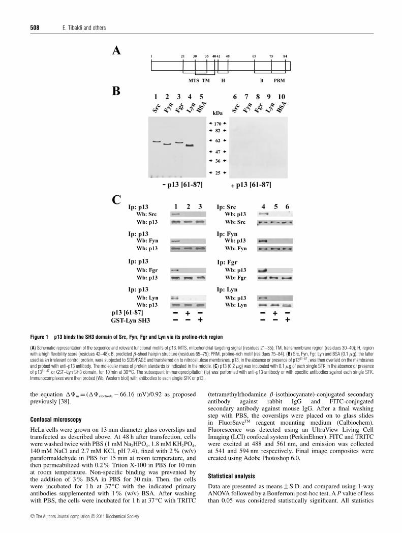
Figure 1 p13 binds the SH3 domain of Src, Fyn, Fgr and Lyn via its proline-rich region
(A) Schematic representation of the sequence and relevant functional motifs of p13. MTS, mitochondrial targeting signal (residues 21–35); TM, transmembrane region (residues 30–40); H, regionwith a high flexibility score (residues 42–48); B, predicted β-sheet hairpin structure (residues 65–75); PRM, proline-rich motif (residues 75–84). (B) Src, Fyn, Fgr, Lyn and BSA (0.1 μg), the latterused as an irrelevant control protein, were subjected to SDS/PAGE and transferred on to nitrocellulose membranes. p13, in the absence or presence of p1361–87, was then overlaid on the membranesand probed with anti-p13 antibody. The molecular mass of protein standards is indicated in the middle. (C) p13 (0.2 μg) was incubated with 0.1 μg of each single SFK in the absence or presenceof p1361–87 or GST–Lyn SH3 domain, for 10 min at 30◦C. The subsequent immunoprecipitation (Ip) was performed with anti-p13 antibody or with specific antibodies against each single SFK.Immunocomplexes were then probed (Wb, Western blot) with antibodies to each single SFK or p13.
the equation ��m = (��electrode − 66.16 mV)/0.92 as proposedpreviously [38].
Confocal microscopy
HeLa cells were grown on 13 mm diameter glass coverslips andtransfected as described above. At 48 h after transfection, cellswere washed twice with PBS (1 mM Na2HPO4, 1.8 mM KH2PO4,140 mM NaCl and 2.7 mM KCl, pH 7.4), fixed with 2 % (w/v)paraformaldehyde in PBS for 15 min at room temperature, andthen permeabilized with 0.2% Triton X-100 in PBS for 10 minat room temperature. Non-specific binding was prevented bythe addition of 3 % BSA in PBS for 30 min. Then, the cellswere incubated for 1 h at 37 ◦C with the indicated primaryantibodies supplemented with 1% (w/v) BSA. After washingwith PBS, the cells were incubated for 1 h at 37 ◦C with TRITC
(tetramethylrhodamine β-isothiocyanate)-conjugated secondaryantibody against rabbit IgG and FITC-conjugatedsecondary antibody against mouse IgG. After a final washingstep with PBS, the coverslips were placed on to glass slidesin FluorSaveTM reagent mounting medium (Calbiochem).Fluorescence was detected using an UltraView Living CellImaging (LCI) confocal system (PerkinElmer). FITC and TRITCwere excited at 488 and 561 nm, and emission was collectedat 541 and 594 nm respectively. Final image composites werecreated using Adobe Photoshop 6.0.
Statistical analysis
Data are presented as means +− S.D. and compared using 1-wayANOVA followed by a Bonferroni post-hoc test. A P value of lessthan 0.05 was considered statistically significant. All statistics
c© The Authors Journal compilation c© 2011 Biochemical Society

HTLV-1 p13 targets SFKs to mitochondria 509
Figure 2 p13 and p1361–87 act as positive modulators of Src, Fyn, Fgr and Lyn
(A) Tyrosine kinase activity of Src, Fyn, Fgr and Lyn was tested on the Src-specific peptide substrate cdc26–20 in the absence (column 1) or presence of increasing concentrations of p13 (columns2–5) or p1361–87 (columns 6–9) alternatively supplemented with the GST–Lyn SH3 domain (columns 4–5 and 8–10) as described in the Experimental section. Results are expressed as means +− S.D.from three separate experiments.**P < 0.001 and *P < 0.05. (B) Elution profile of activity on Src-specific peptide substrate cdc26–20 of Lyn (top panel), Lyn plus p13 (middle panel) and Lyn plusp13 supplemented with GST–Lyn SH3 domain (bottom panel), obtained from a Superdex 75 HR column as described in the Experimental section. Downward arrows indicate the position of molecularmass standards on the Superdex 75 HR column: BSA (66 kDa), ovalbumin (43 kDa), chymotrypsinogen (26 kDa) and lysozyme (14.5 kDa).
were performed using GraphPad Prism (GraphPad Software)statistical software.
RESULTS
p13 interacts with SFKs and stimulates their activity via itsC-terminal proline-rich region
p13 is an 87-amino acid accessory protein of HTLV-1, theorganization of which is shown in Figure 1(A). The primarystructure of p13 contains a series of regions, including a proline-rich motif putatively capable of interacting with SH3 domains[34], albeit not fulfilling a canonical consensus, because of theabsence of a basic amino acid residue upstream or downstream ofthe proline residues [3,13].
To assess whether the C-terminal proline-rich motif of p13was capable of interacting with the SH3 domain of SFKs,some of the SFK members, Src, Fyn, Fgr and Lyn, were runon SDS/PAGE and transferred on to nitrocellulose to undergofar-Western blot analysis. The immobilized enzymes were thenincubated with p13 in the absence or presence of p1361–87, a p13-derived peptide covering its C-terminal proline-rich motif. Asshown in Figure 1(B), p13 was detected with of all the SFKstested (left-hand panel), whereas this interaction was abolishedby the presence of p1361–87, indicating a competition with full-
length p13 and confirming the inability of the anti-p13 antibody torecognize the C-terminal region of p13 (see Supplementary FigureS1 at http://www.BiochemJ.org/bj/439/bj4390505add.htm). Todemonstrate that the SH3 domain of SFKs was implicated inthe interaction with p13, and in particular with its C-terminalproline-rich motif, p13 was incubated with single SFKs (molarratio 3:1) in the absence or presence of p1361–87 or, alternatively,of the GST–Lyn SH3 domain (Figure 1C). By subsequentimmunoprecipitation of p13 or the SFKs and Western blotanalysis with the respective antibodies, the presence of p1361–87 orthe GST–Lyn SH3 domain proved to prevent the formation of theSFK–p13 complex, confirming that this interaction was highlyspecific and mediated by the C-terminus of the viral protein andthe SH3 domain of the SFKs tested. Under these experimentalconditions, the residual quantity of SFKs in the supernatant afterimmunoprecipitating p13 alone was negligible, whereas p13 in thesupernatant after immunoprecipitating SFKs was approximatelytwo-thirds (results not shown).
As a number of viral proteins containing a proline-rich motifin their sequence have been shown to enhance SFK activity afterengaging the SH3 domain of SFKs [11], we evaluated the abilityof p13 to act as a positive regulator of SFKs. For this purpose,SFK activity was tested in vitro using the Src-specific peptidecdc26–20 as a substrate in the presence of increasing concentrationsof full-length p13 or p1361–87, in the absence or presence of the
c© The Authors Journal compilation c© 2011 Biochemical Society

510 E. Tibaldi and others
Figure 3 p13 acts as a carrier of Src and Lyn for mitochondrial import
(A) RLM (50 μg) was incubated with p13 and/or Src in the absence or presence of the GST–LynSH3 domain. Afterwards, mitochondria were spun down and the resulting fractions, mitochondria(M) and supernatant (S), were subjected to Western blot (Wb) analysis with anti-Src antibodyand subsequently with anti-p13 antibody. The membranes were re-probed with anti-aconitaseantibody as loading control. (B) RLM (50 μg) was incubated with p13 and/or Lyn in the absenceor presence of the GST–Lyn SH3 domain, and treated as described in (A). The Figure isrepresentative of four experiments performed in triplicate.
GST–Lyn SH3 domain. Figure 2(A) shows that both p13 andp1361–87 exerted an activatory effect on all of the SFKs assayedin a dose-dependent manner, although the full-length form ofp13 was more potent in stimulating SFK activity, even at muchlower concentrations. On the other hand, the presence of theGST–Lyn SH3 domain blocked the increase in SFK activity dueto the full-length viral protein or its C-terminal-derived peptide,highlighting that the proline-rich motif–SH3 domain interactionhad a role in modulating SFK activity.
To assess the formation of the Lyn–p13 complex, we alsoperformed size-exclusion chromatography, showing that Lyn,detected at the molecular mass of approximately 60 kDa whenalone (Figure 2B, top panel), was eluted at approximately 75 kDawhen pre-incubated with p13, indicating that these two proteinsform a stable heterodimer with a 5-fold increase in the enzymeactivity (Figure 2B, middle panel). In contrast, the presenceof the GST–Lyn SH3 domain reversed the activatory effect bycompeting with Lyn’s SH3 domain and hence disrupting thecomplex (Figure 2B, bottom panel). Similar data were obtainedwith Src, Fyn and Fgr (results not shown).
SFKs traffic to mitochondria when complexed with p13
p13 is known to mainly target mitochondria through theatypical N-terminal MLS (mitochondrial localization sequence)L21RVWRLCTRRLVPHL35. This sequence is predicted to bepart of an α-helix, the amphipathic properties of which areconferred by the four arginine residues, finally localizing tothe mitochondrial inner membrane [34]. In contrast with otherproteins bearing the canonical MLS, the targeting signal of p13 isnot cleaved upon import into mitochondria [34]. After anchoringto the mitochondrial inner membrane, p13 has been recognized totrigger an influx of K+ into the mitochondrial matrix accompaniedby a concentration-dependent decrease in the mitochondrial innermembrane potential (��m) [35]. Since we demonstrated thatp13 forms a stable heterodimer with the SFKs examined, weevaluated the ability of p13 to act as a carrier for mitochondrialimport of SFKs by virtue of its MLS. For this purpose, highlypurified RLM, in which we have previously demonstrated that theprotein level of SFKs is undetectable [33], were incubated withSrc, Fyn, Fgr and Lyn, alternately, in the absence or presenceof p13. After a 5 min incubation, mitochondria were spun downand the resulting fractions, mitochondria (M) and supernatant (S),underwent Western blot analysis with antibodies against p13 andthe SFK being studied. As shown in Figure 3, Src and Lyn werefound in the mitochondrial fraction only after pre-incubation withp13 (lane 4 compared with lane 6, M), whereas they were totallyrecovered in the supernatant fraction when the sample was eitherdevoid of p13 (lane 4 compared with lane 6, S) or pre-incubatedwith p13 in the presence of the GST–Lyn SH3 domain (lane 6compared with lane 7, M and S). Similar results were also achievedwith Fyn and Fgr (results not shown).
The p13–SFK complex localizes to the intermembrane space ofmitochondria
To establish in which mitochondrial compartment p13 and SFKswere localized, RLM were incubated with p13 alone or in thepresence of a slight molar excess of each tyrosine kinase available,and further processed as described below. First, treatment withproteinase K showed susceptibility of p13 and Lyn to the proteaseonly when mitochondria were solubilized with Triton X-100(Figure 4A), indicating that p13 alone or in association with Lynwas localized inside mitochondria. AIF, a structural component ofthe mitochondrial inner membrane, and aconitase, a mitochondrialmatrix protein, were used as intramitochondrial markers.Secondly, after separation of mitochondrial membranes from thesoluble fractions, as described in the Experimental section, p13proved to be bound to the membrane fraction when incubatedalone with mitochondria (Figure 4B, left-hand panel), whereasit was found in the soluble fraction when complexed with Lyn(Figure 4B, right-hand panel). Thirdly, treatment with increasingconcentrations of digitonin resulted in a selective release of p13depending on the absence or presence of Lyn. In particular,p13 was released from mitochondria, if previously incubatedin the absence of Lyn, only at concentrations of digitoninhigher than 0.4 mg/ml, similarly to AIF, which is suggestiveof either a tight binding to the inner mitochondrial membraneor localization in the mitochondrial matrix; on the other hand,concentrations of digitonin as low as 0.1 mg/ml caused releaseof p13, if the latter was pre-incubated with Lyn, suggesting thatthis interaction prevented p13 from reaching the mitochondrialinner membrane, thus segregating it in the intermembrane space.Similar results were obtained using Src, the prototypical memberof the Src-related group (see Supplementary Figure S2 athttp://www.BiochemJ.org/bj/439/bj4390505add.htm).
c© The Authors Journal compilation c© 2011 Biochemical Society

HTLV-1 p13 targets SFKs to mitochondria 511
Figure 4 p13 and Lyn are detected as a component of a soluble complex in the intermembrane space of isolated mitochondria
RLM were incubated with p13 only (left-hand panels) or in the presence of Lyn (right-hand panels) and treated as described below. (A) RLM were incubated in the absence or presence of proteinaseK (PK) and Triton X-100 (TX-100) according to the Experimental section. Aliquots of the mitochondrial lysate were subjected to Western blot (Wb) analysis with anti-Lyn, anti-p13, anti-AIF andanti-aconitase antibodies as indicated. (B) RLM were subjected to treatment to obtain mitochondrial membranes and soluble fractions as described in the Experimental section. Intact mitochondria(Mit.), mitochondrial membranes (Membr.) and soluble fractions were subjected to Western blot analysis with anti-Lyn, anti-p13, anti-AIF and anti-aconitase antibodies as indicated. (C) RLM wereincubated in the presence of increasing concentrations of digitonin. After each treatment, the samples were centrifuged to separate the pellet (P) from the soluble fraction (S). Aliquots of suchfractions after differential digitonin treatment were subjected to Western blot analysis with anti-Lyn, anti-p13, anti-AIF and anti-aconitase antibodies as indicated. The Figure is representative of fourindependent experiments performed in triplicate.
Tyrosine phosphorylation in isolated mitochondria is induced byp13-mediated import of SFKs
To assess whether the association between SFKs and p13 couldchange Lyn’s activity within mitochondria, as occurred in vitro,we incubated purified RLM in the presence of Lyn and p13,as described above, and evaluated the level of mitochondrialtyrosine phosphorylation by Western blot analysis with the anti-pTyr antibody.
Figure 5(A) shows a striking elevation in tyrosinephosphorylation of a number of mitochondrial proteins distributedover a wide range of molecular masses once Lyn translocatedinto mitochondria. The involvement of Lyn in this event wasfurther confirmed by the effect of PP2, a specific SFK inhibitor(Figure 5A, lane 6 compared with lane 7), and its activationwas confirmed by Western blot analysis with the anti-SrcpY416 antibody. To establish whether the level of tyrosinephosphorylation observed inside mitochondria was determinedby the stability of the p13–Lyn complex, mitochondria pre-incubated with p13 and Lyn in the above conditions weretreated with 0.1 mg/ml digitonin. After centrifugation, the
resulting supernatant was incubated in the presence of increasingconcentrations of the GST–Lyn SH3 domain, subsequentlysubjected to immunoprecipitation with anti-p13 antibody andassayed for SFK activity. Figure 5(C) shows that the p13–Lyn interaction was still stable after translocation across themitochondrial outer membrane, with a disruption of the complexoccurring in a manner proportional to the concentration ofthe GST–Lyn SH3 domain (Figure 5B). Since the presence of theGST–Lyn SH3 domain blocked the stimulation of SFK activitydue to p13 (Figure 5C), we presumed that the increase in SFKactivity depended upon the stability of the p13–Lyn interaction.Similar findings were achieved for the other SFKs by examiningFyn, Fgr (results not shown) and Src (Supplementary Figure S3at http://www.BiochemJ.org/bj/439/bj4390505add.htm).
The interaction between p13 and SFKs impairs the ability of p13 toinduce mitochondrial K+ influx
p13 is known to target the inner mitochondrial membrane,bringing about substantial structural and functional changes of
c© The Authors Journal compilation c© 2011 Biochemical Society

512 E. Tibaldi and others
Figure 5 p13-mediated import of Lyn increased tyrosine phosphorylationin isolated mitochondria
(A) RLM (50 μg) were incubated with p13 and/or Lyn in the absence or presence of 1 μMPP2. RLM were subsequently centrifuged and subjected to Western blot (Wb) analysis withanti-p-Tyr (top panel), anti-Src pY416 (middle panel) and anti-Lyn (bottom panel) antibodies.The molecular mass markers are indicated on the right-hand side in kDa. (B) RLM (250 μg) wereincubated with p13 and Lyn. RLM were then spun down and aliquots of 50 μg of mitochondriawere lysed and immunoprecipitated (Ip) with anti-p13 antibody in the absence or presence ofincreasing concentrations of GST–Lyn SH3. The immunocomplexes were probed with anti-Lynand subsequently with anti-p13 antibodies. (C) RLM (250 μg) were incubated with p13 and Lyn.After a 5 min incubation, mitochondria were spun down and aliquots of 50 μg of mitochondriawere analysed for in vitro Lyn activity in the absence or presence of increasing concentrationsof GST–Lyn SH3. The Figure is representative of four independent experiments; results aremeans +− S.D.
these organelles, such as fragmentation in a cellular contextas well as an increase in K+ permeability. These effectsresult in depolarization of the inner mitochondrial membraneand enhancement of respiratory chain activity with concurrentelevation in production of ROS (reactive oxygen species) inisolated mitochondria [34]. Interestingly, we observed that p13together with SFKs via a SH3/proline-rich motif associationin vitro formed a stable complex still capable of crossing theouter mitochondrial membrane of highly purified RLM, butthat it was unable to insert itself into the inner mitochondrialmembrane where it usually exerts its perturbing action (Figure 4).Therefore, we sought to verify whether the change in localizationof p13 also affected the functional effects that it is knownto provoke. For this purpose, RLM were tested for membranepotential (��m) before and after incubation with p13, the latterin turn mixed with either GST–Lyn SH3 or Lyn. Figure 6(A)shows that free p13 caused a sharp drop in ��m, as expectedin accordance with the previously documented increase in thepermeability of the inner mitochondrial membrane to small
cations [35]. Conversely, when p13 was complexed with Lyn,its effect on ��m was dampened in a manner proportional to themolar ratios between the two proteins. In fact, decreasing p13/Lynratios as far as 1:1, as shown in Figure 6(A), progressivelyabolished the capability of p13 to alter ��m. Similar effects wereobtained by using the GST–Lyn SH3 domain as a ligand for p13(Figure 6B), indicating a role for the SH3 domain in affecting bothfunction and localization of p13 itself. These findings also suggesta possible non-catalytic function of SFKs mediated by their SH3domain.
The p13–Lyn complex enters mitochondria in p13-transfected HeLacells
Once we verified that p13 interacted with SFKs to form acomplex capable of translocating to isolated mitochondria, weinvestigated the presence and the possible localization of sucha complex in living cells, using Lyn- and p13-overexpressingcells. Among SFKs, we monitored Lyn expression because it hasalready been shown to translocate from the plasma membrane tomitochondria [33]. Therefore HeLa cells were transfected withexpression vectors for Lyn or p13 singly or in combination,or both empty vectors as a control, in the absence or in thepresence of PP2, as described in the Experimental section. Weexamined the protein level of p13 and Lyn by Western blot analysison the total cell lysate and on highly purified mitochondriaobtained by protocols described previously [33]. Figure 7(A)shows an increase in tyrosine phosphorylation in the whole celllysate (left-hand panel) upon transfection of Lyn (lane 3), asexpected, and of p13 (lane 5), indicating that the latter was capableof eliciting an endogenous SFK-dependent kinase activity, aselucidated by the inhibitory effect of PP2 (lanes 4 and 6). Thiseffect was dramatically enhanced by the co-transfection of bothproteins (lanes 3 and 5 compared with lane 7), further supportingthe model of interaction inferred from the in vitro data. Notably,Lyn, both endogenous and transfected, was detected insidepurified mitochondria only when p13 was present (Figure 7A,right-hand panel, lanes 5–8 compared with lanes 1–4), clearlyparalleling the level of mitochondrial tyrosine phosphorylation(lane 5 compared with lane 1 and lane 7 compared with lane 3).The relative purity of the mitochondrial fraction was assessed byusing antibodies against specific cellular markers.
To examine whether the interaction between Lyn and p13occurred according to the mechanisms previously identified invitro and whether a stable complex persisted within mitochondriain cultured cells, p13 was immunoprecipitated from themitochondrial lysates of HeLa cells transfected with expressionvectors for Lyn and p13, or with both empty vectors. As shown inFigure 7(B), Lyn co-immunoprecipitated with p13 and the GST–Lyn SH3 domain was able to disrupt the p13–Lyn complex (left-hand panel, lane 4 compared with lane 3), highlighting the role ofLyn’s SH3 domain in the interaction with p13. Moreover, the samemitochondrial lysates were assayed for SFK activity in the absenceor presence of the GST–Lyn SH3 domain, demonstrating that Lynin mitochondria displayed a higher activity when associated withp13, further confirming the activatory role of p13 on SFKs (right-hand panel, column 3 compared with column 1).
To corroborate the biochemical data obtained in HeLa cellswith regard to the p13–Lyn interaction and import of Lyn intomitochondria, the subcellular localization of Lyn was analysedin relation with p13 expression by confocal microscopy. Sincethe anti-p13 antibody was useless in the immunofluorescenceassay, we used the anti-c-Myc antibody to follow p13 cellularlocalization. The pcDNA3.1 expression vector for p13 also
c© The Authors Journal compilation c© 2011 Biochemical Society

HTLV-1 p13 targets SFKs to mitochondria 513
Figure 6 Interaction between the SH3 domain of Lyn and p13 proline-rich motif impairs p13 ability to induce a collapse in the inner mitochondrial potential(��m)
RLM were incubated in 200 mM sucrose, 10 mM Hepes, pH 7.4, 5 mM succinate, 1.25 μM rotenone, 1 mM sodium phosphate and protease inhibitors in the presence of p13 only or with increasingconcentrations of Lyn (A) or GST–Lyn SH3 domain (B). The downward arrows indicate the time point at which p13 or p13 complex was added. The Figure is representative of four independentexperiments.
contains a Myc tag. As shown in Figure 8, we observed thatp13 mainly localizes to mitochondria, as visualized by doubleimmunofluorescence staining with the anti-TOM20 antibody asmitochondrial marker and the anti-c-Myc antibody, and thatits subcellular distribution was unaltered, even upon Lyn co-transfection (Figure 8A, merge). In contrast, overexpressed Lynwas shown to co-localize with TOM20 only when p13 wasexpressed, indicating that p13 was the driving force for Lyn tobe recruited to mitochondria (Figure 8B). Finally, as shown inFigure 8(C), p13 and Lyn widely overlap when co-expressed inHeLa cells, supporting the association of the two proteins in vivo.
DISCUSSION
In the present study, we demonstrate that p13, a HTLV-1 accessoryprotein containing an N-terminal MLS, associates with theSH3 domain of SFKs through its C-terminal proline-rich motif,forming a stable heterodimer that migrates into mitochondria andconfers novel functional properties to the single components of thecomplex. It is noted that mitochondria have only been described asa location for SFKs within the last ten years [7]. In fact, althoughSFKs have commonly been thought to be located at the innerleaflet of the plasma membrane to relay extracellular cues fromdifferent types of transmembrane receptors [1–3], accumulatingevidence has been reported for a wider intracellular distributionof this family of protein kinases as well as for peculiar functionsof SFKs associated with their distinct intracellular localizations[4–9]. In this regard, post-translational modifications such aspalmitoylation, and lately the redox state, have been recognizedas crucial factors in driving trafficking and localization of SFKs.In particular, three modes of SFK trafficking have been proposedaccording to the palmitoylation state: (i) the cycling pathwaybetween plasma membrane and endosomes/lysosomes for non-palmitoylated SFKs [8,40,41]; (ii) the secretory pathway from the
Golgi apparatus to the plasma membrane for mono-palmitoylatedSFK [8,42]; and (iii) the direct plasma membrane-targetingpathway for doubly palmitoylated SFKs [8]. The redox state hasbeen recently demonstrated to profoundly regulate the traffickingof non-palmitoylated Src as well, with a remarkable shift ofthis SFK from the plasma membrane to endosomes/lysosomesaccompanied by a decrease in enzyme activity under reducingconditions [36]. In addition to post-translational modifications,protein–protein interactions due to the multimodular structureof SFKs have also been implicated in their targeting to specificsubcellular compartments. Accumulating data show that the SH3domain, besides modulating the SFK activity by binding thePPII motif of SFKs themselves, is directly implicated in the local-ization of SFKs to cellular compartments other than the plasmamembrane by recognizing specific ligands. An example of thismode of trafficking is given by the interaction between Hck,a SFK highly expressed in macrophages, and Nef, a HIV-1multifunctional protein, resulting in the activation of Hck and itsaccumulation to the Golgi apparatus, thereby causing the arrestof the maturation of the nascent cytokine receptor Fms [31].
Our results from the present study corroborate the notion thatthe SH3 domain of SFKs is one of the players involved in directingSFKs themselves to final destinations upon the basis of localiza-tion signals present on interacting partners. In fact, p13, known toenter mitochondria through its MLS, behaves analogously to Nef,by virtue of its C-terminal proline-rich motif theoretically suitablefor binding SH3 domains. In the present study, we demonstratefor the first time that p13 associates indifferently with distinctmembers of the Src family (Src, Fyn, Fgr and Lyn) via SH3domain–proline-rich motif interaction in vitro, hence forminga heterodimer in which the activity of all the SFKs tested aredramatically enhanced (Figures 1 and 2). Importantly, p13 con-serves its ability to translocate into isolated mitochondria,concomitantly participating in a stable complex with SFKs,ultimately carrying them into the mitochondrial intermembrane
c© The Authors Journal compilation c© 2011 Biochemical Society

514 E. Tibaldi and others
Figure 7 The p13–Lyn complex localizes in mitochondria of p13-transfected HeLa cells
(A) HeLa cells were transfected with expression vectors for Lyn (pCMV6-XL4/Lyn, lanes 3–4 and 7–8), p13 (pcDNA3.1/myc-His C/p13, lanes 5–8), or with both empty vectors (pCMV6-XL4 andpcDNA3.1/myc-His C, lanes 1 and 2 respectively). At 36 h after transfection, cells were treated with 10 μM PP2 (lanes 2, 4, 6 and 8) for 12 h. Whole cell lysates (left-hand panels) and wholemitochondrial lysates (right-hand panels) were assayed by Western blot (Wb) analysis with anti-Lyn, anti-p13, anti-p-Tyr, anti-LDH (cytosolic marker), anti-calnexin (microsomal marker), anti-lamin(nuclear marker) and anti-aconitase (mitochondrial marker) antibodies as indicated. (B) Mitochondrial lysates from HeLa cells transfected with expression vectors for Lyn and p13 (lanes 3 and 4),or with both empty vectors (lanes 1 and 2) were analysed for in vitro Lyn activity on SFK-specific peptide substrate cdc26–20 (right-hand panel) or were immunoprecipitated with anti-p13 antibody(left-hand panel) in the absence (lanes 1 and 3) or presence (lanes 2 and 4) of GST–Lyn SH3. The immunocomplexes were probed with anti-Lyn antibody and subsequently with anti-p13antibody. The Figure is representative of three independent experiments.
space. Likewise, both biochemical analysis and confocalmicroscopy provide compelling evidence that Lyn is also detectedinside mitochondria when p13 is co-expressed in HeLa cells, andis associated with p13 (Figures 7 and 8). This data supportsthe hypothesis that p13 may act as a carrier for SFKs intomitochondria, even in living cells. Moreover, the striking elevationin tyrosine phosphorylation observed in either freshly isolatedmitochondria in the presence of synthetic p13 or in p13-overexpressing HeLa cells highlights the role of SFKs, as shownby the use of the specific SFK inhibitor PP2 (Figures 5A and 7A),confirming the ability of p13 to function as positive modulator ofSFKs.
Notably, we observed a similar scenario while investigatingthe role of SFKs, and in particular of Lyn, in the context of earlyliver regeneration, assessing that Lyn translocates from the plasmamembrane to mitochondria, more precisely into the mitochondrialintermembrane space, where it is part of a 230 kDa multi-protein complex [33]. It has been demonstrated only recently that
this complex, similar to the results shown in the present study, isdisrupted by the GST–Lyn SH3 domain, in agreement with themodel proposed. Furthermore, we also showed that Lyn, as part ofthe multiprotein complex, protected the structural and functionalintegrity as well as the bioenergetic competence of mitochondriaby contrasting the potentially harmful effects resulting from ROSelevation and Ca2 + overload, which would fatally lead to celldeath (E. Tibaldi, M. A. Pagano and A. M. Brunati, unpublishedwork).
An experimental setting that exploits p13 as a Trojan horsefor SFKs might provide a useful model to elucidate the roleof SFKs themselves within mitochondria and how tyrosinephosphorylation influences mitochondrial physiology. In thisregard, it is worthwhile to remember that functional studies onisolated mitochondria with full-length synthetic p13 showed thatthis protein inserts itself into the inner mitochondrial membrane,thereby inducing an inward K+ current with a drop in ��m
and activation of the electron transport chain accompanied by
c© The Authors Journal compilation c© 2011 Biochemical Society

HTLV-1 p13 targets SFKs to mitochondria 515
Figure 8 Lyn localizes to the mitochondria of HeLa cells only upon co-expression of p13
HeLa cells were transfected with expression vectors for Lyn (pCMV6-XL4/Lyn), p13(pcDNA3.1/myc-His C/p13), or with both empty vectors. At 48 h post-transfection, cells werefixed, permeabilized and incubated with the indicated antibodies. (A) Cells transfected withp13 only or Lyn and p13 were incubated with anti-c-Myc and anti-TOM20 R (mitochondrialmarker) antibodies followed by FITC-conjugated anti-mouse secondary antibody (green)and with TRITC-conjugated anti-rabbit secondary antibody (red) respectively. (B) Cells trans-fected with Lyn only or Lyn and p13 were incubated with anti-Lyn and anti-TOM20 M antibodies,followed by incubation with TRITC-conjugated anti-rabbit secondary antibody (red) and withFITC-conjugated anti-mouse secondary antibody (green) respectively. (C) Cells transfectedwith expression vectors for Lyn, p13 or Lyn/p13 were probed with anti-Lyn andanti-c-Myc antibodies, followed by the appropriate secondary antibodies, as described above.Co-localization is visualized by the yellow fluorescence appearing after merging of both signals.The Figure is representative of three independent experiments. Scale bar, 10 μm.
increased mitochondrial ROS production [34,35]. In contrast, ashighlighted by our results, the SFK–p13 complex is still takenup by these organelles, but p13 is unable to target the innermitochondrial membrane and to perturb ��m. These observationsare in agreement with studies that recognize a scaffolding rolefor SFKs mediated by the SH3 domain, in our hands resultingin impairment of the activity of p13, indicating a non-catalytic
function of SFKs [42–46]. Importantly, we note that this propertyof SFKs does not prove detrimental to their catalytic action, whichinstead was remarkably increased upon interaction with p13,suggesting that the catalytic and non-catalytic function might notbe mutually exclusive, as is sometimes reported in the literature[46], but might even be independent or synergistic, and potentiallytakes part in pathophysiological conditions.
In summary, the SFK–p13 interaction and the resulting effectsdescribed above lead us to hypothesize a mechanism by whichthe transfer of SFKs to different cellular districts, mediated by theSH3 domain and directed by the localization signals of interactingproteins, may exert a more general action on cell fate by modifyingthe phosphorylation state and hence the function of key substratesresident in the targeted cellular compartments.
AUTHOR CONTRIBUTION
Elena Tibaldi performed the majority of the research, analysed the data and wrote someof the manuscript. Andrea Venerando synthesized and purified the peptides used in thiswork and reviewed the manuscript prior to submission. Francesca Zonta performed invitro research. Carlo Bidoia actively contributed to the setting up of genetic assays. ElisaMagrin performed some of the in vitro research. Oriano Marin purified the antibodyagainst p13. Antonio Toninello provided cultural background and a location for assays onmitochondria. Luciana Bordin performed some of the in vitro research. Veronica Martiniperformed the immunofluorescence assays. Mario Angelo Pagano supported the workintellectually, reviewed all of the data and wrote the manuscript. Anna Maria Brunatidesigned the research, reviewed all of the data, participated in analysis of data and wrotethe manuscript.
ACKNOWLEDGEMENTS
We thank Luc Willems (Gembloux, Belgium) for kindly providing pcDNA3.1/myc-HisC/p13.
FUNDING
This work was funded by Fondazione CARIPARO (Progetto di Eccellenza 2008) (to A.M.B.).
REFERENCES
1 Thomas, S. M. and Brugge, J. S. (1997) Cellular functions regulated by Src familykinases. Annu. Rev. Cell Dev. Biol. 13, 513–609
2 Parsons, S. J. and Parsons, J. T. (2004) Src family kinases, key regulators of signaltransduction. Oncogene 18, 7906–7909
3 Engen, J. R., Wales, T. E., Hochrein, J. M., Meyn, 3rd, M. A., Banu Ozkan, S., Bahar, I. andSmithgall, T. E. (2008) Structure and dynamic regulation of Src-family kinases. Cell. Mol.Life Sci. 65, 3058–3073
4 Mohn, H., LeCabec, V., Fischer, S. and Maridonneau-Parini, I. (1995) The Src-familyprotein-tyrosine kinase p59hck is located on the secretory granules in human neutrophilsand translocates towards the phagosome during cell activation. Biochem. J. 309,657–665
5 Bard, F., Patel, U., Levy, J. B., Jurdic, P., Horne, W. C. and Baron, R. (2002) Molecularcomplexes that contain both c-Cbl and c-Src associate with Golgi membranes. Eur. J.Cell. Biol. 81, 26–35
6 Kasahara, K., Nakayama, Y., Ikeda, K., Fukushima, Y., Matsuda, D., Horimoto, S. andYamaguchi, N. (2004) Trafficking of Lyn through the Golgi caveolin involves the chargedresidues on αE and αI helices in the kinase domain. J. Cell Biol. 165, 641–652
7 Salvi, M., Brunati, A. M. and Toninello, A. (2005) Tyrosine phosphorylation inmitochondria: a new frontier in mitochondrial signalling. Free Radical Biol. Med. 38,1267–1277
8 Sato, I., Obata, Y., Kasahara, K., Nakayama, Y., Fukumoto, Y., Yamasaki, T., Yokoyama,K. K., Saito, T. and Yamaguchi, N. (2009) Differential trafficking of Src, Lyn, Yes and Fynis specified by the state of palmitoylation in the SH4 domain. J. Cell Sci. 122, 965–975
9 Frasson, M., Vitadello, M., Brunati, A. M., La Rocca, N., Tibaldi, E., Pinna, L. A., Gorza, L.and Donella-Deana, A. (2009) Grp94 is Tyr-phosphorylated by Fyn in the lumen of theendoplasmic reticulum and translocates to Golgi in differentiating myoblasts. Biochim.Biophys. Acta 1793, 239–252
c© The Authors Journal compilation c© 2011 Biochemical Society

516 E. Tibaldi and others
10 Williams, J. C., Wierenga, R. K. and Saraste, M. (1998) Insights into Src kinase functions:structural comparisons. Trends Biochem. Sci. 23, 179–184
11 Chong, Y. P., Ia, K. K., Mulhern, T. D. and Cheng, H. C. (2005) Endogenous and syntheticinhibitors of the Src-family protein tyrosine kinases. Biochim. Biophys. Acta 1754,210–220
12 Sicheri, F., Moarefi, I. and Kuriyan, J. (1997) Crystal structure of the Src family tyrosinekinase Hck. Nature 385, 602–609
13 Ingley, E. (2008) Src family kinases: regulation of their activities, levels and identificationof new pathways. Biochim. Biophys. Acta 1784, 56–65
14 Wang, D., Esselman, W. J. and Cole, P. A. (2002) Substrate conformational restriction andCD45-catalyzed dephosphorylation of tail tyrosine-phosphorylated Src protein. J. Biol.Chem. 277, 40428–40433
15 Boonyaratanakornkit, V., Scott, M. P., Ribon, V., Sherman, L., Anderson, S. M., Maller,J. L., Miller, W. T. and Edwards, D. P. (2001) Progesterone receptor contains a proline-richmotif that directly interacts with SH3 domains and activates c-Src family tyrosine kinases.Mol. Cell 8, 269–280
16 Pellicena, P. and Miller, W. T. (2001) Processive phosphorylation of p130Cas by Srcdepends on SH3-polyproline interactions. J. Biol. Chem. 276, 28190–28196
17 Moarefi, I., LaFevre-Bernt, M., Sicheri, F., Huse, M., Lee, C. H., Kuriyan, J. and Miller,W. T. (1997) Activation of the Src-family tyrosine kinase Hck by SH3 domaindisplacement. Nature 385, 650–653
18 Trible, R. P., Emert-Sedlak, L. and Smithgall, T. E. (2006) HIV-1 Nef selectively activatesSrc family kinases Hck, Lyn, and c-Src through direct SH3 domain interaction. J. Biol.Chem. 281, 27029–27038
19 Lerner, E. C. and Smithgall, T. E. (2002) SH3-dependent stimulation of Src-family kinaseautophosphorylation without tail release from the SH2 domain in vivo. Nat. Struct. Biol.9, 365–369
20 Porter, M., Schindler, T., Kuriyan, J. and Miller, W. T. (2000) Reciprocal regulation of Hckactivity by phosphorylation of Tyr527 and Tyr416. Effect of introducing a high affinityintramolecular SH2 ligand. J. Biol. Chem. 275, 2721–2726
21 Bauer, F., Schweimer, K., Meiselbach, H., Hoffmann, S., Rosch, P. and Sticht, H. (2005)Structural characterization of Lyn-SH3 domain in complex with a herpesviral proteinreveals an extended recognition motif that enhances binding affinity. Protein Sci. 14,2487–2498
22 Liang, Y. and Roizman, B. (2006) State and role of Src family kinases in replication ofherpes simplex virus 1. J. Virol. 80, 3349–3359
23 Shelton, H. and Harris, M. (2008) Hepatitis C virus NS5A protein binds the SH3 domainof the Fyn tyrosine kinase with high affinity: mutagenic analysis of residues within theSH3 domain that contribute to the interaction. Virol. J. 11, 5–24
24 Cho, N. H., Choi, Y. K. and Choi, J. K. (2008) Multi-transmembrane protein K15 ofKaposi’s sarcoma-associated herpesvirus targets Lyn kinase in the membrane raft andinduces NFAT/AP1 activities. Exp. Mol. Med. 40, 565–573
25 Solheim, S. A., Torgersen, K. M., Tasken, K. and Berge, T. (2008) Regulation ofFynT function by dual domain docking on PAG/Cbp. J. Biol. Chem. 283,2773–2783
26 Chae, Y. K., Woo, J., Kim, M. J., Kang, S. K., Kim, M. S., Lee, J., Lee, S. K., Gong, G.,Kim, Y. H., Soria, J. C. et al. (2008) Expression of aquaporin 5 (AQP5) promotes tumorinvasion in human non small cell lung cancer. PLoS ONE 3, e2162
27 Samuels, A. L., Klinken, S. P. and Ingley, E. (2009) Liar, a novel Lyn-bindingnuclear/cytoplasmic shuttling protein that influences erythropoietin-induceddifferentiation. Blood 113, 3845–3856
28 Hammond, S., Wagenknecht-Wiesner, A., Veatch, S. L., Holowka, D. and Baird, B. (2009)Roles for SH2 and SH3 domains in Lyn kinase association with activated FcεRI in RBLmast cells revealed by patterned surface analysis. J. Struct. Biol. 168, 161–167
29 Honda, Z., Suzuki, T. and Honda, H. (2009) Identification of CENP-V as a novelmicrotubule-associating molecule that activates Src family kinases through SH3 domaininteraction. Genes Cells 14, 1383–1394
30 Xi, G., Shen, X. and Clemmons, D. R. (2010) p66shc inhibits insulin-like growth factor-Isignaling via direct binding to Src through its polyproline and Src homology 2 domains,resulting in impairment of Src kinase activation. J. Biol. Chem. 285, 6937–6951
31 Trentin, L., Frasson, M., Donella-Deana, A., Frezzato, F., Pagano, M. A., Tibaldi, E.,Gattazzo, C., Zambello, R., Semenzato, G. and Brunati, A. M. (2008)Geldanamycin-induced Lyn dissociation from aberrant Hsp90-stabilized cytosoliccomplex is an early event in apoptotic mechanisms in B-chronic lymphocytic leukemia.Blood 112, 4665–4674
32 Hassan, R., Suzu, S., Hiyoshi, M., Takahashi-Makise, N., Ueno, T., Agatsuma, T., Akari,H., Komano, J., Takebe, Y., Motoyoshi, K. and Okada, S. (2009) Dys-regulated activationof a Src tyrosine kinase Hck at the Golgi disturbs N-glycosylation of a cytokine receptorFms. J. Cell. Physiol. 221, 458–468
33 Gringeri, E., Carraro, A., Tibaldi, E., D’Amico, F. E., Mancon, M., Toninello, A., Pagano,M. A., Vio, C., Cillo, U. and Brunati, A. M. (2009) Lyn-mediated mitochondrial tyrosinephosphorylation is required to preserve mitochondrial integrity in early liver regeneration.Biochem. J. 425, 401–412
34 Silic-Benussi, M., Biasiotto, R., Andresen, V., Franchini, G., D’Agostino, D. M. andCiminale, V. (2010) HTLV-1 p13, a small protein with a busy agenda. Mol. Aspects Med.31, 350–358
35 Silic-Benussi, M., Cannizzaro, E., Venerando, A., Cavallari, I., Petronilli, V., La Rocca, N.,Marin, O., Chieco-Bianchi, L., Di Lisa, F., D’Agostino, D. M. et al. (2009) Modulation ofmitochondrial K+ permeability and reactive oxygen species production by the p13protein of human T-cell leukemia virus type 1. Biochim. Biophys. Acta 1787, 947–954
36 Krasnowska, E. K., Pittaluga, E., Brunati, A. M., Brunelli, R., Costa, G., De Spirito, M.,Serafino, A., Ursini, F. and Parasassi, T. (2008) N-acetyl-L-cysteine fosters inactivationand transfer to endolysosomes of c-Src. Free Radical Biol. Med. 45, 1566–1572
37 Kamo, N., Muratsugu, M., Hongoh, R. and Kobatake, Y. (1979) Membrane potential ofmitochondria measured with an electrode sensitive to tetraphenyl phosphonium andrelationship between proton electrochemical potential and phosphorylation potential insteady state. J. Membr. Biol. 49, 105–121
38 Jensen, B. D., Gunter, K. K. and Gunter, T. E. (1986) The efficiencies of the componentsteps of oxidative phosphorylation. II. Experimental determination of the efficiencies inmitochondria and examination of the equivalence of membrane potential and pH gradientin phosphorylation. Arch. Biochem. Biophys. 248, 305–323
39 Lefebvre, L., Vanderplasschen, A., Ciminale, V., Heremans, H., Dangoisse, O., Jauniaux,J. C., Toussaint, J. F., Zelnik, V., Burny, A., Kettmann, R. and Willems, L. (2002) Oncoviralbovine leukemia virus G4 and human T-cell leukemia virus type 1 p13(II) accessoryproteins interact with farnesyl pyrophosphate synthetase. J. Virol. 76, 1400–1414
40 Silic-Benussi, M., Cavallari, I., Vajente, N., Vidali, S., Chieco-Bianchi, L., Di Lisa, F.,Saggioro, D., D’Agostino, D. M. and Ciminale, V. (2010) Redox regulation of T-cellturnover by the p13 protein of human T-cell leukemia virus type 1: distinct effects inprimary versus transformed cells. Blood 116, 54–62
41 Kasahara, K., Nakayama, Y., Kihara, A., Matsuda, D., Ikeda, K., Kuga, T., Fukumoto, Y.,Igarashi, Y. and Yamaguchi, N. (2007) Rapid trafficking of c-Src, a non-palmitoylatedSrc-family kinase, between the plasma membrane and late endosomes/lysosomes. Exp.Cell Res. 313, 2651–2666
42 Obata, Y., Fukumoto, Y., Nakayama, Y., Kuga, T., Dohmae, N. and Yamaguchi, N. (2010)The Lyn kinase C-lobe mediates Golgi export of Lyn through conformation-dependentACSL3 association. J. Cell Sci. 123, 2649–2662
43 Kaplan, K. B., Swedlow, J. R., Morgan, D. O. and Varmus, H. E. (1995) c-Src enhancesthe spreading of src − / − fibroblasts on fibronectin by a kinase-independentmechanism. Genes Dev. 9, 1505–1517
44 Schwartzberg, P. L., Xing, L., Hoffmann, O., Lowell, C. A., Garrett, L., Boyce, B. F. andVarmus, H. E. (1997) Rescue of osteoclast function by transgenic expression ofkinase-deficient Src in src − / − mutant mice. Genes Dev. 11, 2835–2844
45 Brunton, V. G., Avizienyte, E., Fincham, V. J., Serrels, B., Metcalf, 3rd, C. A., Sawyer, T. K.and Frame, M. C. (2005) Identification of Src-specific phosphorylation site on focaladhesion kinase: dissection of the role of Src SH2 and catalytic functions and theirconsequences for tumor cell behaviour. Cancer Res. 65, 1335–1342
46 Garcıa-Martınez, J. M., Calcabrini, A., Gonzalez, L., Martın-Forero, E., Agullo-Ortuno,M. T., Simon, V., Watkin, H., Anderson, S. M., Roche, S. and Martın-Perez, J. (2010) Anon-catalytic function of the Src family tyrosine kinases controls prolactin-induced Jak2signaling. Cell. Signalling 22, 415–426
Received 7 October 2010/22 June 2011; accepted 7 July 2011Published as BJ Immediate Publication 7 July 2011, doi:10.1042/BJ20101650
c© The Authors Journal compilation c© 2011 Biochemical Society

Biochem. J. (2011) 439, 505–516 (Printed in Great Britain) doi:10.1042/BJ20101650
SUPPLEMENTARY ONLINE DATAInteraction between the SH3 domain of Src family kinases and theproline-rich motif of HTLV-1 p13: a novel mechanism underlying deliveryof Src family kinases to mitochondriaElena TIBALDI*, Andrea VENERANDO*†, Francesca ZONTA*, Carlo BIDOIA‡, Elisa MAGRIN*, Oriano MARIN*,Antonio TONINELLO*, Luciana BORDIN*, Veronica MARTINI†, Mario Angelo PAGANO*1 and Anna Maria BRUNATI*1,2
*Department of Biological Chemistry, University of Padua, Viale G. Colombo 3, 35131 Padua, Italy, †Venetian Institute of Molecular Medicine (VIMM), Via G. Orus, 2, 35129 Padua,Italy, and ‡Centre for Research in Infectious Diseases, School of Medicine and Medical Science, University College Dublin, Belfield, Dublin 4, Ireland
Figure S1 Characterization of the anti-p13 antibody
For the dot blot analysis, various amounts (from 1 ng to 1 μg) of full-length synthetic p13, theN-terminal p13-derived peptide (p139–41) or the C-terminal p13-derived peptide (p1361–87) werespotted on to nitrocellulose membranes. The nitrocellulose membrane was incubated with twodifferent dilutions, 1:500 (A) or 1:2000 (B), of the anti-p13 antibody and developed as describedin the Experimental section. (C) Western blot analysis of HeLa cell lysates either transfected withpcDNA3.1/myc-His C (lane 1) or pcDNA3.1/myc-His C/p13 (lane 2) with anti-p13 antibody.
Figure S2 p13 and Src are detected as a component of a soluble complexin the intermembrane space of isolated mitochondria
RLM were incubated with synthetic p13 only (left-hand panels) or in the presence of purifiedSrc (right-hand panels) and treated as described below. (A) Mitochondria were incubated in theabsence or presence of proteinase K (PK) and Triton X-100 (TX-100) according to the protocolreported in the Experimental section of the main text. Aliquots of the mitochondrial lysate weresubjected to Western blot (Wb) analysis with the indicated antibodies. (B) Mitochondriawere subjected to treatment to obtain mitochondrial membranes and soluble fractions asdescribed in the Experimental section of the main text. Intact mitochondria (Mit.), mitochondrialmembranes (Membr.) and soluble fractions were subjected to Western blot analysis with anti-Srcand anti-p13 antibodies as indicated. (C) Mitochondria were incubated in the presence ofincreasing concentrations of digitonin. After each treatment the samples were spun down toseparate the insoluble fraction (P) from soluble fraction (S). The Figure is representative of fourindependent experiments performed in triplicate.
1 These authors contributed equally to this work.2 To whom correspondence should be addressed (email [email protected]).
c© The Authors Journal compilation c© 2011 Biochemical Society

E. Tibaldi and others
Figure S3 p13-mediated import of Src increased tyrosine phosphorylationin isolated mitochondria
(A) RLM (50 μg) were incubated with p13 and/or Src in the absence or presence of 1 μMPP2. After 5 min of incubation, mitochondria were spun down and subjected to Western blot(Wb) analysis with anti-p-Tyr (top panel), anti-Src pY416 (middle panel) or anti-Src (bottompanel) antibodies. The molecular mass markers are indicated by arrows. (B) RLM (250 μg)incubated with p13 and Src were spun down and aliquots of 50 μg of mitochondria were lysedand immunoprecipitated (Ip) with anti-p13 antibody in the absence or presence of increasingconcentrations of GST–Lyn SH3. The immunocomplexes were probed with anti-Src antibodyand subsequently with anti-p13 antibody. (C) RLM (250 μg) incubated with p13 and Src werespun down and aliquots of 50 μg of mitochondria were analysed for in vitro Src activity on theSrc-specific peptide substrate cdc26–20 in the absence or presence of increasing concentrationsof GST–Lyn SH3. The Figure is representative of four independent experiments; results aremeans +− S.D.
Received 7 October 2010/22 June 2011; accepted 7 July 2011Published as BJ Immediate Publication 7 July 2011, doi:10.1042/BJ20101650
c© The Authors Journal compilation c© 2011 Biochemical Society