Embed Size (px)
Citation preview
David M. Groppe1, Pierre Mégevand1, Zianka Fallil2, Sean T. Hwang2, Jai Hira2, Adetoun Abisogun2, Chao-Gan Yan4, R. Cameron Craddock3,4, Michael P. Milham3,4, Stephan Bickel5, Cynthia Harden2 , Ashesh D. Mehta1
1Dept. of Neurosurgery or 2Neurology, Hofstra North Shore-LIJ School of Medicine and the Feinstein Inst. for Medical Research; 3Child Mind Institute; 4Nathan Kline Inst. for Psychiatric Research; 5Department of Neurology, Albert Einstein College of Medicine;
Question •Can asymmetries in resting brain activity measured by fMRI help identify epileptiform areas?
Participants•Ten individuals with drug resistant epilepsy undergoing evaluation for epilepsy surgery (5 medial temporal epilepsy, 3 non-lesional extra temporal lobe epilepsy, 2 lesional extra temporal lobe epilepsy)
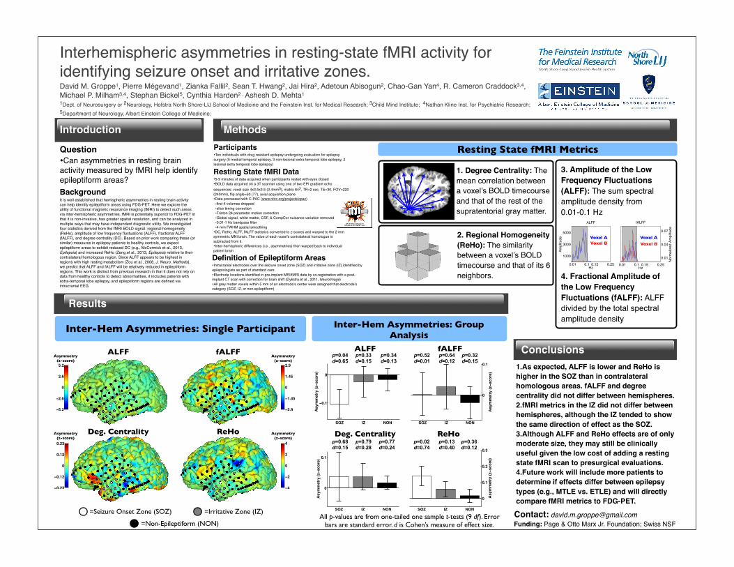
1.As expected, ALFF is lower and ReHo is higher in the SOZ than in contralateral homologous areas. fALFF and degree centrality did not differ between hemispheres.2.fMRI metrics in the IZ did not differ between hemispheres, although the IZ tended to show the same direction of effect as the SOZ.3.Although ALFF and ReHo effects are of only moderate size, they may still be clinically useful given the low cost of adding a resting state fMRI scan to presurgical evaluations.4.Future work will include more patients to determine if effects differ between epilepsy types (e.g., MTLE vs. ETLE) and will directly compare fMRI metrics to FDG-PET.
Interhemispheric asymmetries in resting-state fMRI activity for identifying seizure onset and irritative zones.
Introduction Methods
Results
BackgroundIt is well established that hemispheric asymmetries in resting brain activity can help identify epileptiform areas using FDG-PET. Here we explore the utility of functional magnetic resonance imaging (fMRI) to detect such areas via inter-hemispheric asymmetries. fMRI is potentially superior to FDG-PET in that it is non-invasive, has greater spatial resolution, and can be analyzed in multiple ways that may have independent diagnostic utility. We investigated four statistics derived from the fMRI BOLD signal: regional homogeneity (ReHo), amplitude of low frequency fluctuations (ALFF), fractional ALFF (fALFF), and degree centrality (DC). Based on prior work comparing these (or similar) measures in epilepsy patients to healthy controls, we expect epileptiform areas to exhibit reduced DC (e.g., McCormick et al., 2013, Epilepsia) and increased ReHo (Zeng et al., 2013, Epilepsia) relative to their contralateral homologous region. Since ALFF appears to be highest in regions with high resting metabolism (Zou et al., 2008, J. Neuro. Methods), we predict that ALFF and fALFF will be relatively reduced in epileptiform regions. This work is distinct from previous research in that it does not rely on data from healthy controls to detect abnormalities, it includes patients with extra-temporal lobe epilepsy, and epileptiform regions are defined via intracranial EEG.
Resting State fMRI Metrics
Conclusions
Resting State fMRI Data•5-9 minutes of data acquired when participants rested with eyes closed•BOLD data acquired on a 3T scanner using one of two EPI gradient echo sequences: voxel size 4x3.5x3.5 (3.4mm3), matrix 642, TR=2 sec, TE=30, FOV=220 (240mm), flip angle=50 (77), axial acquisition plane•Data processed with C-PAC (www.nitrc.org/projects/cpac)-first 4 volumes dropped-slice timing correction-Friston 24 parameter motion correction-Global signal, white matter, CSF, & CompCor nuisance variation removed-0.01-1 Hz bandpass filter -4 mm FWHM spatial smoothing
•DC, ReHo, ALFF, fALFF statistics converted to z-scores and warped to the 2 mm symmetric MNI brain. The value of each voxel’s contralateral homologue is subtracted from it.•Inter-hemispheric differences (i.e., asymmetries) then warped back to individual patient brain
Definition of Epileptiform Areas•Intracranial electrodes over the seizure onset zone (SOZ) and irritative zone (IZ) identified by epileptologists as part of standard care•Electrode locations identified in pre-implant MRI/fMRI data by co-registration with a post-implant CT scan with correction for brain shift (Dykstra et al., 2011, NeuroImage)•All grey matter voxels within 5 mm of an electrode’s center were assigned that electrode’s category (SOZ, IZ, or non-epileptiform)
Inter-Hem Asymmetries: Single Participant
All p-values are from one-tailed one sample t-tests (9 df). Error bars are standard error. d is Cohen’s measure of effect size.
Contact: [email protected]: Page & Otto Marx Jr. Foundation; Swiss NSF
fALFFALFFfALFF
ReHoDeg. Centrality
ALFF
=Seizure Onset Zone (SOZ) =Irritative Zone (IZ)
=Non-Epileptiform (NON)
Inter-Hem Asymmetries: Group Analysis
2. Regional Homogeneity (ReHo): The similarity between a voxel’s BOLD timecourse and that of its 6 neighbors.
1. Degree Centrality: The mean correlation between a voxel’s BOLD timecourse and that of the rest of the supratentorial gray matter.
3. Amplitude of the Low Frequency Fluctuations (ALFF): The sum spectral amplitude density from 0.01-0.1 Hz
4. Fractional Amplitude of the Low Frequency Fluctuations (fALFF): ALFF divided by the total spectral amplitude density
0.01 0.1 0.15 0.25
1000
3000
5000
Hz
Ampl
itude
ALFF
0.01 0.1 0.15 0.25
0.01
0.04
0.07
Hz
Nor
mal
ized
Am
plitu
de
fALFF
0.01 0.1 0.15 0.25
1000
3000
5000
Hz
Ampl
itude
ALFF
0.01 0.1 0.15 0.25
0.01
0.04
0.07
Hz
Nor
mal
ized
Am
plitu
de
fALFF
Voxel BVoxel A
Voxel BVoxel A
−0.23
−0.12
0
0.12
0.23
Asymmetry(z-score)
−4
−2
0
2
4
−5.2
−2.6
0
2.6
5.2
−2.9
−1.45
0
1.45
2.9
SOZ IZ NON
−0.1
0
Asym
met
ry (z−s
core
)
alff: G1p=0.04d=0.65
p=0.33d=0.15
p=0.34d=0.13
SOZ IZ NON
0
0.1
Asym
met
ry (z−s
core
)
falff: G1
SOZ IZ NON
0
0.1
Asym
met
ry (z−s
core
)
degcent: G1
Deg. Centrality
SOZ IZ NON
0
0.1
0.2
0.3
Asym
met
ry (z−s
core
)
reho: G1
ReHop=0.02d=0.74
p=0.13d=0.40
p=0.36d=0.12
p=0.52d=0.01
p=0.64d=0.12
p=0.32d=0.15
p=0.68d=0.15
p=0.79d=0.28
p=0.77d=0.24
Asymmetry(z-score)
Asymmetry(z-score)
Asymmetry(z-score)


![Falx and Interhemispheric Fissure on Axial CT: I. falx cerebri and interhemispheric fissure, although recognized early on axial CT [1], received little attention in the literature](https://img.pdfslide.net/doc/110x75/5d35b31788c993ee5c8c0e1d/falx-and-interhemispheric-fissure-on-axial-ct-i-falx-cerebri-and-interhemispheric.jpg)