Embed Size (px)
Citation preview

Posted at the Institutional Resources for Unique Collection and Academic Archives at Tokyo Dental College,
Available from http://ir.tdc.ac.jp/
TitleInterleukin-1β and 6-induced calcium channel
current modulation in MC3T3-E1 cells
Author(s) 小林, 弘史
Journal , (): -
URL http://hdl.handle.net/10130/3405
Right

1
Interleukin-1β and 6-induced calcium channel current modulation in MC3T3-E1 cells
Hiroshi Kobayashi
Department of Orthodontics, Tokyo Dental College, 1-2-2 Masago, Mihama-ku, Chiba
261-8502, Japan
18 pages
4 figures and figure legends
Keywords: Osteoblasts; Interleukin-1β; Interleukin-6; calcium channels

2
Abstract
Interleukin-1β (IL-1β) and -6 (IL-6) are inflammatory cytokines that are involved in
bone resorption under pathological conditions. Recently, the cytokines are also
demonstrated that they involved in bone formation under physiological conditions.
Osteoblasts are known to play a major role in the bone formation. Voltage sensitive Ca2+
channels (VSCCs) serve as crucial mediators of membrane excitability and many
Ca2+-dependent functions such as growth of bone, regulate proliferation, differentiation,
enzyme activity and gene expression. Several studies demonstrated that IL-1β and IL-6
modulated VSCCs in neurons. Previously, we have been reported that hormones and
peptides modulated VSCCs in pre-osteoblast cell line MC3T3-E1. In this study, the
effects of IL-1β and -6 on VSCCs current (ICa) carried by Ba2+ (IBa) in MC3T3-E1 were
investigated using patch-clamp recording. Application of 50 pM, 500 pM 5 nM and 50
nM IL-1β facilitated IBa 22.8 ± 4.0%, 21.7 ± 2.0%, 34.1± 3.9% and 56.7 ± 5.1%,
respectively. Application of 50 pM, 500 pM and 5 nM IL-6 facilitated IBa 15.8±2.7%,
22.9±4.5% and 35.6±4.0%, respectively. In contrast, 50 nM IL-6 inhibited IBa 21.9±
3.4%. In conclusion, IL-1β facilitated VSCCs in MC3T3-E1 cells. In addition, we
acquired two different groups of results in IL-6: IL-6-induced facilitation and inhibition
of VSCCs in MC3T3-E1 cells. We considered that such biphasic effects are depends on
IL-6 receptor's intracellular signals transduction mechanisms.

3
1. Introduction
Interleukin-1β (IL-1β), a potent inflammatory cytokine, is upregulated during
inflammation mainly produced by activated monocytes and macrophages [1,2]. It
stimulates bone resorption in several cell types, including osteoblasts [3]. IL-6 is a
multifunctional cytokine which has diverse effects on bone metabolism. IL-6 has been
shown to affect growth and differentiation in osteoblasts [4-6].
Osteoblasts are known to play a major role in the bone formation. It is well accepted
that osteoblasts express IL-1β and -6 receptors and therefore it plays an important role
in bone remodeling [7,8].
Voltage sensitive Ca2+ channels (VSCCs) serve as crucial mediators of membrane
excitability [9] and many Ca2+-dependent functions such as growth of bone [10],
regulate proliferation [11], differentiation [12], enzyme activity [13] and gene expression
[14]. It also has been demonstrated that osteoblasts express VSCCs [15,16]. Several
reports demonstrated that IL-1β and -6 modulated VSCCs in neuronal cells [17,18].
However, the mechanism of IL-1β and -6 effects on VSCCs in osteoblasts remains
unclear and even controversial.
Consequently, in this study we investigated the effect of IL-1β and -6 on VSCCs in
osteoblast cell line (MC3T3-E1).
2. Materials and Methods
2.1. Cell Culture

4
Murine osteoblastic MC3T3-E1 cells were purchased from Summit Pharmaceuticals
International Corporation (Tokyo, Japan). Cells were cultured at 37°C in a 5%(v/v) CO2
atmosphere with α-modified minimal essential medium (α-MEM; Gibco BRL, Grand
Island, NY, U.S.A.). Unless otherwise specified, the medium contained 10%(v/v)
heat-inactivated fetal bovine serum, 100 U/ml penicillin and 100 mg/ml streptomycin.
Cell culture medium was changed every 2-3 days. For patch-clamp experiments, cells
were harvested using a 0.05% trypsin/0.02% EDTA solution, when cells reached
confluence. Cells were plated at very low density in 35 mm tissue culture dishes. Prior
to recordings, the cells were washed at least three times with Krebs solution of the
following composition (in mM): 136 NaCl; 5 KCl; 2.5 CaCl2; 0.5 MgCl2; 10.9 glucose;
11.9 NaHCO3 and 1.1 NaH2PO4. The pH was 7.3-7.4. Cell culture reagents were
purchased from Sigma (Mo, U.S.A). Our recordings were performed on 10-30 μm in
diameter, since they were well suited for patch clamp recordings.
2.2. Whole-cell patch-clamp recordings
Voltage-clamp recordings were conducted using the whole-cell configuration of the
patch-clamp technique [19]. Fabricated recording pipettes (2-3 MΩ) were filled with the
internal solution of the following composition (in mM): 150 CsCl; 5 EGTA; 10 D-glucose
and 10 HEPES. The pH was adjusted to 7.3 with CsOH. After the formation of a giga
seal, in order to record VSCCs current (ICa) carried by Ba2+ (IBa), the extracellular
solution was replaced from Krebs solution to a solution containing the following (in
mM): 115 BaCl2 and 20 HEPES. The pH was adjusted to 7.4 with tetraethylammonium
hydroxide. Command voltage protocols were generated with a computer software

5
pCLAMP version 10 (Axon Instruments, Union City, CA, U.S.A.) and transformed to an
analogue signal using a DigiData1440A interface (Axon Instruments, Union City, CA,
U.S.A.). The command pulses were applied to cells through an L/M-EPC7 amplifier
(HEKA Elektronik, Lambrecht, Germany). The currents were recorded with the
amplifier and a computer software pCLAMP10 acquisition system. Access resistance (<
15 MΩ) was determined by transient responses to voltage commands.
2.3. Chemicals
IL-1β and 6 were purchased from Sigma (Saint Louis, MO, U.S.A.).
2.4. Analysis
All date analyses were performed using the pCLAMP10 acquisition system. Values in
text and figures are expressed as mean ± S.E.M.
3. Results
3.1. IL-1β facilitated IBa
Fig. 1A and B shows that, in the presence of high external Ba2+, 5 nM IL-1β facilitated
IBa as shown with raw current traces obtained at +10 mV from ―80 mV in the absence
and presence of IL-1β. IBa was evoked every 5 s with a 100 ms depolarizing voltage step
to +10 mV from a holding potential of ―80 mV. As shown in Fig. 1A, application of 5

6
nM IL-1β facilitated IBa from ―161 pA to ―225 pA (39.7% facilitation) in this cell. To
investigate the voltage dependency of facilitation of IBa by IL-1β, we analyzed the
current-voltage relations in the absence and presence of IL-1β. The current-voltage
relations measured before and during application of IL-1β are shown in Fig. 1C. From a
holding potential of ―80 mV, IBa was activated with a peak current amplitude at +20
mV. IL-1β did not alter the current-voltage relationship. These results suggest that 5
nM IL-1β facilitated IBa in MC3T3-E1 cells.
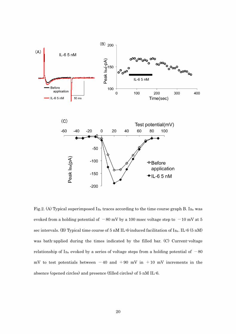
3.2. IL-6 facilitated IBa
Fig. 2A and B shows that 5 nM IL-6 facilitated IBa as shown with raw current traces
obtained at +10 mV from ―80 mV in the absence and presence of IL-6. IBa was evoked
every 5 s with a 100 ms depolarizing voltage step to +10 mV from a holding potential of
―80 mV. As shown in Fig. 2A, application of 5 nM IL-6 facilitated IBa from ―75 pA to
―103 pA (37.3% facilitation) in this cell.
To investigate the voltage dependency of facilitation of IBa by 5 nM IL-6, we analyzed
the current-voltage relations in the absence and presence of 5 nM IL-6. The
current-voltage relations measured before and during application of 5 nM IL-6 are
shown in Fig. 2C. From a holding potential of ―80 mV, IBa was activated with a peak
current amplitude at +20 mV. IL-6 did not alter the current-voltage relationship.
These results suggest that 5 nM IL-6 facilitated IBa in MC3T3-E1 cells.
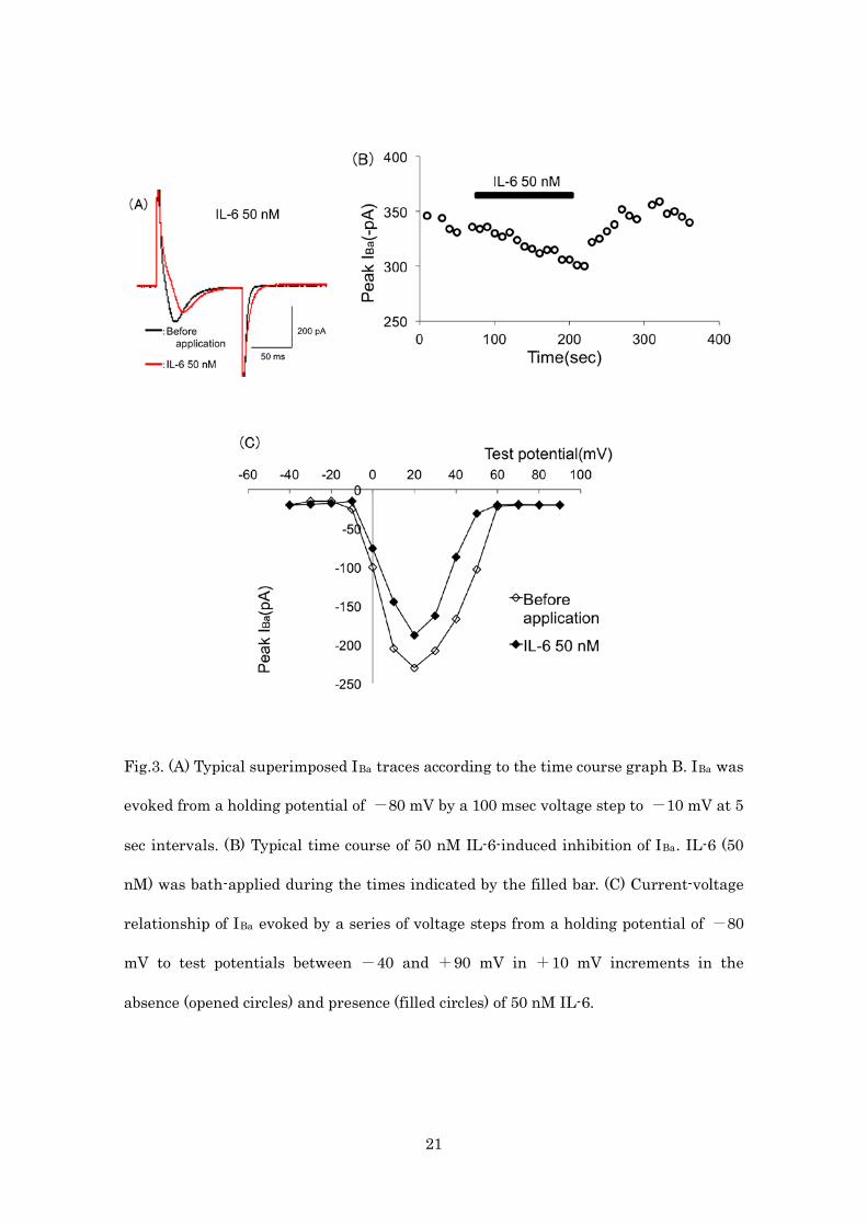
3.3. IL-6 inhibited IBa

7
In addition to facilitation, IL-6-induced inhibition of IBa could be observed. Fig. 3A and
B shows that 50 nM IL-6 inhibited IBa as shown with raw current traces obtained at +
10 mV from ―80 mV in the absence and presence of IL-6. IBa was evoked every 5 s with
a 100 ms depolarizing voltage step to +10 mV from a holding potential of ―80 mV. As
shown in Fig. 3A, application of 50 nM IL-6 inhibited IBa from ―328 pA to ―236 pA
(28.0% inhibition) in this cell.
To investigate the voltage dependency of facilitation of IBa by 50 nM IL-6, we analyzed
the current-voltage relations in the absence and presence of 50 nM IL-6. The
current-voltage relations measured before and during application of 50 nM IL-6 are
shown in Fig. 3C. From a holding potential of ―80 mV, IBa was activated with a peak
current amplitude at +20 mV. IL-6 did not alter the current-voltage relationship.
These results suggest that 50 nM IL-6 inhibited IBa in MC3T3-E1 cells.
3.4. IL-1β and -6 modulated IBa depends on concentration
The concentration-response relationship in the IL-1β and -6-induced modulation of IBa
is shown in Fig. 4A and B, respectively.
Application of 50 pM-50 nM IL-1β facilitated IBa. To generate a concentration-response
curve, IL concentrations were applied randomly, and each MC3T3-E1 cells were exposed
to only a single concentration. Fig. 4A shows that progressive voltage-dependent
facilitations in IL-1β concentrations resulted in a progressively greater facilitation of
IBa.
In addition, application of 50 pM-5 nM IL-6 facilitated IBa. In contrast to above results,
50 nM IL-6 inhibited IBa in MC3T3-E1 cells.
8
4. Discussion
The present study investigated the effects of IL-1β and -6 on IBa in MC3T3-E1 cells.
Application of 50 pM-50 nM IL-1β facilitated IBa. Application of 50 pM-5 nM IL-6
facilitated IBa. In contrast, 50 nM IL-6 inhibited IBa in MC3T3-E1 cells.
In osteoblastic cells, several studies demonstrated that hormones modulated VSCCs,
included in parathyroid hormone (PTH) [20-22], bradykinin [23] and
1,25-dihydroxyvitamin D3 (1,25(OH)2D3) [24-26]. To our knowledge, the data presented
here demonstrate for the first time that IL-1β and -6 modulates VSCCs in osteoblasts.
In contrast to our results, Green et al. reported that IL-6 did not alter basal
intracellular Ca2+ concentration ([Ca2+]i) but inhibited Ca2+ transient induced by PTH,
prostaglandin E2 and endothelin-1 in osteoblast [27].
In this study, we have shown that 50 pM-50 nM IL-1β facilitated IBa. It has been
demonstrated that local treatment with IL-1β (0.5 ng/h) for 72 h resulted in increased
numbers of osteoblasts 14 days as measured histologically [28]. Increased DNA
synthesis in response to IL-1β in vitro has been observed in osteoblast [29] and
especially MC3T3-E1 cells [30]. In contrast, however, it has been reported that IL-1β
inhibits the stimulation of osteocalcin synthesis and has no influence on proliferation
[31]. IL-1β also increases both osteoprotegerin protein release and mRNA levels [32,33].
Recent study demonstrated that treatment with IL-1β resulted in biphasic effects on
osteoblast differentiation. Short-term exposure (2 days) to IL-1β early in culture induces
differentiation. Longer term exposure (6 days) to IL-1β inhibits osteoblast
differentiation [34].

9
In this study, we have shown that 50 pM-5 nM IL-6 facilitated IBa. In contrast, 50 nM
IL-6 inhibited IBa. What is the physiological relevance of IL-6-induced dual modulation
of VSCCs, i.e. both facilitation and inhibition? Several studies indicated that IL-6
enhance in vitro differentiation on osteoblasts [35-40]. In contrast to above reports,
other studies have shown an inhibitory effect of IL-6 on bone formation [41,42]. It can be
considered that such dual effects are depends on IL-6 receptor's intracellular signals
transduction mechanisms. For example, activation of STAT3 is necessary for osteoblast
differentiation and bone formation induced by IL-6 [39]. On the other hand, PKCδ and
ERK1/2 are implicated in IL-6's inhibitory effect on bone formation [43]. It is also
possible that these dual effects depend on the differentiation stage of the osteoblast.
IL-6 would stimulate the first stages of differentiation but on more mature cells, they
would prevent further stimulation [41,42].
In this study, we used 50 pM-50 nM IL-1β and IL-6 to modulate IBa. These IL
concentrations are quite high. In normal state, serum IL-1β concentration is 0.9 pg/ml
approximately [44]. In the electrophysiological study and the immunocytochemistry
study, however, several study demonstrated that high concentration, i.e. 0.57 nM and
4.5 nM, of IL was used to modulate ion channel activity and protein expression in single
neuron [45,46]. IL-1β and -6 were applied by perfusion pressure ejection from perfusion
tubes with a tip diameter of 1 mm placed within 10 mm of the cell surface. Although the
concentration of IL-1β and -6 actually seen by the cell is certainly lower than that
contained in the stock solution, we applied the IL-1β and -6 using same perfusion
system.
We also observed that IBa were recovered to values of peak IBa after IL wash out using
time course graph (Figs. 1B, 2B and 3B). Therefore, it can be considered that 50 pM-50

10
nM IL-1β and IL-6 had no effect on the cell function.
In this study, we used mouse MC3T3-E1 cell line. Chung et al. showed significant
changes in MC3T3-E1 cell morphology, cell proliferation, osteoblastic function, and
responsiveness to various protein after multiple passage (> 65) [47]. In a further study,
cell passage numbers should be defined to prevent change the cell function.
As mentioned above, VSCCs are essential in the regulation of many cellular processes
including gene expression and cell growth. Thus, IL-1β and -6 modulation of somatic
Ca2+ influx via VSCCs could potentially affect Ca2+-dependent gene expression and cell
development [11, 12, 14].
5. Conclusion
In conclusion, IL-1β facilitated VSCCs in MC3T3-E1 cells. In addition, we acquired two
different groups of results in IL-6: IL-6-induced facilitation and inhibition of VSCCs in
MC3T3-E1 cells.

11
References
[1] C.A. Dinarello, Interleukin-1, Cytokine Growth Factor Rev 8 (1997) 253―265.
[2] E. Stylianou, J. Saklatvala, Interleukin-1, Int J Biochem Cell Biol 30 (1998)
1075―1079.
[3] C.A. Dinarello, Biology of interleukin 1, FASEB J 2 (1988) 108―115.
[4] Y. Ishimi, C. Miyaura, C.H. Jin, T. Akasu, E. Abe, Y. Nakamura, A. Yamaguchi, S.
Yoshiki, T. Matsuda, T. Hirano, T. Kishimoto, T. Suda, IL-6 is produced by osteoblasts
and induces bone resorption, J Immunol 145 (1990) 3297―3303.
[5] M.A. Fang, T.J. Hahn, Effects of interleukin-6 on cellular function in UMR-106-01
osteoblast like cells, J Bone Miner Res 6 (1991) 133―139.
[6] F.J. Hughes, G.L. Howells, Interleukin-6 inhibits bone formation in vitro, Bone
Miner 21 (1993) 21―28.
[7] V. Shen, S.-L. Cheng, N.G. Kohler, W.A. Peck, Characterization and hormonal
modulation of IL-1 binding in neonatal mouse osteoblast like cells, J Bone Miner Res 5
(1990) 507―515.
[8] F. Blanchard, L. Duplomb, M. Baud’huin, B. Brounais, The dual role of IL-6-type

12
cytokines on bone remodeling and bone tumors, Cytokine Growth Factor Rev 20 (2009)
19―28.
[9] R.J. Miller, Multiple calcium channels and neuronal function, Science 235 (1987)
46―52.
[10] J. Duriez, B. Flautre, M.C. Blary, P. Hardouin, Effects of the calcium channel
blocker nifedipine on epiphyseal growth plate and bone turnover: a study in rabbit,
Calcif Tissue Int 52 (1993) 120―124.
[11] J. Loza, E. Stephan, C. Dolce, R. Dziak, S. Simasko, Calcium currents in
osteoblastic cells: dependence upon cellular growth stage, Calcif Tissue Int 55 (1994)
128―133.
[12] L. Wen, Y. Wang, H. Wang, L. Kong, L. Zhang, X. Chen, Y. Ding, L-type calcium
channels play a crucial role in the proliferation and osteogenic differentiation of bone
marrow mesenchymal stem cells, Biochem Biophys Res Commun 424 (2012) 439―445.
[13] H. Reuter, Calcium channel modulation by neurotransmitters, enzymes and drugs,
Nature 301 (1983) 569―574.
[14] T.H. Murphy, P.H. Worley, J.M. Baraban, L-type voltage-sensitive calcium channels
mediate synaptic activation of immediate early genes, Neuron 7 (1991) 625―635.

13
[15] R. L. Duncan, K.A. Akanbi, M.C. Farach-Carson, Calcium signals and calcium
channels in osteoblastic cells, Sem Nephrol, 18 (1998) 178―190.
[16] F. McDonald, Ion channels in osteoblasts: A story of two intracellular organelles,
Surg J R Coll Surg Edinb Irel 2, 2 (2004) 63―69.
[17] S.H. Ma, B. Li, H.W. Huang, Y.P. Peng, Y.H. Qiu, Interleukin-6 inhibits L-type
calcium channel activity of cultured cerebellar granule neurons, J Physiol Sci 62 (2012)
385―392.
[18] C. Zhou, H.H. Ye, S.Q. Wang, Z. Chai, Interleukin-1β regulation of N-type
Ca2+ channels in cortical neurons, Neurosci Lett 403 (2006) 181―185.
[19] O.P. Hamill, A. Marty, E. Neher, B. Sakmann, F.J. Sigworth, Improved patch-clamp
techniques for high-resolution current recording from cells and cell-free membrane
patches, Pflugers Arch 391 (1981) 85―100.
[20] D.T. Yamaguchi, T.J. Hahn, A. Iida-Klein, C.R. Kleeman, S.
Muallem, Parathyroid hormone-activated calcium channels in an osteoblast-like clonal
osteosarcoma cell line. cAMP-dependent and cAMP-independent calcium channels, J
Biol Chem 262 (1987) 7711―7718.
[21] J. Green, C.R Kleeman, S. Schotland, C.
Chaimovitz, Acute phosphate depletion dissociates hormonal stimulated second messen

14
gers in osteoblast-like cells, Endocrinology 129 (1991) 848―858.
[22] W. Li, R.L. Duncan, N.J. Karin, M.C. Farach-Carson, 1,25 (OH)2D3 enhances
PTH-induced Ca2+ transients in preosteoblasts by activating L-type Ca2+ channels, Am
J Physiol 273 (1997) E599―E605.
[23] H. Tokuda, J. Kotoyori, Y. Oiso, O. Kozawa, Intracellular signaling mechanism
of bradykinin in osteoblast-like cells: comparison with prostaglandin E2, Endocrinol J 4
(1994) 189―195.
[24] J.J. Bergh, Y. Shao, E. Puente, R.L. Duncan, M.C. Farach-carson, Osteoblast Ca2+
permeability and voltage-sensitive Ca2+ channel expression is temporally regulated by
1,25-dihydroxyvitamin D3, Am J Physiol 290 (2006) C822―C831,
[25] L.P. Zanello, A. Norman, 1α,25(OH)2 vitamin D3 actions on ion channels
in osteoblasts, Steroids 71 (2006) 291―297.
[26] Y. Uchida, T. Endoh, M. Tazaki, K Sueishi, Chronic bradykinin treatment alters
1α,25-dihydroxyvitamin D3-induced calcium current modulation in pre-osteoblasts, Cell
Calcium 51 (2012) 383―392.
[27] J. Green, S. Schotland, Z Sella, C.R. Kleeman, Interleukin-6 attenuates
agonist-mediated calcium mobilization in murine osteoblastic cells, J Clin Invest 93
(1994) 2340―2350.
15
[28] M.L. Olmega, P.S. Landry, K.K. Sadasivan, J.A. Albright, W.D. Meek, R. Routh, A.A.
Marino, Regulation of osteoblast levels during bone healing, J Orthop Trauma 13 (1999)
356―362.
[29] D.B. Evans, R.A. Bunning, J. Van Damme, R.G. Russell, Natural human IL-1 beta
exhibits regulatory actions on human bone-derived cells in vitro, Biochem Biophys Res
Commun 159 (1989) 1242―1248.
[30] E. Ikeda, M. Kusaka, Y. Hakeda, K. Yokota, M. Kumegawa, S. Yamamoto, Effect
of interleukin 1 beta on osteoblastic clone MC3T3-E1 cells, Calcif Tiss Int 43 (1988)
162―166.
[31] R.S. Taichman, P.V. Hauschka, Effects of interleukin-1 beta and tumor necrosis
factor-alpha on osteoblastic expression of osteocalcin and mineralized extracellular
matrix in vitro, Inflammation 16 (1992) 587―601.
[32] L.C. Hofbauer, C.R. Dunstan, T.C. Spelsberg, B.L. Riggs, S. Khosla,
Osteoprotegerin production by human osteoblast lineage cells is stimulated by vitamin
D, bone morphogenetic protein-2, and cytokines, Biochem Biophys Res Commun 250
(1998) 776―781.
[33] L.C. Hofbauer, D.L. Lacey, C.R. Dunstan, T.C. Spelsberg, B.L. Riggs, S. Khosla,
Interleukin-1beta and tumor necrosis factor-alpha, but not interleukin-6, stimulate

16
osteoprotegerin ligand gene expression in human osteoblastic cells, Bone 25 (1999)
255―259.
[34] F.H. Lin, J.B. Chang, M.H. McGuire, J.A. Yee, B.E. Brigman, Biphasic effects of
Interleukin-1β on osteoblast differentiation in vitro, J Orthop Res 28 (2010) 958―964.
[35] X.H. Liu, A. Kirschenbaum, S. Yao, A.C. Levine, The role of the interleukin-6/gp130
signaling pathway in bone metabolism, Vitam Horm 74 (2006) 341―355.
[36] P.K. Wong, I.K. Campbell, P.J. Egan, M. Ernst, I.P. Wicks, The role of the
interleukin-6 family of cytokines in inflammatory arthritis and bone turnover, Arthritis
Rheum 48 (2003) 1177―1189.
[37] N. Franchimont, S. Wertz, M. Malaise, Interleukin-6: an osteotropic factor
influencing bone formation? Bone 37 (2005) 601―606.
[38] T. Bellido, V.Z. Borba, P. Roberson, S.C. Manolagas, Activation of the Janus
kinase/STAT (signal transducer and activator of transcription) signal transduction
pathway by interleukin-6-type cytokines promotes osteoblast differentiation,
Endocrinology 138 (1997) 3666―3676.
[39] S. Itoh, N. Udagawa, N. Takahashi, F. Yoshitake, H. Narita, S. Ebisu, K. Ishihara, A
critical role for interleukin-6 family-mediated Stat3 activation in osteoblast
differentation and bone formation, Bone 39 (2006) 505―512.

17
[40] T. Bellido, C.A. O’Brien, P.K. Roberson, S.C. Manolagas, Transcriptional activation
of the p21 (WAF1, CIP1, SDI1) gene by interleukin-6 type cytokines. A prerequisite for
their pro-differentiating and anti-apoptotic effects on human osteoblastic cells, J Biol
Chem 273 (1998) 21137―21144.
[41] L. Malaval, J.E. Aubin, Biphasic effects of leukemia inhibitory factor on
osteoblastic differentiation, J Cell Biochem 81 (2001) 63―70.
[42] L. Malaval, F. Liu, A.B. Vernallis, J.E. Aubin, GP130/OSMR is the only LIF/IL-6
family receptor complex to promote osteoblast differentiation of calvaria progenitors, J
Cell Physiol 204 (2005) 585―593.
[43] C. Chipoy, M. Berreur, S. Couillaud, G. Pradal, F. Vallette, C. Colombeix, F. Rédini,
D. Heymann, F. Blanchard, Downregulation of osteoblast markers and induction of the
glial fibrillary acidic protein by oncostatin M in osteosarcoma cells require PKCdelta
and STAT3, J Bone Miner Res 19 (2004) 1850―1861.
[44] J. S. Chang, Association between interleukin 1beta and interleukin 10
concentrations: a cross-sectional study in young adolescents in Taiwan, BMC Pediatr,
13(2013) 123.
[45] C. Zhou, H.H. Ye, S.Q. Wang, Z. Chai, Interleukin-1β regulation of N-type
Ca2+ channels in cortical neurons, Neurosci Lett 403 (2006) 181―185.

18
[46] O. Islam, Interleukin-6 and neural stem cells: more than gliogenesis, Mol Biol Cell
20 (2009) 188―99.
[47] C.Y. Chung, A, Iida-Klein, L.E. Wyatt, G.H. Rudkin, K. Ishida, D.T. Yamaguchi, T.A.
Miller, Serial passage of MC3T3-E1 cells alters osteoblastic function and responsiveness
to transforming growth factor-β1 and bone morphogenetic protein-2. Biochem Biophys
Res Commun. (1999), 265, 246-51.
Figure legends

19
Fig.1. (A) Typical superimposed IBa traces according to the time course graph B. IBa was
evoked from a holding potential of -80 mV by a 100 msec voltage step to -10 mV at 5
sec intervals. (B) Typical time course of 5 nM IL-1β-induced facilitation of IBa. IL-1β (5
nM) was bath-applied during the times indicated by the filled bar. (C) Current-voltage
relationship of IBa evoked by a series of voltage steps from a holding potential of -80
mV to test potentials between -40 and +90 mV in +10 mV increments in the
absence (opened circles) and presence (filled circles) of 5 nM IL-1β.
20
Fig.2. (A) Typical superimposed IBa traces according to the time course graph B. IBa was
evoked from a holding potential of -80 mV by a 100 msec voltage step to -10 mV at 5
sec intervals. (B) Typical time course of 5 nM IL-6-induced facilitation of IBa. IL-6 (5 nM)
was bath-applied during the times indicated by the filled bar. (C) Current-voltage
relationship of IBa evoked by a series of voltage steps from a holding potential of -80
mV to test potentials between -40 and +90 mV in +10 mV increments in the
absence (opened circles) and presence (filled circles) of 5 nM IL-6.
21
Fig.3. (A) Typical superimposed IBa traces according to the time course graph B. IBa was
evoked from a holding potential of -80 mV by a 100 msec voltage step to -10 mV at 5
sec intervals. (B) Typical time course of 50 nM IL-6-induced inhibition of IBa. IL-6 (50
nM) was bath-applied during the times indicated by the filled bar. (C) Current-voltage
relationship of IBa evoked by a series of voltage steps from a holding potential of -80
mV to test potentials between -40 and +90 mV in +10 mV increments in the
absence (opened circles) and presence (filled circles) of 50 nM IL-6.

22
Fig.4. (A) Histogram demonstrating the degree of IBa modulation by 50 pM-50 nM IL-1β.
(B) Histogram demonstrating the degree of IBa modulation by 50 pM-50 nM IL-6.