Embed Size (px)
Citation preview
Proc. Natl. Acad. Sci. USAVol. 81, pp. 1117-1121, February 1984Cell Biology
Intermediate filaments in monkey kidney TC7 cells: Focal centersand interrelationship with other cytoskeletal systems
(keratins/vimentin/microtubules/cytochalasin B/microfilaments)
JULIO E. CELIS*, J. VICTOR SMALLt, PETER MOSE LARSEN*, STEPHEN J. FEY*, JAN DE MEYt, ANDARIANA CELIS**Division of Biostructural Chemistry, Department of Chemistry, Arhus University, DK-8000 Arhus C, Denmark; tInstitute of Molecular Biology, AustrianAcademy of Sciences, Salzburg, Austria; and Laboratory of Oncology, Janssen Pharmaceutica Research Laboratories, Beerse, Belgium
Communicated by Diter von Wettstein, October 13, 1983
ABSTRACT Two-dimensional gel electrophoresis of inter-mediate-sized filament-enriched cytoskeletons of epithelialmonkey kidney TC7 cells has shown that they are composed ofat least two keratins (isoelectric focusing 36, Mr = 48,500; IEF46, Mr = 43,500; HeLa protein catalogue number) and vimen-tin. Indirect immunofluorescence as well as immunoelectronmicroscopy using antibodies directed against specific polypep-tides sometimes revealed a discontinuous staining of keratin-containing filaments. Indirect immunofluorescence analysis ofcells stained with keratin or vimentin antibodies also revealeda bright perinuclear staining in 58% of the cells in interphase.Of particular interest were focal centers from which filamentsradiated. Double-label immunofluorescence using tubulin andkeratin antibodies showed that these centers codistributedwith focal arrays of microtubules (most likely centrosomes) ininterphase cells but were not colocalized with centrioles in mi-tosis or, in many cases, with the microtubule organizing cen-ters seen after release from nocodazole treatment. Treatmentof TC7 cells with demecolcine (10 ,ug/ml, 20 hr) resulted in adrastic rearrangement of the keratin and vimentin filaments.Likewise, treatment with cytochalasin B (10 ,ug/ml, 1 hr) pro-duced a star-like arrangement of the keratin and vimentin fila-ments and, in most cases, these codistributed with patches ofactin. The results provide evidence for the interaction of inter-mediate filaments (keratins and vimentin) with both microtu-bules and microfilaments.
Intermediate-sized filaments (7-11 A in diameter) are ubiqui-tous elements (for references, see refs. 1 and 2) that havebeen implicated in various intracellular roles, including nu-clear anchorage (3, 4), organelle interactions (5-12), andgene expression (13); however, the molecular mechanismsunderlying these functions remain largely unknown. To date,much emphasis has been placed on determining their subunitcomposition (for references, see refs. 1, 2 and 14), with lessemphasis on their structural and functional relationshipswith other cytoskeletal systems. In the present study we an-alyzed the composition and distribution of intermediate-sized filaments in epithelial monkey kidney TC7 cells. Theresults, including data on cells treated with either demecol-cine or cytochalasin B, provide evidence for the interactionof intermediate filaments (keratins and vimentin) with bothmicrotubules and microfilaments.
MATERIALS AND METHODSCells. African green monkey kidney TC7 cells (a gift of A.
Graessmann) were grown as monolayer cultures in Dulbec-co's modified Eagle's medium supplemented with 10% fetalcalf serum and antibiotics (penicillin, 100 internationalunits/ml; streptomycin, 50 ,g/ml).
The procedures for labeling cells with [35S]methionine (15,16), two-dimensional gel electrophoresis (17, 18), prepara-tion of low-salt- and high-salt-extracted cytoskeletons (4),and indirect immunofluorescence (10) have been describedin detail elsewhere. The vimentin antibody was a gift from S.Blose.Immunoelectron Microscopy. Immunoelectron microscopy
was performed on unfixed intermediate filament-enrichedcytoskeletons on electron microscope grids (4) using the in-direct immunogold staining procedure according to De Meyet al. (19). After antibody treatment, cytoskeletons werenegatively stained with aqueous uranyl acetate (4).
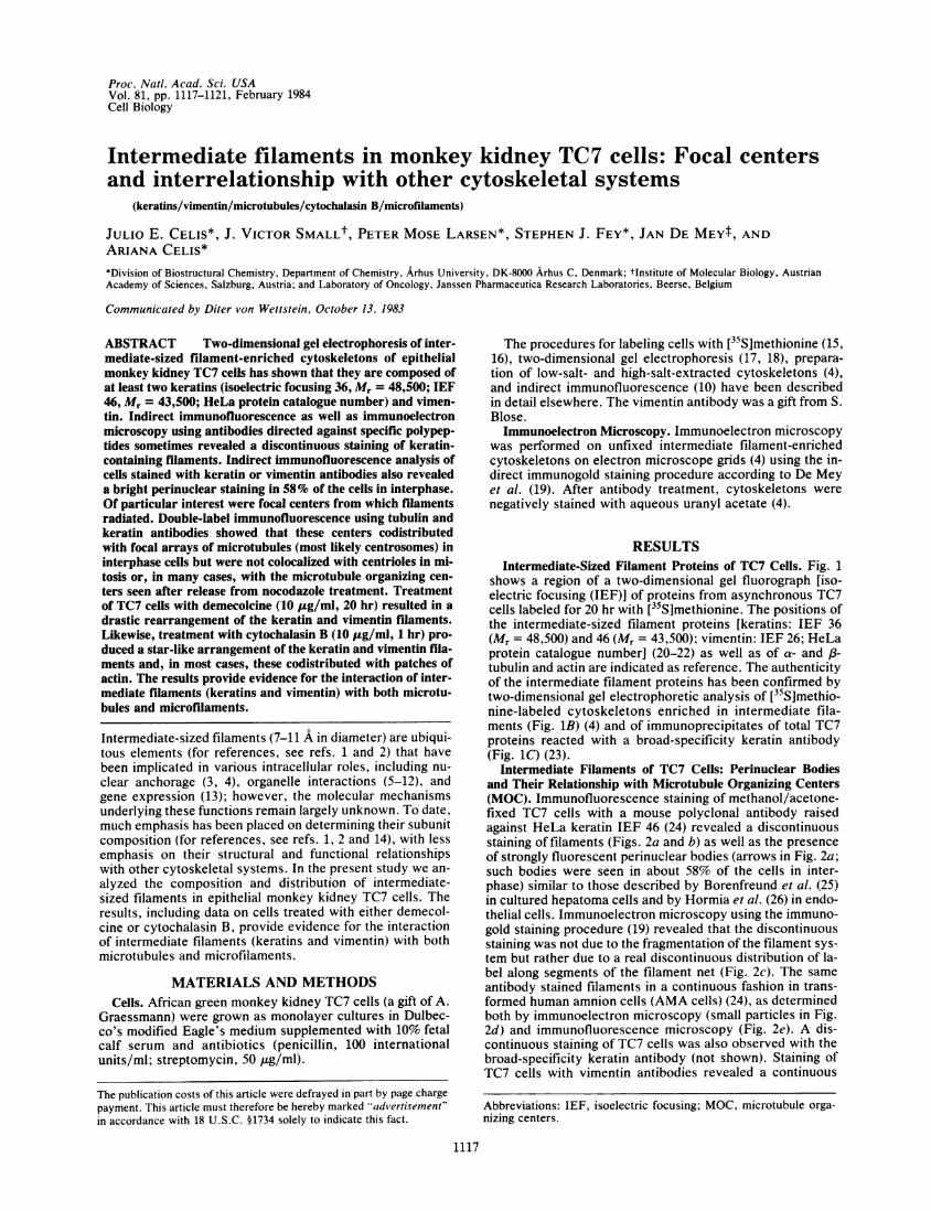
RESULTSIntermediate-Sized Filament Proteins of TC7 Cells. Fig. 1
shows a region of a two-dimensional gel fluorograph [iso-electric focusing (IEF)] of proteins from asynchronous TC7cells labeled for 20 hr with [35S]methionine. The positions ofthe intermediate-sized filament proteins [keratins: IEF 36(Mr = 48,500) and 46 (Mr = 43,500); vimentin: IEF 26; HeLaprotein catalogue number] (20-22) as well as of a- and l3-tubulin and actin are indicated as reference. The authenticityof the intermediate filament proteins has been confirmed bytwo-dimensional gel electrophoretic analysis of [35S]methio-nine-labeled cytoskeletons enriched in intermediate fila-ments (Fig. 1B) (4) and of immunoprecipitates of total TC7proteins reacted with a broad-specificity keratin antibody(Fig. 1C) (23).
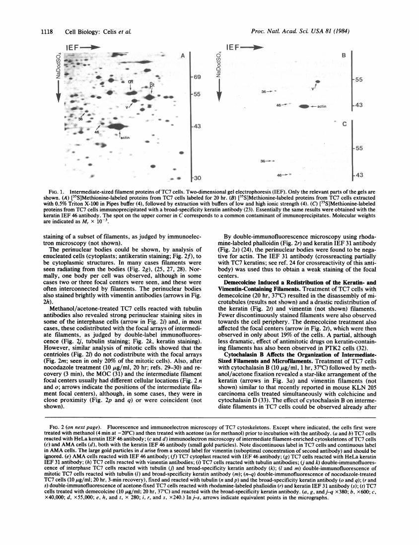
Intermediate Filaments of TC7 Cells: Perinuclear Bodiesand Their Relationship with Microtubule Organizing Centers(MOC). Immunofluorescence staining of methanol/acetone-fixed TC7 cells with a mouse polyclonal antibody raisedagainst HeLa keratin IEF 46 (24) revealed a discontinuousstaining of filaments (Figs. 2a and b) as well as the presenceof strongly fluorescent perinuclear bodies (arrows in Fig. 2a;such bodies were seen in about 58% of the cells in inter-phase) similar to those described by Borenfreund et al. (25)in cultured hepatoma cells and by Hormia et al. (26) in endo-thelial cells. Immunoelectron microscopy using the immuno-gold staining procedure (19) revealed that the discontinuousstaining was not due to the fragmentation of the filament sys-tem but rather due to a real discontinuous distribution of la-bel along segments of the filament net (Fig. 2c). The sameantibody stained filaments in a continuous fashion in trans-formed human amnion cells (AMA cells) (24), as determinedboth by immunoelectron microscopy (small particles in Fig.2d) and immunofluorescence microscopy (Fig. 2e). A dis-continuous staining of TC7 cells was also observed with thebroad-specificity keratin antibody (not shown). Staining ofTC7 cells with vimentin antibodies revealed a continuous
Abbreviations: IEF, isoelectric focusing; MOC, microtubule orga-nizing centers.
1117
The publication costs of this article were defrayed in part by page chargepayment. This article must therefore be hereby marked cadv'ertisement"in accordance with 18 U.S.C. §1734 solely to indicate this fact.
Proc. Natl. Acad. Sci. USA 81 (1984)
IEF
O. 4 e.
_0
40.g / 4.
36 *&
4.
*>1 4 46
A
4;.6 CO
. .c
*O *V/ - a
-4: f
actin
a
6Co00
-69 z
-55 *
IEF I-
B
43
30
FIG. 1. Intermediate-sized filament proteins of TC7 cells. Two-dimensional gel electrophoresis (IEF). Only the relevant parts of the gels areshown. (A) [35S]Methionine-labeled proteins from TC7 cells labeled for 20 hr. (B) [15S]Methionine-labeled proteins from TC7 cells extractedwith 0.5% Triton X-100 in Pipes buffer (4), followed by extraction with buffers of low and high ionic strength (4). (C) [35S]Methionine-labeledproteins from TC7 cells immunoprecipitated with a broad-specificity keratin antibody (23). Essentially the same results were obtained with thekeratin IEF 46 antibody. The spot on the upper corner in C corresponds to a common contaminant of immunoprecipitates. Molecular weightsare indicated as Mr x 10-3.
staining of a subset of filaments, as judged by immunoelec-tron microscopy (not shown).The perinuclear bodies could be shown, by analysis of
enucleated cells (cytoplasts; antikeratin staining; Fig. 2f), tobe cytoplasmic structures. In many cases filaments wereseen radiating from the bodies (Fig. 2g), (25, 27, 28). Nor-mally, one body per cell was observed, although in somecases two or three focal centers were seen, and these wereoften interconnected by filaments. The perinuclear bodiesalso stained brightly with vimentin antibodies (arrows in Fig.2h).
Methanol/acetone-treated TC7 cells reacted with tubulinantibodies also revealed strong perinuclear staining sites insome of the interphase cells (arrow in Fig. 2i) and, in mostcases, these codistributed with the focal arrays of intermedi-ate filaments, as judged by double-label immunofluores-cence (Fig. 2j, tubulin staining; Fig. 2k, keratin staining).However, similar analysis of mitotic cells showed that thecentrioles (Fig. 21) do not codistribute with the focal arrays(Fig. 2m; seen in only 20% of the mitotic cells). Also, afternocodazole treatment (10 pug/ml, 20 hr; refs. 29-30) and re-covery (3 min), the MOC (31) and the intermediate filamentfocal centers usually had different cellular locations (Fig. 2 nand o; arrows indicate the positions of the intermediate fila-ment focal centers), although, in some cases, they were inclose proximity (Fig. 2p and q) or were coincident (notshown).
By double-immunofluorescence microscopy using rhoda-mine-labeled phalloidin (Fig. 2r) and keratin IEF 31 antibody(Fig. 2s) (24), the perinuclear bodies were found to be nega-tive for actin. The IEF 31 antibody (crossreacting partiallywith TC7 keratins; see ref. 24 for crossreactivity of this anti-body) was used thus to obtain a weak staining of the focalcenters.
Demecolcine Induced a Redistribution of the Keratin- andVimentin-Containing Filaments. Treatment of TC7 cells withdemecolcine (20 hr, 37'C) resulted in the disassembly of mi-crotubules (results not shown) and a drastic redistribution ofthe keratin (Fig. 2t) and vimentin (not shown) filaments.Fewer discontinuously stained filaments were also observedtowards the cell periphery. The demecolcine treatment alsoaffected the focal centers (arrow in Fig. 2t), which were thenobserved in only about 19% of the cells. A partial, althoughless dramatic, effect of antimitotic drugs on keratin-contain-ing filaments has also been observed in PTK2 cells (32).
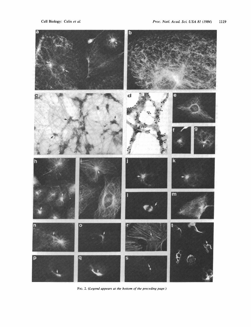
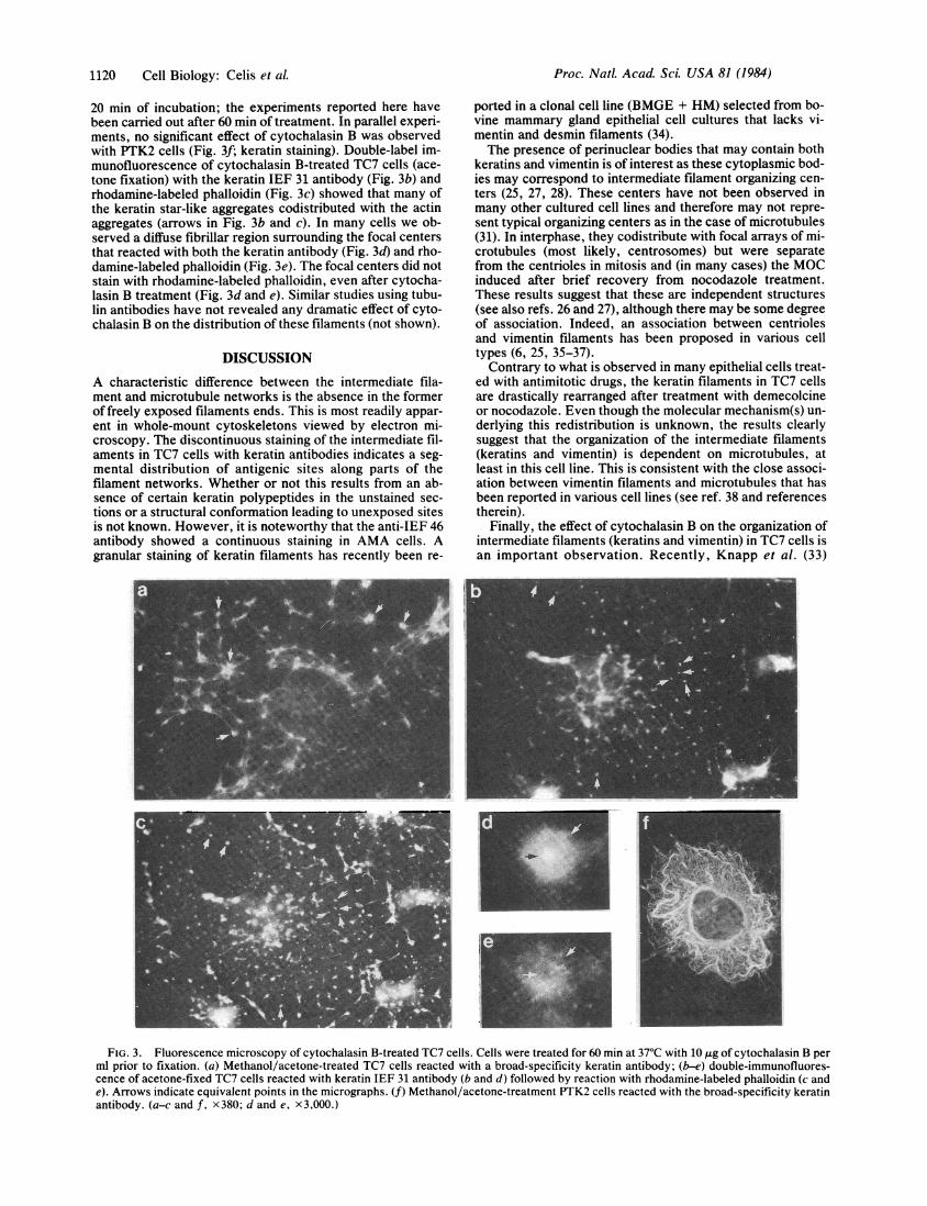
Cytochalasin B Affects the Organization of Intermediate-Sized Filaments and Microfilaments. Treatment of TC7 cellswith cytochalasin B (10 tkg/ml, 1 hr, 37'C) followed by meth-anol/acetone fixation revealed a star-like arrangement of thekeratin (arrows in Fig. 3a) and vimentin filaments (notshown) similar to that recently reported in mouse KLN 205carcinoma cells treated simultaneously with colchicine andcytochalasin D (33). The effect of cytochalasin B on interme-diate filaments in TC7 cells could be observed already after
FIG. 2 (on next page). Fluorescence and immunoelectron microscopy of TC7 cytoskeletons. Except where indicated, the cells first weretreated with methanol (4 min at -200C) and then treated with acetone (as for methanol) prior to incubation with the antibody. (a and b) TC7 cellsreacted with HeLa keratin IEF 46 antibody; (c and d) immunoelectron microscopy of intermediate filament-enriched cytoskeletons of TC7 cells(c) and AMA cells (d), both with the keratin IEF 46 antibody (small gold particles). Note discontinuous label in TC7 cells and continuous labelin AMA cells. The large gold particles in d arise from a second label for vimentin (suboptimal concentration of second antibody) and should beignored. (e) AMA cells reacted with IEF 46 antibody; (f) TC7 cytoplast reacted with IEF 46 antibody; (g) TC7 cells reacted with HeLa keratinIEF 31 antibody; (h) TC7 cells reacted with vimentin antibodies; (l) TC7 cells reacted with tubulin antibodies; (j and k) double-immunofluores-cence of interphase TC7 cells reacted with tubulin (j) and broad-specificity keratin antibody (k); (I and m) double-immunofluorescence ofmitotic TC7 cells reacted with tubulin (1) and broad-specificity keratin antibody (m); (n-q) double-immunofluorescence of nocodazole-treatedTC7 cells (10 ,ug/ml; 20 hr, 3-min recovery), fixed and reacted with tubulin (n and p) and the broad-specificity keratin antibody (o and q); (r ands) double-immunofluorescence of acetone-fixed TC7 cells reacted with rhodamine-labeled phalloidin (r) and keratin IEF 31 antibody (s); (t) TC7cells treated with demecolcine (10 tug/ml; 20 hr, 370C) and reacted with the broad-specificity keratin antibody. (a, g, andj-q x 380; b, x600; c,x40,000; d, x55,000; e, h, and t, X 280; i, r, and s, x240.) Inj-s, arrows indicate equivalent points in the micrographs.
Co00
z
I
C
55
43
55
43
1118 Cell Biology: Celis et aL
I46-- ~ -*-actin
A|_ * s
Proc. NatL. Acad. Sci. USA 81 (1984) 1119
A'.
FIG. 2. (Legend appears at the bottom of the preceding page.)
Cell Biology: Celis et aL
I
A:
.0
i'm
uiA
Proc. Natl. Acad. Sci. USA 81 (1984)
20 min of incubation; the experiments reported here havebeen carried out after 60 min of treatment. In parallel experi-ments, no significant effect of cytochalasin B was observedwith PTK2 cells (Fig. 3f; keratin staining). Double-label im-munofluorescence of cytochalasin B-treated TC7 cells (ace-tone fixation) with the keratin IEF 31 antibody (Fig. 3b) andrhodamine-labeled phalloidin (Fig. 3c) showed that many ofthe keratin star-like aggregates codistributed with the actinaggregates (arrows in Fig. 3b and c). In many cells we ob-served a diffuse fibrillar region surrounding the focal centersthat reacted with both the keratin antibody (Fig. 3d) and rho-damine-labeled phalloidin (Fig. 3e). The focal centers did notstain with rhodamine-labeled phalloidin, even after cytocha-lasin B treatment (Fig. 3d and e). Similar studies using tubu-lin antibodies have not revealed any dramatic effect of cyto-chalasin B on the distribution of these filaments (not shown).
DISCUSSION
A characteristic difference between the intermediate fila-ment and microtubule networks is the absence in the formerof freely exposed filaments ends. This is most readily appar-ent in whole-mount cytoskeletons viewed by electron mi-croscopy. The discontinuous staining of the intermediate fil-aments in TC7 cells with keratin antibodies indicates a seg-mental distribution of antigenic sites along parts of thefilament networks. Whether or not this results from an ab-sence of certain keratin polypeptides in the unstained sec-tions or a structural conformation leading to unexposed sitesis not known. However, it is noteworthy that the anti-IEF 46antibody showed a continuous staining in AMA cells. Agranular staining of keratin filaments has recently been re-
ported in a clonal cell line (BMGE + HM) selected from bo-vine mammary gland epithelial cell cultures that lacks vi-mentin and desmin filaments (34).The presence of perinuclear bodies that may contain both
keratins and vimentin is of interest as these cytoplasmic bod-ies may correspond to intermediate filament organizing cen-ters (25, 27, 28). These centers have not been observed inmany other cultured cell lines and therefore may not repre-sent typical organizing centers as in the case of microtubules(31). In interphase, they codistribute with focal arrays of mi-crotubules (most likely, centrosomes) but were separatefrom the centrioles in mitosis and (in many cases) the MOCinduced after brief recovery from nocodazole treatment.These results suggest that these are independent structures(see also refs. 26 and 27), although there may be some degreeof association. Indeed, an association between centriolesand vimentin filaments has been proposed in various celltypes (6, 25, 35-37).
Contrary to what is observed in many epithelial cells treat-ed with antimitotic drugs, the keratin filaments in TC7 cellsare drastically rearranged after treatment with demecolcineor nocodazole. Even though the molecular mechanism(s) un-derlying this redistribution is unknown, the results clearlysuggest that the organization of the intermediate filaments(keratins and vimentin) is dependent on microtubules, atleast in this cell line. This is consistent with the close associ-ation between vimentin filaments and microtubules that hasbeen reported in various cell lines (see ref. 38 and referencestherein).
Finally, the effect of cytochalasin B on the organization ofintermediate filaments (keratins and vimentin) in TC7 cells isan important observation. Recently, Knapp et al. (33)
FIG. 3. Fluorescence microscopy of cytochalasin B-treated TC7 cells. Cells were treated for 60 min at 37°C with 10 ,ug of cytochalasin B perml prior to fixation. (a) Methanol/acetone-treated TC7 cells reacted with a broad-specificity keratin antibody; (b-e) double-immunofluores-cence of acetone-fixed TC7 cells reacted with keratin IEF 31 antibody (b and d) followed by reaction with rhodamine-labeled phalloidin (c ande). Arrows indicate equivalent points in the micrographs. (f) Methanol/acetone-treatment PTK2 cells reacted with the broad-specificity keratinantibody. (a-c and f, x380; d and e, x3,000.)
1120 Cell Biology: Celis et A
Proc. NatL. Acad. Sci. USA 81 (1984) 1121
showed that treatment ofKLN 205 carcinoma cells with col-chicine together with cytochalasin D converted the keratincytoskeleton into a series of star-like structures, which theysuggested are maintained by multiple membrane attachmentsites. In our studies (using only cytochalasin B), we havefurther shown that many of the star-like structures codistrib-ute with actin patches, a fact that would suggest that inter-mediate filaments may interact with microfilaments at leastin certain regions of the cell.
In summary, our studies point towards a close relationship(structural or functional or both) between the various cyto-skeletal systems in TC7 cells and add support to the notionthat cytoskeletal interactions may be different in various celltypes.
We thank 0. Jensen for photography and K. Dejgaard and M.Hattenberger for assistance. We also thank Prof. Th. Wieland for agenerous gift of rhodamine-labeled phalloidin. P.M.L. is a recipientof a fellowship from the Arhus University. S.J.F. is a recipient of afellowship from the Danish Cancer Foundation. This work was sup-ported by grants from the Danish Natural Science and Medical Re-search Councils, the Danish Cancer Foundation, the CarlsbergFoundation Euratom and NOVO to J.E.C. and by a grant from theAustrian Science Research Council to J.V.S.
1. Lazarides, E. (1980) Nature (London) 283, 249-256.2. Albrecht-Buehler, G. & Watson, J. D. (1982) Cold Spring
Harb. Symp. Quant. Biol. 46.3. Letho, V. P., Virtanen, J. & Kurki, P. (1978) Nature (London)
272, 175-177.4. Small, J. V. & Celis, J. E. (1978). J. Cell Sci. 31, 393-409.5. Lee, C. S., Morgan, G. & Wooding, F. B. P. (1979) J. Cell Sci.
38, 125-135.6. Goldman, R. D., Zackroff, R. V., Starger, J. M. & Whitman,
M. (1979) J. Cell Biol. 83, part 2, 343 (abstr.).7. David-Ferreira, K. L. & David-Ferreira, J. F. (1980) Cell Biol.
Int. Rep. 4, 655-662.8. Pharie-Washington, L., Silverstein, S. C. & Wang, E. (1980) J.
Cell Biol. 92, 575-578.9. Chen, L. B., Summerhayes, I. C., Johnson, L. V., Walsh,
M. L., Bernal, S. D. & Lampidis, T. J. (1982) Cold SpringHarbor Symp. Quant. Biol. 46, 141-151.
10. Mose Larsen, P., Bravo, R., Fey, S. J., Small, J. V. & Celis,J. E. (1982) Cell 31, 681-692.
11. Mose Larsen, P., Fey, S. J., Bravo, R. & Celis, J. E. (1983)Electrophoresis 4, 247-256.
12. Tokuyasu, K. T., Dutton, A. H. & Singer, S. J. (1983) J. CellBiol. 96, 1727-1735.
13. Traub, P., Nelson, J. W., Kuhn, S. & Vorgias, C. (1983) J.Biol. Chem. 258, 1456-1466.
14. Moll, R., Franke, W. W., Schiller, D. L., Geiger, B. &Knepler, R. (1982) Cell 31, 11-24.
15. Bravo, R., Fey, S. J., Small, J. V., Mose Larsen, P. & Celis,J. E. (1981) Cell 25, 195-202.
16. Celis, J. E. & Bravo, R. (1981) Trends Biochem. Sci. 6, 197-202.
17. Bravo, R., Small, J. V., Fey, S. J., Mose Larsen, P. & Celis,J. E. (1982) J. Mol. Biol. 154, 121-142.
18. Bravo, R. (1983) in Two Dimensional Gel Electrophoresis ofProteins, Methods and Applications, eds. Celis, J. E. & Bra-vo, R. (Academic, New York), p. 4-34.
19. De Mey, J., Moerermans, M., Geuens, G., Nuydens, R. & DeBrabander, M. (1981) Cell Biol. Int. Rep. 5, 889-899.
20. Bravo, R., Bellatin, J. & Celis, J. E. (1981) Cell Biol. Int. Rep.5, 93-96.
21. Bravo, R. & Celis, J. E. (1982) Clin. Chem. (Winston-Salem,N.C.) 28, 766-781.
22. Bravo, R. & Celis, J. E. (1984) in Two Dimensional Gel Elec-trophoresis ofProteins, Methods and Applications eds. Celis,J. E. & Bravo, R. (Academic, New York), p. 445-475.
23. Fey, S. J., Mose Larsen, P. & Celis, J. E. (1983) FEBS Lett.157, 165-169.
24. Bravo, R., Fey, S. J., Mose Larsen, P., Coppard, N. & Celis,J. E. (1983) J. Cell Biol. 96, 416-423.
25. Borenfreund, E., Schmid, E., Bendich, A. & Franke, W. W.(1980) Exp. Cell Res. 177, 215-235.
26. Hormia, M., Linder, E., Letho, V.-P., Vartio, T., Badley,R. A. & Virtanen, I. (1982) Exp. Cell Res. 138, 159-166.
27. Eckert, B. S., Daley, R. A. & Parysek, L. M. (1982) J. CellBiol. 192, 575-578.
28. Fey, S. J., Mose Larsen, P., Bravo, R., Celis, A. & Celis,J. E. (1983) Proc. Natl. Acad. Sci. USA 80, 1905-1909.
29. De Brabander, M. J., Van de Veire, R. M. L., Aerts,F. E. M., Borges, M. & Janssen, P. A. J. (1976) Cancer Res.36, 905-916.
30. Hoebeke, J., Van Nijen, G. & De Brabrander, M. (1976) Bio-chem. Biophys. Res. Commun. 69, 319-324.
31. Pickett-Heaps, J. D. (1969) Cytobios 1, 257-280.32. Klymkowski, M. W. (1982) EMBO J. 1, 161-165.33. Knapp, L. W., O'Guin, W. M. & Sawyer, R. H. (1983) Sci-
ence 219, 501-503.34. Schmid, E., Franke, W. W., Grund, C., Schiller, D., Kolb, H.
& Paweletz, N. (1983) Exp. Cell Res. 146, 309-328.35. Wang, E., Connolly, J. A., Kalnins, U. K. & Choping, P. W.
(1979) Proc. NatI. Acad. Sci. USA 76, 5719-5723.36. Aubin, J. E., Osborn, M., Franke, W. W. & Weber, K. (1980)
Exp. Cell Res. 129, 149-165.37. Blose, S. H. (1981) Cell Motility 1, 417-431.38. Geuens, G., de Brabander, M., Nuydens, R. & De Mey, J.
(1983) Cell Biol. Int. Rep. 1, 35-47.
Cell Biology: Celis et aL