Embed Size (px)
Citation preview

Internal Medicine
Board Review - Cardiology
February/March 2011
Joe M. Moody, Jr, MD
UTHSCSA and STVHCS
Corroborates MKSAP 15 Cardiovascular Medicine Module
No new material – only standard stuff
This follows ABIM blueprint
No disclosures

Topics in Cardiology
• Epidemiology
• Heart Tests
• Coronary
Artery Disease
• Heart Failure
• Myocardial Disease
• Adult Congenital Disease
• Aortic Disease
• Peripheral Arterial Disease
• Pregnancy
• Arrhythmias
• Pericardial
Disease
• Valvular
Disease

Topics in Cardiology
• Heart Tests
• Coronary
Artery Disease
• Heart Failure
• Myocardial Disease
• Adult Congenital Disease
• Aortic Disease
• Peripheral Arterial Disease
• Pregnancy
• Arrhythmias
• Pericardial
Disease
• Valvular
Disease
• Epidemiology

Epidemiology of Cardiovascular
Disease (CVD) - 1
• About 1/3 of adults have CVD, almost ½ are >60 y.o. (increasing with age)
• In women, onset is average 10 years later, but CVD is leading cause of death in women too
• More prevalent in Hispanic and American and Alaskan Indian populations due to risk factors

Epidemiology of Cardiovascular
Disease (CVD) – 2 DM
• DM (10% US pop, esp in some ethnics)
is risk factor (2-4x) for MI, HF, stroke,
esp with microalbuminuria
• Metabolic Syndrome (3 of these:
FBG>110, HDL<40, TG>150, Waist
>35-40 in, BP>130 or 85 mmHg) is risk
factor for both DM and CVD, Present in
24% of whites, 32% of Hispanics
Epidemiology of Cardiovascular
Disease (CVD) – 3 Cancer
• Radiation-related coronary disease & valvular and myocardial and pericardial fibrosis, even decades after treatment
• Chemo - anthracycline-doxorubicin>500 mg/m2 18%HF with 50% mortality
• Chemo - trastuzumab esp with anthracycline high risk of HF – close monitoring

Epidemiology of Cardiovascular
Disease (CVD) – 4 InflammationSystemic Lupus E (S) Pcard, endocard, regurg, CAD
Rheumatoid Arth (S) Pcard, CAD, leaf fibros, LVdias
Ankylosing Spondylitis Aortitis, regurg, cond dz, LVdia
Systemic Sclerosis Pcard, Htn, PHtn, myo fibros,
Takayasu Arteritis (L) Aneur, sten-occl, CAD, AR, Htn
Giant Cell Arteritis (L) PAD, stroke, MI
Polyart Nodosa (M) Cardiomyopathy
Kawasaki Disease (M) Coronary artery aneurysm, occl
Behcet disease AR, myo-pericard, cond dz
Sarcoidosis Cardiomyopathy, cond dz,SCD
Epidemiology of Cardiovascular
Disease (CVD) – 5 Lifestyle
• Smoking – 22% US adults
• Alcoholism – 4% of DCMs (>5y heavy)
• Cocaine – CP, spasm, thrombosis, LVH,
dissection, arrhythmia
(methamphetamines similar)
• Sedentary – 40% of adults
• IV drugs – endocarditis TV>MV>AoV

Topics in Cardiology
• Epidemiology
•
• Coronary
Artery Disease
• Heart Failure
• Myocardial Disease
• Adult Congenital Disease
• Aortic Disease
• Peripheral Arterial Disease
• Pregnancy
• Arrhythmias
• Pericardial
Disease
• Valvular
Disease
• Heart Tests

Heart Tests – Structural Disease
• Murmur-valve, DCM or HCM or HF, congenital, Pulm Htn, Pcarditis, Ao dz
• Echo for murmur: all diastolic, systolic ≥3/6 or not mid systolic, or CV sx
• Transthoracic echo is the standard test
• Handheld echo requires training
• TEE when TTE not adequate (AFib with LA appendage thrombus; vegetation; Ao dissection; prosthetic valve dysfunction)
• MUGA, Cath, CT scan, cardiac MRI (CMR)
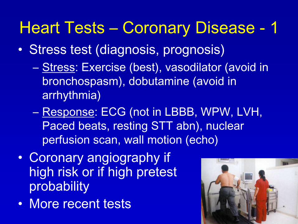
Heart Tests – Coronary Disease - 1
• Stress test (diagnosis, prognosis)
– Stress: Exercise (best), vasodilator (avoid in
bronchospasm), dobutamine (avoid in
arrhythmia)
– Response: ECG (not in LBBB, WPW, LVH,
Paced beats, resting STT abn), nuclear
perfusion scan, wall motion (echo)
• Coronary angiography if high risk or if high pretest probability
• More recent tests

Stress ECG Testing
≥1 mm horizontal ST
segment depression

Nuclear Perfusion Imaging

Heart Tests – Coronary Disease - 2
• Coronary Artery Calcium (CAC) score
– Asymptomatic and intermediate risk of CAD (10-20% 10-yr risk) >400 intensify rx
– Symptomatic and low risk of CAD (score <100 has high neg predict value)
• CT Coronary Angiography (Iodine +)
– Anomalous coronary (also cardiac MR)
– Acute chest pain, intermediate risk, neg Trop
– Maybe Sx + (intermed prob – abn ECG or can’t exercise or prior equivocal test)
• PET Scan, alternative to Nuc, less rad’n

Coronary Artery Calcium

CT Coronary Angiography
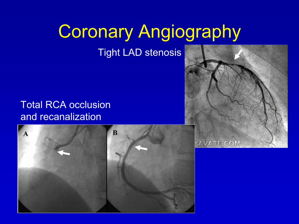
Coronary AngiographyTight LAD stenosis
Total RCA occlusion
and recanalization

Cardiac PET Scan

Heart Tests – Coronary Disease
– Radiation Exposure
Procedure # CXRs
Stress echocardiography 0
Cardiac MR Angiography 0
Coronary artery Calcium score 20-40
Coronary angiography (diagnostic) 200-500
Nuclear perfusion imaging 100-500
PET perfusion imaging 100-400
CT coronary angiography 700-2100
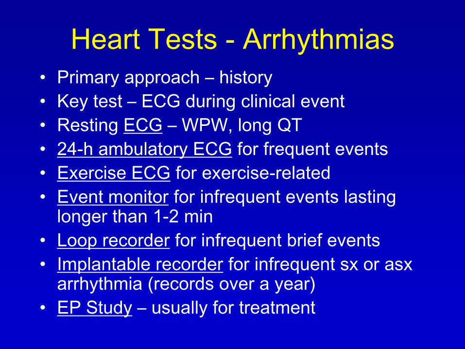
Heart Tests - Arrhythmias
• Primary approach – history
• Key test – ECG during clinical event
• Resting ECG – WPW, long QT
• 24-h ambulatory ECG for frequent events
• Exercise ECG for exercise-related
• Event monitor for infrequent events lasting longer than 1-2 min
• Loop recorder for infrequent brief events
• Implantable recorder for infrequent sx or asx arrhythmia (records over a year)
• EP Study – usually for treatment

ECG - WPW

ECG – Long QT

ECG – Ambulatory Monitor

Exercise Arrhythmia

Event Recorders

Implantable
Loop Recorder

Topics in Cardiology
• Coronary
Artery
Disease
• Adult Congenital Disease
• Aortic Disease
• Peripheral Arterial Disease
• Pregnancy
• Arrhythmias
• Pericardial
Disease
• Valvular
Disease
• Epidemiology
• Heart Tests
• Heart Failure
• Myocardial Disease

Conventional CAD Risk Factors
• Framingham (10-yr)
• Age
• Male
• Hypertension
• Hyperlipidemia
• Smoking
• DM
• Family History
• Known PAD, CVD, DM = risk equivalent
• CKD (<60 ml/min) or proteinuria = risk
• Metabolic syndrome doubles risk (due to individual factors)

CAD Risk Factors- 2• Conditional Risk factors (“emerging”) not
routine
– Homocysteine (risk not reduced by folate)
– Elevation of Lipoprotein (a), hs-CRP (intmed risk), apo-B, or small LDL particle size
• Treatment: ASA (men 45-79), control BP and lipids, smoking, diet, exercise (BMI<25)
– Supplements – no: Vit E, Vit C, β-carotene
– Fish oil – maybe
• Depression – refer for psychiatric evaluation; SSRI are safe in CAD

Question 1
• 51 yo woman with sharp pain lasting 2 minutes provoked by exertion and relieved by rest for the last 4 months. ECG normal. Which test is recommended?
A. No test, reassure, primary prevention targets
B. Exercise tolerance test with ECG
C. Exercise tolerance test with perfusion scan
D. Coronary angiography
E. Vasodilator stress with perfusion scan

Definition of Angina
ACC/AHA Clinical Practice Guideline, Chronic Stable Angina, 2002.

Diagnosis: Pretest Probability of
Obstructive Disease at Catheterization
ACC/AHA Guideline Exercise Testing 2002, p. 7.
Diagnostic testing is appropriate - intermediate pretest probability

Stable CAD: Diagnostic Tests
• ECG normal and able to exercise = ETT
• ECG abnl and able to exercise = ETT
with imaging (nuc-perfusion or echo-wall
motion)
– Exception: LBBB, use vasodilator
• Unable to exercise = vasodilator or
dobutamine stress
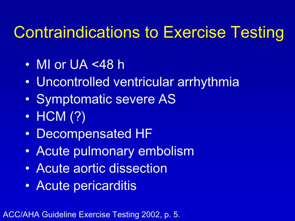
Contraindications to Exercise Testing
• MI or UA <48 h
• Uncontrolled ventricular arrhythmia
• Symptomatic severe AS
• HCM (?)
• Decompensated HF
• Acute pulmonary embolism
• Acute aortic dissection
• Acute pericarditis
ACC/AHA Guideline Exercise Testing 2002, p. 5.

Stable CAD: Low Risk Test Results
• ECG result: Low risk Duke treadmill score (≥5)
– Number of minutes of Bruce protocol
– Minus 5 times number of mm ST depression
– Minus 4 times angina score (0=none, 1=some, 2=limiting)
• Nuclear result: normal, or small perfusion defect at rest or stress
• Stress Echo result: Normal wall motion or no change in limited resting wall motion abnormalities with stress
ACC/AHA Guideline, Stable Angina, 2002

Stable CAD: Strongly Positive (High
Risk) Test Results
• Markedly positive result = coronary angio
• ECG result: Significant ST depression at
low workload, ST elevation, low BP (Duke
treadmill score ≤-11)
• Nuclear result: TID, lung uptake,
multizone ischemia, EF<35%
• Stress Echo result: Fall in EF with stress,
multizone hypokinesis, EF<35%

Coronary Angiography Indications
• Lifestyle-limiting angina despite medical
therapy
• High-risk (markedly positive) stress
testing
• Resuscitation from sudden cardiac death
• Documented VT
• Uncertain diagnosis with recurrent
hospitalization for chest pain
• Angina and heart failure
ACC/AHA Clinical Practice Guideline, Chronic Stable Angina, 2002.

Antianginal Treatment of Stable CAD
• Sublingual NTG drug of choice for episode
• Beta-blocker (1st line)
• Calcium-blocker if beta-blocker not achieve
painfree (not nondihyrdopyridine in HF)
• Long-acting nitrates – avoid tolerance by 8-12h
free interval and avoid PDE-5 inhibitor
• Ranolazine (late sodium channel blocker) new,
if still symptomatic but not with dilt or verapamil
• Refractory: EECP and spinal cord stimulation
– Not currently recommended, may help

Protective Treatment of Stable
CAD
• Aspirin reduces stroke, MI, SCD, vascular
death by 33%, 75-90 mg/da
• Clopidogrel controversial
• ACE-I controversial unless EF<40%, DM,
Htn, and CKD; caution in Cr>2.5, cough is
side effect
• Statin – goal LDL<100 or in high risk <70
– If TG>200, goal is non-HDL of <130 or <100
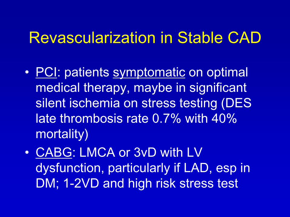
Revascularization in Stable CAD
• PCI: patients symptomatic on optimal
medical therapy, maybe in significant
silent ischemia on stress testing (DES
late thrombosis rate 0.7% with 40%
mortality)
• CABG: LMCA or 3vD with LV
dysfunction, particularly if LAD, esp in
DM; 1-2VD and high risk stress test

Chronic Follow-up Issues in CAD
• No routine ECG or stress test
• Drug-eluting stent (DES): ASA 325/da plus clopidogrel 1 yr or more
• Bare-metal stent (BMS): ASA 325/da plus clopidogrel 1 month
• Noncardiac surgery: Wait 6 weeks after BMS, 1 yr after DES
• Emergency surgery: continue ASA through procedure and restart clopidogrel ASAP

Acute Coronary Syndromes• Classic
• Heartburn
• Silent, SOB
• Sharp, nonchest
Acute Chest pain
ECG
ST elevation No ST elevation
Biomarkers
positive
Biomarkers
positive
Biomarkers
negative
STEMI NSTEMI Unstable
Angina

Question
• 63 yo man in ER with 1 hour midsternal chest pressure. No prior CAD. BP 156/98, P 80, S4. ECG – anterior ST elevation up to 4mm. Which is best next management step?
1. CT, PE protocol
2. Cardiac biomarker evaluation (troponin)
3. Emergent cath and PCI
4. Parenteral thrombolytic therapy
5. Beta-blocker, heparin and noninvasive risk stratification

Biomarkers in Acute Coronary
Syndromes
• Creatine kinase MB fraction (CK-MB) – prior
standard, but may elevate with skeletal
muscle injury, may be useful in re-infarction
• Cardiac troponins T and I are specific to heart
(TnT and TnI) – highly sensitive and specific;
elevation may occur as early as 2 hours and
as late as 12 hr, peaks at 1-2 days, and may
remain elevated up to 14 days
• Higher values indicate higher risk
ACC/AHA Guideline NSTEMI, 2007.

Non-ACS Positive Biomarkers
• Tachyarrhythmia
• Cardiac trauma by interventions
• Chest trauma
• Heart failure
• LVH
• Myocarditis, pericarditis
• Sepsis, burns, respiratory failure, acute neurologic disease, pulmonary embolism, pulmonary hypertension, drug toxicity, cancer chemotherapy, renal insufficiency
ACC/AHA Guideline NSTEMI, 2007.
Necrosis is present, but not acute coronary change

TIMI Risk Score in UA/NSTEMITIMI = Thrombolysis in Myocardial Infarction
1. Age > 65 yo
2. >3 traditional risk factors
3. Prior CAD >50%
4. ST deviation
5. ≥2 anginal episodes in 24 h
6. ASA in past week
7. Elevated biomarkers
• Low risk – stress test, cath if not low risk or sx or low EF
• Intermediate or high - cath
• 0-2 = low risk
• 3-4 = intermediate
• 5-7 = high risk
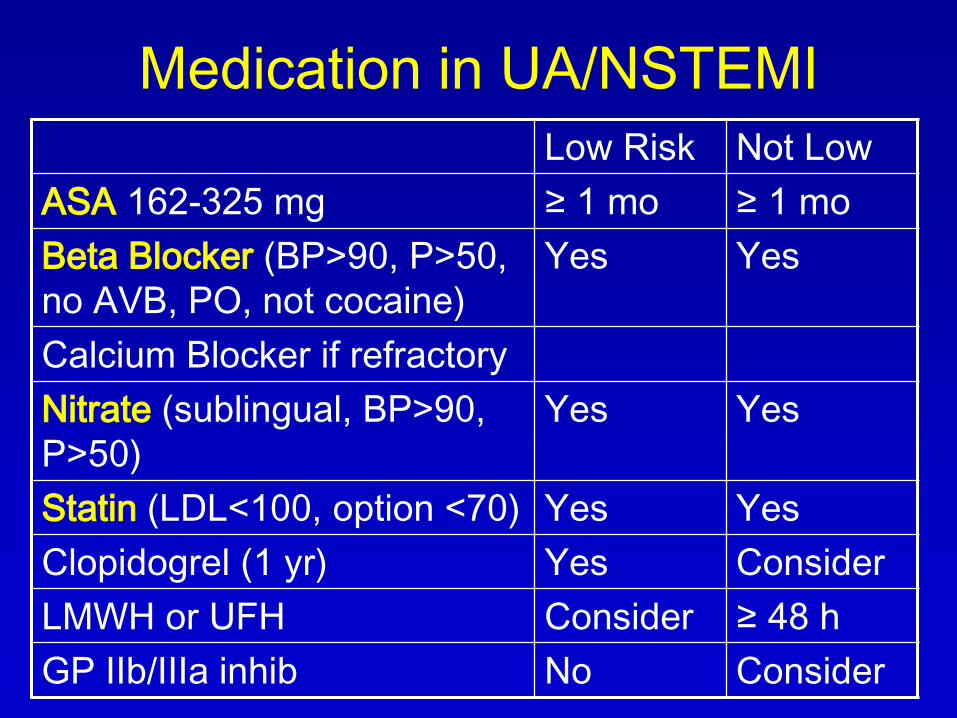
Medication in UA/NSTEMI
Low Risk Not Low
ASA 162-325 mg ≥ 1 mo ≥ 1 mo
Beta Blocker (BP>90, P>50,
no AVB, PO, not cocaine)
Yes Yes
Calcium Blocker if refractory
Nitrate (sublingual, BP>90,
P>50)
Yes Yes
Statin (LDL<100, option <70) Yes Yes
Clopidogrel (1 yr) Yes Consider
LMWH or UFH Consider ≥ 48 h
GP IIb/IIIa inhib No Consider

Unfractionated vs Low Molecular
Weight Heparin
• UFH problems:
– Stable aPTT not easy
– Freq lab testing
– Hep-induced-thrombyocytopenia
• UFH preferred:
– Early invasive strategy
– Increased bleeding risk
– Renal insufficiency
• Bivalirudin (thrombin inhibitor)– data unclear

ST Elevation MI
• Timely recognition
• Exclude pericarditis, pulmonary embolism, dissection (can occlude RCA)
• *High risk: Age>75, LBBB, anterior MI, HF, shock, vent arrhyth
• Reperfusion strategy
– Duration of symptoms
– High-risk features
– Contraindications to thrombolysis
– Time to balloon inflation
– Diagnosis in doubt
• Most receive thrombolysis
*Morrow DA et al. Circulation 2000;102:2031.

ACC/AHA Guidelines for the Management of ST Elevation MI, August 2004
Fibrinolysis <30 min
Generally Preferred
• Early presentation (≤3 h
symptoms) and delay to
invasive strategy
• Invasive strategy not
option (cath lab or skilled
PCI not available, vascular
access difficulty)
• Delay to invasive strategy
(prolonged transport >60
min to balloon, or door to
balloon >90 min)
PCI <90 min
Generally Preferred
• Late presentation (>3 h
symptoms)
• Skilled PCI lab available
with surgical backup (door
to balloon <90 min)
• Cardiogenic shock or
Killip class 3-4
• Contraindications to
fibrinolysis
• Diagnosis of STEMI in
doubt
• Also Rescue PCI for failed
fibrinolysis

Question 2
• Which of the following is an
ABSOLUTE contraindication to
fibrinolysis?
A. Systolic BP on presentation >180 mmHg
B. Suspected aortic dissection
C. CPR lasting more than 10 minutes
D. Noncompressible vascular puncture
E. Pregnancy

Question 2
• Which of the following is an
ABSOLUTE contraindication to
fibrinolysis?
A. Systolic BP on presentation >180 mmHg
B. Suspected aortic dissection
C. CPR lasting more than 10 minutes
D. Noncompressible vascular puncture
E. Pregnancy

Absolute Contraindication to
Fibrinolysis in ST Elevation MI• Any prior intracranial hemorrhage
• Known structural CV lesion (AVM) or malignant CNS neoplasm
• Stroke < 3 mo except stroke <3h
– (>3 mo=relative contraindication)
• Suspected Ao dissection
• Active bleeding or bleeding diathesis (except menses; active peptic ulcer is relative contraindication)
• Significant closed-head or facial trauma within 3 mo.
ACC/AHA Guidelines for the Management of ST Elevation MI, August 2004
CNS

Relative Contraindication to Fibrinolysis in STEMI
• Hypertension
– chronic severe poorly
controlled
– on presentation
(SBP>180 or DBP >110)
• Prior stroke >3 mo,
dementia, or known other
intracranial pathology
• Traumatic or prolonged
(>10 min) CPR or major
surgery (<3 wk)
• Recent (<2-4 wk) internal
bleeding
• Noncompressible vascular punctures
• For streptokinase/ anistreplase: prior exposure (>5 da) or prior allergic reaction
• Pregnancy
• Active peptic ulcer
• Current use of anticoagulants: the higher the INR, the higher the risk
ACC/AHA Guidelines for the Management of ST Elevation MI, August 2004

Common Thrombolytic Agents Alteplase Reteplase Tenecteplase
Dose <100 mg/90
min
10U/2min x
2 @ 30min
30-50 mg
Bolus No Yes Yes
Allergic rxn No No No
TIMI 2/3 flo ~75% ~83% ~83%
Rate of ICH 0.4-0.7% 0.8% 0.9%
Fibrin spec +++ + ++++
Need for
hep
Yes Yes Yes

Evidence of Reperfusion
• Uncertain, 3 features of reperfusion
– Relief of pain
– Reduction of ST elevation >50%
in 1 hr
– Reperfusion arrhythmias – AIVR,
NSVT (in inferior MI, sinus brady
and hypotension)

Medical Therapy of STEMI
• ASA*, Analgesics, Nitrates*, oxygen
• IV beta-blocker if no contraindication (metoprolol 5 mg IV q5min x3) – then PO*
• ACE-I* if low EF or with HF (<24h oral)
• Statin*
• UFH (if PCI) or LMWH
• Clopidogrel*
• Gp IIb/IIIa inhibitor in primary PCI
*=also long term

Complications of STEMI
• ICH from lytics – <1%, mortality >50%, risks
• Hypotension – important to define etiology –volume loading, rhythm control
• RV infarction – inferior STEMI with proximal RCA occlusion, hypotension, elevated JVP, clear lungs, STEL in V4R – echo is helpful –hypotension is provoked by NTG because RV needs preload (treat with preload ± inotrope)
• Pulmonary congestion – O2, morphine, ACE-I, NTG and diuretic
• Cardiogenic shock – inotropes and IABP
ACC/AHA Guidelines for the Management of ST Elevation MI, August 2004
Hem
odynam
ic c
om
plic
ations

ACC/AHA Guidelines for the Management of ST Elevation MI, August 2004
Drug Therapy for Hemodynamic
Compromise in STEMI
SBPSigns of
ShockTherapy
>100 -/+ NTG, ACE-I
70-100 -Dobutamine
(2-20 mcg/kg/min)
70-100 +Dopamine
(5-15 mcg/kg/min)
<70 +Norepinephrine
(0.5-30 mcg/min)

Question 3
• Which of the following requires
antiarrhythmic therapy during acute
STEMI?
A. PVCs
B. Asymptomatic nonsustained VT
C. Accelerated idioventricular rhythm
D. Atrial bigeminy
E. None of the above

Question 3
• Which of the following requires
antiarrhythmic therapy during acute
STEMI?
A. PVCs
B. Asymptomatic nonsustained VT
C. Accelerated idioventricular rhythm
D. Atrial bigeminy
E. None of the above
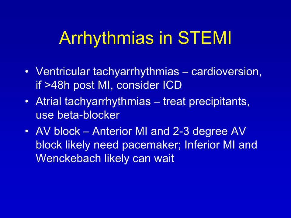
Arrhythmias in STEMI
• Ventricular tachyarrhythmias – cardioversion,
if >48h post MI, consider ICD
• Atrial tachyarrhythmias – treat precipitants,
use beta-blocker
• AV block – Anterior MI and 2-3 degree AV
block likely need pacemaker; Inferior MI and
Wenckebach likely can wait

Mechanical Complications of STEMI
• Occur most frequently at <24h or at 3-5da:
• Emergencies, need emergency cath and surgery, temporize with IABP or nitroprusside except in free wall rupture
ACC/AHA Guidelines for the Management of ST Elevation MI, August 2004
Anterior InferiorSeptal

Left Ventricular Thrombus
• 10-20% of Anterior MI, even in modern
era
• Usually LV apex, diagnose with echo
• Anticoagulation for 3-6 months

Procedures in Management of ST
Elevation MI - Indications
• Swan-Ganz: hypotension unresponsive to fluid or with congestion, suspected mechanical complication if no echo done
• Art line: BP<80, cardiogenic shock or need for inotropes or nitroprusside
• Echo: BP<90, low output state, urgent for pulmonary congestion, possible RV MI, stroke as complication of MI
• IABP: cardiogenic shock not quickly responsive to meds
ACC/AHA Guidelines for the Management of ST Elevation MI, August 2004

Later Management in ST Elevation MI• Risk Stratification – if no cath- noninvasive test,
and cath for a moderate or high risk result
• Pericarditis –use ASA 650 q4-6h, alternative is colchicine 0.6 mg q12h or acetaminophen 500 q6h, steroids last resort; avoid indomethacin
• Evaluate LV systolic function if not known prior (Echo, LV gram with catheterization, MUGA)
• Late post MI:
– LV aneurysm with intractable arrhythmia, failure, or angina may warrant aneurysmectomy
– LV enlargement with remodeling is lessened by medical therapy, particularly ACE-I
ACC/AHA Guidelines for the Management of ST Elevation MI, August 2004

Coronary Disease in Women
• Women present with CAD about 10 years laterthan men and have more comorbidities (DM, Htn, DHF) and more atypical symptoms and worse outcomes post MI
• Stress tests have more false positives but ETT still the recommended test
• Medical therapy for CAD same as for men, but higher bleeding rate with lytics or IIb/IIIa agents –dose for pt weight and eGFR
• Estrogen replacement therapy +/- progesterone are not CV protective; xs thromboembolism
• In ACS and high risk, women benefit from PCI but in ACS and low risk, PCI likely carries xs risk

Coronary Disease in Diabetics
• Optimize control of glc, BP, FLP, daily ASA if >40 yo or add’l risk factors (not clopidogrel)
• CAD and DM often atypical symptoms
• Stress ECG same sens and spec as nondiabetic
• Asymptomatic diabetics – no routine stress test, even if before exercise program
• Medications same as nondiabetic, beta-blocker is fine
• Hospitalized: glu<150 initially; fasting <110 subsequently
• Cath –worse dz; PCI or CABG for refractory symptoms

Topics in Cardiology
• Epidemiology
• Heart Tests
• Coronary Artery Disease
• Myocardial Disease
• Adult Congenital Disease
• Aortic Disease
• Peripheral Arterial Disease
• Pregnancy
• Arrhythmias
• Pericardial
Disease
• Valvular
Disease
• Heart Failure

Limitation none slight marked
NYHA Class: I II-III III-IV
Annual Mortality: 5-10% 15-30% 50-60%
ACC/AHA Guideline - heart failure update 2009.

HF Evaluation and Diagnosis
• H&P in all: PND, S3; less likely if no DOE or
crackles; reliability of JVP not good
• Echo in all
• ECG in all (etiology, CRT)
• BNP good neg pred value if <100 pg/mL
– Not measure of severity
– Increased with age, renal failure, femaleness
– Decreased with obesity
• Cath or noninvasive ischemic workup important
if chest pain or other evidence of ischemia
(viability) – CABG may prolong life

HF Evaluation and Diagnosis – 2
• Handheld echo for IVC size as JVP estimate
(HF but also RV-COPD, Tamponade)
• PA catheter for unclear volume status in
critically ill
• Cardiopulmonary exercise testing clarifies
cause of dyspnea and quantifies impairment
– essential in transplant evaluation
• Endomyocardial biopsy – unexplained new
HF with shock, or suspicion of treatable
cause, also routine post transplant
• MUGA; Cardiac MRI; OSA evaluation

Heart Failure: Medical Therapy
• ALL (NYHA I-IV)
– ACE-I
– Beta-blocker
– Diuretic (current or prior congestion)
• NYHA III-IV (moderate to severe symptoms)
– Spironolactone (eplerenone for gynecomastia)
– Hydralazine/ISDN (for black patients or more)
– Digoxin

Question 4In using beta blockade for heart failure from systolic dysfunction, which of the following is true?
A. Clinical response occurs within 2-3 days
B. Hyperkalemia is a common and dangerous complication
C. Asymptomatic systolic dysfunction (EF<45) should receive beta blockade
D. Patients on beta blockers admitted with volume overload must have beta blockers discontinued
E. If patients develop worsening dyspnea or edema on beta blockade, they should discontinue and not resume therapy

Therapy in Stage C HF
• Na restriction and daily weight
• Flu and pneumococcus vaccine
• Exercise and conditioning are safe
• Problem drugs: antiarrhythmics (except amiodarone), Ca blocker (except dihydropyridine), NSAID (fluid retention & vasoconstriction & antagonism of diuretic and ACE-I effect)
• Monitor serum K (3.8 to 5.2) and Mg
• Monitor adherence, patient education
ACC/AHA Guideline - heart failure update 2009.

ACE Inhibitors in HF Management
• Not proved in BP<90, Cr>2.5, or nl LVEF
• Not in prior angioedema or anuric RF,
pregnancy
• Caution in SBP<80, Cr>3.0, bilateral renal art
stenosis, K>5.5, or near cardiogenic shock
• Start low dose, check electrolytes in 1-2 weeks
• Response: maybe 2-3 days, usually weeks to
months; withdrawal may result in deterioration
• Unstable patients – ACE-I may antagonize
diuretics and inotropes, may require interruption
ACC/AHA Guideline - heart failure update 2009.

ACE Inhibitors in HF Management - 2• ↓BP: ↓ dose only if Sx or renal deterioration
• ↑K+ in worsening renal function or with K+
replacement or K-sparing diuretics, esp. DM, no concomitant ARB
• Cough in 5-10% (50% in Chinese), stops in 1-2 weeks after cessation and returns in 1-2 days on rechallenge – exclude HF exacerbation as cause, encourage toleration of cough, substitute ARB if cough intolerable
• Angioedema in <1% (more if black) and life-threatening, don’t rechallenge, no ARB
• If can’t use ACE-I due to hyperkalemia or creatinine rise, hydralazine/ISDN is reasonable
ACC/AHA Guideline - heart failure update 2009.

ARBs in Class C Heart Failure
• Common ARB choices: losartan (off label), valsartan, candesartan
• Beneficial in ACE-intolerant - cough (or maybe angioedema – extreme caution in starting), but not hypotension, hyperkalemia, or renal insufficiency
• ARB plus ACE-I not proved beneficial
• BP<80, low Na, DM, or CKD merit caution like with ACE-I; monitor initiation like ACE-I
ACC/AHA Guideline - heart failure update 2009.

Beta Blockade in HF• Improve symptoms, LVEF, reduce death (30%)
and hospitalization
• All stable patients with systolic dysfunction with resting HR>65 or SBP>85
• Not patients on recent IV inotropes, volume overload or depleted
• Start low dose, adjust diuretics; double every 2-4 weeks to target
• Response in 2-3 months; avoid abrupt withdrawal
• Clinical deterioration in patients on beta-blocker
– If mild, adjust other medications
– If severe with hypoperfusion or need for IV inotropes (prefer milrinone), prudent to suspend and reintroduce when restabilized
ACC/AHA Guideline - heart failure update 2009.

Recommended Agents in HF
Drug Initial Dose Target Dose
Bisoprolol* 1.25 mg/da 10 mg/da
Carvedilol 3.125 mg bid25 mg bid
(50 bid if >85kg)
Metoprolol
succinate12.5-25 mg/da 200 mg/da
ACC/AHA Guideline - heart failure update 2009.
*=off label

Beta Blockade in HF – Adverse
Reactions
• Worsening HF and fluid retention – may increase diuretic dose
• Fatigue which is usually self-limiting after several weeks but if with hypoperfusion must discontinue
• Bradycardia and heart block (pacemaker?)
• Hypotension (may administer ACE-I and beta-blocker at different times of the day or relax diuretics if too dry)
ACC/AHA Guideline - heart failure update 2009.

Diuretic Use in Stage C HF• “Loop diuretics” (furosemide, bumetanide,
torsemide) for all with current fluid retention and most with prior retention
• Initiate low dose (& Na restriction <3-4 gm/da), goal is loss of 1-2 lb/da, normal JVP and no edema– Inadequate diuresis – less ACE-I response and more β-
blocker risk
– Excessive – more hypotension with ACE-I and vasodilator, more renal insufficiency with ACE-I and ARB
• Problems and risks: Electrolyte disturbances (↓Na, ↓Mg may be eased by using with ACE-I or aldo antagonist)– Diuretic resistance (IV route, fluid restriction, combine
with metolazone or positive inotrope in stage D)
ACC/AHA Guideline - heart failure update 2009.

Digitalis (Class IIa)• Symptomatic but not acutely decompensated HF
• Dose 0.125 to 0.25, no loading dose, lower dose in >70 yo or smaller patient or renal insufficiency, generally do not monitor level (optimal level 0.5-0.9 ng/ml), unless toxicity is concern
• Toxicity: ectopic and reentrant arrhythmias and block, GI and neurologic complaints
• Levels increased by quinidine, verapamil, flecainide, amiodarone, and spironolactone
• Toxicity increased by hypomagnesemia, hypokalemia, or hypothyroidism
• Caution in post MI patients, not with ongoing ischemia
ACC/AHA Guideline - heart failure update 2009.

Aldosterone Antagonists• Weak diuretic but may potentiate other diuretics
• Start spironolactone at 12.5-25/da or eplerenone at 25/da and stop K+ supplements, avoid foods high in K unless prior problem with severe hypokalemia, avoid NSAID and COX-2, stop if diarrhea episode
• Check K+ at 3 and 7 da and at least monthly for 3 months; restart monitoring if alter ACE-I or ARB
• AVOID: triple ACE-I PLUS ARB PLUS Aldo- Antag
• Indication: Moderately severe or severe symptoms despite other agents, not if Cr>2.0-2.5 or K>5.0, not if CCr<30 ml/min, particularly if diabetic. If K rises to >5.5, ↓ dose or stop (unless on K suppl)
ACC/AHA Guideline - heart failure update 2009.

Hydralazine/Nitrate in Class C Heart
Failure
• Hydralazine and Nitrates: Not as good as ACE-I in CHF, no recommendation for hydralazine alone or nitrate alone
• Benefit in self-identified black patients who are moderately to severely symptomatic, added to ACE-I or beta blocker or intolerance to ACE-I or beta blocker (A-HEFT trial 2005) – maybe others too
• Reasonable in ACE-intolerant with hypotension or renal insufficiency
ACC/AHA Guideline - heart failure update 2009.

Not Good in Class C Heart Failure
• Calcium blockers
– Diltiazem and verapamil are adverse
– Felodipine and amlodipine for angina or
hypertension, may be useful in addition to
beta-blockade, are neutral in mortality in HF
• Anticoagulation solely for HF is not
indicated
• Avoid: intermittent inotropes or nutritional
supplements or dynamic cardiomyoplasty
ACC/AHA Guideline - heart failure update 2009.

Heart Failure with Normal EF
• HF is a clinical diagnosis
• 20-60% of HF patients have preserved systolic function (EF> ?40%) and no severe valvular disease, especially women and elderly and hypertensive
• Treatment: control BP, control ventricular rate in atrial fibrillation, diuretics to relieve pulmonary congestion and edema, revascularization for coronary disease when ischemia is considered to cause diastolic dysfunction (IIa)
• ARB may reduce hospitalizations (candesartan)
ACC/AHA Guideline - heart failure update 2009.

Device Indications in Heart Failure
• Context: optimal medical therapy for 4-6 months with persistent low EF, and a reasonable expectation of survival with good functional status for >1 year
• ICD indications:
– Secondary prevention (prior cardiac arrest, VF, or unstable VT)
– Primary prevention with LV EF <35% at least 40 da post MI (or nonischemic) and NYHA class II-III
ACC/AHA Guideline update: heart failure in the adult. 2009

Biventricular Pacing in Heart Failure• Wide QRS (esp LBBB or ventricular paced beat)
= poor synchrony of LV contraction and impaired LV function
• Simultaneous pacing of RV apex and LV lateral wall (via lateral cardiac vein from coronary sinus) improves synchrony of contraction and may relieve symptoms and improve MR and has shown decreased mortality
• Indication: NYHA class III-IV and EF<35% and NSR and QRS widening >0.12 sec (often much wider)
• Also reasonable: AFib, or likely to be frequently ventricular paced
ACC/AHA Guideline update: heart failure in the adult. 2009

HF Serial Assessment
• Symptoms: functional capacity, specific activities
– “What activities have you have had to stop?”
– Six minute walk test (<300 m = high risk mortality)
– Cardiopulmonary exercise testing is more precise
• Volume assessment: weight, BP sit & stand,
***JVP***, abd compression, rales (more in acute),
hepatic congestion, edema
• Signs of poor perfusion (abrupt or marked ↓ C.O.):
narrow pulse pressure, cool extremities, altered
mentation, Cheyne-Stokes, ↑HR, ↑BUN/Cr
ACC/AHA Guideline - heart failure update 2009.

Laboratory Follow-up
• Lytes BUN/Cr regularly
• CXR, echo - only for significant change
• PA catheter - only for specific clinical or
hemodynamic question
• BNP is helpful in in initial diagnosis, but final role
in management is still to be determined
– NT-pro-BNP similar, roughly 6 times higher numbers
• Prognostic assessment: age, gender, SBP, clinical
status and functional capacity, LVEF, Na, BUN/Cr,
Hct
ACC/AHA Guideline - heart failure update 2009.
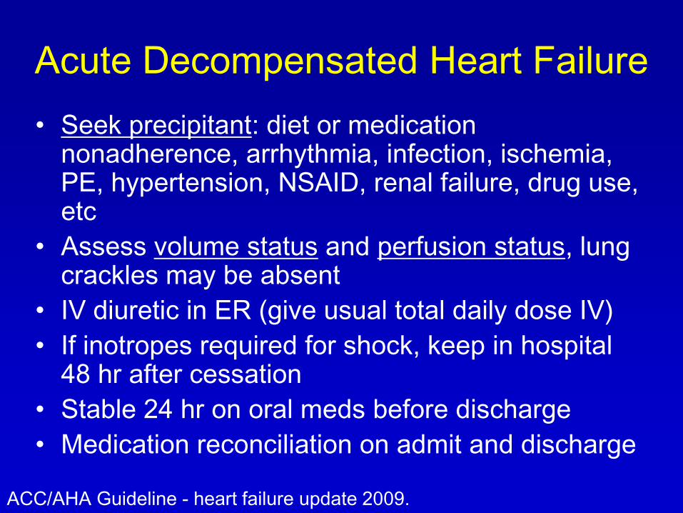
Acute Decompensated Heart Failure
• Seek precipitant: diet or medication nonadherence, arrhythmia, infection, ischemia, PE, hypertension, NSAID, renal failure, drug use, etc
• Assess volume status and perfusion status, lung crackles may be absent
• IV diuretic in ER (give usual total daily dose IV)
• If inotropes required for shock, keep in hospital 48 hr after cessation
• Stable 24 hr on oral meds before discharge
• Medication reconciliation on admit and discharge
ACC/AHA Guideline - heart failure update 2009.
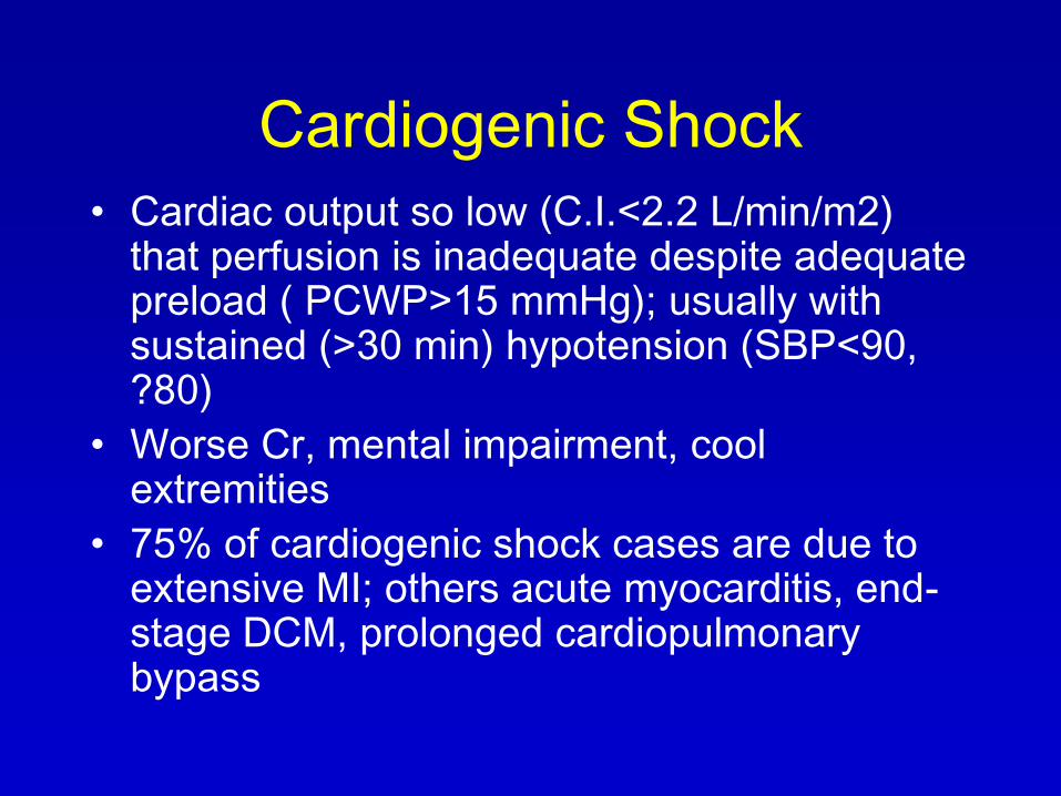
Cardiogenic Shock
• Cardiac output so low (C.I.<2.2 L/min/m2) that perfusion is inadequate despite adequate preload ( PCWP>15 mmHg); usually with sustained (>30 min) hypotension (SBP<90, ?80)
• Worse Cr, mental impairment, cool extremities
• 75% of cardiogenic shock cases are due to extensive MI; others acute myocarditis, end-stage DCM, prolonged cardiopulmonary bypass

Cardiogenic Shock Treatment
• Determine cause: ECG, Echo
• Hypovolemia – give volume
• Uncertain volume or perfusion – PA catheter
• Inotropic support (hours to days) with diuresis – exit strategy important
• Urgent revascularization for AMI
• Urgent surgery for ventricular rupture or acute MR
• Pericardiocentesis for tamponade
• IABP or LVAD if unresponsive

Stage D HF – Frequent Exacerbations• Diuretic resistance – IV loop diuretic or add
metolazone or inotrope, nesiritide or ultrafiltration or dialysis (may restore diuretic responsiveness); inpt till euvolemic and stable oral doses, Na ≤ 2 g/da
• No ACE-I or beta blocker if SBP<80 or peripheral hypoperfusion
• IV inotropes (dobutamine, dopamine, milrinone) and vasodilators (NTG and NTP) –inpatient for 48 hours after weaning; unweanable –home IV inotrope is IIb (may decrease survival)
• Surgery – transplantation prolongs life; mitral valve surgery in selected patients benefit in some reports; LVAD as bridge or even chronically; not Batista (LV otomy) or cardiomyoplasty
ACC/AHA Guideline - heart failure update 2009.

Transplantation in HF – Indications
• Hemodynamic compromise due to HF
– Refractory cardiogenic shock
– Dependence on IV inotrope
– Peak VO2 <10 ml/kg/min/m2 (3 METs)
• Severe refractory angina (non-revascularizable) or symptomatic ventricular arrhythmia
• Relative: Major limitation of daily activity, frequent readmissions for ADHF or UA
• Inadequate: Prior NYHA Class III or IV HF
ACC/AHA Guideline - heart failure update 2009.

Contraindications to Transplant• Diabetes with end-organ damage (nephropathy,
retinopathy)
• Major chronic disabling illness, such as SLE, severe arthritis
• Severe pulmonary hypertension
• Severe peripheral vascular disease
• Active infection
• Renal failure, cirrhosis, COPD
• Active substance abuse, incl smoking
• Obesity
• Mental or psychosocial instability (social support)
• Active or recent malignancy
ACC/AHA Guideline - heart failure update 2009.

Results of Heart Transplantation
• 1 year survival 85%, then 3-4% death
per year
• Average survival about 7-10 years
• Immunosuppression is risk for
malignancy
• Transplant vasculopathy with coronary
stenosis and ischemia
ACC/AHA Guideline - heart failure update 2009.

Other-than-standard Therapies for HF
(Discussed in guidelines but not
recommended)
• CPAP
• Vasopressin-receptor antagonists
improves hyponatremia (renal V2
receptor)
• External counterpulsation – theoretic
afterload reduction and enhancement of
coronary perfusion
ACC/AHA Guideline - heart failure update 2009.

Specific Cardiomyopathies
• Takotsubo Cardiomyopathy, stress cardiomyopathy, apical ballooning (women, CP, ST elevation, +biomarker), reversible
• Acute myocarditis, immunologic, +biomarker, steroid therapy is controversial
• Tachycardia-mediated cardiomyopathy – slow the ventricular rate
• Arrhythmogenic RV dysplasia/cardiomyopathy –fibrofatty infiltration on MRI, SCD, progressive HF
• Giant cell myocarditis – rare, young to middle-aged adults, presents fulminant cardiogenic shock, may recur in transplant
Topics in Cardiology
• Epidemiology
• Heart Tests
• Coronary Artery Disease
• Heart Failure
• Adult Congenital Disease
• Aortic Disease
• Peripheral Arterial Disease
• Pregnancy
• Arrhythmias
• Pericardial
Disease
• Valvular
Disease
• Myocardial Disease

Restrictive Cardiomyopathy• Most are idiopathic: increased LV thickness
without dilation, significant biatrial enlargement
• Supraventricular arrhythmias common
• Signs and symptoms typical for HF (incl MR),
disproportionate RV failure, Kussmaul sign may
be present
• RV endomyocardial biopsy may diagnose
amyloid or hemochromatosis (false negatives)
• Prognosis is poor
• Treatment: fluid balance is a tightrope; treat
hypertension and diabetes, ischemia

Restrictive Cardiomyopathy
• Noninfiltrative – idiopathic, scleroderma
• Infiltrative – amyloidosis, sarcoidosis, hemochromatosis
• Storage diseases – Fabry disease -accumulation of globotriaosylceramide (a glycosphingolipid)
• Endomyocardial disorders –endomyocardial fibrosis, eosinophilic cardiomyopathy (Loeffler endocarditis), toxicity of anthracycline, radiation

Amyloidosis• Presentation: HF, neuropathy, marked proteinuria,
hepatomegaly
– ECG: low voltage considering the thick LV walls
– Echo: Thick MV and TV with pericardial effusion
– Amyloid on biopsy of abdominal fat, rectum, gingiva
• Treatment: avoid digitalis and calcium blockers,
usually no benefit with beta blocker
– Primary amyloid (AL): chemotherapy
– Familial amyloidosis with mutant transthyretin: stem-cell
or liver transplant (not heart transplant – it will recur in
graft)
– Inflammatory amyloid (AA): specific for cause

Sarcoidosis
• Skin, joint or eye lesions
• CXR: Hilar adenopathy
• Arrhythmias and conduction block
• MRI with late gadolinium enhancement for
localized myocardial high-intensity areas
• Endomyocardial biopsy only 20% sensitive
• Treatment: corticosteroids
– Second line: chloroquine, hydroxychloroquine,
cyclosporine, methotrexate

Hemochromatosis• Primary (HFE defect) or secondary
• High ferritin and high transferrin saturation
• Presentation is HF in only 15%, early usually fatigue and arthralgia
• Restrictive cardiomyopathy on echo plus known hemochromatosis – presumptive etiol, but DCM more likely than restrictive
• MRI: low signal intensity due to iron effects
• Treatment: primary – phlebotomy or heart transplant; secondary – iron chelation
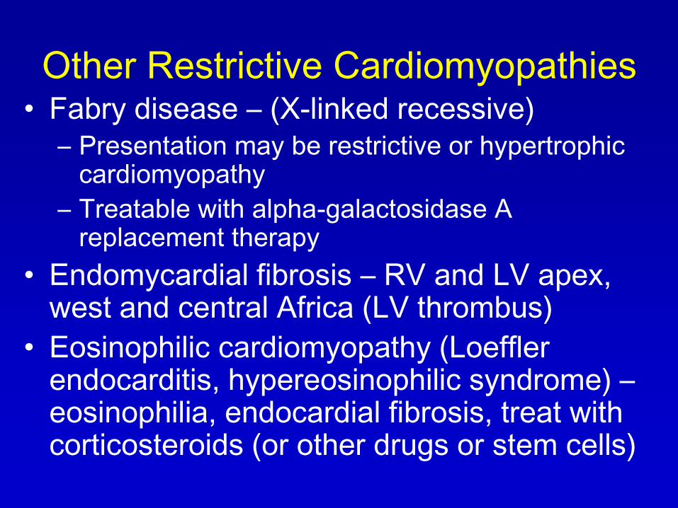
Other Restrictive Cardiomyopathies• Fabry disease – (X-linked recessive)
– Presentation may be restrictive or hypertrophic cardiomyopathy
– Treatable with alpha-galactosidase A replacement therapy
• Endomycardial fibrosis – RV and LV apex, west and central Africa (LV thrombus)
• Eosinophilic cardiomyopathy (Loeffler endocarditis, hypereosinophilic syndrome) –eosinophilia, endocardial fibrosis, treat with corticosteroids (or other drugs or stem cells)

Hypertrophic Cardiomyopathy - 1• Frequency 1/500 population (2x: men, blacks)
• Genetic – beta myosin heavy chain and 10 other genes, usually sarcomeric mutations, SCD 1%/year (variable penetrance and new mutations)
• Presentation: most asymptomatic (angina, HF, palpitations, syncope)
• Screening – by PE, ECG, echo in relatives, periodically, especially late adolescence; not gene testing
• Diagnosis: murmur ↑ with Valsalva; ECG LVH and LAE and T abnormality; Echo asymmetric hypertrophy of LV and normal EF, about 25% show dynamic LVOT obstruction by Doppler and abnormal diastolic relaxation
AHA/ACC/ESC Guidelines, Vent Arrh and SCD, 2006.

Hypertrophic Cardiomyopathy - 2• Asymptomatic or no gradient: no competitive
sports or intense isometrics, maybe β-blocker
• Symptomatic patients with gradients: β-blocker or verapamil or disopyramide, cautious diuretic for congestion
• ICD indicated for: prior SCD, sust VT or NSVT, FH SCD <40 y.o., syncope, severe LVH (>30mm), abnormal BP response to exercise (rise <20 or drop in SBP)
• Class III-IV symptoms – myectomy or catheter-based septal ablation if IVS>18, risk for pacemaker, not good for bad MR or diffuse hypertrophy
• Afib – cardioversion, rhythm control
AHA/ACC/NASPE Guidelines for Device Therapy for Arrhythmias, 2008.

HCM vs Athlete’s Heart
HCM Ath
Fam Hx HCM Yes No
Unusual ECG pattern Yes No
Unusual Echo LVH distribution Yes No
Echo LV enlargement (>55mm) No Yes
Echo LV nl cavity (<45mm) Yes No
Marked Echo LA enlargement Yes No
Abnormal LV Doppler filling Yes No
LV thickness decrease with
deconditioningNo Yes
Maximal O2 Consumption >100% No Yes

Cardiac Tumors• Most are metastatic – lung and breast (met and
direct extension), renal and hepatocellular and adrenal (venous extension)
• Benign – 50% are myxomas, generally LA arising from IAS, may simulate MS – surgically remove all, survey for recurrence
• Papillary fibroelastoma, lipomatous hypertrophy of IAS
• Malignant – sarcoma (lesser angiosarcoma, rhabdomyosarcoma, osteosarcoma)
• Echo is helpful in diagnosis but other imaging (TEE, CT or CMR) often needed
• Treatment: benign = resection; malignant = poor prognosis

Topics in Cardiology
• Epidemiology
• Heart Tests
• Coronary
Artery Disease
• Heart Failure
• Myocardial Disease
• Adult Congenital Disease
• Aortic Disease
• Peripheral Arterial Disease
• Pregnancy
• Arrhythmias
• Pericardial
Disease
• Valvular
Disease

Bradycardia• Failure of impulse formation – sinus bradycardia,
drugs, hypothyroidism, sick sinus syndrome –brady-tachy syndrome
• Failure of impulse conduction – AV block
– First degree, constant and long PR interval
– Second degree
• Wenckebach, generally AV node, narrow QRS
• Mobitz II, generally infranodal, wide QRS
• 2:1 block, can’t distinguish Wenckebach or Mobitz II
– Third degree, no beats conduct, escape rhythm
• Junctional (narrow QRS, rate 45-60)
• Ventricular (wide QRS, rate usually < 45-50)

ECG in AV Block (not AV Dissociation)
Degree of
Block
Which
ConductPR interval RR interval
First AllConstant
and longRegular
Wenckebach
(Mobitz I)Some
Variable,
progressive
Grouped
beating
2:1 Some Constant Regular
Mobitz II Some Constant Pauses
Third NoneVariable,
randomRegular

Pacemaker Therapy: Indications
• Symptomatic sinus bradycardia – relieves symptoms but doesn’t prolong life (HR<40 without clear correlation with symptoms)
• Third degree or advanced second degree (2 consecutive blocked P waves) heart block with symptoms, Mobitz II usually needs pacer
• Iatrogenic AV block with symptoms when negative dromotropic meds are needed
• No symptoms: AV block with 3.0 sec pause in awake, HR<40 awake
• Congenital heart block: Wide QRS or LV dysfunction, long QT, unknown in adults, many implant
AHA/ACC/NASPE Guidelines for Device Therapy for Arrhythmias, 2008.

Tachycardia Diagnosis
• Wide QRS: VT (80%, AV dissociation), SVT with aberrancy (20%), antidromic AVRT (preexcitation, 5%)
• Narrow QRS and regular
– P wave normal – sinus tach
– P wave absent or at end of QRS – AVNRT
– P wave shortly after QRS – AVRT
– P wave abnormal, before QRS – atrial tach
• Narrow QRS and irregular – Aflutter (can be regular at 150 or 75), afib or MAT

Tachycardia Therapy
• Unstable: synchronized cardioversion
• WCT: usually presume VT; avoid adenosine and calcium blockers
• Look at ECG post reversion for diagnostic clues
– LVH, prior MI, long QT
– Echo also helpful
– ETT if exercise-related
– Many need coronary angiography
• Referral to EP for consideration for ablation
• Antiarrhythmic agents have limitations

Antiarrhythmic Drugs
AHA/ACC/ESC Guidelines for SV Arrhythmias. 2003
Vaughan Williams Example Uses
IA – Fast Na
blocker, ↑QRS
Procainamide AF-WPW, SVT,
ventric arrhy
IB – Fast Na
Blocker, ↓QRS
Lidocaine Ventric arrhy, esp
w/ isch
IC – Fast Na
Blocker, slow
conduction
Flecainide,
propafenone
SVT, PAF, AFlut
II – beta blocker Atenolol, etc Many
III – K blocker Amio, Sotalol VT, SVT, AF, AFL
IV – Ca++ blocker Verap, dilt Many

Antiarrhythmic Drugs
• Proarrhythmia is an issue with class I and III, 5-10% (less with amiodarone)
• IC agents in AFib should use with AV nodal -blocker
• IC agents should not be used in pts with CAD
• Prolonged QT and torsades de pointes
• Generally initiate as inpatient (esp class III)
• Others not of Vaughan Williams class– Digoxin – for rate control of AF in elderly
sedentary
– Adenosine – for SVT using AV node

Question 5• 77 yo hypertensive diabetic man with mild
palpitations for 5 days. ECG shows afib, rate 100. Which of the following is preferred?
A. Warfarin to INR 2.0-3.0
B. Aspirin alone 325 mg/da
C. Aspirin plus clopidogrel
D. Cardioversion
E. TEE and possible cardioversion

Question 5• 77 yo hypertensive diabetic man with mild
palpitations for 5 days. ECG shows afib, rate 100. Which of the following is preferred?
A. Warfarin to INR 2.0-3.0
B. Aspirin alone 325 mg/da
C. Aspirin plus clopidogrel
D. Cardioversion
E. TEE and possible cardioversion

Atrial Fibrillation• A Fib is the most common sustained atrial
arrhythmia; chaotic, fibrillation waves
• Types: paroxysmal (<7d), persistent (>7d), permanent
• A Fib increases with age, over 6% of subjects over 80 yo, more in white, less than 12% of patients with AF have no structural disease; lone AF is people <60 with no disease by clinical and echo
• Associations: hypertension, valve disease, coronary disease, cardiomyopathy of any morphology, preexcitation, alcohol, thyroid, myocarditis or pericarditis, postoperative
• In elderly consider subclinical hyperthyroidism
AHA/ACC/ESC Practice Guidelines, Atrial Fibrillation, 2006.

Symptoms in Atrial Fibrillation• Asymptomatic
• Palpitations, fatigue, exercise intolerance,
dyspnea, dizziness, syncope, HF, stroke:
related to loss of atrial contraction, slow or
fast rate, irregularity
• Tachycardia-related cardiomyopathy if
HR>130/min for several weeks
• Angina if higher rate or lower blood
pressure cause coronary flow demand to
exceed supply
AHA/ACC/ESC Practice Guidelines, Atrial Fibrillation, 2006.

AFib Acute Management
• Unstable (severe symptoms) not responding to rate control and support: cardioversion acutely even before anticoagulation
• Stable patient: if >48 hr – decide between rate or rhythm control, anticoagulation (INR 2.0-3.0 for 3 wk prior and 4 wk after cversion, TEE-Cardioversion is reasonable alternative)
• Antiarrhythmic drugs, only for severe symptoms (hypotension, unstable ischemia, acute HF)
• Persistent AF: Cardiovert, sooner is more successful; 5-15% AF patients over 48 hr have thrombus on TEE (LA appendage)
• If AFib for >3 mo, use antiarrhythmic before cardioversion, continue about a month after
AHA/ACC/ESC Practice Guidelines, Atrial Fibrillation, 2006.

Afib: Rate Control and Rhythm Control
• Rate control (60-80 rest, 90-115 exerc):
– Beta blocker or calcium blocker is preferable (digoxin less effective)
– Pre-excitation: avoid usual drugs and use procainamide or amiodarone (to slow conduction in accessory pathway)
• Rhythm control strategy for patients still symptomatic on rate control, or if rate control is inadequate (Class III or IC), 20-50% recur in ≥1y
• Cardioversion OK with Pacer or ICD

Atrial Fibrillation and Stroke
• Major concern in AFib is thromboembolism,
particularly stroke, about 5%/yr in nonrheumatic,
about 2-7x more likely than NSR
• 75% of strokes are from the AF, 25% other
causes
• Warfarin lowers stroke rate 60% (1-2% major
bleed per year); aspirin only 20% lowering
• CHADS2 score (Age>75, HF, Htn, DM, prior
stroke (=2)): warfarin for 3 or more, ASA for 0,
judgment for 1-2
AHA/ACC/ESC Practice Guidelines, Atrial Fibrillation, 2006.

Device Management of Atrial Fibrillation
• AVN ablation and pacemaker: need anticoagulation, for medically refractory to rate control
• Catheter ablation – technique still developing, 80% success; “reasonable alternative to prevent recurrence in symptomatic pts with little or no LAE”, only in disabling symptoms, pulmonary vein isolation and adjunctive ablation areas (complications 6%, PV stenosis, atrioesophageal fistula, embolism)
• Surgical Maze (during CABG or Valve Surgery) success 70-95%
AHA/ACC/ESC Practice Guidelines, Atrial Fibrillation, 2006.

Atrial Flutter
• Macro-reentrant circuit usually in right atrium with pathway including the cavotricuspid isthmus (tissue between IVC and tricuspid annulus)
• Atrial rate 240-340/min – sawtooth negative in II, III, and aVF, positive in V1
• Associations: congenital heart disease, pulmonary disease, Htn, DM, Obesity, Thyroid disorders, sick sinus, major cardiac surgery
• Ventricle often responds 2:1 at rate about 150
AHA/ACC/ESC Practice Guidelines, Atrial Fibrillation, 2006.

Atrial Flutter Management• Patients with rapid ventricular response may
not respond well to rate control (using beta blocker or nondihyrdopyridine calcium blocker)
• Early synchronized cardioversion often needed
• Class III agents (amiodarone, sotalol) generally more effective to revert to NSR
• Anticoagulation decisions are identical to those with atrial fibrillation
• Radiofrequency ablation is first line for recurrent Aflutter, 90% success, 1-2% complications … 6 month recurrence 60%
AHA/ACC/ESC Practice Guidelines, Atrial Fibrillation, 2006.

Paroxysmal Reentrant SVT• AVNRT: Most common SVT (60% SVTs), regular,
rate 140-250; in typical AVNRT the retrograde P is buried in QRS or at end of QRS (pseudo-S in II, and pseudo R prime in V1)
– Sx: palpitations and SOB, syncope infrequent
– Rx: vagal maneuvers – Valsalva, immersion in ice water, CSM, adenosine (bolus, more if theophylline, less if dipyridamole, not if severe asthma)
– Prevention with beta blocker or calcium blocker; avoid class IC or III agents, avoid triggers
– Catheter ablation if severe sx or med intolerance (95% success, 1-2% complication)
AHA/ACC/ESC Guidelines for SV Arrhythmias. 2003

Paroxysmal Reentrant SVT
AHA/ACC/ESC Guidelines for SV Arrhythmias. 2003
Pseudo R prime in V1 and pseudo S in II

Preexcitation Syndromes - 1• Accessory pathway straddles the AV groove at
mitral or tricuspid annulus
• Most have manifest preexcitation (short PR and delta wave and wide QRS and ST-T changes), some are concealed with only retrograde conduction
• Most common (95%) reentrant arrhythmia is Atrioventricular reentrant tachycardia (AVRT) , usually (90%) narrow complex (“orthodromic AVRT”); antidromic AVRT looks like VT
• Up to 1/3 of WPW patients have Atrial Fib, dangerous if accessory pathway conducts rapidly leading to VF
• Sudden Cardiac Death in patients with WPW is 0.1-0.4%, usually due to AF degenerate to VF
AHA/ACC/ESC Guidelines for Supraventricular Arrhythmias. 2003
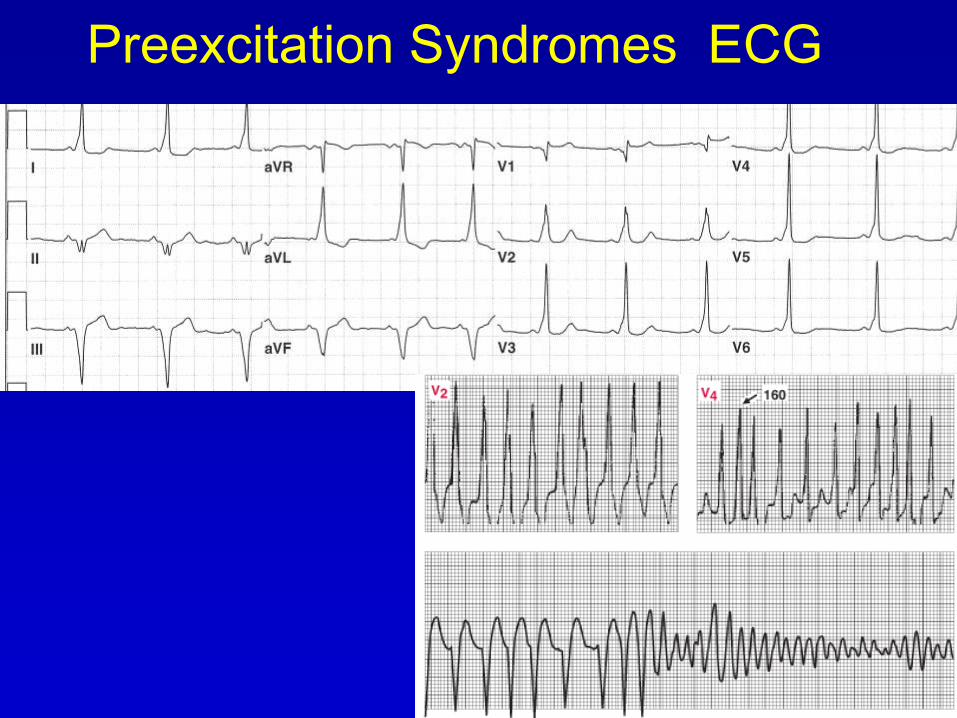
Preexcitation Syndromes ECG

Preexcitation Syndromes ECG

Preexcitation Syndromes - 2• Management: orthodromic responds to
adenosine; antidromic should not receive AVN blockers, can make arrhythmia unstable
• Unstable tachycardia: cardioversion regardless of mechanism or arrhythmia
• Drugs that block accessory pathway acutely: ibutilide, procainamide, propafenone, flecainide, amiodarone; oral agents for chronic use Ia, Ic, III and possibly added beta-blocker
• Catheter ablation recommended as first line therapy when symptomatic arrhythmia is present, unless tachycardia is uncommon and minimally symptomatic
• WPW pattern on ECG and no symptoms – no treatment
AHA/ACC/ESC Guidelines for Supraventricular Arrhythmias. 2003

Atrial Tachycardia• Not very common (10% of SVTs), usually benign
unless incessant, then may produce tachycardia-induced cardiomyopathy
• Atrial tachycardia with block may be due to digitalis excess (worse with hypokalemia)
• ECG – usually long R-P
• Acutely can slow with AVN blockade and many will revert with adenosine or have AV block
• Chronic therapy with nondihydropyridine Ca Blocker or Beta-blocker may reduce occurrences
• Cardioversion – high likelihood of early recurrence
• Ablation of atrial focus is an option for drug failure
• Multifocal atrial tachycardia – 3 different P waves – treat underlying disease
AHA/ACC/ESC Guidelines for Supraventricular Arrhythmias. 2003

Ventricular Ectopy• PVC: generally benign, minimal significance if EF
is OK, but if EF is low, assoc with increased mortality, but treatment doesn’t improve outcome; so complex ventricular ectopy deserves workup for structural heart disease … treat PVC with beta-blocker or HR-lowering Ca blocker only if symptomatic (palpitation, fatigue, near syncope), avoid antiarrhythmic agents
• Nonsustained VT (<30sec) prevalent in HF, not bad prognosis in normal heart or in first 48 hr of MI, but bad later after MI – treat ischemia, and if EF<35% .. ICD, or if EF 35-55% and inducible sustained VT in EPS … Med rx is beta-blocker and HR-lowering Ca blocker … can use amio or sotalol … ablation for drug failure
AHA/ACC/ESC Guidelines, Vent Arrh and SCD, 2006.

Sustained Ventricular Tachycardia
• Usually reentrant mechanism
• Prognosis of monomorphic VT depends on underlying heart disease – high rate of recurrence
• Polymorphic VT is most often genetic defect and dangerous
• Idiopathic ventricular tachycardia (RV outflow tract tachycardia) is usually benign … exclude arrhythmogenic RV cardiomyopathy, may try Ca or beta blocker, ablation success >90%
AHA/ACC/ESC Guidelines, Vent Arrh and SCD, 2006.

Sudden Cardiac Death - 1
• Cause: 75% are CAD (50% of SCD have acute coronary pathology), VT / VF in 75-80%, bradycardia in 15-20%
• Underlying factors (most victims are low risk): DM, Htn, HLP, FH CAD, smoking, obesity, inactivity, HF, LVH, heavy EtOH
• Triggers: Electrolyte abn, drugs (cocaine, QT-prolonging, antiarrhythmics), exercise, emotional stress
• Familial DCM, ARVC, HCM, Channelopathy, Long QT syndrome, Brugada syndrome, short QT syndrome
AHA/ACC/ESC Guidelines, Vent Arrh and SCD, 2006.

Sudden Cardiac Death - 2• Primary prevention: ICD superior in CAD and low
EF, and also in DCM, class II or III and EF<35%
• Acute treatment of VF: one shock; epi or vasopressin, amiodarone; after return of spontaneous circulation hypothermia improves outcomes
• Secondary prevention (30% recur in 1 yr), improved with CABG
• Don’t use ICD: Severe class IV heart failure, expected survival <1 y, incessant or frequent nonsuppressible VT/VF
• Inappropriate ICD shocks from lead problem, SVT, sinus tach, electromagnetic interference
AHA/ACC/ESC Guidelines, Vent Arrh and SCD, 2006.

Pulseless Electrical Activity - Asystole
• Look for potentially reversible causes
– Pulmonary Embolism
– MI
– Tamponade
– Tension pneumothorax
– Acidosis, drug overdose, hyperkalemia,
hypothermia, hypoxia
• Use atropine and epi to increase HR
AHA/ACC/ESC Guidelines, Vent Arrh and SCD, 2006.

Topics in Cardiology
• Epidemiology
• Heart Tests
• Coronary
Artery Disease
• Heart Failure
• Myocardial Disease
• Adult Congenital Disease
• Aortic Disease
• Peripheral Arterial Disease
• Pregnancy
• Pericardial Disease
• Arrhythmias
• Valvular
Disease

Question 6
• 59 yo diabetic woman with pleuritic chest pain for 2 days. Normal exam. ECG-diffuse ST elevation. What is the best choice for this patient?
A. Prednisone for 6 weeks
B. Aspirin for 4 weeks
C. Right and left heart catheterization
D. Colchicine for 3 months
E. Methotrexate weekly for 6 months
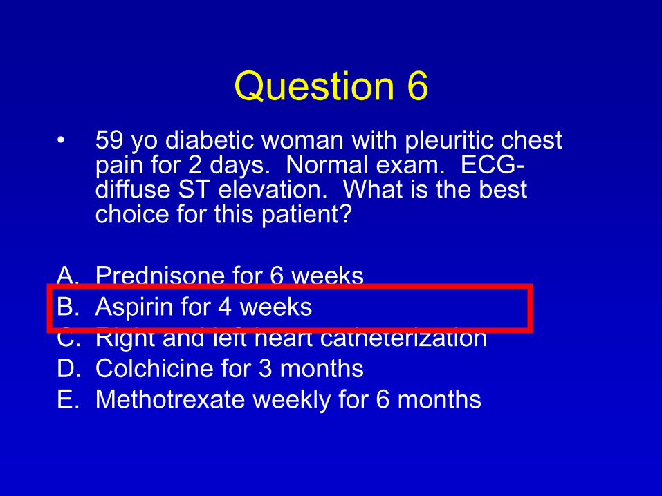

Acute Pericarditis - Causes• Idiopathic* (40%), viral* (20%) – HIV, bacterial
(7%), fungal, TB, uremia (6%)
• Autoimmune*, esp SLE (1/3 of SLE pts eventually
have pericarditis)
• Acute MI (<3d), post MI (3-10 wk), post
pericardiotomy* (>1wk)
• Neoplasm (7%)
• Medication – hydralazine, procainamide, warfarin,
heparin, methylsergide, doxorubicin, penicillins
• Chest irradiation*, chest trauma, asc Ao
dissectionEur Soc Card. Guideline
Pericardial Diseases, 2004.*=Constriction too

Acute Pericarditis - Diagnosis
• Pain, usually sharp for hours or days, better sitting forward, or pleuritic, with diaphoresis, ↑HR, ↑RR
• PE: 3-component friction rub (squeaky, scratchy, high-pitched, swooshing, absence not exclude diagnosis)
• ECG diffuse ST↑ and PR↓ are supportive
• Echo: about 50% have pericardial effusion, seek HF or concomitant problems
• Biomarkers for CAD, ESR, Autoimmune tests for recurrent disease (troponin may be elevated)
Eur Soc Card. Guideline Pericardial Diseases, 2004.
Dia
gno
sis
: 2 o
f th
e 3

ECG in Acute Pericarditis
• ST segment elevation
• PR segment depression

Recurrent Pericarditis
• 10-30% of idiopathic pericarditis
recurs (return of Sx and Sn) within
days to weeks of stopping therapy
(elev ESR)
• More in rheumatologic disorders,
Dressler’s syndrome (post MI
pericarditis), or post pericardiotomy
syndrome
Eur Soc Card. Guideline Pericardial Diseases, 2004.

Pericarditis - Treatment• NSAID in relatively high dose for 3-4 weeks (ASA
650 q4-6h, Ibuprofen 400-800 q6-8h, taper ASA
after a week, ibuprofen after 2 wk) total may be
3-4 weeks
– Pain relief expected in 24 hr, or increase dose
or use alternative
– Alternative: colchicine 0.6 mg bid (tid if >70kg),
(only 1 day if with NSAID) – for 3 months
– Avoid steroids = tend to recur when stopped
• Recurrent: Colchicine 6 months, may add NSAID
for 3 mo (ibuprofen 400 qid)
Eur Soc Card. Guideline Pericardial Diseases, 2004.

Pericardial Effusion
• Etiology same as pericarditis, about 1/3 of large effusions develop tamponade; rapid accumulation of fluid tends to tamponade – dx best with echo, not CXR
• Pericardiocentesis – (by experienced physician) for tamponade or suspected bacterial, TB or systemic inflammatory dz, or if >3 mo.
• Anticoagulation inadvisable – risk of hemopericardium
Eur Soc Card. Guideline Pericardial Diseases, 2004.

Pericardial Tamponade
• Diagnosis – *distant heart sounds, *elevated JVP, *hypotension, tachycardia, dyspnea (JVP has no Y descent)
• Pulsus paradoxus (may be absent if AR, ASD, LVH, PHtn, RVH)
• Confirm with Echo (RV diastolic collapse, increased respiratory change in LV and RV filling velocities, IVC enlargement without respiratory change)
• Management: volume resuscitation, may need vasopressors, pericardiocentesis guided by echo; caution with mechanical ventilation or sedation
*= Beck’s triad.

Constrictive Pericarditis - 1
• Etiology same as other pericardial processes (esp CT surgery and idiopathic pericarditis) plus XRT to chest (breast Ca or Hodgkin’s)
• Sx and Sn of right-heart failure > expected LV or valve effects; fatigue, dyspnea, edema, ascites, JVD (Kussmaul’s sign, brisk X and Y), pericardial knock; afib in 20%
• DDX restrictive cardiomyopathy is difficult
Eur Soc Card. Guideline Pericardial Diseases, 2004.

Constrictive Pericarditis - 2• Echo is best, with Doppler accentuated but brief
early filling; accentuated respiratory change in Doppler and respiratory ventricular septal shift (not seen in restrictive cardiomyopathy)
• CT and MRI for pericardial thickening, but 18% CP have normal thickness
• Cath – equal pressures, “square-root sign” = dip and plateau in diastole RV and LV
• Management, cautious diuretics (CP may resolve spont or with anti-inflamm), pericardiectomy (mortality 6-12%) if NYHA class 2 for at least 3 months, outcomes worse if radiation etiology
Eur Soc Card. Guideline Pericardial Diseases, 2004.

Topics in Cardiology
• Epidemiology
• Heart Tests
• Coronary
Artery Disease
• Heart Failure
• Myocardial Disease
• Adult Congenital Disease
• Aortic Disease
• Peripheral Arterial Disease
• Pregnancy
• Arrhythmias
• Pericardial
Disease
• Valvular
Disease

Valvular DiseaseBicuspid Aortic Valve
with calcified nodules
Flail anterior
mitral leaflet
Valves in
diastole

Question46-year old woman with sudden onset of dyspnea. On 2L NC sats 96%, R-20, BP 132/86; ECG shows atrial fibrillation with rate 124. Echocardiogram shows normal LV function and mitral stenosis with mean gradient 18 mmHg. Which of the following is the most appropriate next step?
A. IV Digoxin
B. IV Diltiazem
C. Open mitral commissurotomy
D. Direct current cardioversion
E. Balloon mitral valvuloplasty
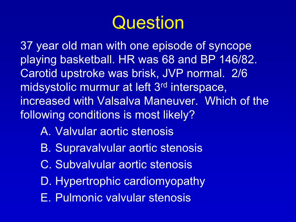
Question37 year old man with one episode of syncope
playing basketball. HR was 68 and BP 146/82.
Carotid upstroke was brisk, JVP normal. 2/6
midsystolic murmur at left 3rd interspace,
increased with Valsalva Maneuver. Which of the
following conditions is most likely?
A. Valvular aortic stenosis
B. Supravalvular aortic stenosis
C. Subvalvular aortic stenosis
D. Hypertrophic cardiomyopathy
E. Pulmonic valvular stenosis

Clinical Evaluation in Valvular
Disease
• Presentation: acute severe heart failure, no
symptoms, mild reduction in activity, murmur
• Murmur: intensity, duration, radiation, timing
in cardiac cycle
• Examination: apical impulse, distal pulses,
timing of S1 and S2, ejection sounds, other
extra sounds, response to Valsalva and other
maneuvers

Midsystolic Murmurs• Turbulent antegrade flow – most are benign
• Aortic stenosis, radiate to apex (Gallavardin), carotid, decreased carotid upstroke (ejection sound in bicuspid valve, subvalvular or supravalvular stenosis has no ejection sound)
• HCM, increase with Valsalva, standing, amyl nitrate inhalation, decrease with squat
• Pulmonic stenosis, LUSB, increase with inspiration (ejection sound in valvular disease)
• Tetralogy of Fallot has RVOT obstruction as cause of murmur (no VSD murmur because RV and LV systolic pressure are equal)
• High flow states (pregnancy, cirrhosis, anemia, thyrotoxicosis, beri-beri, atrial septal defect)
LVOT
RVOT

Holosystolic Murmurs
• Turbulent retrograde flow, murmur generally persists to S2:
– Mitral regurgitation: best at apex, radiate to axilla or back or base (papillary muscle dysfunction murmur and mitral prolapse murmur may begin later in systole and usually persist to S2; acute MR may be early systolic)
– Tricuspid regurgitation: with large V wave in JVP, ↑ with inspiration, pulsatile liver
– Ventricular septal defect: louder murmur if smaller defect and lower RV systolic pressure (small muscular defect may be only early systolic)

Diastolic Murmurs
• Diastolic regurgitant (retrograde flow in diastole):
– Aortic regurgitation: LLSB for leaflet, RLSB for root, longer murmur usually means worse leak
– Pulmonic regurgitation: rarely significant
• Mid-diastolic flow (antegrade turbulent flow):
– Mitral stenosis with usually loud S1, and with opening snap, short A2-OS interval indicates tight stenosis
– Tricuspid stenosis louder with inspiration

Other Murmurs
• Continuous murmur (often peaks around S2): PDA is most likely, AV fistula is a frequent cause of acquired continuous murmur
• Aortic Coarctation may be somewhat continuous and heard at the back
• Pericardial friction rub: may have 1 or 2 or 3 components, systolic component is most common, scratchy, creaking leather

Maneuvers
• All right sided auscultatory events (PS,
PR, TS, TR, S3, S4) increase with
inspiration (except pulmonary ejection
sound which decreases)
• All murmurs decrease with Valsalva
maneuver except HCM which increases
and maybe mitral prolapse

Systolic Murmur EvaluationType Location Radiation Maneuver Associations
Innocent Base Usually no None None
Ao
Sclero
Base
R2icsNone
↓handgrip
↓standingNone
Ao
Stenosis
Base
R2ics
Carotids,
occ apex
↓handgrip
↓standing
↓A2, delay
and ↓pulses
PS L2ics LSB ↑inspirat’n Wide split S2
HCM Base Carotids↓handgrip
↑standingBifid pulses
MR ApexVariable,
axilla↑handgrip
LV
enlargement
TR LLSB LRSB ↑inspirat’n JVP, liver
VSD LSB RSB Thrill LLSB

Other Murmur Evaluation
Type Location Radiation Maneuver Associations
Diastolic
ARLSB or
RSB
Variable,
none or
apex
End-expir
lean
forward
Enlarged apical
impulse, wide pulse
pressure
MS Apex
Localized
to small
area
Left lateral
recumbent
Possible Opening
Snap
PR L2-3ics LSB None RV heave
TS LLSB None ↑ inspPossible Opening
Snap
Continuous
PDA L2-3ics Back

Key Non-Murmur Findings
• Carotid upstroke – bounding in AR,
delayed in AS, double in HCM or AR
• JVP –Large V wave in TR
• Apex – sustained in LVH (aortic stenosis),
displaced leftward and inferiorly in AR and
MR
• Aortic ejection sound (bicuspid aortic
valve)
• Mitral opening snap (MS)

Evaluation of a Murmur
ACC/AHA Guidelines, Valve disease, 2006

Echocardiography in Valve Disease
• To determine lesion etiology and severity and
coexistent lesions, PASP estimate, LA and LV
size and LV function
• Stenosis: transvalvular velocities for calculation of
valve area and pressure gradients
• Regurgitation: color jet size, width of narrowest
regurgitant segment, signal strength, and
calculated quantitation of regurgitant area or
volume
• TEE: selected patients, esp severe MR and
prosthetic valves and endocarditis

Normal Echo Measurements
LV Ejection Fraction >55%
LV end-diastolic dimension <60 mm
LV end-systolic dimension <40 mm
LA dimension <40 mm
PA systolic pressure <30 mmHg
Ascending aortic diameter <3.5 cm

Tests in Valve Disease
• BNP to assess significance of symptoms
• Exercise testing with or without echo
• Dobutamine stress echo for AS with
systolic dysfunction for increase in valve
area (pseudostenosis) or not (true stenosis)
• Cardiac cath only for coronary angiography
or occasionally to clarify discrepant findings

Management of Valve Disease
• Frequency of serial evaluation depends on lesion and severity
• Sports participation is generally OK if no symptoms
• Noncardiac surgery generally OK for asymptomatic patients: careful fluid balance
• Symptomatic patients with mild or moderate valve disease – seek other causes for symptoms
• Symptomatic patients with severe disease –surgery (optimize preload and volume and arrhythmia)
• Asymptomatic patients with severe disease and need for CABG – add valve surgery

Valve Disease Follow-upLesion Criteria by echo Eval Echo
AS-mild V<3m/s, AVA>1.5 Yr 3-5y
AS-mod V 3-4m/s, AVA 1.0-1.5 Yr 1-2y
AS-sev V>4m/s, AVA<1.0 Yr Yr
MS-mild MVA>1.5, MG<5, PA<30 Yr 3-5y
MS-mod MVA 1.0-1.5, MG 5-10 Yr 1-2y
MS-sev MVA<1.0, MG>10, PA>50 Yr Yr
AR-mild VC<0.3cm, ROA <0.10cm2,
RV<30 ml/beat, EF nl
Yr 2-3y
AR-sev LV nl VC>0.6cm, ROA>0.3 cm2,
RV>60 ml/beat, RF>50%
6-12mo Yr
AR-sev LVE 6 mo 6-12mo
MR-mild See mild AR .. EF nl, LV nl Yr For sx
MR-sev VC>0.7cm, ROA>0.4cm2 6-12mo 6-12mo

Aortic Stenosis Etiology
• Bicuspid valve: ~1-2% of population; young person
with an aortic ejection sound needs no endocarditis
prophylaxis and no physical limitations
– Becomes stenotic earlier than tricuspid, age 40-
60; associated with aortic dilation and dissection
• Most aortic stenosis is calcific degeneration of a
tricuspid aortic valve; atherosclerotic risk factors
usually present (aortic sclerosis patients have
increased risk of MI or cardiovascular death)
ACC/AHA Guidelines, Valve disease, 2006

Aortic Stenosis - 1
• Exam: carotid parvus et tardus, single S2, S4,
harsh mid to late peaking midsystolic murmur – get
ECG, CXR, Echo
• Echo – Thick poorly mobile aortic leaflets,
concentric LVH with normal LV size, impaired
diastolic function, Doppler velocities increased
• Treadmill exercise test contraindicated in
symptomatic AS, OK in equivocal symptoms to
diagnose exercise intolerance not obtainable by
history (perform by cardiologist)
ACC/AHA Guidelines, Valve disease, 2006
Aortic Stenosis – 2 – Heyde’s
Syndrome
• Patient with GI bleeding from arteriovenous malformations
– About 20% of pts with severe AS have clinical bleeding, e.g, epistaxis, ecchymosis, GI bleed
• Acquired von Willebrand syndrome
• Disruption of von Willebrand multimers during turbulent passage through the stenotic aortic valve
ACC/AHA Guidelines, Valve disease, 2006

Aortic Stenosis - 3• Symptoms: none, angina, HF (exercise
intolerance, usually gradual, may be subtle), syncope (rare)
• Acute increase in symptoms if atrial fibrillation due to loss of atrial kick
• Valve replacement (3-4% periop mort) for – severe symptomatic AS, or
– severe AS with LVEF<50 or
– severe or moderate AS without symptoms but with surgery for something else (MVR or aortic root surgery, or possibly CABG)
• Percutaneous valve replacement is new, promising (not valvotomy)
ACC/AHA Guidelines, Valve disease, 2006

Mitral Stenosis - 1
• Cause = RHD; 60% w/ hx ARF, women 2:1
• Presentation: dyspnea with exercise, emotional
stress, infection, pregnancy, AF with RVR (may
give pulm edema); systemic emboli/stroke in
atrial fibrillation
– Adaptive changes in pulmonary vasculature
and alveoli and lymphatics may ameliorate
symptoms
– Presentation in US generally in 40s and 50s,
younger in other areas
ACC/AHA Guidelines, Valve disease, 2006
Mitral Stenosis - 2
• Exam: Loud S1, opening snap, low-
pitched diastolic rumble with presystolic
accentuation
• CXR: may show straightened left heart
border, congested lungs
• Echocardiogram: Thickened MV with
fused commissures, calcification, diastolic
doming, subvalvular disease, turbulent LV
inflow, look for concomitant regurgitation
ACC/AHA Guidelines, Valve disease, 2006

Mitral Stenosis –
Medical Therapy
• β-blocker or HR-lowering Ca++ blocker may relieve symptoms during high HR
• Diuretics and Na restriction for congestion
• Anticoagulation for paroxysmal atrial fibrillation and control of ventricular rate (AV node blocker)
• Emergent cardioversion if hemodynamically unstable
• Treat recurrence of AF with IC (and negative dromotropic agents) or Class III antiarrhythmic
ACC/AHA Guidelines, Valve Disease, 2006
Mitral leaflet doming

Mitral Stenosis - Intervention• Favorable anatomy for balloon valvuloplasty
(percutaneous valvotomy): no LA thrombus on TEE, no 3-4+ MR, pliable noncalcified leaflets, not severe subvalvular disease and no commissural fusion
• Valvuloplasty for favorable anatomy and: Asymptomatic if MVA<1.5 and rest PASP>50 or exercise PASP>60 or PAWP>25 or gradient >15; maybe for new afib
• Valvuloplasty complications: MR, systemic embolization, tamponade (1% mort)
• Valve repair/replacement: unfavorable anatomy and MVA<1.5 and NYHA 3/4 (MAZE maybe)
ACC/AHA Guidelines, Valve disease, 2006

Acute Left Sided Valvular Regurgitation
• Pathophysiology: sudden severe regurgitation gives sudden LV volume load, LV is unable to acutely dilate, so diastolic pressures rise –tachycardia, pulmonary edema, respiratory failure, and/or shock
• Presentation – sudden pulmonary edema or cardiogenic shock, worse with prior Htn or AS (concentric LVH)
• Physical Exam less reliable, tachycardia, softer murmur
• CXR: No cardiomegaly but severe pulmonary edema is characteristic
• Treatment: Afterload reduction with nitroprusside if normotensive, inotropes if hypotensive, Surgery
ACC/AHA Guidelines, Valve disease, 2006

Acute Aortic Regurgitation
• Etiology (trauma, IE, dissection)
• Physical Exam less reliable, tachycardia,
softer murmur (absent S1 indicates surg)
• Echo for diagnosis; other imaging if
dissection is suspected (TEE, CT, MRI)
• Treatment if severe
– Temporize with nitroprusside, inotropes,
(NO IABP)
– Emergent AVR +/- aortic repair
ACC/AHA Guidelines, Valve disease, 2006

Acute Mitral Regurgitation• Etiology: Infective endocarditis, ruptured
chordae, ruptured papillary muscle (Acute MI
with sudden pulmonary edema)
• PE less reliable, tachycardia, softer MR murmur
maybe only early systole, +S3, +S4
• Echo for diagnosis, usually TEE to plan mitral
repair vs. replacement
• Treatment: nitroprusside if normotensive,
inotropes if hypotensive, IABP if hypotensive;
definitive treatment is emergent MVR
ACC/AHA Guidelines, Valve disease, 2006

Chronic Left Sided Valve Regurgitation
• Mild-moderate usually asymptomatic and most do not progress to severe
• Severe produces compensatory dilation
• Eventual exercise intolerance but symptoms not reliable
• Surgery for NHYA II-IV and severe AR or MR
• Surgery for severe AR and MR without symptoms if enlargement is excessive or systolic function is subnormal
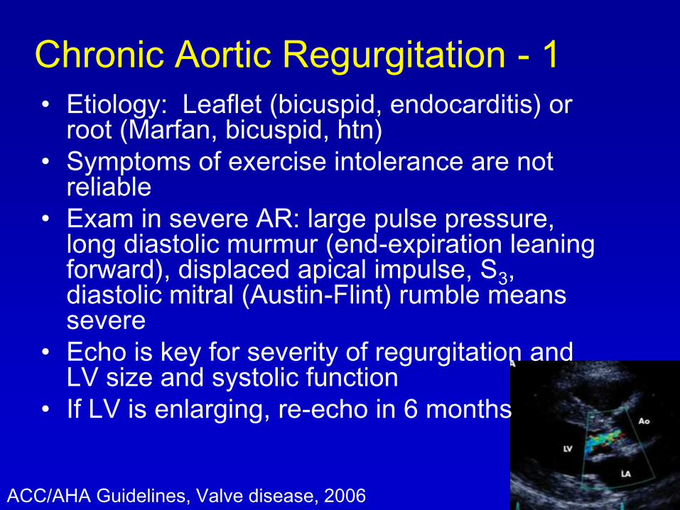
Chronic Aortic Regurgitation - 1
• Etiology: Leaflet (bicuspid, endocarditis) or root (Marfan, bicuspid, htn)
• Symptoms of exercise intolerance are not reliable
• Exam in severe AR: large pulse pressure, long diastolic murmur (end-expiration leaning forward), displaced apical impulse, S3, diastolic mitral (Austin-Flint) rumble means severe
• Echo is key for severity of regurgitation and LV size and systolic function
• If LV is enlarging, re-echo in 6 months
ACC/AHA Guidelines, Valve disease, 2006

Chronic Aortic Regurgitation - 2
• Med Rx – for severe AR with htn or LV enlargement: hydralazine, nifedipine, ACE-I
• Surg only for severe – normal EF and NYHA class 3-4 HF (class 2?) or CCS 2-4 angina, EF 25-50 and HF, asymptomatic with EF<50 x 2, LVEDD>75 mm, LVESD>55 mm (less in smaller patients)
• At surgery: check aortic root dilation (Marfan’s, dissection, chronic post-stenotic or hypertensive), reconstruct aortic root if >50mm diameter (45 mm if bicuspid valve)
ACC/AHA Guidelines, Valve disease, 2006

Question 7
In asymptomatic chronic severe mitral regurgitation, which of the following is an indication for surgery?
A. LV ejection fraction 60%
B. Grade IV/VI murmur
C. Frequent PACs
D. Echo LV systolic dimension >40 mm
E. Concomitant mild mitral stenosis

Question 7
In asymptomatic chronic severe mitral regurgitation, which of the following is an indication for surgery?
A. LV ejection fraction 60%
B. Grade IV/VI murmur
C. Frequent PACs
D. Echo LV systolic dimension >40 mm
E. Concomitant mild mitral stenosis

Chronic MR - Diagnosis
• Etiologies: MV prolapse, infective endocarditis, LV enlargement, CAD
• Clinical evaluation:
– Symptoms: DOE/exercise intolerance
– Examination: apical murmur, apical displacement, diastolic rumble, loud P2, RV lift
• Echo is key for cause and severity of MR, for LAE and for LV function and size (end-systolic dimension) and PASP; low-pressure leak means LV EF should be >60%
• Other tests: ECG-LVH, CXR-cardiomegaly
ACC/AHA Guidelines, Valve disease, 2006

Chronic MR – Management
• Organic MR: vasodilator (ACE-I) logical but unproved, control BP, management of AF
• Functional MR (from LV dilation or akinesis): diuresis and β-blocker and ACE-I may reduce MR severity, also CRT (resynchronization therapy)
• AFib and MR: warfarin with INR 2-3
• Surgery only for severe MR, repair if technically feasible (minimal calcification, posterior leaflet involvement, non-rheumatic cause)
• Replacement better results with chordal preservation
• Surgery for asymptomatic if EF<60 or LVESD>40mm; atrial fibrillation; even earlier if likelihood of repair >90%
ACC/AHA Guidelines, Valve disease, 2006

MV Prolapse• Common, 1-2½% population, MV bulging into LA
• Exam: midsystolic click and late systolic murmur, longer with Valsalva
• Most have benign course, but MVP is most common predisposing cause of endocarditis and most common reason for MR surgery
– Ruptured chordae tendinae, endocarditis, arrhythmia, heart failure
• Medical therapy: beta-blocker for chest pain or palpitations, also stop caffeine, EtOH, cigarettes; liberalization of sodium intake for orthostasis; aspirin 75-325/da for TIA
• Surgery: mitral repair or replacement indications are the same as for MR of any other cause
ACC/AHA Guidelines, Valve disease, 2006

Tricuspid Valve Disease• TS: rare, rheumatic (carcinoid, Fabry’s,
methylsergide)
• TR (normal finding with echo): usually secondary to LV dz, or to RV pressure load (>55 PHTN or cor pulmonale) or volume load (primary from endocarditis or RHD, where PASP often <40);
• TR Exam: large JVP V wave, systolic murmur ↑-inspiration, pulsatile liver
• Echo for severity and cause and PASP and RV and RA size
• Diuretics; annuloplasty ring usually only with other surgery, bioprosthetic possible
ACC/AHA Guidelines, Valve disease update, 2008

Endocarditis - 1• Suspect – murmur esp with fever and with prior
valve dz or prosthesis
• Strep and Staph and HACEK (Haemophilus, Actinobacillus, Cardiobacterium, Eikenella, Kingella), culture neg, fungi, TB
• Dx: 2 major or 1 major and 3 minor or 5 minor
• Major: Persistent pos blood cultures with typical organism; New valve regurg; positive echo for vegetation
• Minor: Predisposing condition or IVDU, fever, embolic vascular phenomena, immunologic phenomena (GN, RhF), other pos blood culture
ACC/AHA Guidelines, Valve disease update, 2008

Endocarditis - 2• TTE 50-80% sens, TEE 95% and better, esp for
abscess
• Blood cultures before antibiotics, esp with
indwelling hardware
• Management: antibiotics; emergency valve
surgery if unstable hemodynamics (caveat such
as CNS damage)
• Surgery more likely with worse valve destruction,
paravalvular abscess, heart block, resistant
organisms, larger vegetations, recurrent emboli
• Prefer valve repair if poss
ACC/AHA Guidelines, Valve disease update, 2008

Endocarditis Prophylaxis for
Dental Procedures
• Prosthetic heart valves or other cardiac
prosthetic material
• Prior endocarditis
• Congenital disease
– Shunts and conduits
– Unrepaired cyanotic congenital disease
– Prosthetic material for first 6 months
• Heart transplant patient with valve disease
AHA Guideline. Prevention of Infective Endocarditis. Circulation. 2007.
Prosthetic Valves
• Complications: infection (20% of endocarditis cases), bleeding, thrombosis and embolism (rarely hemolytic anemia)
• Valve selection
– Repair is preferable to replacement if feasible
– Lifestyle preferences of patient are often overriding consideration
– Mechanical for long life span, renal dialysis, already requiring anticoagulation
– Biologic for avoid anticoagulation
• AVR if >65 yo
• MVR if >65 yo and no risk factors for thromboembolism
ACC/AHA Guidelines, Valve disease, 2006

Mechanical Prosthetic Valves
• More durability but more anticoagulation is required and risk of bleeding is higher
• Preferred for >10-15 year life expectancy
• Types:
– Ball in cage (Starr-Edwards)
– Tilting disc (Bjork-Shiley; Medtronic Hall)
– Bileaflet (St. Jude)
ACC/AHA Guidelines,
Valve disease, 2006

Biological
Prosthetic Valves
• Less thromboembolism, less anticoagulation but less durable and more reoperation
• Options
– Autograft (pulmonic to aortic .. Ross procedure)
– Autologous or autogenous pericardial (crafted at surgery)
– Homograft (allograft – human)
– Heterograft or xenograft is sterilized and nonviable thus bioprosthesis, bovine pericardial, porcine valve (degeneration is more likely after 5 yr for MVR and after 8 yr for AVR, if regurg is noted, echo q 3-6 mo)
ACC/AHA Guidelines, Valve disease, 2006

Anticoagulation of Mechanical
Valvular Prostheses
Prosthesis Type INR ASA, mg
Mechanical
Caged-ball Aortic valve 2.5-3.5 75-100
Ao valve low risk (St Jude,
Carbomedics, Medtronic-Hall)
2.0-3.0 75-100
Ao valve + risk factors* 2.5-3.5 75-100
Mitral valve (with or without risk
factors*)
2.5-3.5 75-100
*Risk factors = AFib, LV systolic dysfunction, prior TE event or
hypercoagulable state
ACC/AHA Guidelines, Valve disease, 2006

Anticoagulation of Biologic Valvular
ProsthesesProsthesis Type INR ASA, mg
Tissue
<3 mo after replacement 2.0-3.0 75-100
≥3 mo after replacement
Ao or Mitral valve none 75-100
Ao or Mitral valve + risk
factors*
2.0-3.0 75-100
*Risk factors = AFib, LV systolic dysfunction, prior TE event or
hypercoagulable state
ACC/AHA Guidelines, Valve disease, 2006

Topics in Cardiology
• Epidemiology
• Heart Tests
• Coronary
Artery Disease
• Heart Failure
• Myocardial Disease
• Aortic Disease
• Peripheral Arterial Disease
• Pregnancy
• Arrhythmias
• Pericardial
Disease
• Valvular
Disease
• Adult Congenital Disease
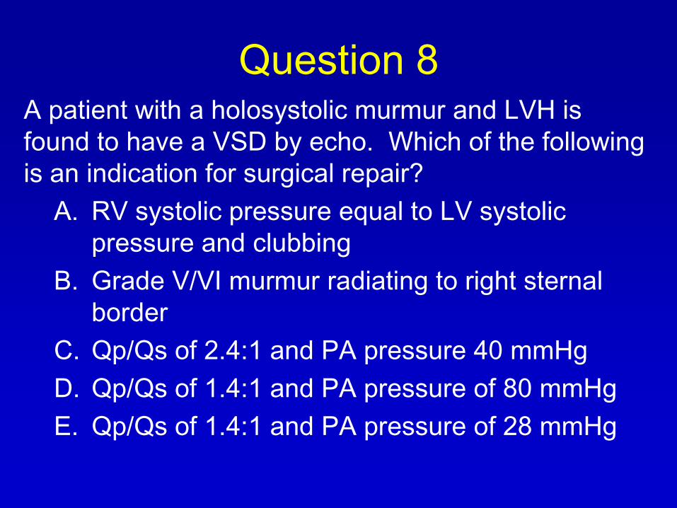
Question 8A patient with a holosystolic murmur and LVH is
found to have a VSD by echo. Which of the following
is an indication for surgical repair?
A. RV systolic pressure equal to LV systolic
pressure and clubbing
B. Grade V/VI murmur radiating to right sternal
border
C. Qp/Qs of 2.4:1 and PA pressure 40 mmHg
D. Qp/Qs of 1.4:1 and PA pressure of 80 mmHg
E. Qp/Qs of 1.4:1 and PA pressure of 28 mmHg
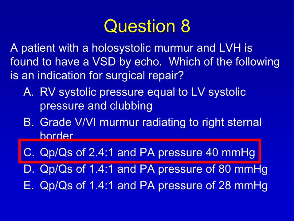

Atrial Septal Defect• A common adult congenital heart disease; 70% are
isolated abnormality; PHtn develops in 5-10%
• Left-to-right shunt with RV volume overload
• Aging and LV stiffening can increase the shunt and give RV failure
• Atrial fibrillation is common in older patients, with increased risk for paradoxical embolism and stroke
• Percutaneous closure: TEE to determine suitability, only for ostium secundum, unknown comparison to surgery – safe short term
• Indication for closing ASD is Qp/Qs>1.7 or RVVO
• No BE prophylaxis before surgery, but yes for 6 months after surgery or device implantation (and ASA)
Pulmonary flow murmur
RV Heave Fixed split S2
ACC/AHA Guidelines for Adults with CHD. 2008.

Atrial Septal Defect - Tests
• ECG – incomplete RBBB in secundum, plus left axis deviation in primum, and maybe LAE
• CXR – RA and RV enlargement, increased pulmonary vascularity, large main PA
• Echo – RA and RV enlargement, estimate PASP (agitated saline with Valsalva release)
• TEE – confirm location and size and PAPVR
• CMR – good for RV volume, good alternative to TEE for PAPVR
ACC/AHA Guidelines for Adults with CHD. 2008.

3 Main Types of ASD
Sinus Venosus
ASD 5-10%,
anomalous
pulmonary vein
Interatrial
septum
viewed from
RA
SVC
Secundum ASD – 75%
Primum ASD – 15-20%
cleft mitral leaflet
IVCRV
TV
ACC/AHA Guidelines for Adults with CHD. 2008.
Primum ASD in
Down’s syndrome

Patent Foramen Ovale - PFO• 25% of population, (normal PE) associated with atrial
septal aneurysm (ASA), diagnose with bubble contrast echo during Valsalva release or cough
• Optimal treatment for patients with PFO and cryptogenic stroke is still undetermined, but device closure is now feasible
• Other syndromes from PFO that might warrant closure:
– Orthodeoxia-platypnea syndrome (cyanosis in upright position)
– Severe decompression illness in divers
– Hypoxemia and right heart failure – RV MI or big PE
– Doubtful that closure might help migraine
• Low dose aspirin therapy is therefore optional

Ventricular Septal Defect
• Shunt is usually small in adult; usually perimembranous near TV, typically loud HSM indicates small defect and normal PVR, muscular VSD may close spontaneously
• PE with big shunt may show displaced apical impulse, mitral rumble, S3, echo with LVVO (echo RVH suggests pulmonary hypertension or PS)
• Large VSD may result in pulmonary vascular obstructive disease and PHtn and right-to-left shunt and cyanosis (Eisenmenger’s syndrome)
• Closure of VSD is not indicated to prevent endocarditis, but for progressive AR or TR or for LV volume load or occasionally for recurrent IE
Holosystolic murmur at LLSB
ACC/AHA Guidelines for Adults with CHD. 2008.

Aortic Coarctation
• CONSIDER in young with hypertension; 50% have associated bicuspid AoV (ejection sound and SEM)
• PE: radial-femoral delay and lower BP in legs, murmur radiates to interscapular area of back
• CXR: classic “3” sign and rib notching lower margin from dilated intercostal arteries that provide collateral blood to descending aorta
• Doppler: gradient across coarct
• MRI: excellent anatomic definition
• Repair if gradient >20 and proximal hypertension
• Short length coarctation may be intervened percutaneously, longer may need surgery
Midsystolic or longer murmur, diminished femoral pulses
ACC/AHA Guidelines for Adults with CHD. 2008.
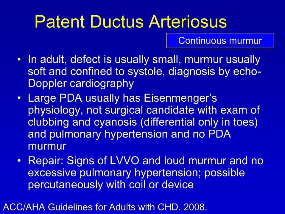
Patent Ductus Arteriosus
• In adult, defect is usually small, murmur usually soft and confined to systole, diagnosis by echo-Doppler cardiography
• Large PDA usually has Eisenmenger’s physiology, not surgical candidate with exam of clubbing and cyanosis (differential only in toes) and pulmonary hypertension and no PDA murmur
• Repair: Signs of LVVO and loud murmur and no excessive pulmonary hypertension; possible percutaneously with coil or device
Continuous murmur
ACC/AHA Guidelines for Adults with CHD. 2008.

Pulmonic Valve Disease
• PS: congenital, usually well tolerated, if severe can cause R→L atrial shunt and cyanosis and paradoxic emboli; Echo is definitive (severe means Echo peak gradient >60, Cath peak-to-peak gradient >40); Usually percutaneous balloon valvuloplasty for Cath peak gradient >40
• PR: mild by echo is normal finding; seen clinically after PS valvuloplasty or repair of Tetralogy of Fallot, recommendations for management are controversial, more indicated if symptomatic with RV enlargement
ACC/AHA Guidelines, Valve disease, 2006; Congenital disease, 2008
Midsystolic murmur, pulmonic ejection sound

Postoperative ASD, VSD, Coarct
• If no residual shunt, no need for endocarditis
prophylaxis after endothelialization of implanted
prosthetic material (6 mo)
• Residual disease should be followed in
specialized centers for adult congenital heart
disease
• ASD – atrial arrhythymias
• VSD – residual shunt, pulmonary hypertension
• Coarctation – persistent or recurrent Htn, re-
coarctation, aneurysm (reoperation is common)
ACC/AHA Guideline: Adult Congenital Heart Disease 2008.

Tricuspid Valve Disease
Ebstein’s anomaly of the tricuspid valve
• Congenital apical displacement of septal and posterior leaflets
• RA pressure ↑ from TR and stiff RV
• Possible cyanosis across ASD or PFO
• Association with WPW (15-25%)
• May see very impressive RAE on ECG
• Surgery for severe TR with NYHA Class III symptoms or cyanosis – tricuspid repair or replacement
ACC/AHA Guidelines, Valve disease update, 2008

Tetralogy
of Fallot
• Most common cyanotic congenital heart disease, 25% associated right aortic arch; microdeletion (15%) in 22q11.2 means 50% risk for ToF in child
• Repair: most have total intracardiac repair –pulmonary regurgitation with right-heart dilation is common, unclear when to replace pulmonary valve; occasional prior palliative Ao-Pulm shunt
• Risk after 25 years is greater, usually SCD, more with QRS (RBBB) duration >180ms, so maybe replace pulmonary valve at that time
RVH
RVOT obstruction
VSD
Overriding aorta
ACC/AHA Guideline: Adult Congenital Heart Disease 2008.

Cyanotic Heart Disease
• Hyperviscosity: usually hct >65, phlebotomy only to
treat symptoms, iron deficiency is associated with
stroke; gout, cerebral abscess, paradoxic embolus
• If dehydration and HF - rehydrate with D5W
• Pregnancy strictly contraindicated, maternal
mortality <50% usually early post-partum
– If no uncontrolled arrhythmias or pulmonary
vascular disease and functional class I,
pregnancy usually well tolerated
• Risk for endocarditis, p-doxic embolism (IV filters),
arrhythmias, SCD and HF (pulmonary vasodilator,
lung or heart-lung transplant)
Follow in
Specialty Center

Eisenmenger Syndrome
• Eisenmenger syndrome is severe
pulmonary vascular disease due
to prior large shunt (ASD, VSD,
PDA)
• Lung transplant is option
• Vasodilator therapy may improve
symptoms
Follow in
Specialty Center

Other Cyanotic
Disease
• Transposition of the Great Arteries -arterial switch procedure (Jatene) and reimplant coronary artery ostia; previously atrial switch procedure (Mustard or Senning) – so RV becomes systemic ventricle and risk for HF
– SSS and atrial arrhythmias will frequently require pacemaker or ablation or antiarrhythmic drugs
dTGA

Topics in Cardiology
• Epidemiology
• Heart Tests
• Coronary
Artery Disease
• Heart Failure
• Myocardial Disease
• Aortic
Disease
• Arrhythmias
• Pericardial
Disease
• Valvular
Disease
• Adult Congenital Disease
• Peripheral Arterial Disease
• Pregnancy

Aortic Atheroembolic Disease
• Plaque more than 4 mm into the lumen is risk
for embolic disease, including stroke,
peripheral embolism, renal embolism
• Ulcerated plaque and mobile debris are
associated findings in thoracic aorta
• Pedunculated mobile plaque is highest risk
• Warfarin reduces recurrence of peripheral
embolic events in patients with mobile
atheroma, may be superior to aspirin

Abdominal Aortic Aneurysm
• Def: dilation (>1.5x normal) or >3 cm
• Risks: smoking, white, male, age>60, CAD, FH
• Most are asymptomatic – expansion → abd or back pain, distal embolism or local compression
• Rupture risk high: >5.5cm or >0.5cm ↑ in 6 months
• PE good if >5.0cm and waist<100cm (39.4 inches)
• Screening US if >60 + FH, or 65-75 + ever smoked
• AAA 4.0-5.4 cm needs CT or US q 6 mo
AHA Guidelines, Peripheral Arterial Disease. 2005.

Abdominal Aortic Aneurysm - 2
• Repair ≥5.5 cm or expansion>5mm/y
(maybe smaller in women, not known) or
symptomatic
• Repair thoracic AA 5.0 cm ascending or 6.0
cm descending or rapid expansion (>1 cm in
1 y)
• Endovascular repair has high success rate
and low mortality and is option (IIa for high
surg risk, or IIb)
AHA Guidelines, Peripheral Arterial Disease. 2005.

Thoracic Aortic Aneurysm
• Symptoms in minority (indication for
surgery): hoarseness, dysphagia, back pain
• Higher rupture risk with larger aneurysms
• Medical therapy: beta-blocker, ARB
• Repair thoracic AA 5.0 cm ascending or 6.0
cm descending or rapid expansion (>1 cm in
1 y), surgical risk higher than AAA surgery
• Endovascular repair is an option

Marfan Syndrome• Cause: mutations in fibrillin-1 gene (FBN-1),
autosomal dominant or sporadic (abnl connective tissue and abnl TGF-beta signalling)
• Aortic disease is major morbidity and mortality (dissection and rupture)
• β-blockers (propranolol) may reduce rate of dilation and risk of dissection (calcium blocker if intolerant); ARB may improve outcomes
• Dilation in up to 50% of children with Marfan and progresses – yearly echo, repair when 45-50 mm or rapid expansion >5mm/y, maybe earlier in woman desiring pregnancy – avoid burst activity
• Surgery: composite root and usually prosthetic valve, 83% 20-yr survival
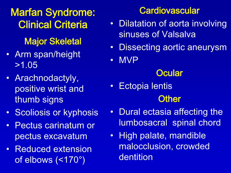
Marfan Syndrome:
Clinical Criteria
Major Skeletal
• Arm span/height
>1.05
• Arachnodactyly,
positive wrist and
thumb signs
• Scoliosis or kyphosis
• Pectus carinatum or
pectus excavatum
• Reduced extension
of elbows (<170°)
Cardiovascular
• Dilatation of aorta involving
sinuses of Valsalva
• Dissecting aortic aneurysm
• MVP
Ocular
• Ectopia lentis
Other
• Dural ectasia affecting the
lumbosacral spinal chord
• High palate, mandible
malocclusion, crowded
dentition

Arteritis
• Takayasu Arteritis: large artery involvement, frequently young women, constitutional symptoms
• Pathology similar to giant cell arteritis with thickening, stenosis, aneurysms
• Claudication arm or leg, ulcerations
• Involvement of carotid, vertebral, upper extrem obstruction, coronary ostium

Acute Aortic Syndromes• Acute aortic dissection (intimal tear and flap,
false lumen)
• Aortic intramural hematoma treat like dissection (isolated crescent of fresh hematoma contained in media – may be rupture of vasa vasorum, precursor of dissection)
• Penetrating atherosclerotic ulcer likely need surgery (avulsion of mural plaque, erosion of media, pseudoaneurysmal adventitial containment)
• Each of these syndromes presents similarly with severe sharp chest discomfort, ripping, tearing, may be migrating – dx mainly suspicion and imaging
Tsai TT et al. Circulation 2005;112:3802.

Aortic Dissection
• Risk: htn, younger often Marfan, other CT disease,
bicuspid AoV, coarctation, trauma, pregnancy
• Evaluation: most have sudden pain onset, minority
have AR, pulse deficits (BP in both arms)
• Imaging is mandatory: TEE and contrast CT are
preferred for acute, MRI for chronic, aortography
only for coexisting ischemia or localization of tear
• Aggressive BP: β-blocker to SBP ,120 and HR 60
(metoprolol, propranolol, labetolol), or dilt or
verapamil, not nitroprusside alone (fenoldopam,
enalaprilat)
Tsai TT et al. Circulation 2005;112:3802.

Aortic Dissection Management• Stanford Type A – involves ascending, generally
surgery
• Type B –descending only, generally medicine and reimaging (1, 3, 6 months), unless recur or persist pain or progressive expansion or contained rupture or large (20mm) or deep (10 mm) ulcer or with intramural hematoma, or peripheral ischemia –endovascular stenting is an option
• Hypotension (contained rupture with tamponade, exsanguination, cardiogenic shock from AR, MI) -rapid surgery
• Long-term: control BP, avoid strenuous exercise, 10-yr survival 50%, watch dilation with 1-2/yr MRI (surg for 5 or 6 cm)
Tsai TT et al. Circulation 2005;112:3802.

Topics in Cardiology
• Epidemiology
• Heart Tests
• Coronary
Artery Disease
• Heart Failure
• Myocardial Disease
• Adult Congenital Disease
• Aortic Disease
• Arrhythmias
• Pericardial
Disease
• Valvular
Disease
• Pregnancy
• Peripheral Arterial Disease

Question 9• 74 yo man with severe left leg pain, absent
left DP and PT pulses, no sensory deficit, no
weakness. What is the best management?
A. Bedrest, aspirin, heparin
B. Urgent revascularization
C. Amputation
D. Dihydropyridine calcium blocker
E. Cilostazol

Question 9• 74 yo man with severe left leg pain, absent
left DP and PT pulses, no sensory deficit, no
weakness. What is the best management?
A. Bedrest, aspirin, heparin
B. Urgent revascularization
C. Amputation
D. Dihydropyridine calcium blocker
E. Cilostazol

Peripheral Arterial Disease
• Prevalence of ABI <0.9 is 12% among older adults and only 10-35% have claudication and 1-2% critical limb ischemia (pain in distal foot at rest or ischemic ulceration or gangrene)
• H and P are suggestive, confirmation requires ABI<0.9
• Patients with PAD are at 20-60% increased risk for MI, 40% increased risk for stroke, and 2-6-fold increased risk of coronary death; sixfold for cardiovascular and 3-6%/year for total mortality
• Claudication remains stable in about 75% over 5 years (10-20% worsen), 1% risk of limb loss
AHA Guidelines, Peripheral Arterial Disease. 2005.

Peripheral Arterial Disease• Risks are usual, esp. DM and Tob
• Screening: carotid, brachial, femoral and pedal
pulses plus abd bruit; hair loss, skin coloration
• Claudication: with walking (hip/buttock =
aortoiliac; thigh = common femoral or aortoiliac;
upper calf = superficial femoral; lower calf =
popliteal), not just standing (spinal stenosis)
• Reduced pulses - measure ABI
• Ischemic ulcer: toes, foot, painful, does not bleed,
irregular margins (venous are malleolar, less
painful)
AHA Guidelines, Peripheral Arterial Disease. 2005.

Screening for PAD
• Diabetic age <50 + smoking or HLP or HTN or hyperhomocysteinemia
• Age 50-69 + smoking hx or DM
• Age>70
• Leg symptoms suggesting claudication or rest ischemic pain
• Abnormal LE pulse exam
• Known CAD, carotid or renal disease

Ankle-Brachial Index
• SBP in both arms and ankles with CW Doppler,
ankles normally 10-15 mmHg higher than brachial
• Result is highest ankle pressure (L and R PT and
DP) divided by highest brachial pressure
– Normal: 1.00-1.29 (0.91-0.99 equivocal)
– Mild-to-moderate PAD: 0.41-0.90
– Severe PAD: 0.00-0.40
– >1.30 noncompressible vessels (calcified
vessels, long DM or advanced age)
– If >1.30, toe-brachial index <0.70 indicates PAD
AHA Guidelines, Peripheral Arterial Disease. 2005.

Other diagnostic tests in PAD
• Segmental limb pressure recordings, thigh, calf, ankle to plan revascularization– Abnormal = 20 mmHg drop
• MR angiography (contraindicated in pacemaker, etc; gadolinium nephrogenic systemic fibrosis)
• Contrast enhanced multidetector CT
• Invasive angiography for planned revascularization

Treatment of PAD – 1
• Smoking cessation with pharmacologic support should be offered, may slow progression to critical leg ischemia and reduce MI and vascular death, aids maintaining patency of angioplasty or vascular surgery
• DM goal A1c<7.0% prevents microvascular complications, uncertain peripheral vascular outcomes
• Lipid-lowering therapy reduces risk of PAD and symptoms of claudication – goal LDL<100 (optional <70) and TG<150, statin first, then fibric acid derivative (statins improve claudication)
• Hypertension – ACE-I beneficial, β-blockers safe in claudication, not really adverse, good pre-op
AHA Guidelines, Peripheral Arterial Disease. 2005.

Treatment of PAD - 2
• Antiplatelet – ASA recommended at 75-325, clopidogrel is an alternative to ASA in PAD, the only agent approved by FDA in PAD with no MI or stroke
• Exercise training 3/week for 30-45 minutes minimum 12 weeks, supervised, almost as good as surgery, and maybe better than angioplasty
• Cilostazol 100 mg bid is indicated and improves symptoms, but not in HF where it increases mortality
• Pentoxifylline 400 tid is second-line (IIb), may not be effective
• Not proven: L-arginine, proprionyl-L-carnitine, maybe gingko biloba
• Not helpful: beraprost, iloprost, vitamin E, chelation
AHA Guidelines, Peripheral Arterial Disease. 2005.

PAD: Angioplasty and
Vascular Surgery• Critical leg ischemia - revascularization for limb
salvage (healing, relief of ischemic rest pain, prevent amputation)
• Revascularization for significantly disabling claudication despite optimal therapy (if not excessive comorbidity) and favorable anatomy
• Aortoiliac disease: primary stenting preferred, especially with short lesions
• Common femoral or deep femoral: endarterectomy and patch angioplasty or bypass
• Popliteal and tibial: bypass
AHA Guidelines, Peripheral Arterial Disease. 2005.
Acute limb ischemia:
heparin, consider
thrombolysis

Critical Limb Ischemia
• Spontaneous or iatrogenic embolism, or in-situ thrombosis
• Cholesterol embolism – inflammation, renal failure, eosinophilia, livedo reticularis, distal necrosis (treatment is supportive)
• 6 P’s: pulseless, painful, pallor, paresthesia, paralysis, poikilothermy
• Class I: no rest pain (>50mmHg)– several days
• Class II: rest pain (<40 mmHg)– hospitalize, urgent intervention, (lytics; urgent endovascular or open therapy if muscle weakness)
• Class III: muscle rigor, anesthesia, paralysis -amputation
AHA Guidelines, Peripheral Arterial Disease. 2005.
Acute limb ischemia:
heparin, consider
thrombolysis

Topics in Cardiology
• Epidemiology
• Heart Tests
• Coronary
Artery Disease
• Heart Failure
• Myocardial Disease
• Adult Congenital Disease
• Aortic Disease
• Peripheral Arterial Disease
• Arrhythmias
• Pericardial
Disease
• Valvular
Disease
• Pregnancy

The Heart in Normal Pregnancy
• Normal physical findings of pregnancy may be misinterpreted as abnormal
– Prominent P2 and persistent S2 splitting, S3 in >80%, early pulmonary flow murmur in >90%, HR increase 20-30%
– Abnormal: orthopnea, PND, cough, S4, murmur ≥3/6, diastolic murmur, fixed split S2, pulmonary edema
• Cardiac output increases by 30-50% and about 80% during labor and delivery (bad for AS and MS), decreased systemic resistance (not bad for MR and AR)

Risk in Pregnancy in Heart DiseaseRisk Factors:
• Prior cardiac event or arrhythmia (HF, TIA, stroke, arrhythmia)
• Baseline NYHA Class II-IV or cyanosis
• Left heart obstruction
• Reduced LV function
Risk for Pulmonary Edema, Embolic stroke, Tachyarrhythmia, cardiac death:
• 0: 4% (close follow-up, community hospital OK)
• 1: 31% (regional center)
• >1: 69% (regional center)Consider
preconception cardiac
intervention

Pulmonary Hypertension in
Pregnancy
• Severe pulmonary hypertension (PAP >70%
systemic) has 30-50% maternal mortality
• Absolute contraindication to pregnancy
• “If pregnancy occurs in a patient with
established severe pulmonary hypertension,
she should be counseled against continuing
the pregnancy. If the pregnancy is continued,
meticulous medical monitoring is
recommended”

Peripartum Cardiomyopathy
• Definition: HF onset last trimester or <6 mo postpartum in absence of other recognized cause, usually first month postpartum, higher in twin, multiparous, mom >30 yo, black women, gestational Htn, tocolytic agents
• 50% of women with peripartum cardiomyopathy improve in LV EF in 6 mo; standard HF therapy (no ACE-I or ARB or Aldo-antag till delivery)
• Consider IV immune globulin and pentoxifylline (inhibits TNF alpha)
• Recurrence is common so repeat pregnancy is contraindicated
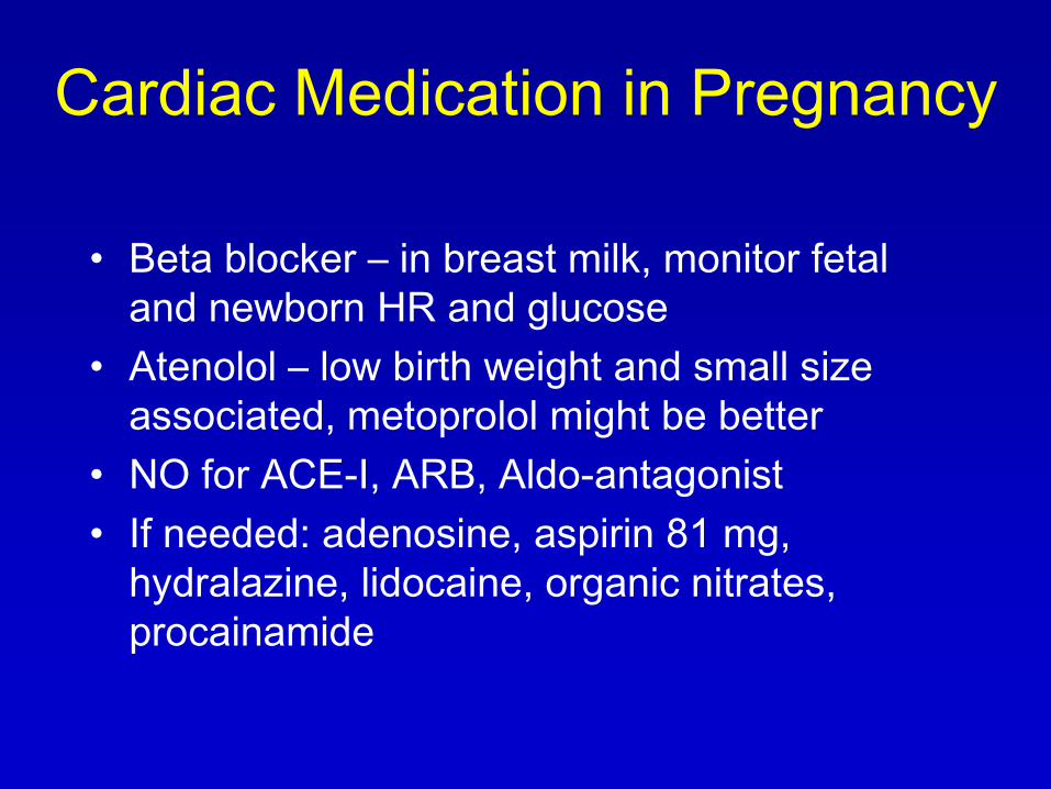
Cardiac Medication in Pregnancy
• Beta blocker – in breast milk, monitor fetal
and newborn HR and glucose
• Atenolol – low birth weight and small size
associated, metoprolol might be better
• NO for ACE-I, ARB, Aldo-antagonist
• If needed: adenosine, aspirin 81 mg,
hydralazine, lidocaine, organic nitrates,
procainamide

Pregnancy and Valve Prosthesis - 1
• Heparin (does not cross placenta) may not prevent mechanical valve thrombosis (12-24% risk of thromboembolic complications with subcutaneous route)
• Prolonged heparin therapy IV or SQ: thrombocytopenia, osteoporosis, alopecia
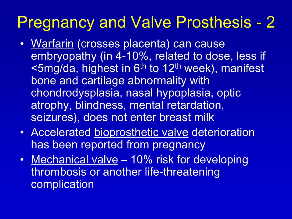
Pregnancy and Valve Prosthesis - 2
• Warfarin (crosses placenta) can cause embryopathy (in 4-10%, related to dose, less if <5mg/da, highest in 6th to 12th week), manifest bone and cartilage abnormality with chondrodysplasia, nasal hypoplasia, optic atrophy, blindness, mental retardation, seizures), does not enter breast milk
• Accelerated bioprosthetic valve deterioration has been reported from pregnancy
• Mechanical valve – 10% risk for developing thrombosis or another life-threatening complication

Anticoagulation Recommendations• Week 0-6: Warfarin
• Week 6-12: UFH (IV or SQ) or LMWH (SQ) or warfarin – (↑fetal risk)
• Week 13-37: UFH (IV or SQ) or LMWH (SQ) or warfarin
• Week 37-delivery: Stop LMWH, warfarin or SQ UFH, start continuous IV UFH, planned delivery
• Post-partum: restart warfarin when bleeding controlled, continue IV UFH till INR therapeutic
• Monitoring:– UFH: aPTT at least 2x control
– LMWH: anti Xa 0.7-1.2 U/mL 4-h post dose
– Warfarin: INR 3 (range 2.5-3.5)
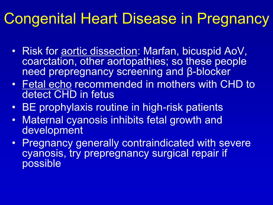
Congenital Heart Disease in Pregnancy
• Risk for aortic dissection: Marfan, bicuspid AoV, coarctation, other aortopathies; so these people need prepregnancy screening and β-blocker
• Fetal echo recommended in mothers with CHD to detect CHD in fetus
• BE prophylaxis routine in high-risk patients
• Maternal cyanosis inhibits fetal growth and development
• Pregnancy generally contraindicated with severe cyanosis, try prepregnancy surgical repair if possible