Embed Size (px)
Citation preview

Int.J.Curr.Microbiol.App.Sci (2016) 5(12): 240-255
240
Original Research Article http://dx.doi.org/10.20546/ijcmas.2016.512.026
Curcumin Boosts up the Efficacy of Imatinib Mesylate in Chronic Myelogenic
Leukemia Cell Line K-562 by Modulation of Various Markers
A. Mukherjee1, R. Sarkar
1, S. Mukherjee
1, J. Biswas
2 and M. Roy
1*
1Department of Environmental Carcinogenesis & Toxicology,
2Director
Chittaranjan National Cancer Institute, 37, S P Mukherjee Road, Kolkata 700 026, India *Corresponding author
A B S T R A C T
Introduction
Leukemia, a type of malignancy, originates
in the bone marrow and results in increased
proliferation of abnormal white blood cells.
Leukemia may be acute or chronic, and
various types have been identified (Udensi
and Tchounwou, 2014). Among different
types, chronic myelogenous leukemia
(CML) is the most prevalent form in Asian
countries (Anand et al., 2012), which may
be divided into three phases, namely an
initial chronic phase followed by accelerated
and blastic phases (Valent, 2007).
The Philadelphia chromosome, resulting
from a reciprocal translocation between the
long arms of chromosomes 9 and 22, is a
very common feature of CML. It has
constitutive tyrosine kinase activity (Anand
et al., 2012). There are some endogenous
proteins or metabolites called biomarkers,
which are aberrantly expressed in cancer and
are hence considered as predictive
indicators. A group of cytokines, regulating
proliferation of blast cells are biological
markers in leukemia. Tumor necrosis factor
International Journal of Current Microbiology and Applied Sciences ISSN: 2319-7706 Volume 5 Number 12 (2016) pp. 240-255
Journal homepage: http://www.ijcmas.com
Imatinib mesylate(IM) is the most commonly used drug for therapy of chronic
myeloid leukemia (CML). It exerts its effect by targeting a constitutively activated
tyrosine kinase Bcr-Abl, a characteristic feature of CML. This anti-tumor drug also
influences various other proteins like cytokines, transcription factors and their
associated signalling pathways that contribute to the development of
leukemogenesis. However, it poses serious side effects. Thus attention should be
given to non-toxic means of leukemia control. Phytochemicals like curcumin
possess a plethora of anti-cancer properties, show minimal toxicity and may
therefore come to the rescue. The present study aims to explore the potential of
curcumin to boost up the efficacy of IM, and to minimize the drug dose and
associated toxicity in human chronic myelogenous leukemia cell line K-562. The
drug IM was found to modulate the expression of markers deregulated in CML.
However, curcumin refines the efficacy of IM, especially when added prior to drug
treatment. Similar trend was reflected in induction of programmed cell death. Thus
the phytochemical curcumin may enhance the potency of IM used in leukemia
therapy, thereby suggesting its role in an adjuvant therapy for CML.
K e y w o r d s
Chronic myelogenous
leukemia,
imatinib mesylate,
curcumin,
cytokines,
Bcr-Abl,
apoptosis.
Accepted:
12 November 2016
Available Online: 10 December 2016
Article Info

Int.J.Curr.Microbiol.App.Sci (2016) 5(12): 240-255
241
alpha (TNFα), belonging to the TNF/TNFR
cytokine superfamily is an important
mediator of inflammation and associated
disorders including cancer (Wang and Lin,
2008). TNFα, on the other hand activates
Nuclear factor kappa B (NF-kB), which is a
transcription factor mediating antiapoptotic
signals in malignancy. NF-κB, consisting of
five subunits normally resides in the
cytoplasm, which upon phosphorylation and
subsequent ubiquitination and proteasomal
degradation of IκB translocates to the
nucleus, where it is active. p50 and p65 are
the two most important subunits of NF-kB
(Lawrence, 2009). Cytokines may be pro
and anti-inflammatory. NF-kB regulates the
pro-inflammatory cytokine interleukin-8
(IL-8) which plays an important role in cell
proliferation and survival
(Elliott et al.,
2001). Production of IL-8 is known to be
increased by various stimuli including
TNFα. IL-10, a multifunctional anti-
inflammatory cytokine is also
transcriptionally controlled by NF-κB
(Driessler et al., 2004). Cancer cells
encounter various forms of stresses,
including oxidative stress, due to which an
important group of proteins called Heat
Shock Proteins (HSPs) get induced (Nahleh
et al., 2012). Of all types of HSPs, HSP 90
is a major regulator of signal transduction
and cellular proliferation and it is
abundantly expressed in leukemia (Flandrin
et al., 2008). Furthermore HSP 90 plays an
important role in NF-κB mediated inhibition
of apoptosis (Lewis et al., 2000; Sarkar et
al., 2014). Survivin, a member of the
inhibitor of apoptosis protein family (IAP)
plays a pivotal role in carcinogenesis by
suppressing apoptosis and promoting cell
division
(Kelly et al., 2011). TNF-α
upregulates the expression of survivin via
NFκB activated signaling cascade (Ahmeda
et al., 2012). All these markers may be
considered as potent targets in leukemia
therapy.
During the last decade, there has been a
considerable advancement in CML therapy,
leading to better prognosis. Commonly used
treatment modalities for CML are
chemotherapy (busulfan and hydroxyurea
etc), targeted therapy (Imatinib Mesylate
etc), immunotherapy, radiotherapy and bone
marrow transplantation
(Henkes et al.,
2008). Imatinib Mesylate (IM) nowadays is
considered to be the standard therapy of
myeloid leukemia which works by targeting
these biomarkers aberrantly expressed in
leukemia and is also a specific inhibitor of
the Bcr-Abl tyrosine kinase activity (An et
al., 2010).
However IM shows severe side effects like
higher susceptibility to infections, skin rash,
fatigue, muscle cramps, and diarrhea. Apart
from these, IM is also reported to be
myelosuppressive, hepatotoxic and
nephrotoxic
(Henkes et al., 2008). To
overcome the problem of IM toxicity, it is
desirable to reduce the drug dose so that the
associated toxicity can be minimized. The
anticancer drugs target actively proliferating
cells in general, they cannot discriminate the
normal healthy cells from the cancerous
ones. Therefore, besides killing the cancer
cells, the healthy cells are also damaged
leading to adverse side effects (Singh et al.,
2008). There are certain plant derived
molecules that can destroy cancer cells,
sparing the normal ones. These molecules,
coined as phytochemicals may have some
differential effect on cancer cells and act as
chemo-enhancer (Gopalakrishnan and Tony
kong, 2008; Sak, 2012). The golden spice
Curcumin, obtained from the rhizomes of
the plant Curcuma longa may be helpful in
this regard as it possesses antioxidant,
chemo-preventive, chemotherapeutic, and
chemo-sensitizing activities and it acts by
targeting some of the key molecules
involved in the carcinogenesis (Jiao et al.,
2009). Curcumin though bestowed with

Int.J.Curr.Microbiol.App.Sci (2016) 5(12): 240-255
242
various disease healing properties, have poor
bioavailability; another plant ingredient
piperine has been found to take care of this
problem
(Suresh and Srinivasan, 2010).
Present study aims to assess the potential of
curcumin in sensitizing the leukemic cells
towards the anti-cancer drug IM. This study
may shed some light on the putative role of
Curcumin as a chemo-enhancer in leukemia
by targeting certain biomarkers involved in
the process of leukemogenesis.
Materials and Methods
Cell culture media RPMI-1640 was
procured from GIBCO-BRL India Pvt. Ltd,
New Delhi, India. Acrylamide, N, N’-
methylenebisacrylamide, fetal bovine serum
(FBS) and ELISA kits to detect the levels of
cytokines TNF-α, IL-8 and IL-10 were
purchased from Invitrogen BioServices
India Pvt. Ltd., Bangalore, India.
Phytohaemagglutinin (PHA) was obtained
from GIBCO-BRL India Pvt. Ltd, New
Delhi, India. Histopaque, dithiothreitol
(DTT), bovine serum albumin (BSA),
Ponceau S, ethylene glycol-O,-O’-bis,(2-
aminoethyl) N,N,N’,N’-tetra acetic acid
(EGTA), 4-(2-hydroxyethyl)-1-
piperazineethanesulfonic acid (HEPES),
CHAPS, RNase A, proteinase K , IM and
propidium iodide (PI) were procured from
Sigma Chemical Co, St. Louis, MO, USA.
Curcumin was obtained from Sigma-
Aldrich, St Louis, MO, USA. Goat anti-
mouse IgG-alkaline phosphatase conjugate
and 5-bromo-4-chloro-3-indolyl phosphate/
nitro blue tetrazolium (BCIP-NBT) were
obtained from Bangalore Genei, India.
Nitrocellulose membrane was procured from
Hybond ECL, Amersham Biosciences, UK.
Tris, sodium dodecyl sulfate (SDS) and
glycine were obtained from Amresco, Ohio,
USA. Assay kits from Millipore, Billerica,
MA were used to detect activities of
caspases 3, 8 and 9. Antibodies against
TNF-α and survivin (Biorbyt), IL-8 and IL-
10 (Abcam), NF-κB(p50) and NF-κB(p65)
and HSP90 (Santa Cruz) were used for
western blotting technique. Anti-BCR-ABL
Monoclonal Antibody [7C6] (MA1-153)
was purchased from Pierce; Thermo
Scientific, USA. Other reagents of analytical
grade are purchased locally. The instruments
used were Gel doc apparatus (BioRad),
fluorescence microscope (Leica),
spectrofluorimeter (Varian), ELISA plate
reader (Tecan) and spectrophotometer
(Varian).
Methods
Cell culture
Chronic myelogenous leukemia cell line K-
562 (human origin) were cultured as per
laboratory protocol (Roy et al., 2015). They
were grown at 37˚C in a humidified 5% CO2
atmosphere in RPMI 1640, with 10% FBS
and antibiotics to prevent any unwanted
bacterial contamination.
Isolation and culture of lymphocytes
Lymphocytes obtained from healthy human
donor were used as control for the study.
They were maintained as per laboratory
protocol (Sarkar et al., 2014) and seeded in
RPMI-1640 supplemented with 10% FBS,
antibiotics and 20μg/ml Phyto-
haemagglutinin (PHA). The lymphocytes
thus isolated are grown in an ambient
condition of 37˚C in a humidified
atmosphere of 5% CO2.
Treatment protocol
Exponentially growing cells were treated
with different concentrations of IM (0.5, 1,
2.5 and 5 µM) in absence and presence of
Curcumin (30 µM). Different modes of
treatment were followed: (I) treatment with
IM alone, (II) simultaneous mode, where
exponentially growing cells were treated
Int.J.Curr.Microbiol.App.Sci (2016) 5(12): 240-255
243
with Curcumin and IM simultaneously for
24 hours and (III) pre-treatment mode,
where cells were treated with Curcumin for
24 hours prior to treatment with IM for the
next 24 hours.
Assessment of cell cytotoxicity
Cytotoxicity of antitumor drug IM to K-562
was measured by MTT assay. Reduction of
3-(4, 5-dimethythiazol-2-yl)-2,5-diphenyl
tetrazolium bromide (MTT) by the
mitochondrial dehydrogenase of live cells
are measured spectrophotometrically. The
colour developed, hence the OD gives an
estimate of viable cells. The formazan
product formed are soluble in DMSO giving
a purple coloration and the absorbance was
measured at 570 nm.
Enzyme-linked immunosorbent assay
The levels of cytokines (TNF-α, IL-8, and
IL-10) were studied using ELISA kits
employing a technique called quantitative
sandwich immunoassay. Samples were
added to microtiter plate pre-coated with an
antibody specific to the interleukins along
with a biotin-conjugated antibody
preparation and incubated as prescribed. In
order to determine the amount of the
specific interleukin present, the sample was
incubated with avidin conjugated
Horseradish Peroxidase (HRP) and a TMB
(3, 3’, 5, 5' tetramethyl-benzidine) substrate
solution was added. The colour change was
measured spectrophotometrically at a
wavelength of 450nm.
Western blotting analysis
Expression of NF-κB (p50 and p65
subunits), survivin, HSP90 and p210Bcr-Abl
,
were assessed by Western blot (WB)
analysis using corresponding antibodies,
following the protocol of Sarkar et al (Sarkar
et al., 2013). β-actin and TBP were used as
loading control.
Assessment of apoptosis by staining with
PI
Treated cells were harvested, washed with
PBS, stained with Propidium Iodide solution
(50µg/ml) and incubated for 10 min in the
dark at room temperature. Cells taken on a
glass slide were visualized under a
fluorescence microscope (Leica) for
assessment of apoptotic features.
Assessment of caspase activity
Treated cells were harvested. Cell lysates
were prepared to assess the activities of
caspases 3, 8 and 9 as per manufacturer’s
protocol. These assays employ synthetic
substrates DEVD-AFC, IETD-AFC and
LEHD-AFC respectively for caspases 3, 8
and 9. A fluorophore AFC (7-amino-4-
trifluoromethyl coumarin) is tagged with
these synthetic peptides. Caspases release
free AFC upon cleavage of the synthetic
substrate, which emits a yellow green
fluorescence at 480-520 nm (peak at 505
nm) upon excitation at 400 nm. The
fluorescence intensity was estimated by a
spectrofluorimeter (Varian).
DNA Fragmentation
Extraction of fragmented DNA was done
following the protocol of Hermann et al
(Hermann and Frischauf, 1987). Treated
cells were harvested, washed and collected
by centrifugation. Then cells were exposed
to lysis buffer and centrifuged. 1% SDS was
then added to the collected supernatant. The
mixture was incubated overnight with 5
µg/ml RNase A at 560C. The mixture was
further digested with 2.5 µg/ml proteinase K
for 4 hours at 37ºC. 0.5 volume of 10 M
ammonium acetate was added and DNA was
precipitated using 2.5 volume of cold
ethanol. The extracted DNA was air-dried
and resuspended in loading buffer and

Int.J.Curr.Microbiol.App.Sci (2016) 5(12): 240-255
244
resolved on 1.5% agarose gel. Fragments of
DNA were visualized using Gel doc
apparatus (BioRad).
PARP degradation
Immunocytochemistry was performed to
determine PARP degradation by caspases
during apoptosis. Treated cells were
harvested, seeded on coverslips; air dried
and fixed using formaldehyde solution. They
were then permeabilized with ice-cold
acetone and blocked with BSA. The cells
were incubated with anti-PARP monoclonal
primary antibody, washed with PBS and
incubated with FITC conjugated secondary
antibody at 37°C in a dark humid chamber.
The cells were viewed under a fluorescence
microscope and photographed.
Statistical Analysis
SPSS 10.0 (one way ANOVA followed by
Dunett t-test) was used for statistical anlysis.
Results and Discussion
Determination of toxicity of IM and
Curcumin
MTT assay has been carried out to assess the
cytotoxicity of IM and Curcumin towards K-
562 cells and peripheral blood lymphocytes
(PBL) isolated from healthy donor. Results
reveal that IM is toxic to both the cells,
though the extent of killing is more in K-562
than PBL (Fig 1A). On the other hand,
Curcumin shows its cytotoxic effect towards
K-562 cells only; very little toxicity towards
PBL has been noticed at the higher
concentrations (Fig 1B). As shown in the
graph, only 20% K-562 cells survive when
treated with 50 µM Curcumin for 24 hours,
therefore for all subsequent experiments, 30
µM has been chosen as the highest dose of
Curcumin where 50% cell killing is
achieved.
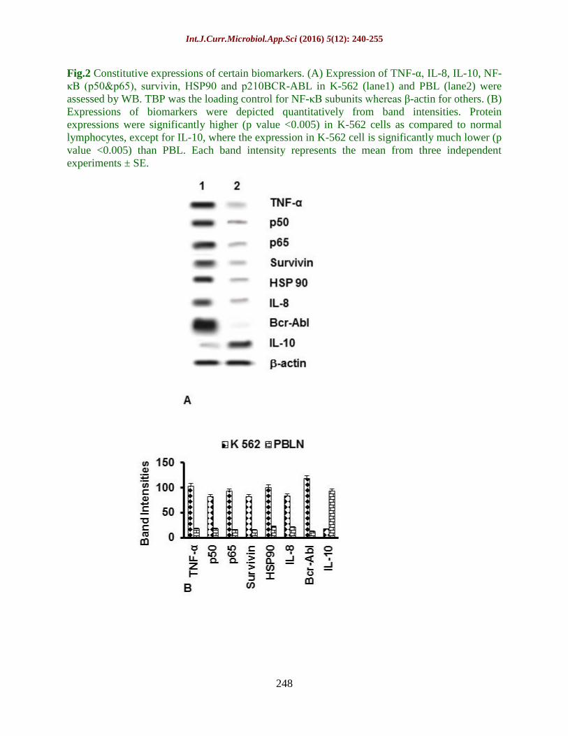
Aberrant expressions of tumor markers
in K-562 cells
Western blot analysis has been performed to
assess the constitutive expression of tumor
markers cytokines (TNF-α, IL-8, IL-10),
NF-κB (p50 & p65 subunits), survivin, HSP
90 and p210BCR-ABL
in leukemic cells K-562
and PBL isolated from healthy donor.
Results reveal that cytokines (TNF-α, IL-8),
NF-κB (p50 and p65), survivin, HSP 90 and
p210Bcr-Abl
are constitutively high in K-562
cells compared to the PBL. Expression of
anti-inflammatory cytokine IL-10 however
is low in the leukemia cells than normal
lymphocytes (Fig 2).
Modulation of cytokines and other tumor
markers by IM in absence and presence
of Curcumin
K-562 cells have been treated with
increasing concentrations of IM (0, 0.5, 1,
2.5, 5 µM) for 24 hours, either alone or in
conjunction with 30 µM Curcumin. ELISA
has been carried out to assess the levels of
cytokines in these cells. Pro-inflammatory
cytokines, TNF-α (Fig 3A) and IL-8 (Fig
3B) are found to be downregulated dose-
dependently by IM both in absence and
presence of 30 µM Curcumin. IM hardly
shows any effect on the levels of IL-10.
However, in presence of curcumin, the
marker has been found to be up-regulated
(Fig 3C). Western blot analysis has been
performed to investigate the modulation of
other leukemic markers by IM in absence
and presence of 30µM Curcumin.
Transcription factor NF-κB subunits p50
and p65, survivin, HSP90 and p210Bcr-Abl
are
found to be negatively regulated by the drug
IM alone as well as when used in the
combinatorial treatment modality. The
corresponding band intensities are given in
the bar diagram and fold change have been
calculated based on band intensity values,

Int.J.Curr.Microbiol.App.Sci (2016) 5(12): 240-255
245
which represent the mean from three
independent experiments (Fig 4). The fold
change of leukemic biomarkers achieved by
treatment with curcumin has been tabulated
in Table 1.
Induction of programmed cell death in
leukemia cells
Treatment of cells with a combination of IM
and Curcumin has been found to induce
apoptosis as evident from fluorescence
microscopic examination of morphological
features of the cells. Representative
micrographs show presence of typical
features of apoptosis, namely shrinkage of
cells, condensation of chromatin,
fragmented nuclei, membrane blebbing and
formation of apoptotic bodies (Fig 5A). The
apoptotic index (i.e the ratio of the apoptotic
to the non apoptotic cells) has been
calculated and are depicted in the bar
diagram (Fig 5B). Apoptotic indices are
higher in presence of curcumin and that too
in pretreatment modality.
Activation of caspases 3, 8 and 9
Caspases, a group of cysteine-dependent
aspartate-directed proteases serve as key
regulators of the apoptotic pathway. Cells
are treated as described before and the
activities of caspases 3, 8 and 9 have been
assessed by fluorimetric analysis. Results
show that IM increases the activity of
caspases in K-562; presence of curcumin
along with IM enhances the effect further,
especially in the pre-treatment modality (Fig
6A).
It is clear from the results that fold increase
in caspase 3 and 9 are maximum (data not
shown) compared to caspase 8, indicating
that induction of programmed cell death is
facilitated mainly by the intrinsic pathway.
Study of DNA Fragmentation, a hallmark
of apoptosis
DNA fragmentation is a characteristic
hallmark of apoptosis. DNA fragmentation
is assessed by subjecting extracted DNA to
gel electrophoresis. During apoptosis,
activated endonucleases cleave chromatin
DNA into 180 bp nucleosomal unit
fragments or multiple of that. Characteristic
fragmentation of DNA has been depicted in
Fig 6B, showing that laddering, which is an
indicator of internucleosomal fragments is
maximum when cells are treated with IM
and curcumin in pretreatment modality.
Results are at par with those from apoptotic
index values.
Degradation of PARP
K-562 cells, after treatment with either IM,
alone or in combination with Curcumin are
incubated with anti-PARP antibody and
labeled with FITC conjugated secondary
antibody. The cells have been viewed under
a fluorescence microscope and
representative photographs are given in Fig
6C. Expression of PARP is higher in
untreated K-562 cells as evident from the
fluorescent intensity. The intensity has been
found to be diminished when treated with
IM; extent of decrease in fluoresence
intensity is more in presence of curcumin
and that is even higher in pre-treatment
modality, indicating degradation of PARP.
Several proteins called tumor markers are
aberrantly expressed in cancer and their
deregulation leads to carcinogenesis.
Treatment strategy is based on targeting
these markers (Magee et al., 2013). Drugs,
used in the treatment of cancer often show
severe toxicity and associated side effects
(Naidu et al., 2004), though they differ in
their mechanism of action and impact on an
individual. Apart from their desired action
Int.J.Curr.Microbiol.App.Sci (2016) 5(12): 240-255
246
on cancer cells, anticancer drugs often cause
severe and sometimes painful side effects. It
would have been the ideal situation if the
anticancer drugs could bind only to the
specific proteins they are meant to target,
but, in reality, in addition to these proteins,
they exert their action on other proteins as
well.
This is a problem in cancer therapy. Imatinib
Mesylate is a drug of choice for the
treatment of leukemia, which besides
targeting leukemia cells, also harm the
normal healthy cells (Mughal and Schrieber,
2010). This poses serious concern to control
leukemogenesis, by making an individual
sick and leading to poor prognosis.
Therefore, non-toxic means of leukemia
control needs considerable attention.
Curcumin, a phytochemical with potent anti-
cancer properties may be helpful in this
context (Tuorkey, 2014). This plant derived
molecule may be employed with a curative
intent, so that the toxicity of antitumor
drugs, hence side effects may be minimized.
These phytochemicals can aid in reducing
the drug dose. Curcumin showed minimal
toxicity in peripheral blood lymphocytes
isolated from normal healthy individuals,
but, is potent to modulate the target proteins,
aberrantly expressed in chronic myelogenic
leukemia cell K-562. Through amelioration
of target proteins, curcumin makes the cells
vulnerable to killing by anticancer drugs.
Hence lesser dose of drug can achieve the
same extent of cell killing in presence of
plant derived molecules. In order to prove
the efficacy of curcumin, it is worthwhile to
understand the mechanism.
Previous findings from our laboratory
showed that curcumin down-regulates HSPs
with a concomitant up-regulation of pro-
apoptotic proteins Bax, Bad, Bid, and AIF
and decrease in the expression of anti-
apoptotic protein Bcl-2, resulting in release
of cytochrome c from mitochondria and
activation of caspases (Sarkar et al., 2014).
The anticancer drugs work with the ultimate
aim of achieving programmed cell death, but
the normal healthy cells are not spared.
Therefore, drug dose minimization is
important in cancer therapeutics. Curcumin
aids in diminution of drug dose.
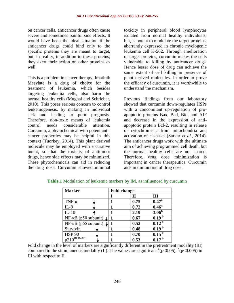
Table.1 Modulation of leukemic markers by IM, as influenced by curcumin
Marker Fold change
I II III
TNF-α 1 0.75 0.47a
IL-8 1 0.72 0.46a
IL-10 1 2.19 3.06b
NF-κB (p50 subunit) 1 0.67 0.19 b
NF-κB (p65 subunit) 1 0.52 0.12 b
Survivin 1 0.48 0.19 b
HSP 90 1 0.70 0.15 b
p210BCR-ABL
1 0.53 0.17 b
Fold change in the level of markers are significantly different in the pretreatment modality (III)
compared to the simultaneous modality (II). The values are significant a(p<0.05),
b(p<0.005) in
III with respect to II.

Int.J.Curr.Microbiol.App.Sci (2016) 5(12): 240-255
247
Fig.1 Effect of IM and Curcumin on proliferation of K-562 cells and human PBL. (A) K-562
cells and human lymphocytes (PBL) from healthy individuals were treated with IM (0, 0.5, 1,
2.5, 5 μM) for 24 hours and subjected to MTT assay. (B) K-562 cells and human lymphocytes
were treated with curcumin (0, 0.5, 1, 10, 30, 50 µM) for 24 hours. Results from MTT assay
show that IM was toxic to both the cells, though toxicity in K-562 is more, whereas curcumin is
preferentially toxic to leukemia cells, not human PBL.
Int.J.Curr.Microbiol.App.Sci (2016) 5(12): 240-255
248
Fig.2 Constitutive expressions of certain biomarkers. (A) Expression of TNF-α, IL-8, IL-10, NF-
κB (p50&p65), survivin, HSP90 and p210BCR-ABL in K-562 (lane1) and PBL (lane2) were
assessed by WB. TBP was the loading control for NF-κB subunits whereas β-actin for others. (B)
Expressions of biomarkers were depicted quantitatively from band intensities. Protein
expressions were significantly higher (p value <0.005) in K-562 cells as compared to normal
lymphocytes, except for IL-10, where the expression in K-562 cell is significantly much lower (p
value <0.005) than PBL. Each band intensity represents the mean from three independent
experiments ± SE.

Int.J.Curr.Microbiol.App.Sci (2016) 5(12): 240-255
249
Fig.3 Effect of IM on biomarkers as influenced by Curcumin. Cells were treated with IM alone
(I), along with Curcumin(30μM), both in simultaneous(II) and pre-treatment modality(III); levels
of TNF-α, IL-8, IL-10 were shown in A,B,C respectively as assessed by ELISA. TNF-α & IL-8
were down-regulated by IM, alone as well as in presence of curcumin (II & III), extent of
decrease is more in the later. Levels of IL-10 however were hardly affected by IM, but presence
of Curcumin increased the level, and that too is more in III. Results are mean of three
independent experiments ± SE and values are significant a(p<0.05), b(p<0.005) and c(p<0.0005)
with respect to the cells treated with IM alone.

Int.J.Curr.Microbiol.App.Sci (2016) 5(12): 240-255
250
Fig.4 Effect of IM on other leukemic biomarkers as influenced by Curcumin. (A) Cells were
treated with IM alone (I) and in combination with Curcumin(30μM), both in simultaneous(II) &
pre-treatment modality (III). WB was performed to assess the expressions of NF-κB (p50&p65),
survivin, HSP90 and p210BCR-ABL. WB bands are shown in the figure. (B) Corresponding
band intensities (average of 3 independent experiments ± SE) were shown. IM downregulated
the expressions of the markers both in absence and presence of Curcumin, however, better effect
was observed when cells were pre-treated with curcumin. Values are significant a(p<0.05),
b(p<0.005) and c(p<0.0005) with respect to only IM treated cells.

Int.J.Curr.Microbiol.App.Sci (2016) 5(12): 240-255
251
Fig.5 Effect of IM on induction of apoptosis as influenced by Curcumin. Cells were treated with
IM alone (I) and in combination with Curcumin(30μM), both in simultaneous(II) and pre-
treatment modality(III). (A) Images of PI stained cells are shown: (Con) shows untreated cells;
(I), (II), (III) respectively represent cells after treatment with 5μM IM alone, IM with Curcumin,
simultaneously and pretreated. Apoptotic features were studied under a fluorescence microscope
and apoptotic cells counted. (B) The apoptotic index (mean of three independent experiments ±
SE) is shown. Values are significant a(p<0.05), b(p<0.005) and c(p<0.0005) with respect to the
cells treated with IM alone.

Int.J.Curr.Microbiol.App.Sci (2016) 5(12): 240-255
252
Fig.6 Effect of IM on apoptosis as influenced by Curcumin. Cells treated with IM(I) and with curcumin,
simultaneously(II) and pretreatment(III) were studied for apoptosis. (A) Fluorimetric analysis reveal
increased activities of caspases in all the treatment modalities, effect is highest in III. Values are
significant a(p<0.05), b(p<0.005) and c(p<0.0005) with respect to IM control. (B) DNA fragmentation,
hallmark of apoptosis after gel electrophoresis. (C) Representative micrographs showing PARP
degradation. (Con) shows untreated cells, (I), (II), (III) respectively represent cells after treatment with
5μM IM alone, IM with Curcumin, simultaneously & pretreated.
Elevated expression of tumor markers like
cytokines (TNF-α, IL-8), NF-κB (p50 and
p65), survivin, p210Bcr-Abl
and HSP 90 and
diminished level of anti-inflammatory
cytokine IL-10 needs to be earmarked to
control leukemogenesis. Present findings

Int.J.Curr.Microbiol.App.Sci (2016) 5(12): 240-255
253
support the notion that proteins mentioned in
the study are involved in development of
leukemia, and hence their modulation by a
combination of IM and curcumin may
ultimately lead to cell killing. Activation of
caspases 3 & 9 are more than caspase 8,
indicating that intrinsic pathway of
apoptosis is mainly responsible for death of
leukemia cells by this combinatorial
treatment modality. Activation of caspases
leads to degradation of an important protein
poly ADP ribose polymerase (PARP), which
is more pronounced in presence of
curcumin. Previous reports from our
laboratory indicated enhancement of
efficacy of IM by curcumin. Among several
treatment modalities, best result was
obtained when the cells were pre-treated
with curcumin prior to IM administration
(Sarkar et al., 2014). Present study aims to
elucidate the detailed mechanism behind the
chemo-enhancement of IM by curcumin.
Present study suggests that curcumin is
capable of augmenting the efficiency of IM
through modulation of cytokines and
associated proteins. The combinatorial
treatment showed better efficacy in
modulating biomarkers than the drug alone.
Prior treatment with curcumin induces
higher extent of apoptosis than IM alone as
revealed by increased apoptotic index,
activation of caspases, DNA ladder
formation and PARP degradation.
In a nut shell, IM efficiently modulated the
aberrant expression of the proteins, but the
concentration required to get the desired
effect is associated with severe toxicity.
Presence of curcumin along with IM comes
to the rescue as similar effect could be
achieved at a much lower concentration of
drug IM. It was reported that curcumin
(Reuter et al., 2009) and IM (Zhang et al.,
2014; Ciarcia et al., 2012) individually are
capable to regulate the expression of these
proteins. Present study emphasized the
synergistic effect of curcumin and IM in
leukemia therapy. Sensitizing the cancer
cells with curcumin, prior to drug
administration could be a better modality.
This is in accordance with other studies
which show that phytochemicals act as
chemo-enhancers
(Doughari et al., 2009).
Therefore, in order to tackle the severe side
effects of conventional anti-cancer drugs,
adjuvant therapy could be a helpful effective
modality. Further research is warranted to
establish the role of curcumin in
combinatorial treatment in leukemia.
Acknowledgement
Authors are indebted to DST for partial
funding of the project. Thanks are due to
Director, CNCI for providing infrastructural
facilities.
References
Ahmeda, M.B., H.H. Shehataa, M. Moussaa,
and Ibrahim T.M. 2012. Prognostic
significance of survivin and tumor
necrosis factor-alpha in adult acute
lymphoblastic leukemia. Clin Biochem.,
45(1-2): 112-116.
An, X., A.K. Tiwari, Y. Sun, P.R. Ding, C.R.
Ashby JR, and Chen, Z.S. 2010. BCR-
ABL tyrosine kinase inhibitors in the
treatment of Philadelphia chromosome
positive chronic myeloid leukemia: a
review. Leuk. Res., 34(10): 1255-1268.
Anand, M.S., N. Varma, S. Varma, K.S. Rana,
and Malhotra, P. 2012. Cytogenetic &
molecular analyses in adult chronic
myelogenous leukaemia patients in
north India. Indian J Med Res., 135, 42-
48.
Ciarcia, R., M.T. Vitiello, M. Galdiero, C.
Pacilio, V. Iovane, D. d'Angelo, D.
Pagnini, G. Caparrotti, D. Conti, V.
Tomei, S. Florio, and Giordano, A.
2012. Imatinib treatment inhibit IL-6,
IL-8, NF-KB and AP-1 production and
modulate intracellular calcium in CML
Int.J.Curr.Microbiol.App.Sci (2016) 5(12): 240-255
254
patients. J. Cell Physiol., 227 (6): 2798-
2803.
Doughari, J.H., I.S. Human, S. Bennade, and
Ndakidemi, P.A. 2009. Phytochemicals
as chemotherapeutic agents and
antioxidants: Possible solution to the
control of antibiotic resistant
verocytotoxin producing bacteria. J.
Med. Plants Res., 3(11), 839-848.
Driessler, F., K. Venstrom, R. Sabat, K.
Asadullah, and Schottelius, A.J. 2004.
Molecular mechanisms of interleukin-
10-mediated inhibition of NF-κB
activity: a role for p50. Clin. Exp.
Immunol., 135(1), 64–73.
Elliott, C.L., V.C. Allport, J.A. Loudon, G.D.
Wu, and Bennett, P.R. 2001. Nuclear
factor-kappa B is essential for up-
regulation of interleukin-8 expression in
human amnion and cervical epithelial
cells. Mol. Hum. Reprod., 7(8), 787-
790.
Flandrin, P., D. Guyotat, A. Duval, J.
Cornillon, E. Tavernier, N. Nadal, and
Campos, L. 2008. Significance of heat-
shock protein (hsp) 90 expression in
acute myeloid leukemia cells. Cell
Stress Chaperones.,13(3): 357–364.
Gopalakrishnan, A., and Tony kong, A.N.
2008. Anticarcinogenesis by dietary
phytochemicals: cytoprotection by Nrf2
in normal cells and cytotoxicity by
modulation of transcription factors NF-
kappa B and AP-1 in abnormal cancer
cells. Food Chem. Toxicol., 46(4), 1257-
1270.
Henkes, M., H. Van der kuip, and Aulitzky
W.E. 2008. Therapeutic options for
chronic myeloid leukemia: focus on
imatinib (Glivec®, Gleevec™). Ther
Clin. Risk Manag., 4(1), 163–187.
Hermann, B.G., and Frischauf, A.M. 1987.
Isolation of genomic DNA. Methods
Enzymol (US). 152, 180–183.
Jiao, Y., J. Wilkinson, X. Di, W. Wang, H.
Hatcher, N.D. Kock, R. D'Agostino Jr,
M.A. Knovich, F.M. Torti, and Torti,
S.V. 2009. Curcumin, a cancer
chemopreventive and chemotherapeutic
agent, is a biologically active iron
chelator. Blood, 113(2): 462–69.
Kelly, R.J., A. Lopez-Chavez, D. Citrin, J.E.
Janik, and Morris J.C. 2011. Impacting
tumor cell-fate by targeting the inhibitor
of apoptosis protein survivin. Mol.
Cancer, 10, 35-45.
Lawrence, T. 2009. The Nuclear Factor NF-
κB Pathway in Inflammation. Cold
Spring Harb Perspect Biol., 1(6),
a001651.
Lewis J, A. Devin, A. Miller, Y. Lin, Y.
Rodriguez, L. Neckers, and Liu, G.
2000. Disruption of hsp90 function
results in degradation of the death
domain kinase, receptor-interacting
protein (RIP), and blockage of tumor
necrosis factor-induced nuclear factor-
kappaB activation. J. Biol. Chem., 275
(14): 10519-10526.
Magee, M.S., A.E. Snook, G.P. Marszalowicz,
and Waldman, S.A. 2013.
Immunotherapeutic strategies to target
prognostic and predictive markers of
cancer. Biomark Med., 7(1), 23–35.
Mughal, T.I., and Schrieber, A. 2010.
Principal long-term adverse effects of
imatinib in patients with chronic
myeloid leukemia in chronic phase.
Biologics, 4, 315–323.
Nahleh, Z., A. Tfayli , A. Najm , A. El Sayed,
and Nahle, Z. 2012. Heat shock
proteins in cancer: targeting the
'chaperones'. Future Med. Chem., 4(7),
927-935.
Naidu, M.U., G.V. Ramana, P.U. Rani, I.K.
Mohan, A. Suman, and Roy, P. 2004.
Chemotherapy-Induced and/or
Radiation Therapy-Induced Oral
Mucositis—Complicating the Treatment
of Cancer. Neoplasia, 6(5), 423–431.
Reuter, S., J. Charlet, T. Juncker, M.H. Teiten,
M. Dicato, and Diederich, M. 2009.
Effect of curcumin on nuclear factor
kappaB signaling pathways in human
chronic myelogenous K562 leukemia
cells. Ann. N.Y. Acad. Sci., 1171, 436-
447.

Int.J.Curr.Microbiol.App.Sci (2016) 5(12): 240-255
255
Roy, M., R. Sarkar, A. Mukherjee, and
Mukherjee, S. 2015. Inhibition of
crosstalk between Bcr-Abl and PKC
signaling by PEITC, augments imatinib
sensitivity in chronic myelogenous
leukemia cells. Chem. Biol. Interact.,
242,195-201.
Sak, K. 2012. Chemotherapy and Dietary
Phytochemical Agents. Chemother. Res.
Pract., 2012, 1-11.
Sarkar, R., A. Mukherjee, R. Biswas, J.
Biswas, and Roy M. 2014.
Sulphoraphane, by virtue of its
antioxidant potential down-regulates
HSP90 in leukemia cells. Int. J. Curr.
Microbiol. App. Sci., 3(1): 476-486.
Sarkar, R., A. Mukherjee, S. Mukherjee, R.
Biswas, J. Biswas, and Roy, M. 2014.
Curcumin augments the efficacy of
antitumor drugs used in leukemia by
modulation of heat shock proteins via
HDAC6. J. Environ. Pathol. Toxicol.
Oncol., 33(3), 247-263.
Sarkar, R., S. Mukherjee, and Roy, M. 2013.
Targeting heat shock proteins by
phenethyl isothiocyanate results in cell-
cycle arrest and apoptosis of human
breast cancer cells. Nutr. Cancer, 65(3),
480-493.
Singh, Y., M. Palombo, and Sinko, P.J. 2008.
Recent Trends in Targeted Anticancer
Prodrug and Conjugate Design. Curr.
Med. Chem., 15(18), 1802–1826.
Suresh, D., and Srinivasan, K. 2010. Tissue
distribution & elimination of capsaicin,
piperine & curcumin following oral
intake in rats. Indian J. Med. Res., 131,
682-691.
Tuorkey, M.J. 2014. Curcumin a potent cancer
preventive agent: Mechanisms of cancer
cell killing. Interv. Med. Appl. Sci., 6(4),
139–146.
Udensi, U.K., and Tchounwou, P.B. 2014.
Dual effect of oxidative stress on
leukemia cancer induction and
treatment. J. Exp. Clin. Cancer Res., 33,
106-121.
Valent, P. 2007. Imatinib-resistant chronic
myeloid leukemia (CML): Current
concepts on pathogenesis and new
emerging pharmacologic approaches.
Biologics, 1(4), 433–448.
Wang, X., and Lin, Y. 2008. Tumor necrosis
factor and cancer, buddies or foes? Acta
Pharmacol. Sin., 29(11), 1275–1288.
Zhang, L., J.H.L. Fok, and Davies, F.E. 2014.
Heat shock proteins in multiple
myeloma. Oncotarget, 5(5), 1132–1148.
How to cite this article:
Mukherjee, A., R. Sarkar, S. Mukherjee, J. Biswas and Roy, M. 2016. Curcumin Boosts up the
Efficacy of Imatinib Mesylate in Chronic Myelogenic Leukemia Cell Line K-562 by
Modulation of Various Markers. Int.J.Curr.Microbiol.App.Sci. 5(12): 240-255.
doi: http://dx.doi.org/10.20546/ijcmas.2016.512.026