Embed Size (px)
Citation preview
International Journal of Stem Cells Vol. 3, No. 2, 2010
ORIGINAL ARTICLE
154
Accepted for publication June 3, 2010Correspondence to Alaa Ismail
Surgery Department, Faculty of Medicine, Ain Shams University, 1 Tadhyia Square, Cairo, Egypt Tel: +20-122142526, Fax: +20-122142526E-mail: [email protected]
Migration of Human Umbilical Cord Blood Cells into Rat Liver
Alaa Ismail1, Ehsan Hassan2, Mohamed I Seleem3, Medhat Hassan3, Firas Z ElDeen1, Ahmed Salah1, Abdulhafez A Selim4
1Surgery Department, Faculty of Medicine, Ain Shams University, Cairo, Egypt, 2Pathology Department and 3Surgery Department, National Hepatology and Tropical Medicine Research Institute, Cairo, Egypt, 4Research and Development, Osteotech Inc., NJ, USA
Background and Objectives: Cell therapy provides an effective strategy for the treatment of an impaired liver. Human umbilical cord blood progenitor cells have the potential to differentiate into hepatocytes. Progenitor cells transplanted into the spleen could migrate directly into the liver through portal circulation. To track migration of human umbilical cord blood progenitor cells in cirrhotic rat liver after intrasplenic transplantation and to prove the possibility similar behavior of human umbilical cord blood nucleated cells in humans.Methods and Results: Umbilical cord blood samples from full-term deliveries will be collected after obtaining an in-formed consent from the mother. The collection procedure will be conducted after completion of delivery and will not interfere with the normal obstetric procedures. Adult male Sprague Dawley rats were subjected to liver cirrhosis by intraperitoneal injection of thioacetamide. Cirrhotic rats were treated with human umbilical cord blood nucleated cells by intra-splenic transplantation. Migration of intrasplenic transplanted human umbilical cord blood cells to the liver was successfully documented with Immunohistochemistry. The liver and spleen from recipient animals were removed. Histopathological and immunohistochemical analysis were performed 20 weeks after intrasplenic injection of the cells. Intrasplenically injected cells migrate to the liver of recipient animals.Conclusions: Human cord blood nucleated cells have the potential to differentiate into hepatocytes and substantially improve the histology and function of the cirrhotic liver in rats. Relocation into liver after intrasplenic transplantation could be detected by immunohistochemistry. Transdifferentiated cells could be efficiently stained with antihuman hepatocytes.
Keywords: Cord blood, Progenitor cells, Intrasplenic transpl, Liver regeneration
Introduction
Cell transplantation provides an effective strategy for the treatment of an impaired liver or liver failure. When compared with orthotopic liver transplantation, stem cell transplantation has the advantages of lower cost, lower risk, and simpler manipulation of the procedure. Autolo-gous cell transplantation helps prevent immunologic re-
jection, which is always a problem for orthotopic liver transplantation, and it bears great potential usefulness as a transient therapy before liver transplantation (1). Exploitation of high regeneration and differentiation of stem cells may solve this problem. Transdifferentiation of different kinds of stem cells into hepatocytes has been pre-viously demonstrated with embryonic stem cells, hepato-blasts (mostly in fetal liver), hepatic oval cells (mostly in mature liver), pancreatic progenitor cells, bone marrow hematopoietic stem cells, and mesenchymal stem cells (MSCs) (2-5). Bone marrow-derived stem cells exhibited better prospects in terms of practical applicability as sur-rogates of hepatocytes for transplantation in MSCs. Both in vitro and in vivo, studies appeared to support bone MSCs (BMSCs) as being more potent in hepatocytic trans-
Alaa Ismail, et al: Migration of Human Umbilical Cord Blood Cells into Rat Liver 157
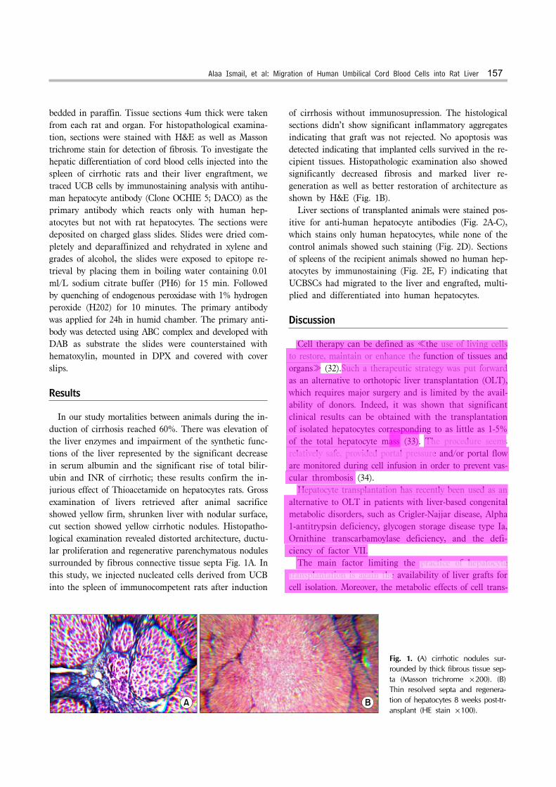
Fig. 1. (A) cirrhotic nodules sur-rounded by thick fibrous tissue sep-ta (Masson trichrome ×200). (B) Thin resolved septa and regenera-tion of hepatocytes 8 weeks post-tr-ansplant (HE stain ×100).
bedded in paraffin. Tissue sections 4um thick were taken from each rat and organ. For histopathological examina-tion, sections were stained with H&E as well as Masson trichrome stain for detection of fibrosis. To investigate the hepatic differentiation of cord blood cells injected into the spleen of cirrhotic rats and their liver engraftment, we traced UCB cells by immunostaining analysis with antihu-man hepatocyte antibody (Clone OCHIE 5; DACO) as the primary antibody which reacts only with human hep-atocytes but not with rat hepatocytes. The sections were deposited on charged glass slides. Slides were dried com-pletely and deparaffinized and rehydrated in xylene and grades of alcohol, the slides were exposed to epitope re-trieval by placing them in boiling water containing 0.01 ml/L sodium citrate buffer (PH6) for 15 min. Followed by quenching of endogenous peroxidase with 1% hydrogen peroxide (H202) for 10 minutes. The primary antibody was applied for 24h in humid chamber. The primary anti-body was detected using ABC complex and developed with DAB as substrate the slides were counterstained with hematoxylin, mounted in DPX and covered with cover slips.
Results
In our study mortalities between animals during the in-duction of cirrhosis reached 60%. There was elevation of the liver enzymes and impairment of the synthetic func-tions of the liver represented by the significant decrease in serum albumin and the significant rise of total bilir-ubin and INR of cirrhotic; these results confirm the in-jurious effect of Thioacetamide on hepatocytes rats. Gross examination of livers retrieved after animal sacrifice showed yellow firm, shrunken liver with nodular surface, cut section showed yellow cirrhotic nodules. Histopatho-logical examination revealed distorted architecture, ductu-lar proliferation and regenerative parenchymatous nodules surrounded by fibrous connective tissue septa Fig. 1A. In this study, we injected nucleated cells derived from UCB into the spleen of immunocompetent rats after induction
of cirrhosis without immunosupression. The histological sections didn’t show significant inflammatory aggregates indicating that graft was not rejected. No apoptosis was detected indicating that implanted cells survived in the re-cipient tissues. Histopathologic examination also showed significantly decreased fibrosis and marked liver re-generation as well as better restoration of architecture as shown by H&E (Fig. 1B). Liver sections of transplanted animals were stained pos-itive for anti-human hepatocyte antibodies (Fig. 2A-C), which stains only human hepatocytes, while none of the control animals showed such staining (Fig. 2D). Sections of spleens of the recipient animals showed no human hep-atocytes by immunostaining (Fig. 2E, F) indicating that UCBSCs had migrated to the liver and engrafted, multi-plied and differentiated into human hepatocytes.
Discussion
Cell therapy can be defined as ≪the use of living cells to restore, maintain or enhance the function of tissues and organs≫ (32).Such a therapeutic strategy was put forward as an alternative to orthotopic liver transplantation (OLT), which requires major surgery and is limited by the avail-ability of donors. Indeed, it was shown that significant clinical results can be obtained with the transplantation of isolated hepatocytes corresponding to as little as 1-5% of the total hepatocyte mass (33). The procedure seems relatively safe, provided portal pressure and/or portal flow are monitored during cell infusion in order to prevent vas-cular thrombosis (34). Hepatocyte transplantation has recently been used as an alternative to OLT in patients with liver-based congenital metabolic disorders, such as Crigler-Najjar disease, Alpha 1-antitrypsin deficiency, glycogen storage disease type Ia, Ornithine transcarbamoylase deficiency, and the defi-ciency of factor VII. The main factor limiting the practice of hepatocyte transplantation is again the availability of liver grafts for cell isolation. Moreover, the metabolic effects of cell trans-
158 International Journal of Stem Cells 2010;3:154-160
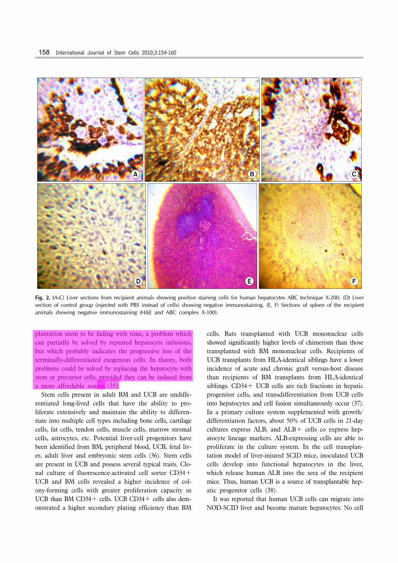
Fig. 2. (A-C) Liver sections from recipient animals showing positive staining cells for human hepatocytes ABC technique X-200. (D) Liver section of control group (injected with PBS instead of cells) showing negative immunostaining. (E, F) Sections of spleen of the recipientanimals showing negative immunostaining (H&E and ABC complex X-100).
plantation seem to be fading with time, a problem which can partially be solved by repeated hepatocyte infusions, but which probably indicates the progressive loss of the terminally-differentiated exogenous cells. In theory, both problems could be solved by replacing the hepatocyte with stem or precursor cells, provided they can be isolated from a more affordable source (35). Stem cells present in adult BM and UCB are undiffe-rentiated long-lived cells that have the ability to pro-liferate extensively and maintain the ability to differen-tiate into multiple cell types including bone cells, cartilage cells, fat cells, tendon cells, muscle cells, marrow stromal cells, astrocytes, etc. Potential liver-cell progenitors have been identified from BM, peripheral blood, UCB, fetal liv-er, adult liver and embryonic stem cells (36). Stem cells are present in UCB and possess several typical traits. Clo-nal culture of fluorescence-activated cell sorter CD34+
UCB and BM cells revealed a higher incidence of col-ony-forming cells with greater proliferation capacity in UCB than BM CD34+ cells. UCB CD34+ cells also dem-onstrated a higher secondary plating efficiency than BM
cells. Rats transplanted with UCB mononuclear cells showed significantly higher levels of chimerism than those transplanted with BM mononuclear cells. Recipients of UCB transplants from HLA-identical siblings have a lower incidence of acute and chronic graft versus-host disease than recipients of BM transplants from HLA-identical siblings. CD34+ UCB cells are rich fractions in hepatic progenitor cells, and transdifferentiation from UCB cells into hepatocytes and cell fusion simultaneously occur (37). In a primary culture system supplemented with growth/ differentiation factors, about 50% of UCB cells in 21-day cultures express ALB, and ALB+ cells co express hep-atocyte lineage markers. ALB-expressing cells are able to proliferate in the culture system. In the cell transplan-tation model of liver-injured SCID mice, inoculated UCB cells develop into functional hepatocytes in the liver, which release human ALB into the sera of the recipient mice. Thus, human UCB is a source of transplantable hep-atic progenitor cells (38). It was reported that human UCB cells can migrate into NOD-SCID liver and become mature hepatocytes. No cell

160 International Journal of Stem Cells 2010;3:154-160
Takimoto R, Iyama S, Matsunaga T, Ohtani S, Matsuura A, Hamada H, Niitsu Y. Human mesenchymal stem cells xenografted directly to rat liver are differentiated into hu-man hepatocytes without fusion. Blood 2005;106:756-763
17. Hua L, Aoki T, Jin Z, Nishino N, Yasuda D, Izumida Y, Morohara K, Koizumi T, Shimizu Y, Murai N, Kusano M. Elevation of serum albumin levels in nagase analbuminemic rats by allogeneic bone marrow cell transplantation. Eur Surg Res 2005;37:111-114
18. Avital I, Inderbitzin D, Aoki T, Tyan DB, Cohen AH, Ferraresso C, Rozga J, Arnaout WS, Demetriou AA. Isola-tion, characterization, and transplantation of bone marr-ow-derived hepatocyte stem cells. Biochem Biophys Res Commun 2001;288:156-164
19. am Esch JS 2nd, Knoefel WT, Klein M, Ghodsizad A, Fuerst G, Poll LW, Piechaczek C, Burchardt ER, Feifel N, Stoldt V, Stockschläder M, Stoecklein N, Tustas RY, Eisen-berger CF, Peiper M, Häussinger D, Hosch SB. Portal ap-plication of autologous CD133+ bone marrow cells to the liver: a novel concept to support hepatic regeneration. Stem Cells 2005;23:463-470
20. Okumoto K, Saito T, Hattori E, Ito JI, Suzuki A, Misawa K, Sanjyo M, Takeda T, Sugahara K, Saito K, Togashi H, Kawata S. Expression of Notch signalling markers in bone marrow cells that differentiate into a liver cell lineage in a rat transplant model. Hepatol Res 2005;31:7-12
21. Vogels BA, Maas MA, Bosma A, Chamuleau RA. Significant improvement of survival by intrasplenic hepatocyte trans-plantation in totally hepatectomized rats. Cell Transplant 1996;5:369-378
22. Kobayashi N, Miyazaki M, Fukaya K, Noguchi H, Tanaka N, Namba M. Intrasplenic transplantation of immortalized human fetal hepatocytes prolongs the survival of 90% hep-atectomized rats. Transplant Proc 2000;32:2365-2367
23. Kim WH, Lee JH, Han SU, Wang HJ, Kim MW. Can clamping of splenic vessels prevent abrupt increase of portal vein pressure and migration of transplanted hepatocytes to the liver after intrasplenic hepatocyte transplantation? Hepatogastroenterology 1998;45:2425-2429
24. Ryu KH. Liver stem cells derived from the bone marrow and umbilical cord blood. Int J Stem Cell 2009;2:97-101
25. Sell S. Stem cells handbook. Totowa, NJ: Humana Press; 2004
26. Reubinoff BE, Pera MF, Fong CY, Trounson A, Bongso A. Embryonic stem cell lines from human blastocysts: somatic differentiation in vitro. Nat Biotechnol 2000;18:399-404
27. Thomas ED. Bone marrow transplantation from the person-al viewpoint. Int J Hematol 2005;81:89-93
28. Bianco P, Riminucci M, Gronthos S, Robey PG. Bone mar-row stromal stem cells: nature, biology, and potential applications. Stem Cells 2001;19:180-192
29. Herzog EL, Chai L, Krause DS. Plasticity of marrow-de-rived stem cells. Blood 2003;102:3483-3493
30. Orlic D, Kajstura J, Chimenti S, Jakoniuk I, Anderson SM, Li B, Pickel J, McKay R, Nadal-Ginard B, Bodine DM, Leri A, Anversa P. Bone marrow cells regenerate infarcted myocardium. Nature 2001;410:701-705
31. Hori N, Okanoue T, Sawa Y, Mori T, Kashima K. Hemody-namic characterization in experimental liver cirrhosis indu-ced by thioacetamide administration. Dig Dis Sci 1993;38: 2195-2202
32. Sipe JD. Tissue engineering and reparative medicine. Ann N Y Acad Sci 2002;961:1-9
33. Lake JR. Hepatocyte transplantation. N Engl J Med 1998;338:1463-1465
34. Muraca M, Neri D, Parenti A, Feltracco P, Granato A, Vilei MT, Ferraresso C, Ballarin R, Zanusso GE, Giron G, Rozga J, Gerunda G. Intraportal hepatocyte transplantation in the pig: hemodynamic and histopathological study. Transplan-tation 2002;73:890-896
35. Stéphenne X, Najimi M, Sibille C, Nassogne MC, Smets F, Sokal EM. Sustained engraftment and tissue enzyme activ-ity after liver cell transplantation for argininosuccinate lyase deficiency. Gastroenterology 2006;130:1317-1323
36. Tang XP, Zhang M, Yang X, Chen LM, Zeng Y. Differen-tiation of human umbilical cord blood stem cells into hep-atocytes in vivo and in vitro. World J Gastroenterol 2006;12: 4014-4019
37. Tanabe Y, Tajima F, Nakamura Y, Shibasaki E, Wakejima M, Shimomura T, Murai R, Murawaki Y, Hashiguchi K, Kanbe T, Saeki T, Ichiba M, Yoshida Y, Mitsunari M, Yoshida S, Miake J, Yamamoto Y, Nagata N, Harada T, Kurimasa A, Hisatome I, Terakawa N, Murawaki Y, Shiota G. Analyses to clarify rich fractions in hepatic progenitor cells from human umbilical cord blood and cell fusion. Biochem Biophys Res Commun 2004;324:711-718
38. Kakinuma S, Tanaka Y, Chinzei R, Watanabe M, Shim-izu-Saito K, Hara Y, Teramoto K, Arii S, Sato C, Takase K, Yasumizu T, Teraoka H. Human umbilical cord blood as a source of transplantable hepatic progenitor cells. Stem Cells 2003;21:217-227
39. Rocha V, Wagner JE Jr, Sobocinski KA, Klein JP, Zhang MJ, Horowitz MM, Gluckman E. Graft-versus-host disease in children who have received a cord-blood or bone marrow transplant from an HLA-identical sibling. Eurocord and International Bone Marrow Transplant Registry Working Committee on Alternative Donor and Stem Cell Sources. N Engl J Med 2000;342:1846-1854
40. Madrigal JA, Cohen SB, Gluckman E, Charron DJ. Does cord blood transplantation result in lower graft-versus-host disease? It takes more than two to tango. Hum Immunol 1997;56:1-5
Differentiation of Hematopoietic Stem Cells into Hepatocytesin Liver Fibrosis in Rats
Y. Zhan, Y. Wang, L. Wei, H. Chen, X. Cong, R. Fei, Y. Gao, and F. Liu
ABSTRACT
It has been reported that hematopoietic stem cells (HSC) can differentiate into hepato-cytes in the normal liver and in some pathologic environments. The aim of this study wasto investigate whether HSC can differentiate into hepatocytes in cases of established liverfibrosis. Rat liver fibrosis was induced by subcutaneous injection of tetrachloride (CCl4).Thy�CD3�CD45RA� HSC in bone marrow cells, which had been enriched by fluorescence-activated cell sorting (FACS), were labeled with PKH26-GL, and autologously trans-planted into CCl4-treated rats. The expressions of albumin (Alb), cytokeratin 8 (CK8), and�-smooth muscle actin (SMA) were determined by immunofluorescence methods. ThePKH26-GL labeled Thy�CD3�CD45RA� HSC expressed the hepatocyte-specific markersAlb and CK8, but did not express �-SMA in liver fibrosis. Thy�CD3�CD45RA� HSCdifferentiated into hepatocytes, but not into hepatic stellate cells. In conclusion, autologousstem cell transplantation may be helpful to treat hepatic fibrosis.
STEM CELLS have been a focus in life science stud-ies.1–4 Recent studies have shown that mice and hu-
man bone marrow stem cells can differentiate into hepato-cytes in normal livers. Furthermore, rat bone marrow stemcells turned into hepatocytes in severely damaged liverswith suppression of hepatocyte proliferation.5–9 Hepaticfibrosis is a common pathological consequence of variouschronic liver diseases. It has not been known whether HSCcan differentiate into hepatocytes in liver fibrosis. In thepresent experiment, we studied the differentiation of ratHSC in a model of established liver fibrosis.
MATERIALS AND METHODSAnimals
Male Sprague-Dawley rats weighing 170 to 190 g were obtainedfrom Academy of Military Medical Science. They were bred andmaintained on standard laboratory chow using 12-hour light/darkcycles. The studies met the national guidelines for animal usage inresearch.
Induction of Hepatic Fibrogenesis
Rat liver fibrosis was induced using subcutaneous injection of 40%CCl4 dissolved in olive oil, twice a week for 6 weeks, as previouslydescribed.10
Thy�CD3�CD45RA� HSC Enrichment
After the last CCl4 injection, the rats were anesthetized using anintraperitioneal injection of sodium pentobarbital (30 mg/kg body
weight). Under general anesthesia, bone marrow was aspiratedfrom a tibia with a syringe containing 1 mL heparin with an18-gauge needle. The marrow cells, transferred to a sterile tube,were mixed with 10 mL culture medium (RPM1640 with 10% fetalbovine serum, penicillin G100u/mL, and streptomycin 100 ug/mL).Red blood cells in bone marrow were depleted using erythrolysin.After three times washing with phosphate-buffered saline (PBS),the cells were incubated at 4°C for 30 minutes with fluoresceinisothiocyanate (FITC)-conjugated anti-Thy mAb (Pharmingen),phycoerythrin (PE)-conjugated anti-CD3 mAb (Becton Dickin-son), or PE-conjugated anti-CD45RA mAb. Cells washed threetimes were resuspended in medium. Labeled cells were analyzedand separated with the FASC-vantage apparatus (Becton Dickin-son, San Jose, Calif). Gating was based on Thy-positive, CD3-negative, and CD45RA-negative.
From Peking University Hepatology Institute, Peking Univer-sity People’s Hospital, Beijing, China.
Supported by the National High Technology Research andDevelopment Program of China (863 Program) (2001AA216031)from Administration of Science and Technology of China, Chi-nese Basic Research Foundation (973) (2005CB522902) andMinistry of Education of P. R. China (20040001133).
Address reprint requests to Lai Wei, MD, Professor, Hepa-tology Institute, Peking University People’s Hospital, No.11Xizhimen South Street, Beijing 100044, China. E-mail: [email protected]
0041-1345/06/$–see front matter © 2006 by Elsevier Inc. All rights reserved.doi:10.1016/j.transproceed.2006.08.132 360 Park Avenue South, New York, NY 10010-1710
3082 Transplantation Proceedings, 38, 3082–3085 (2006)
marrow stem cells differentiated into hepatocytes in thehepatic fibrogenesis environment. The PKH26-GL-labelingand Alb-expressing cells were 0.17 � 0.02% among allhepatocytes.
Immunofluorescence Method for �-SMA
We found that myofibroblasts in fibrotic liver sections werepositive for �-SMA (Fig 5A). PKH26-GL-labeled cells inthe fibrotic liver did not express �-SMA (Fig 5). The resultindicated that bone marrow stem cells did not differentiateinto myofibroblasts in the hepatic fibrogenesis environment.
DISCUSSION
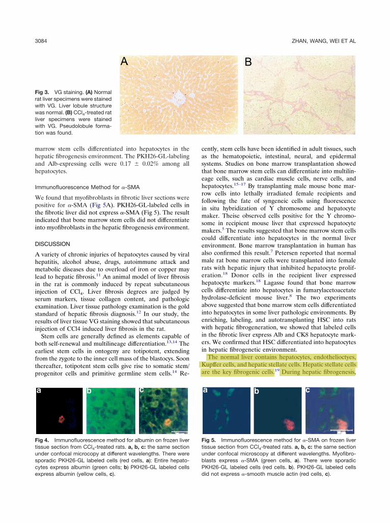
A variety of chronic injuries of hepatocytes caused by viralhepatitis, alcohol abuse, drugs, autoimmune attack andmetabolic diseases due to overload of iron or copper maylead to hepatic fibrosis.11 An animal model of liver fibrosisin the rat is commonly induced by repeat subcutaneousinjection of CCl4. Liver fibrosis degrees are judged byserum markers, tissue collagen content, and pathologicexamination. Liver tissue pathology examination is the goldstandard of hepatic fibrosis diagnosis.12 In our study, theresults of liver tissue VG staining showed that subcutaneousinjection of CCl4 induced liver fibrosis in the rat.
Stem cells are generally defined as elements capable ofboth self-renewal and multilineage differentiation.13,14 Theearliest stem cells in ontogeny are totipotent, extendingfrom the zygote to the inner cell mass of the blastocys. Soonthereafter, totipotent stem cells give rise to somatic stem/progenitor cells and primitive germline stem cells.14 Re-
cently, stem cells have been identified in adult tissues, suchas the hematopoietic, intestinal, neural, and epidermalsystems. Studies on bone marrow transplantation showedthat bone marrow stem cells can differentiate into multilin-eage cells, such as cardiac muscle cells, nerve cells, andhepatocytes.15–17 By transplanting male mouse bone mar-row cells into lethally irradiated female recipients andfollowing the fate of syngeneic cells using fluorescencein situ hybridization of Y chromosome and hepatocytemaker. Theise observed cells positive for the Y chromo-some in recipient mouse liver that expressed hepatocytemakers.5 The results suggested that bone marrow stem cellscould differentiate into hepatocytes in the normal liverenvironment. Bone marrow transplantation in human hasalso confirmed this result.7 Petersen reported that normalmale rat bone marrow cells were transplanted into femalerats with hepatic injury that inhibited hepatocyte prolif-eration.18 Donor cells in the recipient liver expressedhepatocyte markers.18 Lagasse found that bone marrowcells differentiate into hepatocytes in fumarylacetoacetatehydrolase-deficient mouse liver.8 The two experimentsabove suggested that bone marrow stem cells differentiatedinto hepatocytes in some liver pathologic environments. Byenriching, labeling, and autotransplanting HSC into ratswith hepatic fibrogeneration, we showed that labeled cellsin the fibrotic liver express Alb and CK8 hepatocyte mark-ers. We confirmed that HSC differentiated into hepatocytesin hepatic fibrogenetic environment.
The normal liver contains hepatocytes, endothelioctyes,Kupffer cells, and hepatic stellate cells. Hepatic stellate cellsare the key fibrogenic cells.19 During hepatic fibrogenesis,
Fig 5. Immunofluorescence method for �-SMA on frozen livertissue section from CCl4-treated rats. a, b, c: the same sectionunder confocal microscopy at different wavelengths. Myofibro-blasts express �-SMA (green cells, a). There were sporadicPKH26-GL labeled cells (red cells. b). PKH26-GL labeled cellsdid not express �-smooth muscle actin (red cells, c).
Fig 3. VG staining. (A) Normalrat liver specimens were stainedwith VG. Liver lobule structurewas normal. (B) CCl4-treated ratliver specimens were stainedwith VG. Pseudolobule forma-tion was found.
Fig 4. Immunofluorescence method for albumin on frozen livertissue section from CCl4-treated rats. a, b, c: the same sectionunder confocal microcopy at different wavelengths. There weresporadic PKH26-GL labeled cells (red cells, a): Entire hepato-cytes express albumin (green cells; b) PKH26-GL labeled cellsexpress albumin (yellow cells, c).
3084 ZHAN, WANG, WEI ET AL
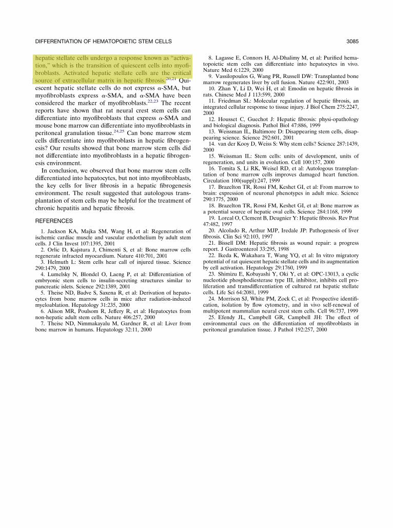
hepatic stellate cells undergo a response known as “activa-tion,” which is the transition of quiescent cells into myofi-broblasts. Activated hepatic stellate cells are the criticalsource of extracellular matrix in hepatic fibrosis.20,21 Qui-escent hepatic stellate cells do not express �-SMA, butmyofibroblasts express �-SMA, and �-SMA have beenconsidered the marker of myofibroblasts.22,23 The recentreports have shown that rat neural crest stem cells candifferentiate into myofibroblasts that express �-SMA andmouse bone marrow can differentiate into myofibroblasts inperitoneal granulation tissue.24,25 Can bone marrow stemcells differentiate into myofibroblasts in hepatic fibrogen-esis? Our results showed that bone marrow stem cells didnot differentiate into myofibroblasts in a hepatic fibrogen-esis environment.
In conclusion, we observed that bone marrow stem cellsdifferentiated into hepatocytes, but not into myofibroblasts,the key cells for liver fibrosis in a hepatic fibrogenesisenvironment. The result suggested that autologous trans-plantation of stem cells may be helpful for the treatment ofchronic hepatitis and hepatic fibrosis.
REFERENCES
1. Jackson KA, Majka SM, Wang H, et al: Regeneration ofischemic cardiac muscle and vascular endothelium by adult stemcells. J Clin Invest 107:1395, 2001
2. Orlic D, Kajstura J, Chimenti S, et al: Bone marrow cellsregenerate infracted myocardium. Nature 410:701, 2001
3. Helmuth L: Stem cells hear call of injured tissue. Science290:1479, 2000
4. Lumelsky N, Blondel O, Laeng P, et al: Differentiation ofembryonic stem cells to insulin-secreting structures similar topancreatic islets. Science 292:1389, 2001
5. Theise ND, Badve S, Saxena R, et al: Derivation of hepato-cytes from bone marrow cells in mice after radiation-inducedmyeloablation. Hepatology 31:235, 2000
6. Alison MR, Poulsom R, Jeffery R, et al: Hepatocytes fromnon-hepatic adult stem cells. Nature 406:257, 2000
7. Theise ND, Nimmakayalu M, Gardner R, et al: Liver frombone marrow in humans. Hepatology 32:11, 2000
8. Lagasse E, Connors H, Al-Dhalimy M, et al: Purified hema-topoietic stem cells can differentiate into hepatocytes in vivo.Nature Med 6:1229, 2000
9. Vassilopoulos G, Wang PR, Russell DW: Transplanted bonemarrow regenerates liver by cell fusion. Nature 422:901, 2003
10. Zhan Y, Li D, Wei H, et al: Emodin on hepatic fibrosis inrats. Chinese Med J 113:599, 2000
11. Friedman SL: Molecular regulation of hepatic fibrosis, anintegrated cellular response to tissue injury. J Biol Chem 275:2247,2000
12. Housset C, Guechot J: Hepatic fibrosis: physi-opathologyand biological diagnosis. Pathol Biol 47:886, 1999
13. Weissman IL, Baltimore D: Disappearing stem cells, disap-pearing science. Science 292:601, 2001
14. van der Kooy D, Weiss S: Why stem cells? Science 287:1439,2000
15. Weissman IL: Stem cells: units of development, units ofregeneration, and units in evolution. Cell 100:157, 2000
16. Tomita S, Li RK, Weisel RD, et al: Autologous transplan-tation of bone marrow cells improves damaged heart function.Circulation 100(suppl):247, 1999
17. Brazelton TR, Rossi FM, Keshet GI, et al: From marrow tobrain: expression of neuronal phenotypes in adult mice. Science290:1775, 2000
18. Brazelton TR, Rossi FM, Keshet GI, et al: Bone marrow asa potential source of hepatic oval cells. Science 284:1168, 1999
19. Loreal O, Clement B, Deugnier Y: Hepatic fibrosis. Rev Prat47:482, 1997
20. Alcolado R, Arthur MJP, Iredale JP: Pathogenesis of liverfibrosis. Clin Sci 92:103, 1997
21. Bissell DM: Hepatic fibrosis as wound repair: a progressreport. J Gastroenterol 33:295, 1998
22. Ikeda K, Wakahara T, Wang YQ, et al: In vitro migratorypotential of rat quiescent hepatic stellate cells and its augmentationby cell activation. Hepatology 29:1760, 1999
23. Shimizu E, Kobayashi Y, Oki Y, et al: OPC-13013, a cyclicnucleotide phosphodiesterase type III, inhibitor, inhibits cell pro-liferation and transdifferentiation of cultured rat hepatic stellatecells. Life Sci 64:2081, 1999
24. Morrison SJ, White PM, Zock C, et al: Prospective identifi-cation, isolation by flow cytometry, and in vivo self-renewal ofmultipotent mammalian neural crest stem cells. Cell 96:737, 1999
25. Efendy JL, Campbell GR, Campbell JH: The effect ofenvironmental cues on the differentiation of myofibroblasts inperitoneal granulation tissue. J Pathol 192:257, 2000
DIFFERENTIATION OF HEMATOPOIETIC STEM CELLS 3085
ORIGINAL ARTICLE
Isolation and Characterization of a NovelPopulation of Progenitor Cells fromUnmanipulated Rat LiverM. Behnan Sahin,1 Robert E. Schwartz,1 Shannon M. Buckley,1 Yves Heremans,1 Lucas Chase,1
Wei-Shou Hu,2 and Catherine M. Verfaillie1
1Stem Cell Institute and Department of Medicine and 2Department of Chemical Engineering, University ofMinnesota, Minneapolis, MN
Widespread use of liver transplantation in the treatment of hepatic diseases is restricted by the limited availability of donatedorgans. One potential solution to this problem would be isolation and propagation of liver progenitor cells or stem cells. Here,we report on the isolation of a novel progenitor cell population from unmanipulated (that is, no prior exposure to chemicals andno injury) adult rat liver. Rat liver cells were cultured following a protocol developed in our laboratory to generate a uniqueprogenitor cell population called liver-derived progenitor cells (LDPCs). LDPCs were analyzed by fluorescence-activated cellsorting, real-time polymerase chain reaction (RT-PCR), immunostaining and microarray gene expression. LDPCs were alsodifferentiated into hepatocytes and biliary epithelium in vitro and examined for mature hepatic markers and urea and albuminproduction. These analyses showed that, LDPCs expressed stem cell markers such as cluster domain (CD)45, CD34, c-kit,and Thy 1, similar to hematopoietic stem cells, as well as endodermal/hepatic markers such as hepatocyte nuclear factor(HNF)3�, hematopoietically-expressed homeobox gene-1, c-met, and transthyretin. LDPCs were negative for OV-6,cytokeratins (CKs), albumin, and HNF1�. The microarray gene expression profile demonstrated that they showed somesimilarities to known liver progenitor/stem cells such as oval cells. In addition, LDPCs differentiated into functional hepatocytesin vitro as shown by albumin expression and urea production. In conclusion, LDPCs are a population of unique liver progenitorsthat can be generated from unmanipulated adult liver, which makes them potentially useful for clinical applications, especiallyfor cell transplantation in the treatment of liver diseases. Liver Transpl 14:333-345, 2008. © 2008 AASLD.
Received July 4, 2007; accepted September 30, 2007.
Diseases of the liver are common causes of morbidityand mortality in the world.1 Despite the high incidenceof liver diseases that result in liver dysfunction andfailure, current medical therapies are limited to sup-portive care, rather than curative approaches, with thepossible exception of liver transplantation.
Liver transplantation is considered to be the standardtreatment for end-stage liver disease.2 Unfortunately,its extensive application is restricted by the limitedavailability of donor organs. In addition, liver trans-
plantation is associated with significant morbidity andmortality. As most liver disorders result from hepato-cyte dysfunction, there has been great interest in trans-plantation of isolated hepatocytes. However, their clin-ical application is also dependent on the availability ofgood quality donor livers.
To overcome the problem of limited donor organs andto make hepatocytes available for other applications,several approaches to isolate and propagate liver stemcells or progenitor cells have been developed.3,4 It is
This article contains Supplementary Material available at http://www.mrw.interscience.wiley.com/suppmat/1527-6465/suppmat.Abbreviations: AAF, acetylaminofluorene; Ab, antibody; AFP, alpha-fetoprotein; BM, bone marrow; CD, cluster domain; CK, cytoker-atin; ELISA, enzyme-linked immunoassay; FBS, fetal bovine serum; hex, hematopoietically-expressed homeobox gene; HNF, hepa-tocyte nuclear factor; LDPC, liver-derived progenitor cell; mRNA, messenger RNA; QRT-PCR, quantitative real-time polymerase chainreaction; RNA, ribonucleic acid; RT-PCR, real-time polymerase chain reaction.Supported by the National Institutes of Health (U19 DK61244 and R01 HL073221) and by an award from the Alpha 1-AntitrypsinFoundation (to C.M.V.).Address reprint requests to Catherine Verfaillie, M.D., Stem Cell Institute and Department of Medicine, University of Minnesota, 420 DelawareStreet SE, MMC 716, Minneapolis, MN 55455. Telephone: 612-626-4916; FAX: 612-624-2436; E-mail: [email protected]
DOI 10.1002/lt.21380Published online in Wiley InterScience (www.interscience.wiley.com).
LIVER TRANSPLANTATION 14:333-345, 2008
© 2008 American Association for the Study of Liver Diseases.
REVIEW
Stem cells in liver regeneration and therapy
Tobias Cantz & Michael P. Manns & Michael Ott
Received: 15 June 2007 /Accepted: 18 July 2007 / Published online: 28 September 2007# Springer-Verlag 2007
Abstract The liver has adapted to the inflow of ingestedtoxins by the evolutionary development of unique regener-ative properties and responds to injury or tissue loss by therapid division of mature cells. Proliferation of the paren-chymal cells, i.e. hepatocytes and epithelial cells of the bileduct, is regulated by numerous cytokine/growth-factor-mediated pathways and is synchronised with extracellularmatrix degradation and restoration of the vasculature.Resident hepatic stem/progenitor cells have also beenidentified in small numbers in normal liver and implicatedin liver tissue repair. Their putative role in the physiology,pathophysiology and therapy of the liver, however, is not yetprecisely known. Hepatic stem/progenitor cells also knownas “oval cells” in rodents have been implicated in liver tissuerepair, at a time when the capacity for hepatocyte and bileduct replication is exhausted or experimentally inhibited(facultative stem/progenitor cell pool). Although much morehas to be learned about the role of stem/progenitor cells in thephysiology and pathophysiology of the liver, experimentalanalysis of the therapeutic value of these cells has beeninitiated. Transplantation of hepatic stem/progenitor cells orin vivo pharmacological activation of the pool of hepaticstem cells may provide novel modalities for the therapy ofliver diseases. In addition, extrahepatic stem cells (e.g. bone
marrow cells) are being investigated for their contribution toliver regeneration. Hepatic progenitor cells derived fromembryonic stem cells are included in this review, which alsodiscusses future perspectives of stem cell-based therapies forliver diseases.
Keywords Stem cells . Liver . Hepatocytes .
Hepatic stem/progenitor cells . Transplantation
Stem cells in liver regeneration
Physiological homeostasis of the liver
Under physiological conditions, as few as one out of 2000–3000 hepatocytes divide to maintain the physiological livermass. Liver damage or loss of liver mass can howeverextensively stimulate the regenerative capacity until thetissue mass has been restored by the proliferation of matureparenchymal liver cells (Fausto et al. 2006). Up to 75% ofsurgically removed liver mass can be regenerated within1 week in rodents (Michalopoulos and DeFrances 1997).Accelerated parenchymal regeneration after necrogenic orsurgical loss of liver tissue principally originates from theextensive proliferation of mature parenchymal liver cellsrather than from liver stem/progenitor cell proliferation. In ayoung adult rat or mouse, approximately 95% of hepato-cytes replicate during the first 3 days after partialhepatectomy. This proportion drops to approximately 80%in senescent rats (Tsanev 1975).
The newborn liver contains only diploid hepatocytes butpolyploidisation and binuclearity occurs rapidly after birth.Fractionation of isolated adult rat hepatocytes based on celldensity has yielded subpopulations with “small” mononu-cleated hepatocytes and “large” hepatocytes with higher
Cell Tissue Res (2008) 331:271–282DOI 10.1007/s00441-007-0483-6
T. CantzMax-Planck-Institute for Molecular Biomedicine,Muenster, Germany
M. P. Manns :M. Ott (*)Department of Gastroenterology, Hepatology and Endocrinology,Center of Internal Medicine, Hannover Medical School,Carl-Neuberg-Strasse 1,30625 Hannover, Germanye-mail: [email protected]
ploidy. Hepatocytes with higher ploidy have been shown toreside predominantly in the perivenous areas and to containmore DNA and to exhibit greater maturity. The “smaller”mononucleated hepatocytes are located in the periportal areas,contain less DNA and exhibit greater growth factor respon-siveness. The gradient of less complex cells with higherproliferation potential (in vitro) in periportal areas and moremature hepatocytes in perivenous areas has been interpreted asevidence for the existence of a physiological stem cell com-partment (Sigal et al. 1995). The “streaming liver hypothe-sis”, which suggests that the liver lobule is organised in asimilar way to the intestinal crypt by containing a stem cellpool arising form the periportal area has, however, been dis-proved by the observation that nearly all hepatocytes prolif-erate as a response to injury, regardless of location and ploidy.
Although mature hepatocytes and cholangiocytes repre-sent the first and most important resource for tissue repair,experimental data support the hypothesis that the liver alsocontains or activates a stem cell compartment (Doyle andRoss 2003; Lechner and Habener 2003; Suzuki et al. 2002).
Resident hepatic stem/progenitor cells
Evidence for the existence and activation of a resident hepaticstem/progenitor cells (rHSPC) compartment has been provid-ed from various murine animal models of “oval cell”proliferation (Alison et al. 1997; Fausto 2004; Thorgeirsson1996). The general principle underlying oval cell activationis based on a combination of liver injury and the inability ofhepatocytes to proliferate in response to damage. For ex-ample, if a DNA-damaging agent is administered followed bypartial hepatectomy, mitosis of hepatocytes is blocked, andoval-shaped cells emerge from the portal zone. These ovalcells play a facultative role in liver regeneration, i.e. theycontribute to tissue regeneration only in cases in which adulthepatocyte proliferation is inhibited or exhausted (Fausto andCampbell 2003).
A cell compartment that has not as yet been definedgives rise to these rapid proliferating transient cells thatsubsequently differentiate into mature hepatocytes and bileduct cells. Anatomically, they arise from areas close to theterminal biliary duct (canals of Hering) of the liver, althoughadditional locations have been described. To date, whetheroval cells pre-exist in the tissue or develop from other adultcell types (i.e. bile duct cells) after an injury, is unknown. Therestricted potential to differentiate into hepatocytes andcholangiocytes qualifies oval cells more as progenitor cellsrather than true stem cells. Cell lines with “non-hepatocyte”phenotypes, which can function as lineage-generating pre-cursors, have been derived from livers of rodents exposed totoxic chemicals or carcinogens and from normal liver with nohistological evidence of stem/progenitor cell activation(Azuma et al. 2003; Tsao et al. 1984; Tsuchiya et al. 2007).
The dissection and classification of cell populations onthe basis of their molecular heterogeneity have becomefundamental working tools in cell biology. For example, thedifferentiation state, lineage specificity or functional statusof a given haematopoietic cell population can be preciselydetermined and communicated based on their antigenic andmolecular properties (Mason et al. 2002). In contrast to thehaematopoietic cell lineage, the molecular characterisationof the stem cell compartments in solid organs still suffersfrom the lack of specific markers that unambiguously labelall stem cells and only stem cells, enabling their prospectiveidentification, cultivation and propagation. For a long time,this absence of tissue-specific stem cell markers has posedchallenges to the identification and isolation of the “true”liver stem cell. A systematic understanding of the stem cellcompartment and their respective cell lineages, however, isof fundamental importance for our understanding of phys-iological liver regeneration and liver tissue repair afterinjury and disease. A range of markers has been used tocharacterise the rHSPC compartment including a host ofmonoclonal antibodies against cytoskeletal proteins andunknown surface antigens. However, few appropriate cellsurface markers that permit the fractionation of viable livercell sub-populations by fluorescence-activated cell sortingexist. Some candidates have been suggested by a recentreport on fetal liver epithelial cells (Nierhoff et al. 2005).Another report on adult rat liver cells describes six surfacemarkers unique to adult liver precursor cells, includingCD133 (Yovchev et al. 2007). Nevertheless, even the mostbasic parameters of resident liver stem/progenitor cells, suchas their size, precise location within the liver and morphol-ogy, are not yet known in vary species.
For this reason, the stem/progenitor compartment of theliver is assumed to consist of cells with various phenotypesand multiple molecular markers. Most of the markers havebeen identified by immunohistochemistry from rodent modelsof oval cell activation. In multiple independent studies, theseoval cells have been shown to share molecular markers withadult hepatocytes (albumin, cytokeratins 8 and 18), bile ductcells (cytokeratins 7 and 19, OV-6, A6), fetal hepatoblasts(AFP), and haematopoietic stem cells (Thy -1, Sca-1, c-kit). Ina recently published mouse model with transgenic expressionof the green fluorescence protein (GFP) under transcriptionalcontrol of nestin gene promoter and enhancer elements,Gleiberman and co-workers (2005) have identified, in thenormal liver, GFP-marked cells at low frequencies withcharacteristic properties of oval cells, indicating a pre-formedstem/progenitor cell pool in the liver. Additional molecularmarkers have been identified by genome-wide analysis ofmRNA expression in liver tissue and cells derived fromrodent injury models with oval cell activation.
Such studies are beginning to elucidate the molecularmarkers and signalling pathways involved in stem cell/
272 Cell Tissue Res (2008) 331:271–282
Chemico-Biological Interactions 168 (2007) 16–29
Human hepatocytes: Isolation, cryopreservationand applications in drug development
Albert P. Li a,b,∗a The ADMET Group LLC and In Vitro ADMET Laboratories LLC, 15235 Shady Grove Road,
Suite 303, Rockville, MD 20850, United Statesb Advanced Pharmaceutical Sciences Inc., 9221 Rumsey Road, Suite 8, Columbia, MD 21045, USA
Available online 9 January 2007
Abstract
The recent developments in the isolation, culturing, and cryopreservation of human hepatocytes, and the application of the cells indrug development are reviewed. Recent advances include the improvement of cryopreservation procedures to allow cell attachment,thereby extending the use of the cells to assays that requires prolong culturing such as enzyme induction studies. Applicationsof human hepatocytes in drug development include the evaluation of metabolic stability, metabolite profiling and identification,drug–drug interaction potential, and hepatotoxic potential. The use of intact human hepatocytes, because of the complete, undisruptedmetabolic pathways and cofactors, allows the development of data more relevant to humans in vivo than tissue fractions such ashuman liver microsomes. Incorporation of key in vivo factors with the intact hepatocytes in vitro may help predictive human in vivodrug properties. For instance, evaluation of drug metabolism and drug–drug interactions with intact human hepatocytes in 100%human serum may eliminate the need to determine in vivo intracellular concentrations for the extrapolation of in vitro data to in vivo.Co-culturing of hepatocytes and nonhepatic primary cells from other organs in the integrated discrete multiple organ co-culture(IdMOC) may allow the evaluation of multiple organ interactions in drug metabolism and drug toxicity. In conclusion, humanhepatocytes represent a critical experimental model for drug development, allowing early evaluation of human drug properties toguide the design and selection of drug candidates with a high probability of clinical success.© 2007 Elsevier Ireland Ltd. All rights reserved.
Keywords: Human hepatocytes; Hepatocyte isolation; Hepatocyte cryopreservation; Drug development; Drug metabolism; Drug–drug interactions;Drug toxicity; Toxicogenomics; IdMOC
1. Introduction
Primary cultures of hepatocytes represent an experi-mental tool that has been used extensively in biomedicalresearch, both in academia as well as for commercialpurposes such as drug development. The most excit-ing advances are the successful isolation, culturing,and cryopreservation of hepatocytes from human livers.Human hepatocytes are used routinely in drug develop-
∗ Tel.: +1 410 869 9037; fax: +1 410 869 9560.E-mail address: [email protected].
ment as an experimental model for the evaluation of keyhuman-specific drug properties such as metabolic fate,drug–drug interactions, and drug toxicity. The applica-tions range from the early screening the most appropriatenew chemical entities for further development, to thedetermination of key drug properties for New DrugApplications (e.g. drug–drug interactions) to U.S. FDA.
Successful application of human hepatocytes in drugdevelopment requires a thorough understanding of thestrengths and weaknesses of this valuable experimentalsystem. This review is an effort to present a compre-hensive review on the state-of-the-art of the isolation,cryopreservation, and applications of human hepatocytes
0009-2797/$ – see front matter © 2007 Elsevier Ireland Ltd. All rights reserved.doi:10.1016/j.cbi.2007.01.001
A.P. Li / Chemico-Biological Interactions 168 (2007) 16–29 17
in drug development, with emphasis on the research per-formed in our laboratories in the past 20 years.
2. Human hepatocyte isolation
One of the major advances in human hepatocyte tech-nology is the availability of human livers for research. Inthe United States, livers procured but not used for trans-plantation are allowed to be used in research. The majorreasons that procured livers are not used for transplanta-tion are as follows:
1. unavailability of a matched recipient;2. physical damage to the liver;3. pre-existing liver diseases;4. breach of sterility during the procurement process;5. high liver fat content;6. inappropriate age (too young or too old);7. inappropriate warm ischemic time;8. inappropriate cold storage time.
Livers that are procured for transplantation areusually flushed extensively with a cold preservationsolution, with the University of Wisconsin (UW)or histidine–tryptophan–ketoglutarate (HTK) solutionsbeing the most common [1]. The rule of thumb is thatviable hepatocytes can be obtained from human liverswith up to 24 h of cold preservation. There are, however,cases that highly viable hepatocytes can be obtained fromlivers stored beyond this 24-h period.
Hepatocyte isolation from human livers is nowuniversally performed with a “two-step” collagenaseprocedure developed by Berry and Friend [2]. Origi-nally developed for the isolation of rat hepatocytes, thisprocedure has been modified by various laboratories forthe isolation of hepatocytes from sevreal animal species,including human (e.g. [3,4]). The procedure involves theinitial perfusion of the liver with a warm (37 ◦C) diva-lent ion-free, EGTA-containing, isotonic buffer (Step 1)to remove blood and to loosen cell–cell junctions, fol-lowed by perfusion with a warm, isotonic, collagenasesolution (Step 2) to dissociate the liver parenchyma intosingle cells. In general, a higher amount of collagenaseis required for the isolation of hepatocytes from humanlivers than that required for rat livers. As collagenaseis a mixture of proteases, its composition can affect itseffectiveness in the dissociation of the hepatocytes aswell as its cytotoxicity. It is a common practice to eval-uate multiple lots of collagenase to select the one lotyielding the highest number of viable hepatocytes froma liver. After digestion, the cells are harvested by low-speed centrifugation. A density gradient such as Percollis commonly used to enrich for viable cells. The isolated
cells can be used in suspension for experiments requir-ing a relatively short time duration (hours), plated ontissue culture surfaces pretreated with attachment sub-strates (e.g. collagen; Matrigel) for longer term studies,or cryopreserved for future use.
The method of isolation of human hepatocytes froma human liver is by no means optimized. Currently, a so-called “good” yield of human hepatocytes from a humanliver is approximately 10–30 billion viable cells when awhole liver is perfused. Using an approximation of 1.5 kgas an average weight of a human liver, this leads to a yieldof or approximately 7–20 million hepatocytes per gramof liver (e.g. [5,6]), which is considerably less that thetotal number of hepatocytes (approximately 300 billion)in the human liver. It is to be noted that the yield ofhuman hepatocytes (in terms of number of hepatocytesper gram liver) is in general higher from smaller (e.g.,10 to 300 g) liver fragments then whole livers or lobes.
3. Cryopreservation
Hepatocytes, especially human hepatocytes, are nowroutinely used after they are cryopreserved [7,8]. Thegeneral procedures for hepatocyte cryopreservation havenot deviated extensively from the original procedures[9]. Via the use of equipments to control freezing rates(e.g. programmable control-rate freezer) and appropriatecryopreservation agents (e.g. dimethyl sulfoxide), hepa-tocytes now can be stored in liquid nitrogen (lower than−150 ◦C) for an extensively time period (years) withthe retention of high viability and drug metabolizingenzyme activity [7]. The most recent advancement ofhuman hepatocyte cryopreservation is the ability of thethawed hepatocytes to be plated as monolayer cultures(“plateable” hepatocytes) [10,11]. In our laboratory, weroutinely prepare cryopreserved human hepatocytes withmost of the lots having post-thaw viability of >90%(Table 1) and with approximately half of the lots yield-ing over 50% confluent monolayer cultures when platedonto collagen-coated plates (Table 1; Fig. 1). In ourlaboratory, we believe that the key to successful cryop-reservation is to ensure that the hepatocytes are isolatedfrom the human liver with minimal damages to theplasma membrane.
It is important to fully understand the propertiesof cryopreserved human hepatocytes as compared tofreshly isolated cells. The following are general con-clusions on the drug metabolizing enzyme activities ofcryopreserved human hepatocytes:
1. P450 isoform activities: Since the first publicationshowing similar P450 isoform activities between
Annals of Hepatology 5(2) 2006: 68-76MG
68
edigraphic.com
Annals of Hepatology 2006; 5(2): April-June: 68-76
Annalsof
Hepatology
Abstract
Preliminary experience with clinical hepatocyte trans-plantation during the past decade has provided proof ofconcept that cell therapy can be effective for the treat-ment of some liver diseases. Recent progress in cell bi-ology resulting in the isolation and characterization ofhepatic stem cells and progenitor cells further in-creased the expectation for a new approach to thetreatment of genetic and chronic liver disease. Severalpotential sources have been identified of hepatic stem/progenitor cells exhibiting both differentiation to-wards the hepatic lineage in vitro and hepatic paren-chymal repopulation with liver-specific metabolic ac-tivity in liver-injured animal models. However, a fewof these results proved to be poorly reproducible in dif-ferent laboratories, and it was recognized that someinitial optimistic conclusions were drawn from incor-rect interpretation of experimental data or from insuf-ficient knowledge of the mechanisms involved in tissueregeneration. Moreover, only modest results haveemerged so far from ongoing clinical experience in-volving the use of putative stem cells in liver disease.There is much need for a joined effort to concentratethe resources on a specific cell population, in order tobetter characterize its function, to assess its safety and
Concise Review
The future of stem cells in liver diseases
Maurizio Muraca;1 Ginevra Galbiati;1 Maria Teresa Vilei;2 Aline Sueli Coelho Fabricio;2 Maddalena Caruso1
1 Laboratory Medicine, Department of Laboratories, OspedalePediatrico Bambino Gesù, Rome, Italy.
2 Clinica Medica 1, University of Padova, Italy.
Address for correspondence:Prof. Maurizio Muraca, MD, Ph DIRCCS Ospedale Pediatrico Bambino Gesù, Piazza Sant’Onofrio, 4,00165 Roma, Italy.Tel. +39 06 6859 2210.E-mail: [email protected]
Abbreviations:SHPCs: Small Hepatocyte-like Progenitor Cells; HSCs:Hematopoietic Stem Cells; BM: Bone Marrow; UCB: Umbilical CordBlood.
Grants and Financial supportsThe financial support of Telethon – Italy (grant no. GGP02051), of theItalian Association for the Study of the Liver (AISF) and of theRegione Veneto (Centro Regionale per la Terapia Cellulare delleMalattie Metaboliche and grant no. 03/03/01) is gratefullyacknowledged.
Manuscript received and accepted: 27 April, 2006.
to develop better focused clinical trials. In conclusion,while the biological features of stem cells still justifythe hope for future clinical applications, hepatic stemcell therapy has still a long way to go from bench tobedside.
Key words: Hematopoietic stem cells, umbilical cordblood, metabolic diseases, liver regeneration, liver celltherapy.
Cell therapy can be defined as «the use of living cellsto restore, maintain or enhance the function of tissues andorgans».1 The use of isolated, viable cells has emerged asan experimental therapeutic tool in the past decade, due toprogress in cell biology and particularly in techniques forthe isolation and culture of cells derived from several or-gans and tissues. However, experimental cell therapy has alonger tradition in Hepatology, since it has been knownfor more than 30 years that isolated hepatocytes infusedinto the portal vein engraft into the liver cords and expressnormal cell function. Such a therapeutic strategy was putforward as an alternative to orthotopic liver transplanta-tion (OLT), which requires major surgery and is limited bythe availability of donors. Indeed, it was shown that sig-nificant clinical results can be obtained with the transplan-tation of isolated hepatocytes corresponding to as little as1-5% of the total hepatocyte mass.2-6
The procedure seems relatively safe, provided portalpressure and/or portal flow are monitored during cell in-fusion in order to prevent vascular thrombosis.7 Hepato-cyte transplantation has recently been used as an alter-native to OLT in patients with liver-based congenitalmetabolic disorders, such as Crigler-Najjar disease,8 α-1-antitrypsin deficiency,9 glycogen storage disease typeIa,10 ornithine transcarbamoylase deficiency11,12 and thedeficiency of factor VII.13 The role of hepatocyte trans-plantation in the treatment of acute and chronic liverdisease is less clear,9,14 due to difficulty in organizinglarge-scale clinical trials. Indeed, the main factor limit-ing the practice of hepatocyte transplantation is againthe availability of liver grafts for cell isolation. More-over, the metabolic effects of cell transplantation seemto be fading with time, a problem which can partially besolved by repeated hepatocyte infusions15 but whichprobably indicates the progressive loss of the terminal-ly-differentiated exogenous cells. In theory, both prob-lems could be solved by replacing the hepatocyte with
M Muraca et al. The future of stem cells in liver diseases 69
edigraphic.com
stem or precursor cells, provided they can be isolatedfrom a more affordable source.
Indeed, at present, there is growing interest in thetherapeutic use of stem cells.16,17 A stem cell has the abi-lity to divide for indefinite periods of time, to self-renewand to give rise to many different cell types. Embryonicstem cells originate from the inner cell mass of the mam-malian blastocyst and are totipotent.18 Adult stem cellsare more specialized, being committed to give rise tocells with a particular function within their own specifictissue or organ.19 Precursor/progenitor cells are definedas cells rapidly dividing and already partially deter-mined towards a specific differentiation pathways.20
However, experimental evidence suggests that someadult stem cells are able to develop into different typesof specialized cells (a process also known as transdiffer-entiation), depending on the microenvironment wherethey are homed, including the liver.19,21-30
This review will address a series of major issues on he-patic stem cells, including their origin, their role in liverregeneration and fibrosis, and their possible use in thetreatment of liver disease.
What is the origin and what are the possiblesources of hepatic stem cells?
In mammals, the liver has the unique ability amongsolid organs to regenerate following parenchymal injury.During fetal development, hepatoblasts give rise both tohepatocytes and to cholangiocytes. The hepatocyte istraditionally considered as the cell responsible for liverregeneration, being able to re-enter the cell cycle, prolif-erate and differentiate both into hepatocytic and biliarylineage in response to loss of liver mass. However, an in-tra-hepatic progenitor cell compartment, resident in thecanals of Hering and consisting of precursors known as«oval cells» in rodents (or «hepatic progenitor cells» inhuman), is activated when the replicative capacity of themain epithelial cell compartment is inhibited or exhaust-ed.31-36 These small round cells express phenotypic mark-ers of both fetal hepatocytes and biliary cells35,37-39 andare able to differentiate into hepatocytes, bile ducturalcells and intestinal epithelium.40
SHPCs also appear to be involved in the process of he-patic regeneration. SHPCs are highly proliferative andcan generate mature differentiated hepatocytes in vit-ro.41,42 SHPCs express markers such as albumin, alpha-fe-toprotein, transferrin, form bile canaliculi and store gly-cogen.43 SHPCs also appear to be involved in the processof hepatic regeneration after partial hepatectomy (PH) inrats pre-treated with retrorsine, a pirrolyzidine alkaloidwhich severely impairs the proliferating capacity of ma-ture hepatocytes.41,44,45 In this model, hepatic repopula-tion takes place mainly by proliferation of SHPCs, exhib-iting phenotypic traits in common with fully differentiat-ed hepatocytes, fetal hepatoblast, and oval cells.46 Some
reports suggest that SHPCs don’t originate from ovalcells,42 but that they derive from a pre-existing popula-tion of retrorsine-resistant hepatocytes.47 Indeed, duringliver regeneration, the SHPCs lack hepatic cytocromeP450 protein48 that is responsible for metabolizing retrors-ine to pyrrolic derivatives49 and are thus resistant to thetoxic effect of the drug. Avril et al.47 labeled mature hepa-tocytes using a recombinant retroviral vector harboringthe β-galactosydase “LacZ” gene in the retrorsine/PH ratmodel. During parenchymal regeneration, a similar (4%)proportion of β-galactosydase-positive SHPCs and of ma-ture hepatocytes was observed, suggesting that maturehepatocytes could be the actual progenitors of SHPCs.47
However, more recently, using 3-dimensional image anal-ysis in the retrorsine/PH model of liver regeneration, Viget al. observed that oval cells surround and penetrateSHPCs nodules, suggesting that SHPCs nodules can origi-nate from oval cells.43
The origin of hepatic stem or precursor cells is still amatter of debate. Interestingly, oval cells express markersof Hematopoietic Stem Cells (HSCs), such as Thy-1,CD34, CD45, Sca-1, c-Kit and flt-3.22,50-54 In particular,Thy-1 is a highly conserved protein. It has been found inthe brain and in the hematopoietic system of rat, mouseand humans.53 It is also expressed on stem cells of fetalliver and in bone marrow (BM)-derived cells.55 In addi-tion, the normal adult liver contains hematopoietic cellsthat are phenotypically similar to cells present in theBM.56 These observations originated the hypothesis thatliver stem cells may arise from a population resident inthe BM.22 Petersen et al.22 followed the fate of syngeneicBM cells transplanted into lethally irradiated rats whoselivers were subsequently injured by 2-acetylaminofluo-rene and CCl
4, a regimen known to induce oval cell pro-
liferation. Using the sry gene as a marker for the Y chro-mosome, male donor cells were visualized in female re-cipients. In a separate experiment, didpeptidyl peptidaseIV (DPP IV)-positive hepatocytes were identified in theliver of DPP IV –deficient rats transplanted with BMfrom DPP IV–positive animals.
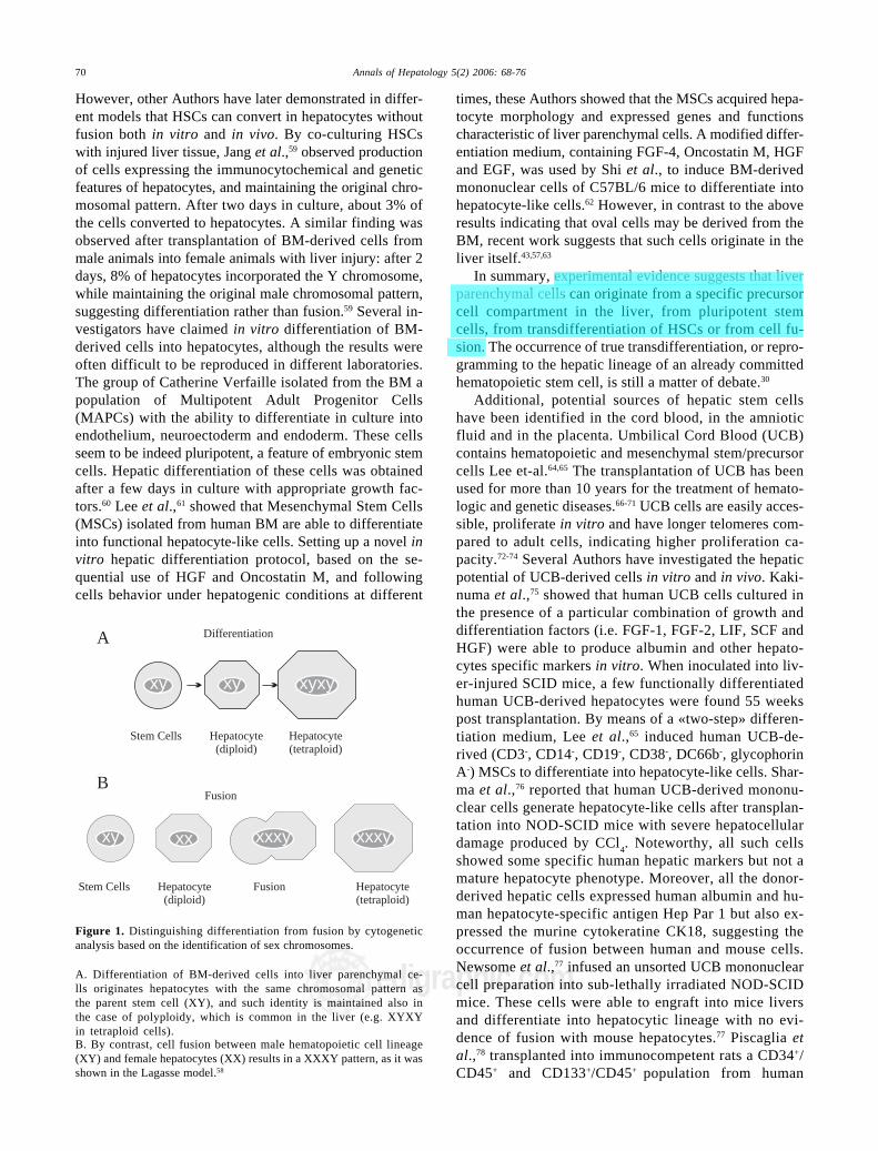
Lagasse et al.,29 transplanted fumarylacetoacetate hy-drolase (FAH)-deficient mouse, an animal model of Ty-rosinemia type I, with BM cells from a non-affected wild-type animal transgenic for the β-galactosidase “LacZ”gene. The liver of the recipient animals was progressivelyrepopulated with hepatocytes harboring both the β-galac-tosidase and the fumarylacetoacetate hydrolase enzyme.Thus, intravenous injection of adult BM cells in the FAH–/– mouse rescued the mouse and restored the biochemicalfunction of its liver. It was later shown that the correctionof the metabolic disorder was not due to transdifferentia-tion of HSCs but rather to a fusion process, probably in-volving macrophages derived from the exogenous he-matopoietic cell lineage and the recipient hepatocytes.57,58
This phenomenon can be demonstrated by cytogeneticanalysis in sex-discordant transplantation (Figure 1).
Annals of Hepatology 5(2) 2006: 68-76MG
70
edigraphic.com
times, these Authors showed that the MSCs acquired hepa-tocyte morphology and expressed genes and functionscharacteristic of liver parenchymal cells. A modified differ-entiation medium, containing FGF-4, Oncostatin M, HGFand EGF, was used by Shi et al., to induce BM-derivedmononuclear cells of C57BL/6 mice to differentiate intohepatocyte-like cells.62 However, in contrast to the aboveresults indicating that oval cells may be derived from theBM, recent work suggests that such cells originate in theliver itself.43,57,63
In summary, experimental evidence suggests that liverparenchymal cells can originate from a specific precursorcell compartment in the liver, from pluripotent stemcells, from transdifferentiation of HSCs or from cell fu-sion. The occurrence of true transdifferentiation, or repro-gramming to the hepatic lineage of an already committedhematopoietic stem cell, is still a matter of debate.30
Additional, potential sources of hepatic stem cellshave been identified in the cord blood, in the amnioticfluid and in the placenta. Umbilical Cord Blood (UCB)contains hematopoietic and mesenchymal stem/precursorcells Lee et-al.64,65 The transplantation of UCB has beenused for more than 10 years for the treatment of hemato-logic and genetic diseases.66-71 UCB cells are easily acces-sible, proliferate in vitro and have longer telomeres com-pared to adult cells, indicating higher proliferation ca-pacity.72-74 Several Authors have investigated the hepaticpotential of UCB-derived cells in vitro and in vivo. Kaki-numa et al.,75 showed that human UCB cells cultured inthe presence of a particular combination of growth anddifferentiation factors (i.e. FGF-1, FGF-2, LIF, SCF andHGF) were able to produce albumin and other hepato-cytes specific markers in vitro. When inoculated into liv-er-injured SCID mice, a few functionally differentiatedhuman UCB-derived hepatocytes were found 55 weekspost transplantation. By means of a «two-step» differen-tiation medium, Lee et al.,65 induced human UCB-de-rived (CD3-, CD14-, CD19-, CD38-, DC66b-, glycophorinA-) MSCs to differentiate into hepatocyte-like cells. Shar-ma et al.,76 reported that human UCB-derived mononu-clear cells generate hepatocyte-like cells after transplan-tation into NOD-SCID mice with severe hepatocellulardamage produced by CCl
4. Noteworthy, all such cells
showed some specific human hepatic markers but not amature hepatocyte phenotype. Moreover, all the donor-derived hepatic cells expressed human albumin and hu-man hepatocyte-specific antigen Hep Par 1 but also ex-pressed the murine cytokeratine CK18, suggesting theoccurrence of fusion between human and mouse cells.Newsome et al.,77 infused an unsorted UCB mononuclearcell preparation into sub-lethally irradiated NOD-SCIDmice. These cells were able to engraft into mice liversand differentiate into hepatocytic lineage with no evi-dence of fusion with mouse hepatocytes.77 Piscaglia etal.,78 transplanted into immunocompetent rats a CD34+/CD45+ and CD133+/CD45+ population from human
Figure 1. Distinguishing differentiation from fusion by cytogeneticanalysis based on the identification of sex chromosomes.
A. Differentiation of BM-derived cells into liver parenchymal ce-lls originates hepatocytes with the same chromosomal pattern asthe parent stem cell (XY), and such identity is maintained also inthe case of polyploidy, which is common in the liver (e.g. XYXYin tetraploid cells).B. By contrast, cell fusion between male hematopoietic cell lineage(XY) and female hepatocytes (XX) results in a XXXY pattern, as it wasshown in the Lagasse model.58
Differentiation
Stem Cells Hepatocyte(diploid)
Hepatocyte(tetraploid)
xy xy xyxy
Stem Cells Hepatocyte(diploid)
Fusion Hepatocyte(tetraploid)
Fusion
xy xx xxxy xxxy
A
B
However, other Authors have later demonstrated in differ-ent models that HSCs can convert in hepatocytes withoutfusion both in vitro and in vivo. By co-culturing HSCswith injured liver tissue, Jang et al.,59 observed productionof cells expressing the immunocytochemical and geneticfeatures of hepatocytes, and maintaining the original chro-mosomal pattern. After two days in culture, about 3% ofthe cells converted to hepatocytes. A similar finding wasobserved after transplantation of BM-derived cells frommale animals into female animals with liver injury: after 2days, 8% of hepatocytes incorporated the Y chromosome,while maintaining the original male chromosomal pattern,suggesting differentiation rather than fusion.59 Several in-vestigators have claimed in vitro differentiation of BM-derived cells into hepatocytes, although the results wereoften difficult to be reproduced in different laboratories.The group of Catherine Verfaille isolated from the BM apopulation of Multipotent Adult Progenitor Cells(MAPCs) with the ability to differentiate in culture intoendothelium, neuroectoderm and endoderm. These cellsseem to be indeed pluripotent, a feature of embryonic stemcells. Hepatic differentiation of these cells was obtainedafter a few days in culture with appropriate growth fac-tors.60 Lee et al.,61 showed that Mesenchymal Stem Cells(MSCs) isolated from human BM are able to differentiateinto functional hepatocyte-like cells. Setting up a novel invitro hepatic differentiation protocol, based on the se-quential use of HGF and Oncostatin M, and followingcells behavior under hepatogenic conditions at different
M Muraca et al. The future of stem cells in liver diseases 73
edigraphic.com
liminary evidence of therapeutic effectiveness has beenprovided in animal models. However, we have to learnmore on the mechanisms of liver regeneration, includingthe role of stem/precursor cells. Definite (and possiblyjoined) protocols for the selection of a specific cell popu-lation and for in vitro expansion/differentiation should bedeveloped, as well as protocols for clinical liver repopula-tion. The long-term fate of the transplanted cells shouldalso be assessed in animal models, with respect to func-tion, possible extra-hepatic localization, genetic/epigenet-ic stability and especially tumorigenesis. The risk is thatsuperficial planning, without adequate consideration andknowledge of the underlying pathophysiology, will resultin poorly focused clinical trials and possible complica-tions, which could in turn originate skepticism on the de-velopment of cell therapy in Hepatology. Even if the bio-logical characteristics of hepatic stem cells still justify thehope for successful future clinical applications, only amore cautious and systematic «bench to bedside» ap-proach will guarantee consistent results.
Acknowledgment
We thank Professor Giuseppe Realdi and ProfessorClaudio Tiribelli for helpful discussion.
References
1. Sipe JD. Tissue Engineering and Reparative Medicine. In: SipeJD, Kelley CA, McNicol LA, eds. Reparative Medicine: GrowingTissues and Organs. Volume 961, Annals of the New York Acad-emy of Sciences, 2002: 1-9.
2. Lake JR. Hepatocyte transplantation. N Engl J Med 1998; 338:1463-5.
3. Gupta S, Bhargava KK, Novikoff PM. Mechanisms of cell en-graftment during liver repopulation with hepatocyte transplanta-tion. Semin Liver Dis 1999; 19: 15-26.
4. Strom SC, Chowdhury JR, Fox IJ. Hepatocyte transplantation forthe treatment of human disease. Semin Liver Dis 1999: 39-48.
5. Grompe M. Liver repopulation for the treatment of metabolicdiseases. J Inherit Metab Dis 2001; 24: 231-44.
6. Horslen SP, Fox IJ. Hepatocyte transplantation. Transplantation2004; 77: 1481-6.
7. Muraca M, Neri D, Parenti A, Feltracco P, Granato A, Vilei MT,Ferraresso C, et al. Intraportal hepatocyte transplantation in thepig. A hemodynamic and histopathological study. Transplanta-tion 2002; 73: 890-6.
8. Fox IJ, Chowdhury JR, Kaufman SS, Goertzen TC, ChowdhuryNR, Warkentin PI, Dorko K, et al. Treatment of the Crigler-Najjarsyndrome type I with hepatocyte transplantation. N Engl J Med1998; 338: 1422-6.
9. Strom SC, Fisher RA, Thompson MT, Sanyal AJ, Cole PE, HamJM, Posner MP. Hepatocyte transplantation as a bridge to ortho-topic liver transplantation in terminal liver failure. Transplanta-tion 1997; 63: 559-69.
10. Muraca M, Gerunda G, Neri D, Vilei MT, Granato A, Feltracco P,Meroni M, et al. Hepatocyte transplantation as a treatment forglycogen storage disease type 1a. Lancet 2002; 359: 1528.
11. Strom SC, Rubinstein WS, Barranger JA, Towbin RB, Charron M,Mieles L, Pisarov LA, et al. Transplantation of human hepato-cytes. Transplantation Proc 1997; 29: 2103-6.
12. Mitry RR, Dhawan A, Hughes RD, Bansal S, Lehec S, Terry C,Heaton ND, et al. One liver, three recipients: segment IV fromsplit-liver procedures as a source of hepatocytes for cell trans-plantation. Transplantation 2004; 77: 1614-6.
13. Dhawan A, Mitry RR, Hughes RD, Lehec S, Terry C, Bansal S,Arya R, et al. Hepatocyte transplantation for inherited factor VIIdeficiency. Transplantation 2004; 78: 1812-4.
14. Bilir BM, Guinette D, Karrer F, Kumpe DA, Krysl J, Stephens J,et al. Hepatocyte transplantation in acute liver failure. Liver Transpl2000; 6: 41-3.
15. Stephenne X, Najimi M, Sibille C, Nassogne MC, Smets F, SokalEM. Sustained engraftment and tissue enzyme activity after livercell transplantation for argininosuccinate lyase deficiency. Gas-troenterology 2006; 130: 1317-23.
16. Rosenthal N. Prometheus’s vulture and the stem-cell promise. NEngl J Med 2003; 349: 267-74.
17. Weissman IL. Translating stem and progenitor cell biology to theclinic: barriers and opportunities. Science 2000; 287: 1442-6.
18. Thomson JA, Itskovitz-Eldor J, Shapiro SS, Waknitz MA, SwiergielJJ, Marshall VS, Jones JM. Embryonic stem cell lines derivedfrom human blastocysts. Science 1998; 282: 1145-7.
19. Korbling M, Estrov Z. Adult stem cells for tissue repair - a newtherapeutic concept? N Engl J Med 2003; 349: 570-82.
20. Laurson J, Selden C, Hodgson HJF. Hepatocyte progenitors inman and in rodents-multiple pathways, multiple candidates. Int JPath 2005; 86: 1-18.
21. Ferrari G, Cusella-De Angelis G, Coletta M, Paolucci E, StornaiuoloA, Cossu G, Mavilio F. Muscle regeneration by bone marrow-derived myogenic progenitors. Science 1998; 279: 1528-30.
22. Petersen BE, Bowen WC, Patrene KD, Mars WM, Sullivan AK,Murase N, Boggs SS, et al. Bone marrow as potential source ofhepatic oval cells. Science 1999; 284: 1168-70.
23. Jackson KA, Mi T, Goodell MA. Hematopoietic potential of stemcells isolated from murine skeletal muscle. Proc Natl Acad SciUSA 1999; 96: 14482-6.
24. Bjornson CR, Rietze RL, Reynolds BA, Magli MC, Vescovi AL.Turning brain into blood: a hematopoietic fate adopted by adultneural stem cells in vivo. Science 1999: 283.
25. Woodbury D, Schwarz EJ, Prockop DJ, Black IB. Adult rat andhuman bone marrow stromal cells differentiate into neurons. JNeurosi Res 2000; 61: 364-70.
26. Mezey E, Chandross KJ. Bone marrow: a possible alternativesource of cells in the adult nervous system. Eur J Pharmacol2000; 405: 297-302.
27. Theise ND, Nimmakayalu M, Gardner R, Illei PB, Morgan G,Teperman L, Henegariu O, et al. Liver from bone marrow inhumans. Hepatology 2000; 32: 11-6.
28. Alison MR, Poulsom R, Jeffery R, Dhillon AP, Quaglia A, JacobJ, Novelli, M, et al. Hepatocytes from non-hepatic adult stemcells. Nature 2000; 406: 257.
29. Lagasse E, Connors H, Al-Dhalimy M, Reitsma M, Dohse M,Osborne L, Wang X, et al. Purified hematopoietic stem cells candifferentiate into hepatocytes in vivo. Nat Med 2000; 6: 1229-34.
30. Fausto N. Liver Regeneration and Repair: Hepatocytes, Progeni-tor Cells, and Stem Cells. Hepatology 2004; 39: 1477-87.
31. Farber E. Similarities in the sequence of early histological changesinduced in the liver of the rat by ethionine, 2-acetylamino-fluo-rene, and 3'-methyl-4-dimethylaminoazobenzene. Cancer Res1956; 16: 142-8.
32. Grisham JW, Hartroft WS. Morphologic identification by elec-tron microscopy of «oval» cells in experimental hepatic degen-eration. Lab Invest 1961; 10: 317-32.
33. Evarts RP, Nagy P, Nakatsukasa H, Marsden E, Thorgeirsson SS.In vivo differentiation of rat liver oval cells into hepatocytes.Cancer Res 1989; 49: 1541-7.
34. Yin L, Lynch D, Ilic Z, Sell S. Proliferation and differentiation ofductular progenitor cells and littoral cells during the regenerationof the rat liver to CCl4/2-AAF injury. Histol Histopathol 2002; 17:65-81.
Differentiating Stem Cells into Liver
ALEJANDRO SOTO-GUTIERREZ1, HESHAM BASMA2 , NALU NAVARRO-ALVAREZ3, BASAK E. UYGUN1, MARTIN L. YARMUSH1, NAOYA KOBAYASHI3, AND IRA J. FOX4*
1 Center for Engineering in Medicine and Department of Surgery, Massachusetts General Hospital, Harvard Medical School, and the Shriners Hospitals for Children, Boston, MA 02114, USA, 2Department of Surgery, University of Nebraska Medical Center, Omaha, Nebraska 68198-3285, USA, 3 Department of Surgery, Okayama University Graduate School of Medicine and Dentistry, 2-5-1 Shikata-cho, Okayama 700-8558, Japan, 4Department of Surgery, Division of Pediatric Transplant Surgery, Children's Hospital of Pittsburgh and McGowan Institute for Regenerative Medicine, University of Pittsburgh School of Medicine, Pittsburgh, PA 15213, USA.
Abstract
Research involving differentiated embryonic stem (ES) cells may revolutionize the study of liver disease, improve the drug discovery process, and assist in the develop-ment of stem-cell-based clinical therapies. Generation of ES cell-derived hepatic tissue has benefited from an understanding of the cytokines, growth factors and biochemical compounds that are essential in liver development, and this knowledge has been used to mimic some aspects of embryonic development in vitro. Although great progress has been made in differentiating human ES cells into liver cells, current protocols have not yet produced cells with the phenotype of a mature hepatocyte. There is a
Biotechnology and Genetic Engineering Reviews - Vol. 25, 149-164 (2008)
To whom correspondence should be addressed ([email protected])
Abbreviations: ES cells, embryonic stem cells; CYP, cytochrome P450; AFP, alpha-fetoprotein; alb, albumin; CK18, cytokerain 18; Oct4, Octamer-4; SSEA-4, Stage-Specific Embryonic Antigen-4; Oxt2, orthodenticle homeobox 2; Hesx 1, HESX homeobox 1; Hex, homeobox; Cdx2, caudal-related homeobox 2; EBs, embroyd bodies; GSC, goosecoid; Foxa 2, forkhead box A2; cxcr4, chemokine C-X-C motif receptor 4; Sox17a/b, sex determining region-Y box 17; VEGFR2, vascular endothelial growth factor receptor-2; PDGFRa, platelet-derived growth factor receptor-a; GATA-4, GATA binding protein 4; EpCAM, epithelial cell adhesion molecule; DPPIV, dipeptidyl peptidase IV; TGFß, Tumor growth factor beta; FGFs, Fibrob-last growth factors; BMPs, Bone marrow proteins; HNF6, Hepatocyte nuclear factor 6; HGF, Hepatocyte growth factor; C/EBPß, CCAAT/enhancer binding protein beta; Foxm1b, Forkhead Box (Fox) m1b; Xbp1, X-box binding protein 1; Dex, Dexamethasone; CYP, cytochrome p450
doi: 10.5661/bger-25-149
Dow
nloa
ded
by [
188.
26.2
0.20
0] a
t 03:
32 1
3 D
ecem
ber
2017
Differentiating stem cells into liver 151
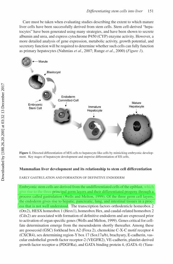
Care must be taken when evaluating studies describing the extent to which mature liver cells have been successfully derived from stem cells. Stem cell-derived “hepa-tocytes” have been generated using many strategies, and have been shown to secrete albumin and urea, and express cytochrome P450 (CYP) enzyme activity. However, a more detailed analysis of gene expression, metabolic activity, growth potential, and secretory function will be required to determine whether such cells can fully function as primary hepatocytes (Nahmias et al., 2007; Runge et al., 2000) (Figure 1).
Figure 1. Directed differentiation of hES cells to hepatocyte-like cells by mimicking embryonic develop-ment. Key stages of hepatocyte development and stepwise differentiation of ES cells.
Mammalian liver development and its relationship to stem cell differentiation
EARLY GASTRULATION AND FORMATION OF DEFINITIVE ENDODERM
Embryonic stem cells are derived from the undifferentiated cells of the epiblast, which give rise to the three principal germ layers and their differentiated progeny through a process called gastrulation (Wells and Melton, 1999). Of the three germ cell layers, the endoderm gives rise to hepatic, pancreatic, lung, and intestinal tissues in a proc-ess that is not well understood. The transcription factors orthodenticle homeobox 2 (Otx2), HESX homeobox 1 (Hesx1), homeobox Hex, and caudal-related homeobox 2 (Cdx2) are associated with formation of definitive endoderm and are expressed prior to activation of organ specific genes (Wells and Melton, 1999). Genes critical for cell-fate determination emerge from the mesendoderm shortly thereafter. Among these are goosecoid (GSC) forkhead box A2 (Foxa 2), chemokine C-X-C motif receptor 4 (CXCR4), sex determining region-Y box 17 (Sox17a/b), brachyury, E-cadherin, vas-cular endothelial growth factor receptor-2 (VEGFR2), VE-cadherin, platelet-derived growth factor receptor-a (PDGFRa), and GATA binding protein 4, (GATA-4) (Yasu-
Dow
nloa
ded
by [
188.
26.2
0.20
0] a
t 03:
32 1
3 D
ecem
ber
2017
152 A. SOTO-GUTIERREZ et al.
naga et al., 2005). An appreciation of the genetic patterns and cellular markers that distinguish definitive endoderm from extraembryonic endoderm, such as EpCAM, CD38, and DPPIV, have been critically important in the process of differentiating and isolating tissue-specific cells from stem cells (Sherwood et al., 2007).
Definitive endoderm can be generated by a variety of means. Definitive endoderm can be produced through formation of embryoid bodies or by culturing ES cells with collagen-sandwiched hepatocytes (Cho et al., 2007). Large numbers of endoderm like-cells can also be produced when mouse ES cells are cultured on fibronectin-coated collagen gels and exposed to follistatin, an Activin binding protein (Parashurama et al., 2007). The common transcriptional machinery present in both definitive and visceral endoderm may, however, limit the extent to which differentiation using these techniques can lead to tissue formation. Generation of a pure population of definitive endoderm from ES cells may require selective inhibition of visceral endoderm.
Recently, investigators have facilitated the development of definitive endoderm from mouse and human ES cells using culture in low concentrations of serum in conjunc-tion with growth in Activin A, a TGFβ family member that binds the same receptors as does Nodal. This finding has facilitated generation of endoderm-derived tissues, such as pancreatic beta cells and hepatocytes (D’Amour et al., 2005; Kubo et al., 2004; Yasunaga et al., 2005) and has resolved the problem of forming a mixture of visceral and definitive endoderm during the differentiation process.
HEPATIC INDUCTION
Growth factor signaling from the cardiac mesoderm and septum transversum mes-enchyme specifies the underlying endoderm to adopt a hepatic fate such that, by the 6–7 somite stage, hepatic gene expression can be detected in the ventral foregut endoderm (Jung et al., 1999; Rossi, 2001). Using a variety of techniques, it has now been shown that fibroblast growth factors (FGFs) can substitute for cardiac mesoderm and bone morphogenic proteins (BMPs) can substitute for the septum transversum mesenchyme to work in concert to induce the ventral endoderm to adopt a hepatic fate (Jung et al., 1999). Studies documenting induction of hepatic specification fol-lowing chick cardiac mesoderm co-culture with ES cells, or following culture in FGF and BMP, illustrate how such developmental principles can assist with stem cell differentiation in vitro (Cai et al., 2007; Fair et al., 2003; Gouon-Evans et al., 2006; Soto-Gutierrez et al., 2007). Factors identified as proposed targets of FGF and BMP signaling include the Foxa and Gata genes, which regulate the competence of foregut endoderm to respond to hepatic inductive signals (Cirillo et al., 2002). In addition, the transcription factor hepatocyte nuclear factor-6 (HNF6) has been shown to play a critical role in the proper morphogenesis of both the intra- and extra-hepatic biliary tree. The mechanism by which HNF6 regulates biliary tree development also appears to involve the related transcription factor, hepatocyte nuclear factor-1 (HNF1) (Clot-man et al., 2005) (Figure 2).
LIVER SPECIFICATION, DEVELOPMENT OF THE LIVER BUD, AND HEPATIC EXPANSION
Hepatocytes and bile duct cells originate from a common precursor, the hepatoblast (Germain et al., 1988). Notch signaling promotes hepatoblast differentiation toward the
Dow
nloa
ded
by [
188.
26.2
0.20
0] a
t 03:
32 1
3 D
ecem
ber
2017
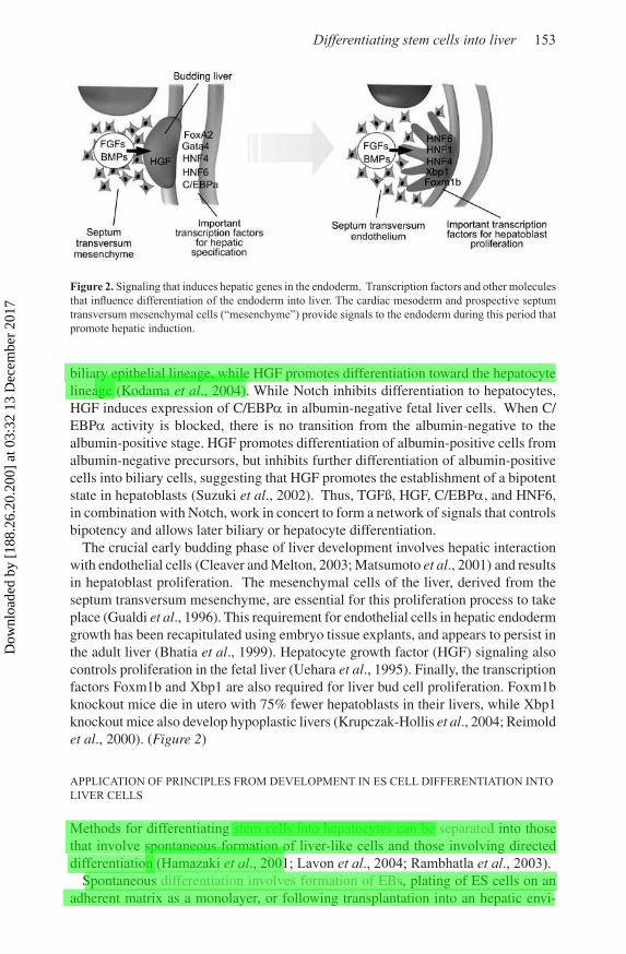
Differentiating stem cells into liver 153
biliary epithelial lineage, while HGF promotes differentiation toward the hepatocyte lineage (Kodama et al., 2004). While Notch inhibits differentiation to hepatocytes, HGF induces expression of C/EBPα in albumin-negative fetal liver cells. When C/EBPα activity is blocked, there is no transition from the albumin-negative to the albumin-positive stage. HGF promotes differentiation of albumin-positive cells from albumin-negative precursors, but inhibits further differentiation of albumin-positive cells into biliary cells, suggesting that HGF promotes the establishment of a bipotent state in hepatoblasts (Suzuki et al., 2002). Thus, TGFß, HGF, C/EBPα, and HNF6, in combination with Notch, work in concert to form a network of signals that controls bipotency and allows later biliary or hepatocyte differentiation.
The crucial early budding phase of liver development involves hepatic interaction with endothelial cells (Cleaver and Melton, 2003; Matsumoto et al., 2001) and results in hepatoblast proliferation. The mesenchymal cells of the liver, derived from the septum transversum mesenchyme, are essential for this proliferation process to take place (Gualdi et al., 1996). This requirement for endothelial cells in hepatic endoderm growth has been recapitulated using embryo tissue explants, and appears to persist in the adult liver (Bhatia et al., 1999). Hepatocyte growth factor (HGF) signaling also controls proliferation in the fetal liver (Uehara et al., 1995). Finally, the transcription factors Foxm1b and Xbp1 are also required for liver bud cell proliferation. Foxm1b knockout mice die in utero with 75% fewer hepatoblasts in their livers, while Xbp1 knockout mice also develop hypoplastic livers (Krupczak-Hollis et al., 2004; Reimold et al., 2000). (Figure 2)
APPLICATION OF PRINCIPLES FROM DEVELOPMENT IN ES CELL DIFFERENTIATION INTO LIVER CELLS
Methods for differentiating stem cells into hepatocytes can be separated into those that involve spontaneous formation of liver-like cells and those involving directed differentiation (Hamazaki et al., 2001; Lavon et al., 2004; Rambhatla et al., 2003).
Spontaneous differentiation involves formation of EBs, plating of ES cells on an adherent matrix as a monolayer, or following transplantation into an hepatic envi-
Figure 2. Signaling that induces hepatic genes in the endoderm. Transcription factors and other molecules that influence differentiation of the endoderm into liver. The cardiac mesoderm and prospective septum transversum mesenchymal cells (“mesenchyme”) provide signals to the endoderm during this period that promote hepatic induction.
Dow
nloa
ded
by [
188.
26.2
0.20
0] a
t 03:
32 1
3 D
ecem
ber
2017
154 A. SOTO-GUTIERREZ et al.
ronment (Lavon et al., 2004; Yamada et al., 2002; Yamamoto et al., 2003). Directed differentiation usually involves addition of growth factors and cytokines to cells in vitro on extracellular matrices (Cai et al., 2007; Rambhatla et al., 2003; Teratani et al., 2005). Combinations of these techniques, involving both formation of EBs with expansion in growth factors and/or co-culture with cells supplying additional factors, have also been successful (Cho et al., 2007; Fair et al., 2003; Soto-Gutierrez et al., 2006; Soto-Gutierrez et al., 2007). In addition, culture in sodium butyrate, a histone deacetylase inhibitor, leads to an increase in the number of cells expressing mature hepatocyte-specific genes.
Selection of hepatocyte-like cells based on the use of liver-specific promoters that drive reporter gene expression has been a relatively successful strategy for selecting a homogeneous population of cells with hepatic characteristics (Lavon et al., 2004). In the majority of settings, the resultant cells have morphological features similar to those of primary hepatocytes and most of the cells express liver-associated proteins (Rambhatla et al., 2003). Whether such cells have the functional characteristics of a mature liver cell will require a more comprehensive analysis.
ES cell differentiation: state of the art
Using a modification of the protocol described earlier using Activin A to enrich for definitive endoderm (D’Amour et al., 2005), and using principles from development, Cai et al. reported that later addition of FGF, BMP, and HGF to cultured cells induces expression of hepatic fate genes, and that further culture in Oncostatin M (OSM) and dexamethasone (Dex) leads to significant differentiation to cells with a large number of hepatocyte-like characteristics. Transplantation of such differentiated cells into mice with chemically-induced liver injury resulted in engraftment of a small number of ES-derived cells in the liver (Cai et al., 2007). Culture of ES cells in Activin A followed by treatment with sodium butyrate and HGF has been also shown to generate cells exhibiting morphologic characteristics of hepatocytes, expression of alpha-fetoprotein and albumin, and cytochrome p450 (CYP) metabolic activity (Hay et al., 2007).
ES cells have also been induced to differentiate into “hepatic-like” cells using other approaches (Cai et al., 2007; Cho et al., 2007; Lavon et al., 2004; Rambhatla et al., 2003; Soto-Gutierrez et al., 2007; Teratani et al., 2005). Development of cells that express hepatocyte markers, such as AFP, Alb, and CK18, inducible expression of cytochrome P450, and morphologic characteristics of an epithelial phenotype has been reported widely (Cai et al., 2007; Lavon et al., 2004; Rambhatla et al., 2003). However, useful stem cell-derived hepatocytes will need to not only express the genes found in mature hepatocytes, but the levels of expression will need to be at or near those found in the normal liver. Furthermore, differentiation will need to be lineage specific.
Evidence of protein production will need to be demonstrated to provide verification of gene expression studies, and the metabolic activity of CYP enzymes will be needed to provide definitive evidence of mature hepatic function. While hepatocytes are the only cell that secretes albumin, inferring that any albumin-secreting cell can, by definition, be regarded as a hepatocyte is not justified. Many stem cell-derived “hepatocytes” express albumin but many do not express other genes that would
Dow
nloa
ded
by [
188.
26.2
0.20
0] a
t 03:
32 1
3 D
ecem
ber
2017

Differentiating stem cells into liver 155
normally constitute normal hepatocyte function. The same can be said for CYP enzyme activity, as p450 expression is not limited to hepatocytes. Inducible CYP activity has been reported by lung, intestinal epithelial cells, and adipose tissue (Baijal et al., 1997; Yoshinari et al., 2004; Zhang et al., 2003).
To adequately assess the extent to which in vitro differentiation of stem cells has been effective, it will be important to clearly demonstrate cellular characteristics and activities that can only be performed by primary hepatocytes.
1. Gene expression by differentiated “hepatocyte-like” cells should be compared to the gene expression profile of human fetal and/or mature liver cells (Hewitt et al., 2007; Li et al., 1990);
2. Evidence of basal and inducible CYP450 isoform function should be assessed (Kostrubsky et al., 1999);
3. Metabolism of xenobiotics or other endogenous substances (hormones and ammonia) should be determined (Duncan et al., 1998; Hewitt et al., 2007);
4. Synthesis and/or secretion of the following should be performed: albumin, clotting factors, complement, transporter proteins, bile acids, and lipids and lipoproteins. (Hewitt et al., 2007; Mita et al., 2006);
5. Evidence of restoration of liver function in appropriate animal models, or evidence of repopulation of the liver by derived “hepatocytes” should be examined.
In summary, derived hepatocytes should demonstrate drug metabolism and detoxification activity by both gene expression and function, and they should express the hepatic transport proteins and transcription factors present in mature hepatocytes. They should also, to some degree, secrete albumin and/or produce bile acids, conjugate bilirrubin, metabolize ammonia, and function in animal models of liver dysfunction after transplantation. Finally, differentiated cells should no longer express transcription factors or other genes characteristic of cells of other cell lineages (Figure 3).
Application of stem cell derived liver cells
The availability of a reliable and homogeneous source of human hepatocytes would be an invaluable tool for liver tissue engineering, for use in cell-based in vitro assays of drug toxicity, for metabolic profiling, for the study of drug–drug interactions, and for use in cell therapy and regenerative medicine (Wobus and Boheler, 2005).
CELL THERAPY
The worldwide shortage of donor organs is likely to increase over the coming dec-ades. As a result, development of alternative methods for treating life-threatening liver disease will become increasingly important. While cell transplantation and other cell-based therapies have great potential for improving the lives of patients with liver failure (Fox et al., 1998; Navarro-Alvarez et al., 2007), the lack of available donors
Dow
nloa
ded
by [
188.
26.2
0.20
0] a
t 03:
32 1
3 D
ecem
ber
2017
Differentiating stem cells into liver 159
of disease models have been examined concerning whether stem cells can correct liver disease. It is a bit premature to conclude that hepatocytes can be generated from non-hepatic cells in culture that will be clinically useful. Standard criteria will need to be developed to assess the extent to which human stem cell-derived hepatocytes have been produced.
References
AZUMA, H., PAULK, N., RANADE, A., DORRELL, C., AL-DHALIMY, M., ELLIS, E., STROM, S., KAY, M. A., FINEGOLD, M., AND GROMPE, M. (2007): Robust expansion of human hepatocytes in Fah(-/-)/Rag2(-/-)/Il2rg(-/-) mice. Nat Biotechnol 25, 903-10.
BAIJAL, P. K., FITZPATRICK, D. W., AND BIRD, R. P. (1997): Phenobarbital and 3-methylcholanthrene treatment alters phase I and II enzymes and the sensitivity of the rat colon to the carcinogenic activity of azoxymethane. Food Chem Toxicol 35, 789-98.
BHATIA, S. N., BALIS, U. J., YARMUSH, M. L., AND TONER, M. (1999): Effect of cell-cell interactions in preservation of cellular phenotype: cocultivation of hepatocytes and nonparenchymal cells. Faseb J 13, 1883-900.
CAI, J., ZHAO, Y., LIU, Y., YE, F., SONG, Z., QIN, H., MENG, S., CHEN, Y., ZHOU, R., SONG, X., GUO, Y., DING, M., AND DENG, H. (2007): Directed differentiation of human embryonic stem cells into functional hepatic cells. Hepatology 45, 1229-39.
CHO, C. H., PARASHURAMA, N., PARK, E. Y., SUGANUMA, K., NAHMIAS, Y., PARK, J., TILLES, A. W., BERTHIAUME, F., AND YARMUSH, M. L. (2007): Homogeneous differentiation of hepatocyte-like cells from embryonic stem cells: applications for the treatment of liver failure. Faseb J.
CIRILLO, L. A., LIN, F. R., CUESTA, I., FRIEDMAN, D., JARNIK, M., AND ZARET, K. S. (2002): Opening of compacted chromatin by early developmental transcription factors HNF3 (FoxA) and GATA-4. Mol Cell 9, 279-89.
CLEAVER, O., AND MELTON, D. A. (2003): Endothelial signaling during development. Nat Med 9, 661-8.
CLOTMAN, F., JACQUEMIN, P., PLUMB-RUDEWIEZ, N., PIERREUX, C. E., VAN DER SMISSEN, P., DIETZ, H. C., COURTOY, P. J., ROUSSEAU, G. G., AND LEMAIGRE, F. P. (2005): Control of liver cell fate decision by a gradient of TGF beta signaling modulated by Onecut transcription factors. Genes Dev 19, 1849-54.
D’AMOUR, K. A., AGULNICK, A. D., ELIAZER, S., KELLY, O. G., KROON, E., AND BAETGE, E. E. (2005): Efficient differentiation of human embryonic stem cells to definitive endoderm. Nat Biotechnol 23, 1534-41.
D’AMOUR, K. A., BANG, A. G., ELIAZER, S., KELLY, O. G., AGULNICK, A. D., SMART, N. G., MOORMAN, M. A., KROON, E., CARPENTER, M. K., AND BAETGE, E. E. (2006): Production of pancreatic hormone-expressing endocrine cells from human embryonic stem cells. Nat Biotechnol 24, 1392-401.
DUNCAN, S. A., NAVAS, M. A., DUFORT, D., ROSSANT, J., AND STOFFEL, M. (1998): Regulation of a transcription factor network required for differentiation and metabolism. Science 281, 692-5.
FAIR, J. H., CAIRNS, B. A., LAPAGLIA, M., WANG, J., MEYER, A. A., KIM, H., HATADA, S., SMITHIES, O., AND PEVNY, L. (2003): Induction of hepatic differentiation in embryonic stem cells by co-culture with embryonic cardiac mesoderm. Surgery 134, 189-96.
Dow
nloa
ded
by [
188.
26.2
0.20
0] a
t 03:
32 1
3 D
ecem
ber
2017
![STEM CELLS EMBRYONIC STEM CELLS/INDUCED PLURIPOTENT STEM CELLS Stem Cells.pdf · germ cell production [2]. Human embryonic stem cells (hESCs) offer the means to further understand](https://img.pdfslide.net/doc/110x75/6014b11f8ab8967916363675/stem-cells-embryonic-stem-cellsinduced-pluripotent-stem-cells-stem-cellspdf.jpg)