Embed Size (px)
Citation preview
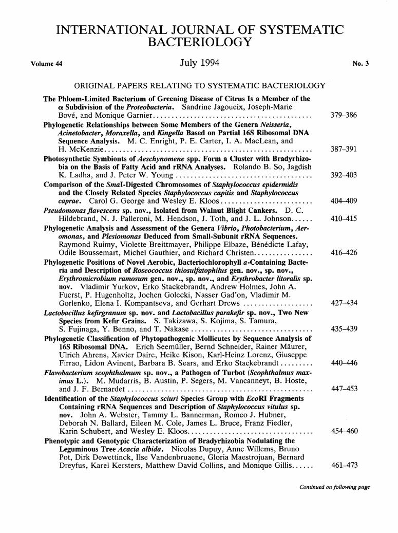
INTERNATIONAL JOURNAL OF SYSTEMATIC BACTERIOLOGY
Volume 44 J u l y 1994 No. 3
O R I G I N A L P A P E R S R E L A T I N G TO S Y S T E M A T I C B A C T E R I O L O G Y
The Phloem-Limited Bacterium of Greening Disease of Citrus Is a Member of the c* Subdivision of the Proteobacteria. Sandrine Jagoueix, Joseph-Marie Bove, and Monique Garnier 379-386
Phylogenetic Relationships between Some Members of the Genera Neisseria, Acinetobacter, M o r a x e l l a , and K i n g e l l a Based on Partial 16S Ribosomal DNA Sequence Analysis. M . C . Enright, P. E . Carter, I. A . MacLean, and H . McKenzie 387-391
Photosynthetic Symbionts of Aeschynomene spp. Form a Cluster with Bradyrhizo-bia on the Basis of Fatty Acid and rRNA Analyses. Rolando B . So, Jagdish K . Ladha, and J . Peter W . Young 392-403
Comparison of the Smal-Digested Chromosomes of Staphylococcus epidermidis and the Closely Related Species Staphylococcus capitis and Staphylococcus caprae. Carol G . George and Wesley E . Kloos 404-409
Pseudomonas flavescens sp. nov., Isolated from Walnut Blight Cankers. D . C . Hildebrand, N . J . Palleroni, M . Hendson, J . Toth, and J . L . Johnson 410-415
Phylogenetic Analysis and Assessment of the Genera V i b r i o , Photobacterium, Aer-omonas, and Plesiomonas Deduced from Small-Subunit rRNA Sequences. Raymond Ruimy, Violette Breittmayer, Philippe Elbaze, Benedicte Lafay, Odile Boussemart, Michel Gauthier, and Richard Christen 416-426
Phylogenetic Positions of Novel Aerobic, Bacteriochlorophyll a-Containing Bacte-ria and Description of Roseococcus thiosulfatophilus gen. nov., sp. nov., E r y t h r o m i c r o b i u m ramosum gen. nov., sp. nov., and Erythrobacter l i t o r a l i s sp. nov. Vladimir Yurkov, Erko Stackebrandt, Andrew Holmes, John A . Fuerst, P. Hugenholtz, Jochen Golecki, Nasser Gad'on, Vladimir M . Gorlenko, Elena I. Kompantseva, and Gerhart Drews 427-434
Lactobacillus k e f i r g r a n u m sp. nov. and Lactobacillus paralcefir sp. nov., Two New Species from Kefir Grains. S. Takizawa, S. Kojima, S. Tamura, S. Fujinaga, Y . Benno, and T. Nakase 435-439
Phylogenetic Classification of Phytopathogenic Mollicutes by Sequence Analysis of 16S Ribosomal DNA. Er ich Seemüller, Bernd Schneider, Rainer Maurer, Ulrich Ahrens, Xavier Daire, Heike Kison , Karl-Heinz Lorenz, Giuseppe Firrao, Lidon Avinent, Barbara B . Sears, and Erko Stackebrandt 440-446
F l a v o b a c t e r i u m scophthalmum sp. nov., a Pathogen of Turbot (Scophthalmus max-imus L.). M . Mudarris, B . Austin, P. Segers, M . Vancanneyt, B . Hoste, and J . F . Bernardet 447-453
Identification of the Staphylococcus sciuri Species Group with E c o W Fragments Containing rRNA Sequences and Description of Staphylococcus vitulus sp. nov. John A . Webster, Tammy L . Bannerman, Romeo J . Hubner, Deborah N . Ballard, Eileen M . Cole, James L . Bruce, Franz Fiedler, Karin Schubert, and Wesley E . Kloos 454-460
Phenotypic and Genotypic Characterization of Bradyrhizobia Nodulating the Leguminous Tree Acacia a l b i d a . Nicolas Dupuy, Anne Willems, Bruno Pot, Dirk Dewettinck, Ilse Vandenbruaene, Gloria Maestrojuan, Bernard Dreyfus, Karel Kersters, Matthew David Collins, and Monique Gillis 461-473
C o n t i n u e d o n f o l l o w i n g page
C o n t i n u e d from p r e c e d i n g page
Nocardiopsis h a l o p h i l a sp. nov., a New Halophilic Actinomycete Isolated from Soil. Amira Mahmoud Al -Ta i and Ji-Sheng Ruan 474-478
Mycoplasma auris sp. nov., Mycoplasma cottewii sp. nov., and Mycoplasma yeatsii sp. nov., New Sterol-Requiring Mollicutes from the External Ear Canals of Goats. A . J . DaMassa, J . G . Tully, D . L . Rose, D . Pitcher, R. H . Leach, and G . S. Cottew 479-484
Transfer of Polychlorophenol-Degrading Rhodococcus chlorophenolicus (Apa-jalahti et al. 1986) to the Genus Mycobacterium as Mycobacterium chlorophe-nolicum comb. nov. Max M . Häggblom, Li isa J . Nohynek, Norberto J . Palleroni, Kaarina Kronqvist, Eeva-Liisa Nurmiaho-Lassila, Mirja S. Salkinoja-Salonen, Stefan Klatte, and Reiner M . Kroppenstedt 485-493
Phylogenetic Evidence for Transfer of Pentachlorophenol-Mineralizing Rhodococcus chlorophenolicus PCP-I T to the Genus Mycobacterium. Maria Briglia, Rik I. L . Eggen, Dirk J . Van Elsas, and Willem M . De Vos 494-498
Classification of Pseudomonas d i m i n u t a Leifson and Hugh 1954 and Pseudomonas vesicularis Büsing, Doli, and Freytag 1953 in Brevundimonas gen. nov. as Brevundimonas d i m i n u t a comb. nov. and Brevundimonas vesicularis comb, nov., Respectively. P . Segers, M . Vancanneyt, B . Pot, U . Torck, B . Hoste, D . Dewettinck, E . Falsen, K . Kersters, and P. De Vos 499-510
Rhizobium ciceri sp. nov., Consisting of Strains That Nodulate Chickpeas {Cicer a r i e t i n u m L.). Sarah M . Nour, Maria P. Fernandez, Philippe Normand, and Jean-Claude Cleyet-Marel 511-522
Phylogenetic Analysis of a New LL-Diaminopimelic Acid-Containing Coryneform Bacterium from Herbage, Nocardioides p l a n t a r u m sp. nov. M . D . Collins, S. Cockcroft, and Sally Wallbanks 523-526
Evolutionary Relationships among Eubacterial Groups as Inferred from GroEL (Chaperonin) Sequence Comparisons. Alejandro M . Viale , Adrian K . Arakaki , Fernando C . Soncini, and Raul G . Ferreyra 527-533
Isolation and Characterization of H a l o t h e r m o t h r i x orenii gen. nov., sp. nov., a Halophilic, Thermophilic, Fermentative, Strictly Anaerobic Bacterium. J . - L . Cayol , B . Ollivier, B . K . C . Patel, G . Prensier, J . Guezennec, and J . - L . Garcia 534-540
Classification of Leptospires of the Pyrogenes Serogroup Isolated from Cattle in Zimbabwe by Cross-Agglutinin Absorption and Restriction Fragment Length Polymorphism Analysis. Sara B . Feresu, Carole A . Bol in , Hans Korver , and Wiepko J . Terpstra 541-546
Pulsed-Field Gel Electrophoresis of Genomic Digests of Thermus Strains and Its Implications for Taxonomic and Evolutionary Studies. A . G . Rodrigo, K . M . Borges, and P. L . Bergquist 547-552
Phylogeny of Helicobacter Isolates from Bird and Swine Feces and Description of Helicobacter pametensis sp. nov. F loyd E . Dewhirst, Charles Seymour, Gayle J . Fräser , Bruce J . Paster, and James G . Fox 553-560
Fatty Acid and Protein Profiles of Streptomyces Scabies Strains Isolated in Eastern Canada. Er ic Paradis, Claudia Goyer, Nancy C . Hodge, Richard Hogue, Robert E . Stall, and Carole Beaulieu 561-564
H a l o a n a e r o b i u m salsugo sp. nov., a Moderately Halophilic, Anaerobic Bacterium from a Subterranean Brine. V . K . Bhupathiraju, A . Oren, P. K . Sharma, R. S. Tanner, C . R. Woese, and M . J . Mclnerney 565-572
Methanolobus t a y l o r i i sp. nov., a New Methylotrophic, Estuarine Methanogen. Ronald S. Oremland and David R. Boone 573-575
C o n t i n u e d o n f o l l o w i n g page
C o n t i n u e d f r o m p r e c e d i n g page
Emendation of the Description of Acidaminococcus fermentans, a fraws-Aconitate-and Citrate-Oxidizing Bacterium. Gregory M . Cook, Frederick A . Rainey, Guangjiong Chen, Erko Stackebrandt, and James B . Russell
Transfer of Propionibacterium innocuum Pitcher and Collins 1991 to P r o p i o n i f e r a x gen. nov. as P r o p i o n i f e r a x i n n o c u a comb. nov. Ak i r a Yokota, Tomohiko Tamura, Mariko Takeuchi, Nobert Weiss, and Erko Stackebrandt
Phylogenetic Analysis of Species of the mew-Diaminopimelic Acid-Containing Genera Brevibacterium and Dermabacter. Junpeng Cai and Matthew D . Collins
M A T T E R S R E L A T I N G T O T H E I N T E R N A T I O N A L C O M M I T T E E O N S Y S T E M A T I C B A C T E R I O L O G Y
Taxonomic Notes Synonymy of Enterobacter cancerogenus (Urosevic 1966) Dickey and Zumoff 1988
and Enterobacter taylorae Farmer et al. 1985 and Resolution of an Ambiguity in the Biochemical Profile. H . C . Sch0nheyder, K . T. Jensen, and W . Frederiksen 586-587
An Aid to Formation of Bacterial Names: Chemical Terminology and Microbio-logical Nomenclature. R. E . Buchanan 588-590
Request for a n Opinion Phylogenetic Placement of S a r c i n a v e n t r i c u l i and S a r c i n a m a x i m a within Group I
C l o s t r i d i u m , a Possible Problem for Future Revision of the Genus C l o s t r i d i u m . A . Willems and M . D . Collins 591-593
M i n u t e s ICSB Subcommittee on the Taxonomy of Staphylococci and Streptococci 594
V a l i d a t i o n List Validation of the Publication of New Names and New Combinations Previously
Effectively Published Outside the IJSB. List N o . 50 595
N o t i f i c a t i o n List Notification that New Names and New Combinations Have Appeared in Volume
44, No. 2, of the USB 596
E R R A T U M
A New Serovar in the Grippotyphosa Serogroup Comprising Leptospiral Isolates from DiflFerent Regions. J . L . Herrmann, P. Bakoss, H . Korver , A . A. Bulu , E . Bellenger, W . J . Terpstra, I. Saint Girons, and G . Baranton 597
576-578
579-582
583-585
C o n t i n u e d o n f o l l o w i n g page

C o n t i n u e d from p r e c e d i n g page
A U T H O R ' S C O R R E C T I O N
16S Ribosomal DNA Sequences of Anaerobic Cocci and Proposal of Ruminococcus hansenii comb. nov. and Ruminococcus productus comb. nov. Takayuki Ezaki , N a L i , Yasuhiro Hashimoto, Hiroaki Miura , and Hiroyuki Yamamoto
INTERNATIONAL JOURNAL OF SYSTEMATIC BACTERIOLOGY, July 1994, p. 4 5 4 ^ 6 0 0020-7713/94/$04.00+0 Copyright © 1994, International Union of Microbiological Societies
Vol. 44, No. 3
Identification of the Staphylococcus sciuri Species Group with E c o R l Fragments Containing rRNA Sequences and
Description of Staphylococcus vitulus sp. nov. J O H N A . W E B S T E R , 1 * T A M M Y L. B A N N E R M A N , 2 R O M E O J. H U B N E R , 1 D E B O R A H N. B A L L A R D , 2
EI L E E N M . C O L E , 1 J A M E S L . B R U C E , 1 F R A N Z F I E D L E R , 3 K A R I N S C H U B E R T , 3
AND W E S L E Y E . K L O O S 2
E . I . d u P o n t de N e m o u r s a n d C o m p a n y , W i l m i n g t o n , D e l a w a r e 1 9 8 8 0 - 0 4 0 2 1 ; N o r t h C a r o l i n a State U n i v e r s i t y , R a l e i g h , N o r t h C a r o l i n a 2 7 6 9 5 - 7 6 1 4 2 ; a n d U n i v e r s i t y of M u n i c h , M u n i c h , G e r m a n y 3
Strains of a new species, Staphylococcus vitulus, were isolated from food and a variety of mammals. This species was recognized on the basis of the results of an analysis of genomic E c o R l fragments containing portions of the rRNA Operons. The patterns of hybridized fragments obtained from strains belonging to the new taxon were sorted into a distinguishable düster and were distinct from the Staphylococcus lentus and Staphylococcus sciuri patterns. The results of DNA-DNA hybridization reactions demonstrated that strains in this Cluster were more closely related to S. lentus and 5. sciuri than to other Staphylococcus species and yet were significantly different. While these strains had some of the phenotypic characteristics of the S. sciuri species group, the newly recognized taxon could be distinguished by its very small colonies on P agar, absence of alkaline Phosphatase activity, and lack of acid production from L-arabinose, maitose, /V-acetylglucosamine, D-mannose, and rafiinose. The type strain of the new species is strain DD 756 (= ATCC 51145).
A general method for distinguishing bacterial species by using restriction fragments containing portions of their r R N A Operons has been described previously (12, 26, 31). This method has been applied to the genus S t a p h y l o c o c c u s (8, 9, 29) and recently was recommended as a way to distinguish a newly described staphylococcus from previously described taxa (7).
In this study, the electrophoretic patterns of restriction fragments labeled by hybridization with an r R N A Operon from E s c h e r i c h i a c o l i were used to characterize organisms belonging to the S t a p h y l o c o c c u s s c i u r i species group. When the patterns were sorted on the basis of similarity by using correlation values, Clusters of strains identified as 5. s c i u r i and S t a p h y l o coccus l e n t u s were formed. We also distinguished another Cluster of novobiocin-resistant, oxidase-positive staphylococci. This third taxon and its relationship to the S. s c i u r i species group are described in this paper.
MATERIALS AND METHODS
Bacterial strains. In this study, strains were identified by their DuPont numbers. Table 1 shows the strains which we studied, other designations of some strains, the species or subspecies to which each strain belongs, and the source of each strain.
Characteristic determinations. The following characteristics were determined as previously described (18, 19, 21, 22): colony morphology and pigment, motility, anaerobic growth in thioglycolate broth, catalase activity, acetylmethylcarbinol (acetoin) production, nitrate reduction, tube coagulase activity, clumping factor, hemolysis of bovine blood, carbohydrate reactions, and susceptibility to various antibiotics. Clumping factor and protein A were detected with a Staph Latex kit (Remel, Lenexa, Kans.). The oxidase test was performed by using a Microdase disk (Remel) (10). Pyrrolidonyl arylamidase
* Corresponding author. Mailing address: E. I. du Pont de Nemours and Company, Central Research & Development, E402/4313, Exper-imental Station, Wilmington, D E 19880-0402. Phone: (302) 695-1613. Fax: (302) 695-8557.
activity was determined by using the Pyr broth and Pyr reagent of Carr-Scarborough Microbiologicals (Stone Mountain, Ga.) for identifkation of group A Streptococci and enterococci (13). Esculin hydrolysis was determined on Aesculin agar (Carr-Scarborough Microbiologicals). Heat-stable nuclease activity was analyzed by using thermonuclease agar supplemented with toluidine blue (Remel) according to the manufacturer's Instructions. Ornithine decarboxylase activity was determined by using a modification of the test of Moeller (25), as described in the M a n u a l of C l i n i c a l M i c r o h i o l o g y (18). Alkaline Phosphatase, urease, ß-galactosidase, ß-glucosidase, and ß-gluc-uronidase activities and arginine utilization were analyzed by using the A P I S T A P H - I D E N T System (bio-Merieux Vitek, Hazelwood, Mo.) . Additional biochemical profile data were obtained by using the S T A P H Trac System (bio-Merieux Vitek).
DNA-DNA hybridization. D N A was isolated and purified by using the procedures of Brenner and coworkers (5), as modi-fled by Kloos and Wolfshohl (23). D N A - D N A hybridization reactions were performed under stringent (70°C) and optimal (55°C) conditions, and Single- and double-stranded D N A s were separated by using the baten procedure for determlning the extent of hybridization and thermal elution of D N A from hydroxyapatite (5).
Patterns: cell and DNA processing. Cells from 3 ml of broth were isolated and lysed in 10 m M Tr i s -HCl -10 m M sodium chloride-50 m M E D T A (pH 8.0) by sequentially adding 30 u,g of N-acetylmuramidase, 600 jxg of lysozyme, 25 U of lyso-staphin, and 10 U of RNase (total volume, 70 JULI). Following ineubation, 800 u,g of achromopeptidase in 40 u,l of water was added. After a second ineubation, 126 JULI of a 10% sodium dodecyl sulfate (SDS) Solution and 1.26 mg of Proteinase K (concentration, 10 mg/ml) were added. The genomic D N A was isolated by phenol-chloroform-water extraction and ethanol preeipitation. The D N A was digested to completion with E C Ö R I .
Patterns: electrophoresis, transfer, denaturation, and UV cross-linking. The D N A fragments were separated by electrophoresis in a 0.8% agarose gel in a minigel apparatus. The
454
V O L . 44, 1994 S T A P H Y L O C O C C U S VITULUS SP. N O V . 455
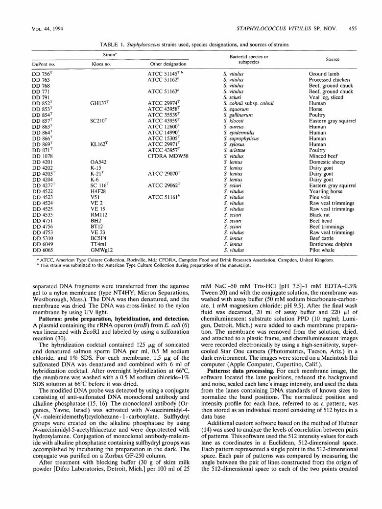
T A B L E 1. Staphylococcus strains used, species designations, and sources of strains
Strain" Bacterial species or Source DuPont no. Kloos no. Other designation subspecies
Source
DD 756T A T C C 51145™ S. v i t u l u s Ground lamb DD 763 A T C C 51162" S. v i t u l u s Processed chicken DD 768 S. vitulus Beef, ground chuck DD 771 A T C C 51163" S. v i t u l u s Beef, ground chuck D D 791 S. sciuri Veal leg, sliced DD 852T GH137 T ATCC 29974T S. c o h n i i subsp. c o h n i i Human D D 853T A T C C 43958T S. e q u o r u m Horse DD 854T A T C C 35539T S. g a l l i n a r u m Poultry DD 857T SC210T A T C C 43959T S. k l o o s i i Eastern gray squirrel DD 8631 ATCC 12600T S. aureus Human DD 864T A T C C 14990T S. epidermidis Human DD 866T ATCC 15305T S. saprophyticus Human DD 869T KL162 T A T C C 29971T S. xylosus Human DD 871T ATCC 43957T S. arlettae Poultry DD 1078 C F D R A MDW58 S. vitulus Minced beef DD 4201 OA542 S. lentus Domestic sheep D D 4202 K-15 S. lentus Dairy goat D D 4203T K-21 T ATCC 29070T S. l e n t u s Dairy goat DD 4204 K-6 S. lentus Dairy goat DD 4277T SC 116T A T C C 29062T S. sciuri Eastern gray squirrel DD 4522 H4F28 S. vitulus Yearling horse DD 4523 V51 A T C C 51161" S. v i t u l u s Pine vole DD 4524 V E 2 S. vitulus Raw veal trimmings DD 4525 V E 15 S. vitulus Raw veal trimmings DD 4535 RM112 S. sciuri Black rat D D 4751 BH2 S. sciuri Beef head DD 4756 BT12 S. sciuri Beef trimmings DD 4753 V E 23 S. vitulus Raw veal trimmings DD 5310 BC5F4 S. l e n t u s Beef cattle D D 6049 TT4ml S. lentus Bottlenose dolphin DD 6065 GMWgl2 S. vitulus Pilot whale
" A T C C , American Type Culture Collection, Rockville, Md.; C F D R A , Campden Food and Drink Research Association, Campden, United Kingdom. h This strain was submitted to the American Type Culture Collection during preparation of the manuscript.
separated D N A fragments were transferred from the agarose gel to a nylon membrane (type N T 4 H Y ; Micron Separations, Westborough, Mass.). The D N A was then denatured, and the membrane was dried. The D N A was cross-linked to the nylon membrane by using U V light.
Patterns: probe preparation, hybridization, and detection. A plasmid containing the r R N A Operon ( r r n B ) from E . c o l i (6) was linearized with E c o R l and labeled by using a sulfonation reaction (30).
The hybridization cocktail contained 125 |mg of sonicated and denatured salmon sperm D N A per ml, 0.5 M sodium Chloride, and 1% SDS. For each membrane, 1.5 fxg of the sulfonated D N A was denatured and combined with 6 ml of hybridization cocktail. After overnight hybridization at 66°C, the membrane was washed with a 0.5 M sodium chloride-1% SDS Solution at 66°C before it was dried.
The modified D N A probe was detected by using a conjugate consisting of anti-sulfonated D N A monoclonal antibody and alkaline Phosphatase (15, 16). The monoclonal antibody (Or-genics, Yavne, Israel) was activated with N-succinimidyl-4-( N - maleimidemethyl)cyclohexane -1 - carboxylate. Sulfhydryl groups were created on the alkaline Phosphatase by using A^succinimidyl-5-acetylthiacetate and were deprotected with hydroxylamine. Conjugation of monoclonal antibody-maleim-ide with alkaline Phosphatase containing sulfhydryl groups was accomplished by incubating the preparation in the dark. The conjugate was purified on a Zorbax GF-250 column.
After treatment with blocking buffer (30 g of skim milk powder [Difco Laboratories, Detroit, Mich.] per 100 ml of 25
m M NaCl-50 m M Tr is -HCl [pH 7.5]-l m M E D T A - 0 . 3 % Tween 20) and with the conjugate Solution, the membrane was washed with assay buffer (50 m M sodium bicarbonate-carbon-ate, 1 m M magnesium Chloride; p H 9.5). After the final wash fluid was decanted, 20 ml of assay buffer and 220 juul of chemiluminescent Substrate Solution P P D (10 mg/ml; Lumigen, Detroit, Mich.) were added to each membrane preparation. The membrane was removed from the Solution, dried, and attached to a plastic frame, and chemiluminescent images were recorded electronically by using a high-sensitivity, super-cooled Star One camera (Photometrics, Tucson, Ariz .) in a dark environment. The images were stored on a Macintosh Ilci Computer (Apple Computer, Cupertino, Calif.).
Patterns: data processing. For each membrane image, the Software located the lane positions, reduced the background and noise, scaled each lane's image intensity, and used the data from the lanes containing D N A Standards of known sizes to normalize the band positions. The normalized position and intensity profile for each lane, referred to as a pattern, was then stored as an individual record consisting of 512 bytes in a data base.
Additional custom Software based on the method of Hubner (14) was used to analyze the levels of correlation between pairs of patterns. This Software used the 512 intensity values for each lane as coordinates in a Euclidean, 512-dimensional Space. Each pattern represented a Single point in the 512-dimensional space. Each pair of patterns was compared by measuring the angle between the pair of lines constructed from the origin of the 512-dimensional Space to each of the two points created
456 WEBSTER ET A L . INT. J . SYST. BACTKRIOI...
T A B L E 2. Results of hybridization of staphylococcal DNAs with [me//?y/-3H]thymidine-labeled DNAs
Species or subspecies
% Relative binding with labeled DNA from:
Pattern type"
Strain D D 756 r DD 771 DD 4525 DD 4523
55°C 70°C 55°C 70°C 55°C 70°C 55°C 70°C
S. vitulus dd 756 DD 756T 100 100 81 84 74 81 81 79 dd 756 DD 4753 87 93 97 93 78 84 76 80 dd 756 DD 6065 74 82 81 88 NT* NT NT NT dd 771 DD 771 74 77 100 100 86 81 86 79 dd 771 DD 1078 76 76 84 84 88 84 80 81 dd 4522 DD 4522 75 75 88 81 88 83 85 84 dd 4522 DD 4525 74 83 87 83 100 100 84 88 dd 4523 DD 4523 79 76 85 81 81 82 100 100
S. sciuri DD 4277T 46 10 53 14 NT NT NT NT 5. l e n t u s DD 4 2 0 3 T 38 7 42 15 NT NT NT NT S. saprophyticus DD 866T 14 7 25 9 NT NT NT NT S. c o h n i i subsp. c o h n i i D D 852T 19 7 28 9 NT NT NT NT-S. xylosus DD 869T 15 5 27 9 NT NT NT NT S. kloosii DD 857T 14 5 29 8 NT NT NT NT S. e q u o r u m DD 853T 16 6 25 9 NT NT NT NT S. arlettae DD 871T 17 8 28 10 NT NT NT NT S. g a l l i n a r u m DD 854T 13 8 14 5 NT NT NT NT
" Each pattern type was named after the strain with the lowest DuPont Company number that exhibited that pattern. h NT, not tested.
from the two patterns being compared. Similar patterns had angles approaching zero degrees and cosines approaching unity (one). While the correlation was defined as the cosine of the angle, in this work we routinely used the correlation value squared.
Cell wall analysis. Staphylococci were grown to the station-ary phase in a medium containing (per liter of distilled water) 10 g of tryptone, 5 g of yeast extract, 5 g of glucose, and 5 g of NaCl (pH 7.3). The cells were harvested by centrifugation at 20,000 X g for 20 min at 4°C and were disintegrated by shaking the preparation with glass beads. Cell walls were purified with 4% SDS by using the procedures of Glauner et al. (11) to determine peptidoglycan composition and teichoic acids. For total amino acid analysis cell walls purified with SDS were hydrolyzed with 4 N H C l for 16 h at 1Ö0°C. The levels of amino acids and amino sugars were determined by using a model LC6001 amino acid analyzer (Biotronik, Maintal, Germany). The peptidoglycan type was determined on the basis of the molar ratio of glutamic acid to L-lysine to L-serine to L-alanine to glycine (28). Teichoic acids were extracted from cell walls purified with SDS by the method of Kaya et al. (17). The teichoic acids were purified by ion-exchange chromatography on a column (1.6 by 17 cm) filled with DEAE-Sephacel (Pharmacia, Uppsala, Sweden), using a flow rate of 20 ml/h, a base mobile phase consisting of 0.01 M Tr i s -HCl (pH 7.0), and a linear 0 to 1 M NaCl gradient. The teichoic acids were hydrolyzed with 60% (wt/vol) hydrofluoric acid for 16 h at 0°C (3). Nonphosphorylated teichoic acid fragments were separated on a column (1.5 by 90 cm) filled with Bio-Gel P-2 (Pharmacia) by using a distilled water mobile phase and a flow rate of 8 ml/h. The sugars in the fractions were determined by gas-liquid chromatography, and glycerol was detected enzy-matically by the method of Bergmeyer (4). The sugars were completely hydrolyzed with 2 N H C l for 3 h at 100°C (3). The hydrolysates were derivatized as described by Albersheim et al. (1) and were subjected to gas-liquid chromatography by using a Packard model G C 438 gas Chromatograph equipped with a flame ionization detector (Packard Instrument Co., Meriden, Conn.) and a column (2 by 1,000 mm) filled with 3% SP 2340 on 100/200 Supelcoport (Supelco, Bellefonte, Pa.). The gas
liquid chromatography column was equilibrated at 140°C and was kept at that temperature for the first 2 min of each analysis. The temperature was increased at a rate of 6°C/min until it reached 270°C, where it was kept for an additional 3 min. Phosphate levels were determined bv the method of Arnes (2).
DNA base composition. The guanine-plus-cytosine content of D N A was determined by A . G . Steigerwalt in the laboratory of D . J. Brenner, Centers for Disease Control, Atlanta, Ga., by using the thermal denaturation method of Marmur and Doty (24).
RESULTS AND DISCUSSION
DNA-DNA hybridization. The D N A relationships among S t a p h y l o c o c c u s v i t u l u s strains that reprcsented diflferent pattern types and between S. v i t u l u s and other S t a p h y l o c o c c u s species are shown in Table 2.
In D N A - D N A hybridization reactions performed under optimal (55°C) and stringent (70°C) conditions, S. v i t u l u s strains exhibited relatively high levels of D N A similarity (82% ± 6% at 55°C and 82% ± 4% at 70°C [means ± Standard deviations]). No significant differences in levels of D N A similarity were found between S. v i t u l u s strains that represented different pattern types. However, the levels of D N A related-ness between the new species and other members of the S. s c i u r i species group, including S. l e n t u s and S. s c i u r i , were significantly lower. For S. l e n t u s , the levels of D N A similarity were 40% ± 2% at 55°C and 11% ± 4% at 70°C. For S. s c i u r i , the levels of D N A similarity were 50% ± 4% at 55°C and 12% ± 2% at 70°C. The levels of D N A relatedness between S. v i t u l u s and other novobiocin-resistant S t a p h y l o c o c c u s species that did not belong to the S. s c i u r i species group were even lower.
Patterns. The raw image from a typical agarose gel gener-ated by using enzyme-triggered chemiluminescence and cap-tured with a customized charge-coupled device camera is shown in Fig. 1. The lanes that are not labeled with a species name were used to separate D N A Standards whose sizes were known. The positions of the Standard bands were used to
V O L . 44, 1994 S T A P H Y L O C O C C U S VITULUS S P . N O V . 457
t 1 1 A M i t • '4 6'. v i t u l u s
i i m S. s c i u r i
1 i
I § t i i i » S. s c i u r i
1 I i ' I i l S. s c i u r i
• l * i
1 U II • S. s c i u r i
I t
1 a i
# ^ 5. s c i u r i
1 • 41 • 1 i : 1
1.8 5.0 3.0 2.3 1.4 kbp
FIG. 1. Electronically recorded image of a membrane containing EcoRl fragments hybridized with a plasmid containing the miß rRNA Operon of E. coli.
correct for both lane-to-lane and gel-to-gel variations in band position.
A processcd image of some of the patterns determined during this study is shown in Fig. 2. The displayed width of each lane pattern was arbitrarily chosen for Fig. 2. The patterns were arranged to maximize the squared correlation values for adjacenl patterns in the display. This sorting re-vealed that the S. v i t u l u s patterns were most similar to the S. lentus and S. s c i u r i patterns and were different from the patterns obtained with the more than 200 other species in our data base. 5. v i t u l u s , S. lentus, and S. s c i u r i could be differen-liated by their patterns cither by visually comparing the patterns or by Computer analysis.
Visually, the qualitative differences among the patterns of the thrce species were obvious. The S. v i t u l u s patterns revealed sets of conserved, frequently occurring restriction fragments that were found in different strains of S. v i t u l u s but were not conserved as a group in strains of other species.
Patterns that were identical (within the reproducibility of the method) were considered members of the same pattern type. Multiple strains belonging to the same species could have the same pattern type. Each pattern type was named after the lowest-numbered DuPont strain that produced that type of pattern. The 11 S. v i t u l u s strains examined produced four distinguishable types of patterns, dd 756, dd 771, dd 4522, and dd 4523.
Computer analysis provided a way to measure the levels of similarity of the patterns by using the squared correlation values obtained from the 512-dimensional space. A l l of the S. v i t u l u s patterns of the same pattern type (for example, dd 756) had squared correlation values with each other that were greater than 0.85. When each of the four S. v i t u l u s pattern types with was correlated with its nearest S. v i t u l u s neighbors in Fig. 2, the squared correlation values ranged from 0.71 to 0.79. No S. v i t u l u s pattern had a squared correlation value with any S. lentus pattern that was greater than 0.56 or a squared correlation value with any S. s c i u r i pattern that was greater than 0.46.
Figures 3 and 4 show two-dimensional representations of the 512-dimensional Space. The patterns of more than 5,500 strains, including members of approximately 200 species, were
6049 (I)
5310 (1)
1201 (1)
1201 (I)
1203 (I)
I078 (I)
77I (1)
1522 (I)
1525 (!)
763 (!)
756 (1)
768 (1)
1753 (!)
1521 (!)
6065 (l)
1523 (!)
1751 (1)
791 (I)
1535 (I)
1756 (I)
1277 (I)
lOKBp lKUp
II
S.lentus
S.lentus
S. lentus
S.lentus
S.lentus
S.vitulus
S.vitulus
S.vitulus
S.vitulus
S.vitulus
S.vitulus
S.vitulus
S. vitulus
S.vitulus
S.vitulus
S.vitulus
S.sciuri
S.sciuri
S.sciuri
S.sciuri
S.sciuri
FIG. 2. Patterns of E c o R l fragments containing rRNA sequences derived from image data. The image data for each lane were processed to normalize band positions relative to Standards, to reduce background, and to scale the band intensity. The pattern numbers are shown on the left, and the species names are shown on the right.
each defined by a Single labeled point in Fig. 3 and 4. In each figure, the squared correlation values with the two reference patterns defined the two dimensions. For Fig. 3 we used the nomenclatural type strain of S. lentus, D D 4203, and the nomenclatural type strain of S. v i t u l u s , D D 756, as the reference strains. For Fig. 4 we used the nomenclatural type strain of S. v i t u l u s , D D 756, and the nomenclatural type strain of S. s c i u r i , D D 4277, as the reference strains. In these two-dimensional representations, the visual distinetions between the unique S, v i t u l u s Cluster and the other species tested are relatively clear. Clusters of points surrounding the reference strains are clearly identified by those reference strains.
Using the complete 512-dimensional-space analysis and the corresponding correlation values, we could clearly distinguish between S. v i t u l u s and all of the other species tested. A n organism could be identified as a member of S. v i t u l u s by the squared correlation value of its pattern obtained from the 512-dimensional-space analysis, even if the pattern did not exactly match one of the pattern types associated with S. v i t u l u s . In this study, squared pattern correlation values greater than 0.71 appeared to be strong evidence that strains belonged to the same species.
Cell wall peptidoglycan and teichoic acid. We also determined the peptidoglycan compositions of S. v i t u l u s D D 756 1 (T = type strain), D D 771, and D D 4523, S. lentus D D 4202, S. s c i u r i D D 4277 1, and Staphylococcus aureus D D 863 T . The peptidoglycan of S. v i t u l u s strains is type L-Lys-L-Ala-Gly 3 _ 3 4
458 WEBSTER ET A L .
FIG. 3. Clustering of patterns as determined by transforming each pattern into a point in 512-dimensional Space and depicting the points in a two-dimensional representation by using S. lentus DD 4203T and S. v i t u l u s DD 756T to define the two dimensions. The strain number is placed in the appropriate location for each point on the figure. Data for approximately 5,550 strains belonging to 200 species are shown.
peptidoglycan. This peptidoglycan type is also present in S. lentus and S. s c i u r i (27).
The teichoic acids of S. v i t u l u s are based on glycerol. Strain D D 756 T has a glycerol teichoic acid with an integrated Af-acetylglucosamine 1-phosphate; strain D D 771 has a glycerol teichoic acid substituted at a high level with N-acetylgalac-tosamine; and strain D D 4523 has a glycerol teichoic acid substituted at a low level with Af-acetylglucosamine.
DNA base composition. The guanine-plus-cytosine content of S. v i t u l u s D D 756 T D N A is 34 mol%; the D N A guanine-plus-cytosine content of strains D D 771 and D D 4523 is 35 mol%; and the S. s c i u r i and S. lentus D N A guanine-plus-cytosine Contents are between 30 and 36 mol% (20).
Description of Staphylococcus vitulus sp. nov. Staphylococcus v i t u l u s (vit'u.lus. N . L . n. v i t u l u s , veal). The description of S. v i t u l u s below is based on the results of a study of 11 strains.
A total of 10 of the 11 strains produce colonies that are less than 3 mm in diameter when the organisms are grown aerobi-
FIG. 4. Clustering of patterns as determined by transforming each pattern into a point in 512-dimensional space and depicting the points in a two-dimensional representation by using S. v i t u l u s DD 756T and S. sciuri DD 4277T to define the two dimensions. The strain number is placed in the appropriate location for each point on the figure. Data for approximately 5,550 strains belonging to 200 species are shown.
INT. J . SYST. BACTERIOL.
T A B L E 3. Variable characteristics of S. v i t u l u s strains
Characteristic No. of strains positive/ no. of strains tested
% of strains positive
Colony diam. of ^3 mm on P agar 10/11 91 Colony pigment 10/11 91 ß-Glucosidase activity3 9/11" 82 Nitrate reduction 10/11 91 Esculin hydrolysis 7/11 (1/1 l ) c 64 (9)c
Clumping factor: Standard slide test 0/11 (2/11) 0(18) Staph Latex kit (Remel)'' 9/11(2/11) 82(18)
Acid produced aerobically from: D-Trehalose'' 0/11(4/11) 0(36) D-Xylose 7/11(1/11) 64 (9) D-Cellobiose 2/11 (1/11) 18(9) Salicin 1/11(2/11) 9(18) D-Ribose 9/11 (2/11) 82(18)
a Activity determined by the API STAPH-IDENT System. b Most strains that produced a dd 756 pattern were positive for ß-glucosidase
activity and did not produce acid from mannose or trehalose (the only exception was weak acid production from trehalose by strain D D 6065).
c Weak positive reactions were not included as positive reactions. The values in parentheses are the number of strains that exhibited weak positive reactions/ total number of strains tested and the percentage of strains that exhibited a weak positive reaction.
d The Staph Latex kit detected both clumping factor and protein A.
cally on P agar (18) at 35°C. Colonies on P agar are usually raised with ulcerated, irregulär centers, are opaque, and often have sectored or irregulär edges; 10 of the 11 strains studied produce cream to yellow pigmentation, and 1 strain produces unpigmented colonies. Colonies grown on tryptic soy agar are much larger (8 to 12 mm in diameter) and not as irregulär as colonies grown on P agar.
Colony growth is reduced or inhibited at 40°C, and none of the strains grows at 45°C. The cells are not motile, and the organisms do not grow anaerobically in thioglycolate semisolid agar. However, they exhibit minimal growth on sheep blood agar and tryptic soy agar plates incubated at 35°C in anaerobic jars. The cells are gram-positive cocci, do not form spores, and occur singly, in pairs, in tetrads, and in Clusters.
A l l strains produce zones of greening on bovine blood agar. A l l strains are negative for staphylocoagulase, thermonuclease, alkaline Phosphatase, pyrrolidonyl arylamidase, Ornithine de-carboxylase, urease, ß-glucuronidase, and ß-galactosidase ac-tivities, arginine utilization, and acetoin production. A l l strains are negative for aerobic production of acid from the following Substrates: mannose, lactose, galactose, melezitose, xylitol, rhamnose, turanose, arabinose, maitose, A^acetyIglucosamine, and raffinose. A l l strains are negative for anaerobic production of acid from glucose and mannitol. A l l strains are positive for catalase activity, modified oxidase activity, and aerobic production of acid from the following Substrates: mannitol, glycerol, sucrose, and fructose. The variable characteristics of S. v i t u l u s are shown in Table 3.
The major A P I profiles are 4200 (six strains) and 4700 (three strains). S T A P H Trac profiles exhibit Variation with respect to acid production from maitose, raffinose, xylose, and trehalose, nitrate reduction, alkaline Phosphatase, and acetoin production, indicating that there is no predominant profile.
E c o R l fragments containing rRNA sequences. The patterns of all S. v i t u l u s strains Cluster on the basis of similarity and are distinguishable from the patterns of all other bacteria exam-ined, including all previously described Staphylococcus species. The species can be recognized by a high level of correlation with the pattern types described in this paper.
V O L . 44, 1994 S T A P H Y L O C O C C U S VITULUS SP. NOV. 459
T A B L E 4. Characteristics that differentiate S. v i t u l u s from other novobiocin-resistant, oxidase-positive Staphylococcus species"
Characteristic S. vitulus S. sciuri S. lentus
(±) d
Alkaline Phosphatase activity - + (±) ß-Glucosidase activity d + + Esculin hydrolysis d + + Acid produced aerobically from:
D-Trehalose (d) + + D-Mannose - (d) (+) D -Tu ran ose - (±) (±) D-Xylose (d) (d) (±) D-Cellobiose (d) + + L-Arabinose - d d Maltose - (d) d /V-acetyl-D-glucosamine - d d Raffinose - - +
" +, 90% or more of the strains are positive; ± , 90% or more of the strains are weakly positive; - , 0 to 10% of the strains are positive; d, 11 to 89% of the strains are positive. Parentheses indicate that a response is delayed.
h The Staph Latex kit detected both clumping factor and protein A. S. vitulus produces greening during this test.
Antibiotic susceptibilities. As determined by agar disk dif-fusion tests, all strains of S. v i t u l u s are susceptible to furazoli-done, penicillin, erythromycin, and Clindamycin. A l l strains are resistant to bacitracin and novobiocin. Three strains that produce pattern type dd 756 ( D D 756 T , D D 4524, and D D 4753) are resistant to tetracycline.
Description of the type strain. The type strain of S. v i t u l u s is A T C C 51145 (= D D 756). It has all of the characteristics of the species described above. In addition, it has the properties described below.
Cells are spherical (diameter, 0.9 to 1.1 jxm) and occur singly, in pairs, in tetrads, and in Clusters.
P agar colonies are raised with ulcerated, irregulär centers, irregulär edges, and cream pigmentation; the colonies are approximately 2 mm in diameter. Colonies on tryptic soy agar are raised with wide, depressed centers; each colony has a slightly irregulär edge, and is pigmented, with a pinkisn center blending to a cream edge. These colonies are approximately 10 to 11 mm in diameter. ß-Glucosidase is produced. Nitrates are reduced to nitrites. Esculin is not hydrolyzed. The Standard slide test is negative for clumping factor. The Staph Latex kit reaction is strongly positive.
A c i d is produced aerobically from D-xylose and D-ribose. No acid is produced aerobically from D-cellobiose, salicin, and D-trehalose.
The type strain is resistant to tetracycline. The guanine-plus-cytosine content of the D N A is 34 mol%. The type strain produces a type dd 756 pattern when it is
studied by using the methods described in this paper. Distinguishing characteristics. S. v i t u l u s can be distin
guished from other bacterial species by examining the pattern of restriction fragments containing r R N A sequences that results when the methods described in this paper are used. S. v i t u l u s can also be distinguished by its novobiocin resistance, positive oxidase reaction, strong positive Staph Latex kit reaction, absence of alkaline Phosphatase activity, and lack of acid production from L-arabinose, maitose, N-acetylglu-cosamine, D-mannose, and raffinose. The major phenotypic
features that are useful for distinguishing S. v i t u l u s from other novobiocin-resistant, oxidase-positive Staphylococcus species are summarized in Table 4.
ACKNOWLEDGMENTS
We thank Bill Bowen and V. Thayer (National Marine Fisheries, Beaufort, N.C.) for obtaining bacterial specimens from dolphins and a pilot whale and Herman Berkhoff (College of Veterinary Medicine, North Carolina State University, Raleigh) for assisting in obtaining bacterial specimens from dairy cattle and horses. We also thank Joe Neubauer and Channeary lern (DuPont Company) for technical assistance, Lucy Ganfield and Dipti Schoop (DuPont Company) for preparing the probe and conjugate, and Charles Robertson and Robert Martin for modifying the Star One camera used.
REFERENCES 1. Albersheim, J. W., D. J. Nevins, P. D. English, and A. Karr. 1967.
A method for the analysis of sugars in plant cell wall Polysaccharide by gas liquid chromatography. Carbohydr. Res. 5:340-345.
2. Arnes, B. N. 1966. Assay of inorganic phosphate, total phosphate and phosphatases. Methods Enzymol. 8:115-118.
3. Anderson, A. J., R. S. Green, and A. R. Archibald. 1977. Specific determination of ribitol teichoic acid in whole bacteria and isolated walls of B a c i l l u s s u b t i l i s W23. Carbohydr. Res. 57:C7-C10.
4. Bergmeyer, H. U. 1974. Methoden der enzymatischen Analyse. Verlag Chemie, Weinheim, Germany.
5. Brenner, D. J., G. R. Fanning, A. Rake, and K. E. Johnson. 1969. A batch procedure for thermal elution of D N A from hydroxyap-atite. Anal. Biochem. 28:447-459.
6. Brosius, J., A. Ullrich, M. A. Raker, A. Gray, T. J. Dull, R. R. Guteil, and H. F. Noller. 1981. Construction and fine mapping of recombinant plasmids containing the r r n B ribosomal RNA Operon of E. c o l i . Plasmid 6:112-118.
7. Chesneau, O., A. Morvan, F. Grimont, H. Labischinski, and N. El Solh. 1993. Staphylococcus p a s t e u r i sp. nov., isolated from human, animal, and food specimens. Int. J. Syst. Bacteriol. 43:237-244.
8. De Buyser, M.-L., A. Morvan, S. Aubert, F. Dilasser, and N. El Solh. 1992. Evaluation of ribosomal R N A gene probe for the identification of species and subspecies within the genus Staphyl o c o c c u s . J. Gen. Microbiol. 138:889-899.
9. De Buyser, M.-L., A. Morvan, F. Grimont, and N. El Solh. 1989. Characterization of Staphylococcus species by ribosomal RNA gene restriction patterns. J. Gen. Microbiol. 135:989-999.
10. Faller, A. H., and K. H. Schleifer. 1981. Modified oxidase and benzidine tests for the Separation of staphylococci from micro-cocci. J. Clin. Microbiol. 13:1031-1035.
11. Glauner, B., J. V. Holtje, and U. Schwarz. 1988. The composition of the murein of Escherichia c o l i . J. Biol. Chem. 263:10088-10095.
12. Grimont, F., and P. A. D. Grimont. 1986. Ribosomal ribonucleic acid gene restriction patterns as potential taxonomic tools. Ann. Inst. Pasteur/Microbiol. (Paris) 137B: 165-175.
13. Hebert, G. A., C. G. Crowder, G. A. Hancock, W. R. Jarvis, and C. Thornsberry. 1988. Characteristics of coagulase-negative staphylococci that help differentiate these species and other members of the family M i c r o c o c c a c e a e . J. Clin. Microbiol. 26:1939-1949.
14. Hubner, R. December 1989. U.S. patent 4,885,697. 15. Ishikawa, E., M. Imagawa, S. Hashida, S. Yoshitake, Y. Hmaguchi,
and T. Ueno. 1983. Enzyme-labeling of antibodies and their fragments for enzyme immunoassay and immunohistochemical staining. J. Immunoassay 4:209-327.
16. Ishikawa, E,, Y. Yamada, S. Yoshitake, and H. Kawaguchi. 1981. A more stable maleimide, N-(4-carboxycyclohexylmethyl) maleimide for enzyme labeling, p. 90-105. In E. Ishikawa, T. Kawai, and K. Miyai (ed.), Enzyme immunoassay. Igaku-Shoin, Tokyo.
17. Kaya, S., Y. Araki, and E. Ito. 1985. Characterization of a novel linkage unit between ribitol teichoic acid and peptidoglycan in Listeria monocytogenes cell walls. Eur. J. Biochem. 146:517-522.
18. Kloos, W. E., and D. W. Lambe, Jr. 1991. Staphylococcus, p. 222-237. I n A. Balows, W. J. Hausler, Jr., K. L. Herrmann, H. D. Isenberg, and H . J. Shadomy (ed.), Manual of clinical microbiol-ogy, 5th ed. American Society for Microbiology, Washington, D.C.
19. Kloos, W. E., and K. H. Schleifer. 1975. Simplified scheme for
Colony size of >6 mm on P agar - + Anaerobic growth - (+) Staph Latex kit reaction + d
(Remel)* Hemolysis of bovine blood ± c ' -Alkaline Phosphatase activity - 4-ß-Glucosidase activity d + Esculin hydrolysis d + Acid produced aerobically from:
D-Trehalose (d) + D-Mannose - (d) D -Tu ran ose - (±) D-Xylose (d) (d) D-Cellobiose (d) + L-Arabinose - d Maltose - (d) yV-acetyl-D-glucosamine - d Raffinose - -
460 WEBSTER ET A L . INT. J . SYST. BACTERIOL.
routine identification of human S t a p h y l o c o c c u s species. J. Clin. Microbiol. 1:82-88.
20. Kloos, W. E., and K. H. Schleifer. 1986. Genus IV. S t a p h y l o c o c c u s , p. 1013-1035. I n P. H . A . Sneath, N . S. Mair, M . E . Sharpe, and J. G. Holt (ed.), Bergey's manual of systematic bacteriology, vol. 2. Williams and Wilkins, Baltimore.
21. Kloos, W. E., K. H. Schleifer, and R. F. Smith. 1976. Character-ization of S t a p h y l o c o c c u s s c i u r i sp. nov. and its subspecies. Int. J. Syst. Bacteriol. 26:22-37.
22. Kloos, W. E., T. G. Tornabene, and K. H. Schleifer. 1974. Isolation and characterization of micrococci from human skin, including two new species, M i c r o c o c c u s lylae and M i c r o c o c c u s k r i s t i n a e . Int. J. Syst. Bacteriol. 24:79-101.
23. Kloos, W. E., and J. F. Wolfshohl. 1979. Evidence for deoxyribo-nucleotide sequence divergence between staphylococci living on human and other primate skin. Curr. Microbiol. 3:167-172.
24. Marmur, J., and P. Doty. 1962. Determination of the base composition of deoxyribonucleic acid from its thermal denatur-ation temperature. J. Mol. Biol. 4:109-118.
25. Moeller, V. 1955. Simplified tests for some amino acid decarboxy-lases and for the arginine dihydrolase System. Acta Pathol. Micro
biol. Scand. 36:158-172. 26. Razin, S., M. Gross, M . Wormser, Y. Pollack, and G. Glasser.
1984. Detection of mycoplasmas infecting cell cultures by D N A hybridization. In Vitro (Rockville) 20:404^08.
27. Schleifer, K. H. 1986. Taxonomy of coagulase-negative staphylococci, p. 11-26. I n P. A . Mardh and K. H . Schleifer (ed.), Coagulase-negative staphylococci. Almqvist and Wiksell International, Stockholm.
28. Schleifer, K. H., and O. Kandier. 1972. Peptidoglycan types of bacterial cell walls and their taxonomic implications. Bacteriol. Rev. 36:407-477.
29. Thomson-Carter, F. M . , P. E. Carter, and T. H. Pennington. 1989. Differentiation of staphylococcal species and strains by ribosomal R N A gene restriction patterns. J. Gen. Microbiol. 135:2093-2097.
30. Verdlov, E. D., G. S. Monastyrakya, L. I. Guskova, T. L. Levitan, V. I. Sheichenko, and E. I. Budowsky. 1974. Modifikation of cytidine residues with a bisulfite-O-methylhydroxylamine mixture. Biochim. Biophys. Acta 340:153-165.
31. Webster, J. A. April 1983. European Patent Office Application 82305061.2.