Embed Size (px)
Citation preview
Interoperability between Anatomic
Pathology Laboratory Information Systems
and Digital Pathology Systems
Authors: Jesus Ellin (Yuma Regional Medical Center), Anna Haskvitz (Sunquest Information Systems, Inc),
Preeti Premraj (Sunquest Information Systems, Inc), Kevin Shields (Genetix Limited), Matthew Smith
(Aurora Interactive, Ltd.), Curtis Stratman (Omnyx, LLC), Mark Wrenn (Aperio Technologies, Inc)
Interoperability between APLIS and Digital Pathology Systems – Page 1
Digital Pathology Association | 2424 American Lane, Madison WI 53704 | Tel: 608.441.8600 | Fax: 608.443.2474 Web: http://www.digitalpathologyassociation.org/
Abstract This white paper offers an overview of the current state of interoperability between Anatomical Pathology
Laboratory Information Systems (APLIS’s) and Digital Pathology Systems (DPS’s). This overview also
includes a brief discussion of future work that will impact interoperability.
Both systems rely on data from the other to efficiently deliver full digital imaging functionality to the
healthcare provider. Anatomic Pathology (AP) departments and patients will benefit most from imaging
workflow when there is a high degree of integration of Digital Pathology information within AP workflow.
Implementations of such data sharing already exist via interfaces and standard communication protocols
between APLIS’s and DPS’s, and work continues on these interface standards to improve the degree to
which these systems can be used together.
The current state of interoperability provides Pathologists with access to images and image analysis data
from within the APLIS or the DPS. This information is then available to the Patient Report.
Interoperability between APLIS and Digital Pathology Systems – Page 2
Digital Pathology Association | 2424 American Lane, Madison WI 53704 | Tel: 608.441.8600 | Fax: 608.443.2474 Web: http://www.digitalpathologyassociation.org/
Contents Abstract ........................................................................................................................................... 1
Introduction ..................................................................................................................................... 3
Benefits of Digital Pathology ........................................................................................................... 4
Geographic Dispersal of Pathologists .......................................................................................... 4
Case Transport ............................................................................................................................. 4
Case Review ................................................................................................................................. 4
Additional Studies........................................................................................................................ 4
Diagnosis Entry ............................................................................................................................ 5
Quality Control ............................................................................................................................ 5
Benefits of Interoperability ............................................................................................................. 5
Digital Pathology within the AP Lab Workflow ............................................................................... 5
Integrated Imaging Workflow ..................................................................................................... 6
Examples of Existing Interoperable APLIS/DPS Implementations ................................................... 9
Metadata Exchange ..................................................................................................................... 9
Encoding Metadata in the Slide Barcode .............................................................................. 10
Sending Metadata between the APLIS and DPS .................................................................... 11
APLIS-Integrated Viewing .......................................................................................................... 12
Communications and Collaboration Networks ............................................................................. 14
Additional Considerations ............................................................................................................. 15
Barcodes .................................................................................................................................... 15
Compatibility vs. Interoperability .............................................................................................. 15
Future Directions in Interoperability ............................................................................................. 15
Scanning speeds ........................................................................................................................ 15
Digital Slide Viewing Tools and Clinical Decision Support ......................................................... 16
Backend Digital Slide Storage .................................................................................................... 16
Multi-Site Communications and Collaboration Applications .................................................... 16
Appendix A – Relevant Industry Standards ................................................................................... 17
Interoperability between APLIS and Digital Pathology Systems – Page 3
Digital Pathology Association | 2424 American Lane, Madison WI 53704 | Tel: 608.441.8600 | Fax: 608.443.2474 Web: http://www.digitalpathologyassociation.org/
Introduction Digital Pathology is much more than just the capturing of a digital image of a glass slide. It is a term that describes the creation, viewing, management, sharing, analysis and interpretation of these images and
includes management and workflow considerations unique to a digital imaging environment.
Some DPS vendors supply all of the functionality described above (end-to-end providers) while others
provide individual components intended to be combined with those of other vendors to create a complete
DPS (best-in-breed providers).
The process of digitizing glass slides (the fundamental building block of DPS’s) has improved dramatically
over the last 15 to 20 years. Several vendors now offer scanning products that support digitizing entire
glass slides – often called Whole Slide Images (WSI’s), digital slides or virtual slides and scan times
continue to improve.
DPS’s provide pathologists and AP laboratories with new tools including:
Diagnostic assistance via the quantitative analysis of tissue samples
Virtual access to samples and subspecialty experts in remote locations
The creation of digital libraries for reference or education
Data-mining of digital slide databases seeking similarities and differences in tissue samples of
various diseases for research
Virtual tumor boards or discussion panels where pathologists share and discuss their cases
Laboratory automation for improved quality control
Easily accessible image archives for future retrieval
Side-by-side viewing of slides for comparison
Precise and accurate measurement tools
Slide annotations for clarity
These kinds of tools are causing rapid growth in the application of DPS’s. Hospitals and laboratories are
looking to Digital Pathology to reduce laboratory expenses, better control quality, improve operational
efficiency, enhance productivity and improve treatment decisions and patient care.
APLIS’s are used by most labs today to manage patient, case, and specimen information. Most pathologists
spend a large part of their work day within the APLIS. It manages their cases, workflows and often their
final diagnostic reports.
Full and efficient utilization of the tools highlighted above cannot be achieved without their integration into
the clinical workflow. Several APLIS vendors and DPS vendors currently support this integration in
various forms and to various degrees.
This whitepaper will describe the impact of DPS’s on laboratory workflow, potential points of APLIS /
DPS data sharing, current approaches to APLIS / DPS data sharing as well as possible future work and the
standards involved.
Interoperability between APLIS and Digital Pathology Systems – Page 4
Digital Pathology Association | 2424 American Lane, Madison WI 53704 | Tel: 608.441.8600 | Fax: 608.443.2474 Web: http://www.digitalpathologyassociation.org/
Benefits of Digital Pathology Before discussing the details of interfacing APLIS’s and DPS’s, it is appropriate to briefly review the
benefits that DPS’s bring to pathology. The tools outlined in the Introduction have a tremendous impact on
several aspects of pathology workflow and collaboration.
Geographic Dispersal of Pathologists
The inherent dependence on physical tissue places many constraints on the workflow. For example, multi-
facility pathology departments must make a trade-off between distributing pathologists to locations near the
labs to reduce case turnaround time and centralizing specialist pathologists away from the labs to facilitate
collaboration.
Digital pathology provides an opportunity to enable the best of both options by eliminating the constraint of
having to transport the glass slides from the lab to the pathologist. Glass slides may be transformed into
digital slides by scanners in the histology lab and the images made immediately available to pathologists at
multiple remote locations.
Case Transport
The virtual delivery of cases eliminates the waiting time slide folders spend in the lab pending pickup, in
transport from the histology lab to the delivery location and in the delivery location until the pathologist
checks for their arrival. Virtual cases also eliminate the need for pathologists to repeatedly check a
delivery location. These together enable a continuous flow in the delivery of cases from the lab to the
pathologist. Delivery of the physical slides is eliminated, resulting in a reduction in shipping and
administration costs and the prevention of lost and/or damaged slides.
Case Review
Digital slides enable capabilities not available with physical slides such as side-by-side viewing of multiple
slides, quantitative image analysis tools, precise and accurate measurements and slide annotations.
The retrieval of prior studies is instantaneous with digital imaging workflow, resulting in improved patient
treatment.
Diagnostic turnaround is improved since digital slides are instantly available without the need for transport.
Access to gross specimen images and/or In Vivo images may also be important in some cases. Several
vendors offer interface capabilities with grossing station imaging systems and PACS hardware in order to
present these images to pathologists in addition to digital slides.
Additional Studies
When additional studies are ordered by the pathologist for a tissue, the case is traditionally put aside and
reviewed again when the results of those studies are available. These studies may either be additional
slides with different chemical stains which the pathologist will review or other procedure types (e.g.
molecular, flow cytometry, electron microscopy, etc.) with the results reviewed and interpreted by another
party. As the studies are received, personnel must currently locate the correct case, check the case for
receipt of all ordered studies, and place the case back into the pathologist’s review queue when complete.
The digital case work list can automate this management of additional studies by placing the case into a
pending status when studies are ordered, automatically matching the studies to the case, and validating the
receipt of all studies to return the case to a complete status. This reduces the manual tracking of studies and
the time personnel need to spend locating cases, as well as reduces administrative time spent on case
management tasks and the risk of unknowingly reviewing an incomplete case.

Interoperability between APLIS and Digital Pathology Systems – Page 5
Digital Pathology Association | 2424 American Lane, Madison WI 53704 | Tel: 608.441.8600 | Fax: 608.443.2474 Web: http://www.digitalpathologyassociation.org/
Diagnosis Entry
Digital slides enable pathologists to associate diagnostic documentation to specific areas of the tissue. This
additional information can be utilized to communicate critical information to the ordering clinician to
support treatment planning. Snapshot images of regions within the digital slide can also be captured for
inclusion in case documentation and reports.
Quality Control
The digitization of slides allows for enhanced opportunities for stain quality control, either offline or prior
to reaching the Pathologist when integrated into the laboratory workflow. Automated analyses can ensure
that stains remain within acceptable limits.
Benefits of Interoperability Interoperability between APLIS’s and DPS’s facilitates the pathologist’s utilization of the DPS tools and
assists in the achievement of the benefits outlined above by automating data exchange between the systems
and providing them within the context of the laboratory workflow. This automation results in a reduction of
manual work, case turnaround time and the likelihood of errors occurring.
Digital Pathology within the AP Lab Workflow Some of the benefits identified so far can be achieved through the creation of digital slides after or in
parallel with the current diagnostic workflow (e.g. QC, education and research). The following diagram
illustrates the creation of digital slides outside the diagnostic workflow.
Other improvements however can only be realized when the pathologist is able to utilize scanned images in
lieu of or in addition to the physical slide, and therefore requires digital slides to be integrated into the
diagnostic workflow1. The following diagram illustrates an integrated workflow.
1 Not yet approved by the FDA for routine surgical cases. For more information refer to the FDA Advisory
Committee: Hematology and Pathology Devices Panel at
http://www.fda.gov/AdvisoryCommittees/CommitteesMeetingMaterials/MedicalDevices/MedicalDevicesA
dvisoryCommittee/HematologyandPathologyDevicesPanel/default.htm.
Interoperability between APLIS and Digital Pathology Systems – Page 6
Digital Pathology Association | 2424 American Lane, Madison WI 53704 | Tel: 608.441.8600 | Fax: 608.443.2474 Web: http://www.digitalpathologyassociation.org/
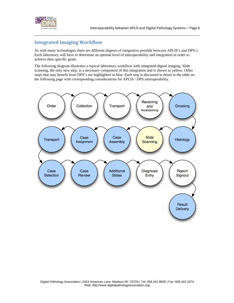
Integrated Imaging Workflow
As with many technologies there are different degrees of integration possible between APLIS’s and DPS’s.
Each laboratory will have to determine an optimal level of interoperability and integration in order to
achieve their specific goals.
The following diagram illustrates a typical laboratory workflow with integrated digital imaging. Slide
scanning, the only new step, is a necessary component of this integration and is shown in yellow. Other
steps that may benefit from DPS’s are highlighted in blue. Each step is discussed in detail in the table on
the following page with corresponding considerations for APLIS / DPS interoperability.
Interoperability between APLIS and Digital Pathology Systems – Page 7
Digital Pathology Association | 2424 American Lane, Madison WI 53704 | Tel: 608.441.8600 | Fax: 608.443.2474 Web: http://www.digitalpathologyassociation.org/
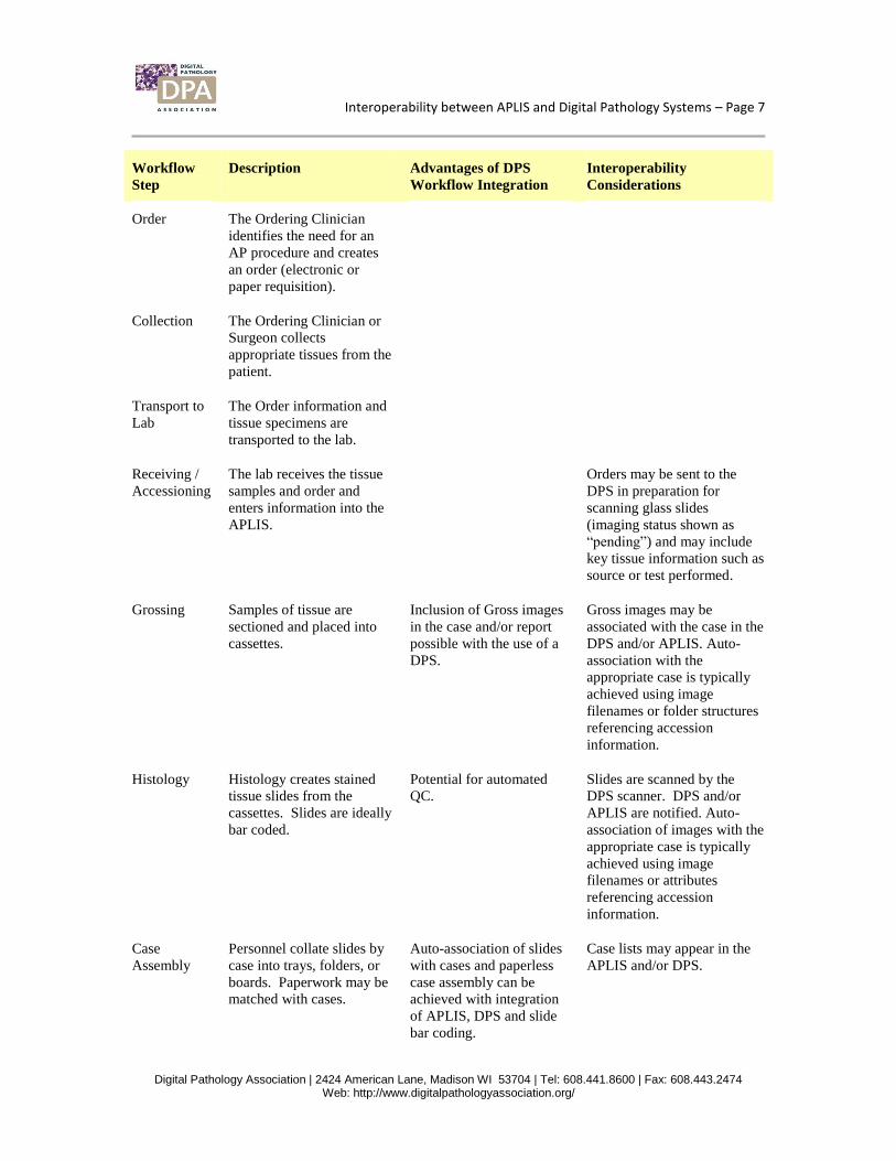
Workflow
Step
Description Advantages of DPS
Workflow Integration
Interoperability
Considerations
Order The Ordering Clinician
identifies the need for an
AP procedure and creates
an order (electronic or
paper requisition).
Collection The Ordering Clinician or
Surgeon collects
appropriate tissues from the
patient.
Transport to
Lab
The Order information and
tissue specimens are
transported to the lab.
Receiving /
Accessioning
The lab receives the tissue
samples and order and
enters information into the
APLIS.
Orders may be sent to the
DPS in preparation for
scanning glass slides
(imaging status shown as
“pending”) and may include
key tissue information such as
source or test performed.
Grossing Samples of tissue are
sectioned and placed into
cassettes.
Inclusion of Gross images
in the case and/or report
possible with the use of a
DPS.
Gross images may be
associated with the case in the
DPS and/or APLIS. Auto-
association with the
appropriate case is typically
achieved using image
filenames or folder structures
referencing accession
information.
Histology Histology creates stained
tissue slides from the
cassettes. Slides are ideally
bar coded.
Potential for automated
QC.
Slides are scanned by the
DPS scanner. DPS and/or
APLIS are notified. Auto-
association of images with the
appropriate case is typically
achieved using image
filenames or attributes
referencing accession
information.
Case
Assembly
Personnel collate slides by
case into trays, folders, or
boards. Paperwork may be
matched with cases.
Auto-association of slides
with cases and paperless
case assembly can be
achieved with integration
of APLIS, DPS and slide
bar coding.
Case lists may appear in the
APLIS and/or DPS.
Interoperability between APLIS and Digital Pathology Systems – Page 8
Digital Pathology Association | 2424 American Lane, Madison WI 53704 | Tel: 608.441.8600 | Fax: 608.443.2474 Web: http://www.digitalpathologyassociation.org/
Workflow
Step
Description Advantages of DPS
Workflow Integration
Interoperability
Considerations
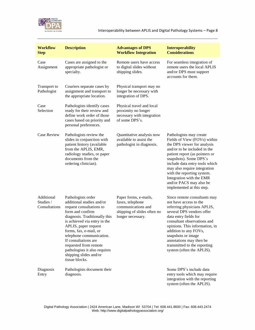
Case
Assignment
Cases are assigned to the
appropriate pathologist or
specialty.
Remote users have access
to digital slides without
shipping slides.
For seamless integration of
remote users the local APLIS
and/or DPS must support
accounts for them.
Transport to
Pathologist
Couriers separate cases by
assignment and transport to
the appropriate location.
Physical transport may no
longer be necessary with
integration of DPS.
Case
Selection
Pathologists identify cases
ready for their review and
define work order of those
cases based on priority and
personal preferences.
Physical travel and local
proximity no longer
necessary with integration
of some DPS’s.
Case Review Pathologists review the
slides in conjunction with
patient history (available
from the APLIS, EMR,
radiology studies, or paper
documents from the
ordering clinician).
Quantitative analysis now
available to assist the
pathologist in diagnosis.
Pathologists may create
Fields of View (FOVs) within
the DPS viewer for analysis
and/or to be included in the
patient report (as pointers or
snapshots). Some DPS’s
include data entry tools which
may also require integration
with the reporting system.
Integration with the EMR
and/or PACS may also be
implemented at this step.
Additional
Studies /
Consultations
Pathologists order
additional studies and/or
request consultations to
form and confirm
diagnosis. Traditionally this
is achieved via entry in the
APLIS, paper request
forms, fax, e-mail, or
telephone communication.
If consultations are
requested from remote
pathologists it also requires
shipping slides and/or
tissue blocks.
Paper forms, e-mails,
faxes, telephone
communications and
shipping of slides often no
longer necessary.
Since remote consultants may
not have access to the
referring physicians APLIS,
several DPS vendors offer
data entry fields for
consultant observations and
opinions. This information, in
addition to any FOVs,
snapshots or image
annotations may then be
transmitted to the reporting
system (often the APLIS).
Diagnosis
Entry
Pathologists document their
diagnosis.
Some DPS’s include data
entry tools which may require
integration with the reporting
system (often the APLIS).
Interoperability between APLIS and Digital Pathology Systems – Page 9
Digital Pathology Association | 2424 American Lane, Madison WI 53704 | Tel: 608.441.8600 | Fax: 608.443.2474 Web: http://www.digitalpathologyassociation.org/
Workflow
Step
Description Advantages of DPS
Workflow Integration
Interoperability
Considerations
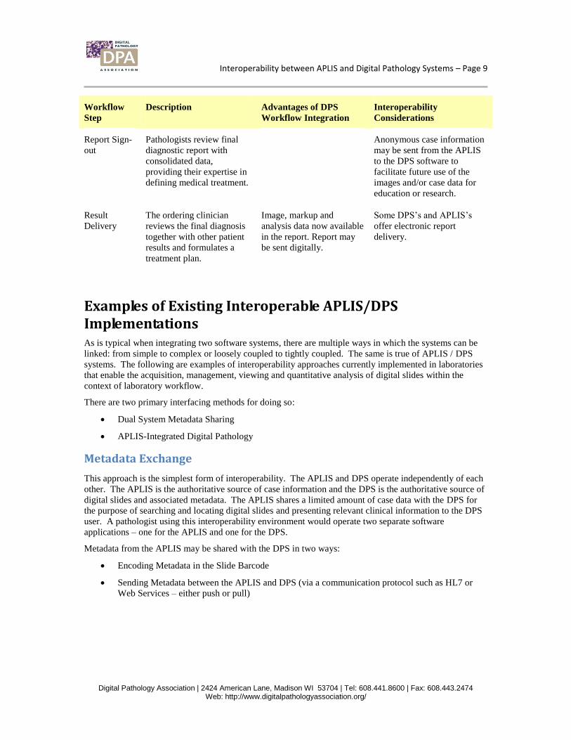
Report Sign-
out
Pathologists review final
diagnostic report with
consolidated data,
providing their expertise in
defining medical treatment.
Anonymous case information
may be sent from the APLIS
to the DPS software to
facilitate future use of the
images and/or case data for
education or research.
Result
Delivery
The ordering clinician
reviews the final diagnosis
together with other patient
results and formulates a
treatment plan.
Image, markup and
analysis data now available
in the report. Report may
be sent digitally.
Some DPS’s and APLIS’s
offer electronic report
delivery.
Examples of Existing Interoperable APLIS/DPS Implementations As is typical when integrating two software systems, there are multiple ways in which the systems can be
linked: from simple to complex or loosely coupled to tightly coupled. The same is true of APLIS / DPS
systems. The following are examples of interoperability approaches currently implemented in laboratories
that enable the acquisition, management, viewing and quantitative analysis of digital slides within the
context of laboratory workflow.
There are two primary interfacing methods for doing so:
Dual System Metadata Sharing
APLIS-Integrated Digital Pathology
Metadata Exchange
This approach is the simplest form of interoperability. The APLIS and DPS operate independently of each
other. The APLIS is the authoritative source of case information and the DPS is the authoritative source of
digital slides and associated metadata. The APLIS shares a limited amount of case data with the DPS for
the purpose of searching and locating digital slides and presenting relevant clinical information to the DPS
user. A pathologist using this interoperability environment would operate two separate software
applications – one for the APLIS and one for the DPS.
Metadata from the APLIS may be shared with the DPS in two ways:
Encoding Metadata in the Slide Barcode
Sending Metadata between the APLIS and DPS (via a communication protocol such as HL7 or
Web Services – either push or pull)
Interoperability between APLIS and Digital Pathology Systems – Page 10
Digital Pathology Association | 2424 American Lane, Madison WI 53704 | Tel: 608.441.8600 | Fax: 608.443.2474 Web: http://www.digitalpathologyassociation.org/
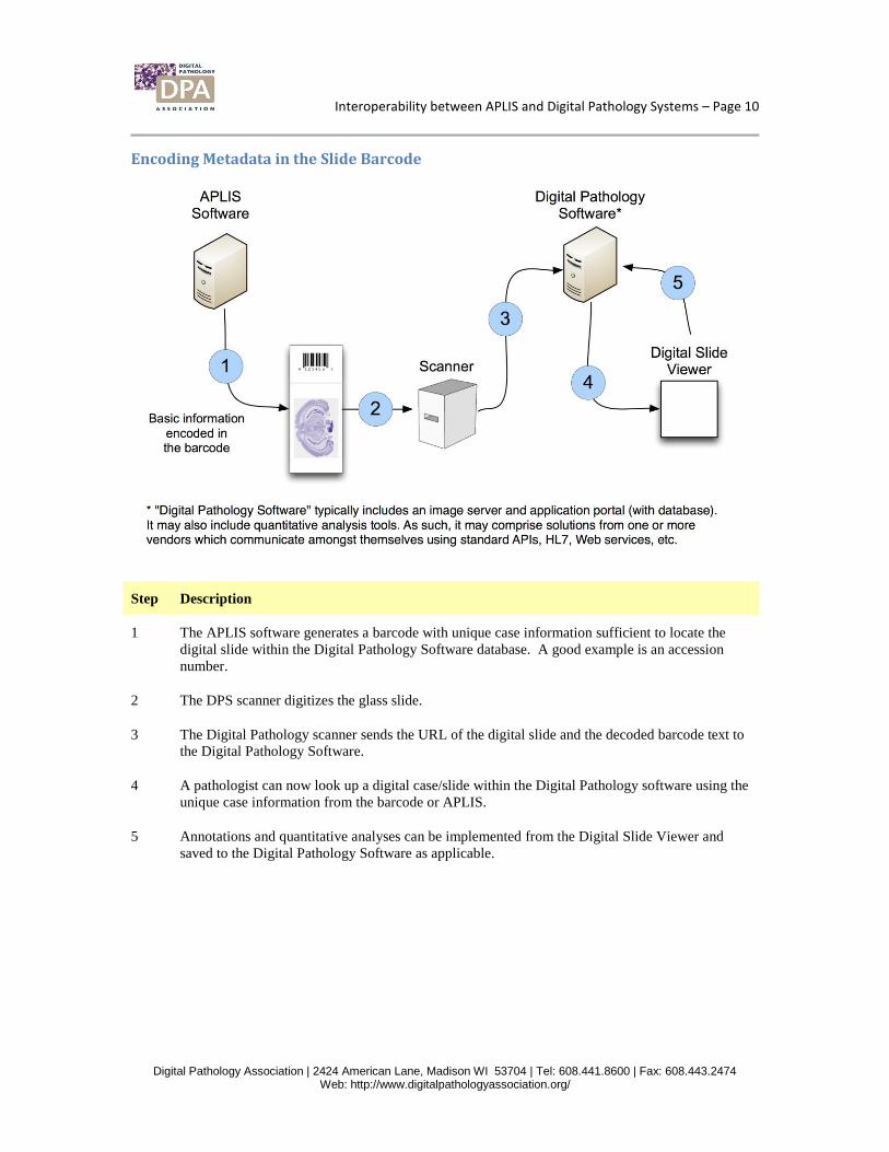
Encoding Metadata in the Slide Barcode
Step Description
1 The APLIS software generates a barcode with unique case information sufficient to locate the
digital slide within the Digital Pathology Software database. A good example is an accession
number.
2 The DPS scanner digitizes the glass slide.
3 The Digital Pathology scanner sends the URL of the digital slide and the decoded barcode text to
the Digital Pathology Software.
4 A pathologist can now look up a digital case/slide within the Digital Pathology software using the
unique case information from the barcode or APLIS.
5 Annotations and quantitative analyses can be implemented from the Digital Slide Viewer and
saved to the Digital Pathology Software as applicable.
Interoperability between APLIS and Digital Pathology Systems – Page 11
Digital Pathology Association | 2424 American Lane, Madison WI 53704 | Tel: 608.441.8600 | Fax: 608.443.2474 Web: http://www.digitalpathologyassociation.org/
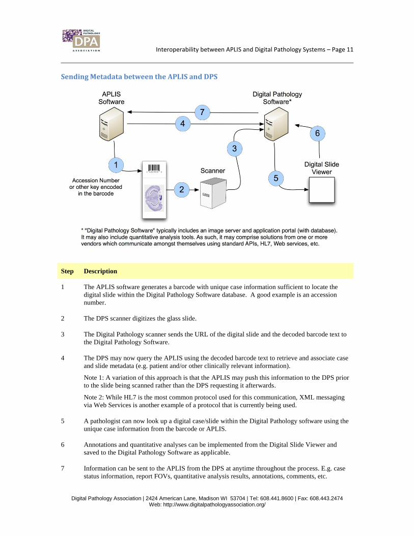
Sending Metadata between the APLIS and DPS
Step Description
1 The APLIS software generates a barcode with unique case information sufficient to locate the
digital slide within the Digital Pathology Software database. A good example is an accession
number.
2 The DPS scanner digitizes the glass slide.
3 The Digital Pathology scanner sends the URL of the digital slide and the decoded barcode text to
the Digital Pathology Software.
4 The DPS may now query the APLIS using the decoded barcode text to retrieve and associate case
and slide metadata (e.g. patient and/or other clinically relevant information).
Note 1: A variation of this approach is that the APLIS may push this information to the DPS prior
to the slide being scanned rather than the DPS requesting it afterwards.
Note 2: While HL7 is the most common protocol used for this communication, XML messaging
via Web Services is another example of a protocol that is currently being used.
5 A pathologist can now look up a digital case/slide within the Digital Pathology software using the
unique case information from the barcode or APLIS.
6 Annotations and quantitative analyses can be implemented from the Digital Slide Viewer and
saved to the Digital Pathology Software as applicable.
7 Information can be sent to the APLIS from the DPS at anytime throughout the process. E.g. case
status information, report FOVs, quantitative analysis results, annotations, comments, etc.
Interoperability between APLIS and Digital Pathology Systems – Page 12
Digital Pathology Association | 2424 American Lane, Madison WI 53704 | Tel: 608.441.8600 | Fax: 608.443.2474 Web: http://www.digitalpathologyassociation.org/
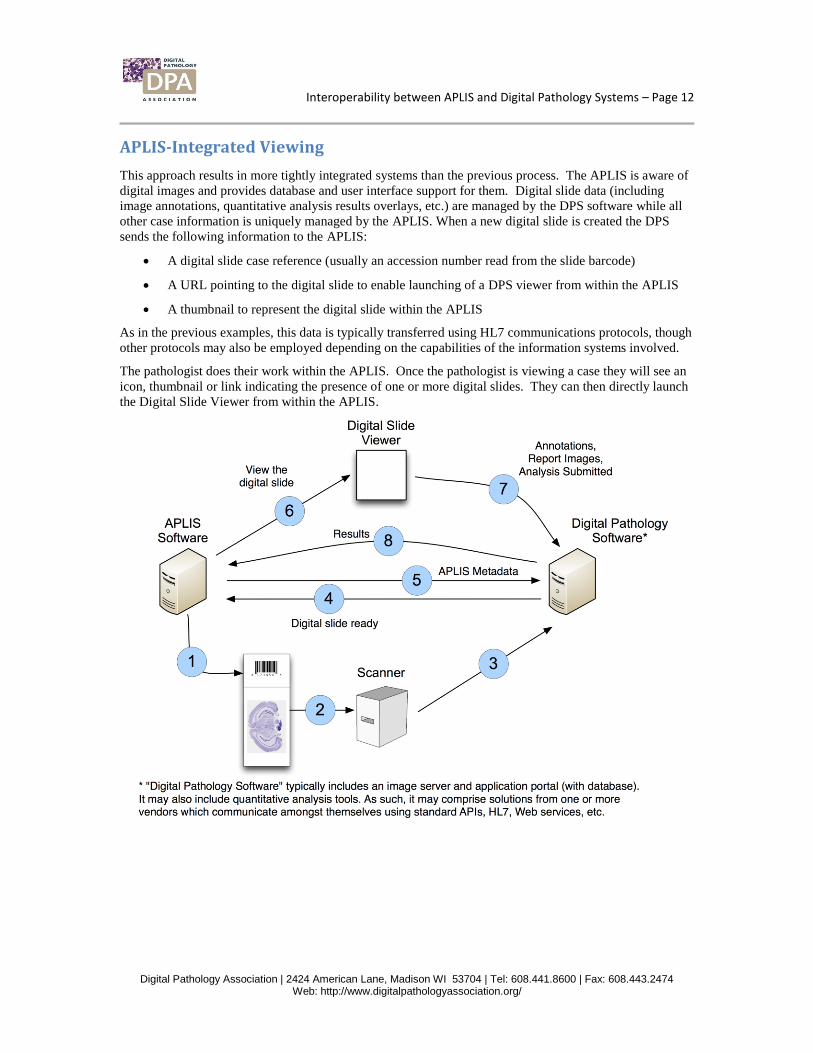
APLIS-Integrated Viewing
This approach results in more tightly integrated systems than the previous process. The APLIS is aware of
digital images and provides database and user interface support for them. Digital slide data (including
image annotations, quantitative analysis results overlays, etc.) are managed by the DPS software while all
other case information is uniquely managed by the APLIS. When a new digital slide is created the DPS
sends the following information to the APLIS:
A digital slide case reference (usually an accession number read from the slide barcode)
A URL pointing to the digital slide to enable launching of a DPS viewer from within the APLIS
A thumbnail to represent the digital slide within the APLIS
As in the previous examples, this data is typically transferred using HL7 communications protocols, though
other protocols may also be employed depending on the capabilities of the information systems involved.
The pathologist does their work within the APLIS. Once the pathologist is viewing a case they will see an
icon, thumbnail or link indicating the presence of one or more digital slides. They can then directly launch
the Digital Slide Viewer from within the APLIS.
Interoperability between APLIS and Digital Pathology Systems – Page 13
Digital Pathology Association | 2424 American Lane, Madison WI 53704 | Tel: 608.441.8600 | Fax: 608.443.2474 Web: http://www.digitalpathologyassociation.org/
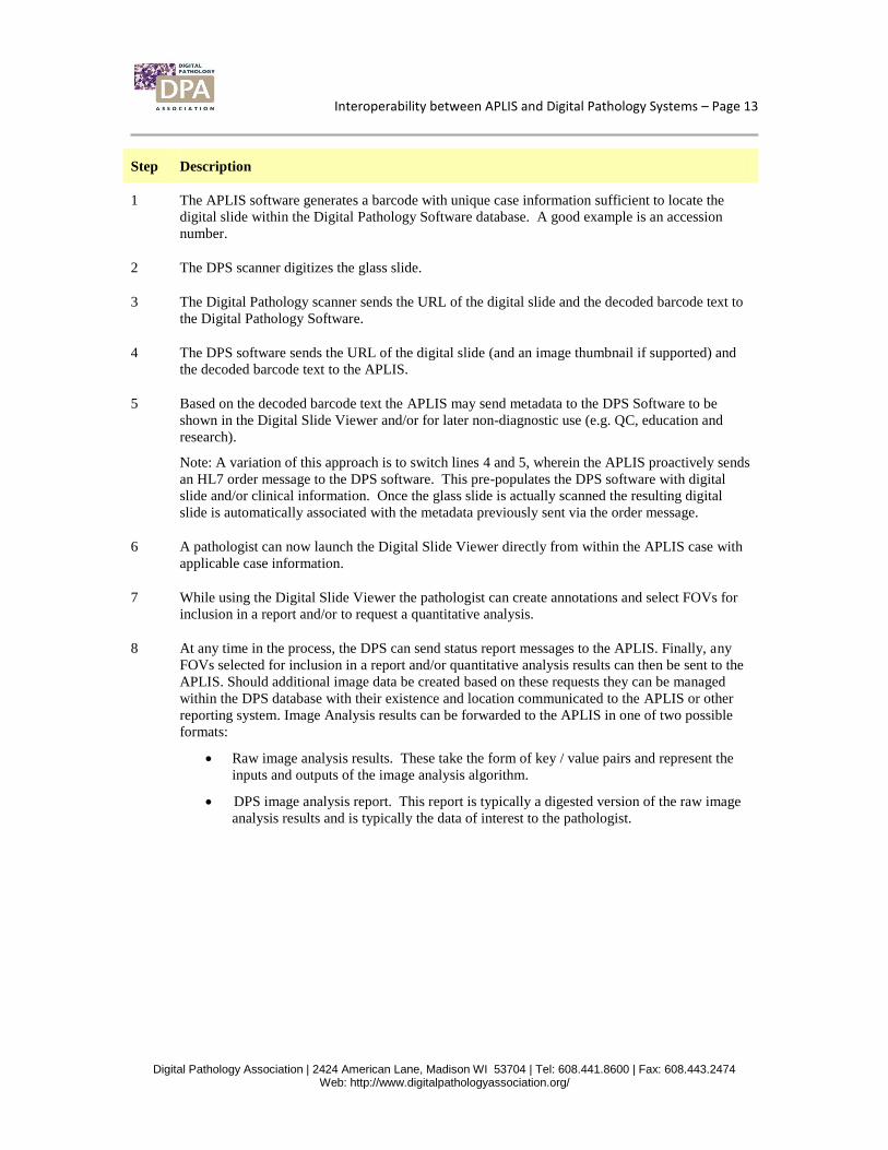
Step Description
1 The APLIS software generates a barcode with unique case information sufficient to locate the
digital slide within the Digital Pathology Software database. A good example is an accession
number.
2 The DPS scanner digitizes the glass slide.
3 The Digital Pathology scanner sends the URL of the digital slide and the decoded barcode text to
the Digital Pathology Software.
4 The DPS software sends the URL of the digital slide (and an image thumbnail if supported) and
the decoded barcode text to the APLIS.
5 Based on the decoded barcode text the APLIS may send metadata to the DPS Software to be
shown in the Digital Slide Viewer and/or for later non-diagnostic use (e.g. QC, education and
research).
Note: A variation of this approach is to switch lines 4 and 5, wherein the APLIS proactively sends
an HL7 order message to the DPS software. This pre-populates the DPS software with digital
slide and/or clinical information. Once the glass slide is actually scanned the resulting digital
slide is automatically associated with the metadata previously sent via the order message.
6 A pathologist can now launch the Digital Slide Viewer directly from within the APLIS case with
applicable case information.
7 While using the Digital Slide Viewer the pathologist can create annotations and select FOVs for
inclusion in a report and/or to request a quantitative analysis.
8 At any time in the process, the DPS can send status report messages to the APLIS. Finally, any
FOVs selected for inclusion in a report and/or quantitative analysis results can then be sent to the
APLIS. Should additional image data be created based on these requests they can be managed
within the DPS database with their existence and location communicated to the APLIS or other
reporting system. Image Analysis results can be forwarded to the APLIS in one of two possible
formats:
Raw image analysis results. These take the form of key / value pairs and represent the
inputs and outputs of the image analysis algorithm.
DPS image analysis report. This report is typically a digested version of the raw image
analysis results and is typically the data of interest to the pathologist.
Interoperability between APLIS and Digital Pathology Systems – Page 14
Digital Pathology Association | 2424 American Lane, Madison WI 53704 | Tel: 608.441.8600 | Fax: 608.443.2474 Web: http://www.digitalpathologyassociation.org/
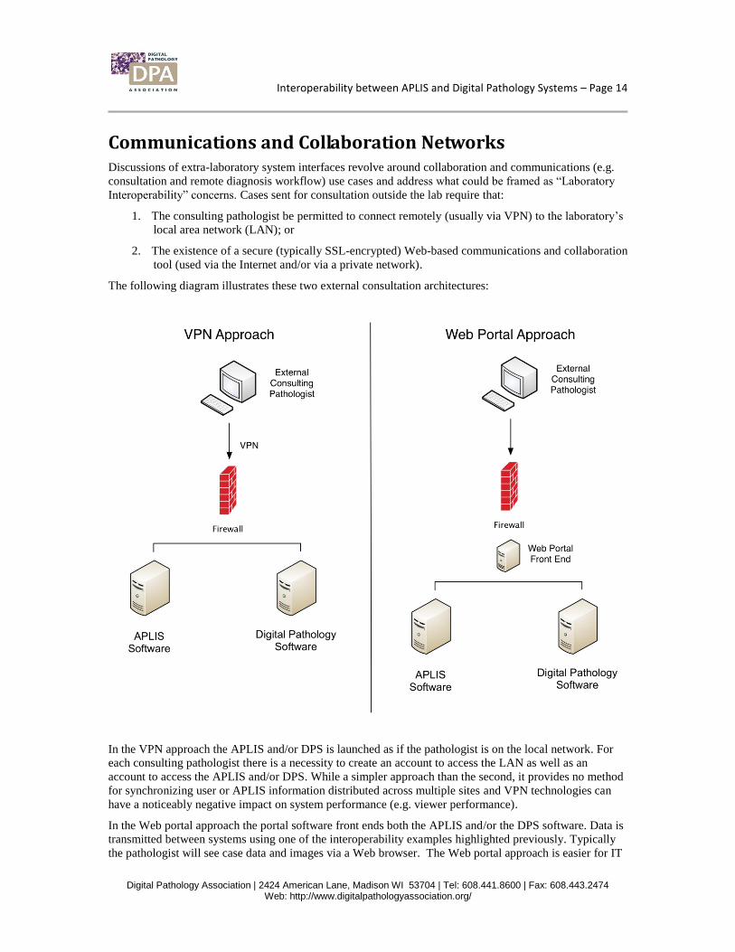
Communications and Collaboration Networks Discussions of extra-laboratory system interfaces revolve around collaboration and communications (e.g.
consultation and remote diagnosis workflow) use cases and address what could be framed as “Laboratory
Interoperability” concerns. Cases sent for consultation outside the lab require that:
1. The consulting pathologist be permitted to connect remotely (usually via VPN) to the laboratory’s
local area network (LAN); or
2. The existence of a secure (typically SSL-encrypted) Web-based communications and collaboration
tool (used via the Internet and/or via a private network).
The following diagram illustrates these two external consultation architectures:
In the VPN approach the APLIS and/or DPS is launched as if the pathologist is on the local network. For
each consulting pathologist there is a necessity to create an account to access the LAN as well as an
account to access the APLIS and/or DPS. While a simpler approach than the second, it provides no method
for synchronizing user or APLIS information distributed across multiple sites and VPN technologies can
have a noticeably negative impact on system performance (e.g. viewer performance).
In the Web portal approach the portal software front ends both the APLIS and/or the DPS software. Data is
transmitted between systems using one of the interoperability examples highlighted previously. Typically
the pathologist will see case data and images via a Web browser. The Web portal approach is easier for IT
Interoperability between APLIS and Digital Pathology Systems – Page 15
Digital Pathology Association | 2424 American Lane, Madison WI 53704 | Tel: 608.441.8600 | Fax: 608.443.2474 Web: http://www.digitalpathologyassociation.org/
departments to deploy and maintain (particularly as external consultation networks grow) and typically
offers better system performance from a user perspective. The Web portal may often provide additional
functionalities such as electronic ordering and/or reporting.
Some DPS vendors currently offer centralized and/or peer-to-peer Web portal applications intended to
enable virtualization, management and user synchronization of pathologists distributed across multiple sites
and APLIS’s as well as workflow tools for use outside the context of a unique laboratory.
Additional Considerations
Barcodes
As indicated in the interoperability examples outlined previously, bar coding is an important aspect of any
real degree of APLIS and DPS integration. While some level of interoperability can be implemented
without them, the realization of the full value and integrated clinical workflow is only achievable with the
use of barcodes that uniquely identify and track each slide.
Compatibility vs. Interoperability
It is also important to discuss the difference between compatibility and interoperability, as it can be easy to
confuse the two. The former refers to the ability of one system to understand and use information created
by another without any communications required between the two (e.g. one DPS vendor’s image viewer
opens another DPS vendor’s image format). The latter refers to the ability of two systems to communicate
with each other to achieve a common goal (e.g. one DPS vendor’s viewer is capable of requesting analysis
from another DPS vendor’s analysis system).
These can be important for users of DPS’s to understand upfront, since different vendors have different
approaches as to which information and tools they do or don’t make available to each other (e.g. images,
image markup data, quantitative analysis tools, etc.). This is particularly important for multi-site
consultation networks which may have multiple DPS’s.
Future Directions in Interoperability Digital Pathology comprises rapidly advancing technologies. Over time we expect improvements to occur
in:
Scanning speeds
Digital slide viewing tools and clinical decision support
Backend digital slide storage
Multi-site communications & collaboration applications
Scanning speeds
To fully integrate Digital Pathology into the mainstream AP workflow, improvements in throughput will be
needed. Undoubtedly hardware image acquisition speeds will reach the point where a slide can be digitized
in less than 30 seconds. Image storage densities will continue to increase to keep up with number of digital
slides that are created and used within the lab.
Interoperability between APLIS and Digital Pathology Systems – Page 16
Digital Pathology Association | 2424 American Lane, Madison WI 53704 | Tel: 608.441.8600 | Fax: 608.443.2474 Web: http://www.digitalpathologyassociation.org/
Digital Slide Viewing Tools and Clinical Decision Support
The digital slide viewing experience will continue to improve. Input techniques that allow the pathologist
to quickly and efficiently review digital slides will evolve and image analysis tools that reduce the number
of slides that have to be reviewed by a pathologist will improve the throughput of the lab.
Backend Digital Slide Storage
In addition to the trend toward denser file storage other protocols will be adopted creating a standardized
environment. For example, Working Group 26, a subcommittee of the DICOM standards body, has ratified
a proposal for storing digital slides within a PACS archive. In the next several years PACS software
vendors and Digital Pathology software vendors will support this standard. This may increase the
interoperability within the AP lab.
Multi-Site Communications and Collaboration Applications
As Digital Pathology enables simpler and more rapid access to sub-specialty opinions, consultation
networks will continue to form and grow. The Web-based tools currently providing a platform for these
multi-site communications will similarly evolve to offer more applications and integrations with APLIS
vendors and other DPS’s.
Interoperability between APLIS and Digital Pathology Systems – Page 17
Digital Pathology Association | 2424 American Lane, Madison WI 53704 | Tel: 608.441.8600 | Fax: 608.443.2474 Web: http://www.digitalpathologyassociation.org/
Appendix A – Relevant Industry Standards
HL7 (www.hl7.org)
Anatomic Pathology Working Group:
http://www.hl7.org/Special/committees/anatomicpath/index.cfm
HL7 Anatomic Pathology Special Interest Group Wiki:
http://wiki.hl7.org/index.php?title=Pathology_SIG
Most commonly used and supported standard for sharing AP data
Uses a case, specimen, child-specimen hierarchy
Supports sending of links and encoded reports (MIME, for example) in addition to structured data
elements
Not designed to send actual images (pointers only) or the 3D information from whole slide images
Fast transfer speed, easy to implement, widely used, standard is owned by HL7 International and
can be purchased
Moves case information from one system to another
DICOM (http://medical.nema.org/)
Working Group 26 – Supplement 145: Newly ratified standard to define a DICOM standard for
whole slide images
Intended to be the standard for sharing of information about pathology images
Can offer the transfer of images and more information about the attributes of slides than in HL7
Adds to the HL7 Specimen model in order to send slide-detail information and images between
imaging systems
Work is being done to improve performance and reduce image size limitations
Moves an image from one system (image-based modality) to another (image-based modality)
IHE (http://www.ihe.net/)
IHE Anatomic Pathology Wiki: http://wiki.ihe.net/index.php?title=Anatomic_Pathology
An initiative, not a standard
Focuses on addressing gaps between the patient information focus of HL7 and the image
information focus of DICOM
Provides “implementation profiles” that show how to use DICOM and HL7 together to achieve
user-focused results
HTTP/HTTPS (http://www.w3.org/Protocols/)
Industry standards used for communications with Web-based software