Embed Size (px)
Citation preview
EPIDEMIC AND PANDEMICALERT AND RESPONSE
WHO/HSE/EPR/2008.3
Interregional meeting onprevention and controlof plague
Antananarivo, Madagascar1 –11 April 2006

Interregional meeting onprevention and controlof plague
Antananarivo, Madagascar1 –11 April 2006

© World Health Organization 2008
All rights reserved.
The designations employed and the presentation of the material in this publication do not imply theexpression of any opinion whatsoever on the part of the World Health Organization concerning the legalstatus of any country, territory, city or area or of its authorities, or concerning the delimitation of itsfrontiers or boundaries. Dotted lines on maps represent approximate border lines for which there maynot yet be full agreement.
The mention of specific companies or of certain manufacturers’ products does not imply that they areendorsed or recommended by the World Health Organization in preference to others of a similar naturethat are not mentioned. Errors and omissions excepted, the names of proprietary products are distin-guished by initial capital letters.
All reasonable precautions have been taken by the World Health Organization to verify the informationcontained in this publication. However, the published material is being distributed without warranty ofany kind, either express or implied. The responsibility for the interpretation and use of the material lieswith the reader. In no event shall the World Health Organization be liable for damages arising from itsuse.

Interregional Meeting on Prevention and Control of Plague – Antananarivo, Madagascar, 7–11 April 2006
– 1 –
Contents
Summary .............................................................................................................................................................. 3 Introduction ......................................................................................................................................................... 9
Plague in the world today: situation and new challenges........................................................................... 9 Session 1. Epidemiology ......................................................................................................................... 11 1.1 Overview of the plague situation in Africa....................................................................................... 11 1.2 Central database on plague in Madagascar................................................................................... 11 1.3 Plague situation in the Democratic Republic of the Congo .......................................................... 12 1.4 Plague situation in Central Asia ....................................................................................................... 13 1.5 Plague situation in the United Republic of Tanzania .................................................................... 14 1.6 Plague situation in Mongolia ............................................................................................................. 15 1.7 Plague situation in China................................................................................................................... 16 1.8 Plague situation in the Americas...................................................................................................... 17 1.9 Contribution of molecular typing to the plague outbreak in Algeria, 2003 ................................. 18 1.10 Plague situation in Peru .................................................................................................................... 19 1.11 Discussion........................................................................................................................................... 21
Session 2. Clinical management and prevention in the human population ................................... 22 2.1 Treatment and prophylaxis .............................................................................................................. 22 2.2 Strategies for plague control in Madagascar.................................................................................. 22 2.3 Field experience in Manjakandriana district, Madagascar ........................................................... 23 2.4 Preliminary results of a multicentre study on the safety and efficacy of gentamicin versus
doxycycline and streptomycin for treatment of naturally occurring human plague ................. 23 2.5 Clinical aspects of plague in Zobia, Democratic Republic of the Congo, 2005......................... 24 2.6 Nosocomial risk: the Indian experience .......................................................................................... 25 2.7 Human case management and prevention in China ..................................................................... 26 2.8 Vaccine development......................................................................................................................... 26 2.9 Discussion ........................................................................................................................................... 27
Session 3. Laboratory diagnosis and strain analysis ......................................................................... 28 3.1 Rapid diagnostic test of the Institut Pasteur, Madagascar ........................................................... 28 3.2 Immunological diagnosis ................................................................................................................... 29 3.3 Current molecular diagnosis ............................................................................................................. 30 3.4 Laboratory strategy during the 2005 outbreak in the Democratic Republic of the Congo....... 30 3.5 National strategy for diagnosis of plague in the USA.................................................................... 31 3.6 Capacity-building for plague diagnosis in Africa ............................................................................ 32 3.7 External quality assessment: the experience of WHO and the National Institute for
Communicable Diseases in Africa .................................................................................................... 32 3.8 Overview of current laboratory techniques ..................................................................................... 33 3.9 Discussion ........................................................................................................................................... 34

Interregional Meeting on Prevention and Control of Plague – Antananarivo, Madagascar, 7–11 April 2006
– 2 –
Session 4. Vector and reservoir...................................................................................................... 35
4.1 Natural foci of plague: are they extending worldwide? ................................................................. 35 4.2 Rodent and vector surveillance in Madagascar ............................................................................. 35 4.3 Rodent and vector surveillance in Brazil ......................................................................................... 36 4.4 Rodent and vector surveillance in the USA .................................................................................... 37 4.5 Rodent and vector surveillance in the northern Caucasus........................................................... 38 4.6 Predictive thresholds for plague surveillance in Kazakhstan....................................................... 38 4.7 Rodent and vector surveillance in India .......................................................................................... 39 4.8 Discussion ........................................................................................................................................... 40
Session 5. Risks of epidemics in urban settings ................................................................................. 42 5.1 Plague surveillance in Odessa, Ukraine ......................................................................................... 42 5.2 Risk factors in Kinshasa, Democratic Republic of the Congo...................................................... 42 5.3 Plague, poverty and people: the socioeconomics of and human behaviour in plague
outbreaks in urban settings................................................................................................................ 43 5.4 Risk factors in urban foci of Mahajanga, Madagascar .................................................................. 44 5.5 Plague situation and control in Antananarivo, Madagascar ......................................................... 45 5.6 Re-emergence of plague in Oran region, Algeria .......................................................................... 46 5.7 Discussion ........................................................................................................................................... 47 6. Expert recommendations .................................................................................................................. 48 6.1 Case management and chemoprophylaxis .................................................................................... 48 6.2 Laboratory strategy, standardization and case definition ............................................................. 51 6.3 Vector and rodent control .................................................................................................................. 53
List of participants ........................................................................................................................................... 55 Annex 1 ............................................................................................................................................................... 61 References ......................................................................................................................................................... 65

Interregional Meeting on Prevention and Control of Plague – Antananarivo, Madagascar, 7–11 April 2006
– 3 –
Summary The interregional meeting on prevention and control of plague, held 7–11 April 2006 in Antananarivo, Madagascar, gathered 70 participants from 24 countries. The meeting was held under the auspices of the World Health Organization (WHO), with the support of the Institut Pasteur of Madagascar and the Kazakh Scientific Centre for Quarantine and Zoonotic Diseases, Almaty, Kazakhstan, for the scientific organization. Since antibiotics became available in the mid-twentieth century, devastating plague epidemics have been considered to be a phenomenon of the past; however, the disease is far from being eradicated and is re-emerging in several countries and regions (e.g. Algeria and the Democratic Republic of the Congo).
The key features of the five plenary sessions are summarized below, and the abstracts of the oral presentations and the conclusions of three expert round tables are compiled in the main body of this document.
Session 1. Epidemiology Plague remains an epidemiological threat and a disease of major public health importance in Africa. The occurrence of recent outbreaks shows that plague can re-emerge after a long period of silence. The most heavily affected African countries are the Democratic Republic of the Congo, Madagascar, Mozambique, Uganda and the United Republic of Tanzania.
In Madagascar, a computerized database has been set up at the Institut Pasteur for surveillance and standardized notification. This tool is valuable for analysing plague trends and for improving the reliability of notification.
Plague is endemic in the Democratic Republic of the Congo and represents a heavy public health burden. During the civil war (2002–2003), the number of human cases increased drastically. In 2005, an outbreak of pulmonary plague occurred outside the known focus of Ituri. Unfortunately, the current lack of specific resources and surveillance for plague increases the risk for extension of the existing foci.
In the United Republic of Tanzania, no human plague cases have been reported since 2003; however, surveillance of vectors and reservoirs shows that the disease has not disappeared.
In 2003, plague re-emerged in Algeria, in two villages south of Oran. Epidemiological and molecular investigations showed that the strains were unrelated to any other Yersinia pestis strain studied. The origin of this outbreak remains unclear.
The Central Asian region has active plague foci in deserts, mountains and steppes. The most active focus is in the Central Asian desert, affecting Kazakhstan, Turkmenistan and Uzbekistan. The only country in Central Asia from which human plague cases are still reported to WHO is, however, Kazakhstan. The disease is transmitted through flea bites and direct contact with infected camels.
Almost 30% of the vast Mongolian territory consists of natural plague foci. Human infection occurs through flea bites and during skinning of marmots. Of the cases of plague, 90% are the bubonic form, and more than 40% of the cases of primary bubonic plague evolve into secondary pneumonic plague. The mortality rate can be up to 70% owing to lack of availability of treatment in remote areas and the low density of health structures.
Plague foci are distributed in 19 provinces and autonomous regions of China, and the incidence has been increasing rapidly since the 1990s. Two main type of foci exist. In the south, Xenopsylla cheopis bites cause bubonic plague, and the lethality rate is low. In the western and northern provinces, contamination occurs during skinning of infected animals, most cases evolve into septicaemic or primary pneumonic plague, and the case-fatality rate is over 50%.
Interregional Meeting on Prevention and Control of Plague – Antananarivo, Madagascar, 7–11 April 2006
– 4 –
Permanent plague foci exist in the Americas among native rodent and flea populations in Bolivia, Brazil, Ecuador, Peru and the United States of America. Extensive epidemics have occurred recently only in Peru; Ecuador experienced a small outbreak of pneumonic plague in 1998. A possible association between an increase in human cases and the ‘El Niño’ effect has been postulated. There may be a risk of plague in the Andes spreading to other regions through trade and travel.
Session 2. Clinical management and prevention in the human population
The widespread availability of antibiotics in the 1940s made possible adequate treatment of plague. Aminoglycosides are effective against Gram-negative bacilli but are associated with poor oral absorption and poor cerebrospinal fluid penetration. The main treatment is with streptomycin, intravenously or intramuscularly, despite its known toxic side-effects. Gentamicin offers the advantage of a single daily administration. Tetracyclines and sulfonamides can be used for patients for whom aminoglycosides are contraindicated. Fluoroquinolones are effective in vitro and in studies in experimental animals, but they have not been studied in humans. Rare resistant strains have been described in the literature, indicating the need to maintain epidemiological and biological surveillance of the susceptibility of Y. pestis to antimicrobial agents.
The indications for chemoprophylaxis are close contact with a patient with pneumonic plague, exposure to Y. pestis-infected fleas or direct contact with Y. pestis. The current chemoprophylactic options are sulfonamides, tetracyclines and chloramphenicol.
Despite the availability of antibiotics, the vaccine option is still valid, because mortality from plague is significant, and immunization is cheaper than treatment. None of the known vaccines, however, confers long-lasting protection again bubonic plague, and they do not protect against pneumonic plague. Most of the vaccines that are being developed are composed of a combination of two antigens, F1 and LcrV. Various forms and galenic formulations have been successfully tested in mice, but the results in primates are mixed. Human trials have passed phase I (toxicity testing).
During the outbreak of pneumonic plague near the Damaseke diamond mine (Democratic Republic of the Congo) in 2005, the clinical diagnosis was confirmed by the therapeutic efficacy of gentamicin and positive results with a rapid diagnostic test for F1 antigen in sputum samples. Within 13 weeks, 134 cases and 57 deaths were recorded (case-fatality rate, 45%). The prophylactic measures included contact tracing and chemoprophylaxis with doxycycline and cotrimoxazole for 7 days after contact.
In 1994, a large plague outbreak affected the cities of Surat and Beed in India. The response to this crisis was inadequate, and panic ensued. The Indian health sector was strengthened subsequent to this episode, and a more efficient response was available to meet the outbreak of pneumonic plague in Himachal Pradesh in 2002.
In China, streptomycin is the antibiotic of choice. Patient isolation and contact tracing and chemoprophylaxis are performed routinely. In some cases, emergency measures like road traffic quarantine are also enforced.

Interregional Meeting on Prevention and Control of Plague – Antananarivo, Madagascar, 7–11 April 2006
– 5 –
Session 3. Laboratory diagnosis and strain analysis
Biological diagnosis of plague remains a challenge because most human cases occur in remote zones, where access to health care is difficult. The current two confirmation techniques are retrospective and require a minimum of 4 days for Y. pestis culture and 7 days for seroconversion, in addition to specimen transport delay. The drawbacks are that culture requires specific equipment and expertise and two serum samples must be obtained from patients, which is often a problem. The main approaches for plague diagnosis are: microscopic observation of a smear after Gram or Wayson staining; F1 antigen detection by a direct fluorescence assay, enzyme-linked immunosorbent assay (ELISA) or rapid diagnostic test; serology (anti-F1 antibody detection) by ELISA, an agglutination test, a rapid diagnostic test or immunoblotting; and Y. pestis culture, identification and screening for antibiotic resistance.
Since the last plague conference in Atlanta, Georgia, USA, in 2000, rapid diagnostic tests for F1 antigen detection have become available and evaluated. These tests, which are compatible with field conditions, are useful for confirming a clinical diagnosis, performing large-scale focus surveys and triggering early public health measures. Although they are easy to use, these biological tests must be performed by trained health staff. After 2 years of routine use of rapid diagnostic tests in Madagascar, the confirmation rate increased from less than 30% to close to 60%, and a trend to a decrease in lethality and in pulmonary forms was observed.
Immunological methods play an important role in confirmation of diagnosis when the causative agent cannot be isolated, as during the Damaseke outbreak (Democratic Republic of the Congo, 2005). Confirmation is provided by evidence of seroconversion, i.e. at least a fourfold increase in the anti-F1 immunoglobulin (Ig) G titre in paired sera. The rapid diagnostic test for plague serodiagnosis is based on detection of anti-F1 IgG. It was designed for use in surveys of plague foci; as it does not recognize IgM, it is not designed for diagnosing human plague.
Molecular tools are commonly used in research laboratories for genotyping Y. pestis. Real-time polymerase chain reaction (PCR) amplification with probes specific for Y. pestis has been shown to be sensitive and reproducible; however, the suitability of PCR and real-time PCR for diagnosing plague in the clinical setting remains to be assessed in an endemic zone, with biological specimens. In 2002, the WHO Regional Office for Africa set up an African network of plague laboratories, involving 16 countries.
A quality control programme was instituted by the National Health Laboratory Service of South Africa and WHO to identify potential problems and needs for training. It is the only multinational external quality assurance scheme in Africa.
Session 4. Vector and reservoir Plague emergence, re-emergence and focus enlargement have complex causes. Associations between human plague and climatic phenomenon like ‘El Niño’ have been proposed. The number of areas at high risk of re-emergence tends to be increasing. Rodent and vector surveillance is useful for detecting plague circulation in reservoir populations, assessing epidemic risk factors and surveying the susceptibility of fleas to insecticides. In addition to routine surveillance activities, new strategies based on use of geographical information systems, climatic and ecological data and mathematical models are improving the prediction of outbreaks. Mathematical modelling has been used to identify potential risk areas in Canada, Kazakhstan, Mexico and the USA, and might be a cheap alternative for plague surveys targeting high-risk foci.

Interregional Meeting on Prevention and Control of Plague – Antananarivo, Madagascar, 7–11 April 2006
– 6 –
The epidemic risk of urban plague is high in districts with low hygiene standards, such as Mahajanga and Antananarivo in Madagascar. A reservoir and vector survey showed that plague is still circulating in the capital. The resistance of fleas to insecticides indicates cautious, appropriate use of these chemicals for efficient eradication campaigns.
In Brazil, nine wild plague foci have been identified. The main vector is Polygenis spp., a flea that does not present the phenomenon of proventricule blocking but has infectious potential. Since 1986, Y. pestis has not been isolated from either rodents or fleas, and few laboratory-confirmed human cases have been reported. Serological surveys indicate, however, that plague is still circulating in sentinel seropositive animals, mainly dogs.
Plague is rare in the USA, with an average of 11 cases per year. All plague cases occur in the western states, after a flea bite or after handling infected rodents, domestic cats or game. The Centers for Disease Control and Prevention and public health programmes routinely analyse samples collected from wild rodents, fleas, rodent-consuming carnivores and domestic cats and dogs.
In the Russian Federation, local epizootic sites have been detected in Chechnya, Dagestan and Stavropol Krai. In 2004, 60 Y. pestis strains were isolated in the central Caucasian focus located in the mountain areas of Karachay–Cherkessia and Kabardino–Balkaria.
Every year, human cases are reported in Kazakhstan, gerbils representing the main reservoir. The plague control programme (active survey and disinfection of burrows) has been successful, but it is labour-intensive. A mathematical model based on estimates of burrow occupancy correctly described the plague situation in 52 out of the past 66 years.
More than 12 million people died of plague in India between 1898 and 1950. The inadequate management of the outbreaks in Beed and Surat (1994) triggered improvements in the plague control system. As a result, the outbreak of pneumonic plague in Shimla (Himachal Pradesh) in 2002 was contained in the shortest possible time, with early diagnosis, antibiotic treatment, mass chemoprophylaxis, quarantine and vector control measures performed in a timely manner.
Session 5. Risks of epidemics in urban settings Most human plague cases occur in rural areas; however, recent urban plague outbreaks (in India and Madagascar) prove that the urban risk should not be underestimated. Global travel and shipment of goods constitute risk factors for urban plague re-emergence, especially in large harbours and towns that maintain regular connections with plague-endemic countries.
During the nineteenth and twentieth centuries, the harbour of Odessa, Ukraine, represented the entry point for plague, with spread of the disease inland. In 2003–2004, 1431 ships arrived from plague-endemic countries. Therefore, close epidemiological and epizootological surveillance is performed in all Odessa ports, with surveys of international transport and rodents and control measures including rat-proofing of buildings and ships. Data for the past 10 years show a strong increase in the number of rodents aboard ships and the re-appearance of Rattus rattus.
An inventory of small mammals and their ectoparasites was carried out in Kinshasa, Democratic Republic of the Congo, in 2003, and it was found that the main vectors of a potential urban plague cycle (fleas and rodents) were present in the capital. Importation to the capital is possible because it is connected by road to the densely populated area of Kisangani and the plague-endemic province of Ituri. Sanitary measures for rodent and flea control are therefore necessary to prevent plague importation along the Congo River and in Kinshasa ports.
Human plague re-emerged in the harbour city of Mahajanga, Madagascar, in 1991. A survey conducted in 1997–2001 showed that the abundance of the shrew Suncus murinus and its fleas

Interregional Meeting on Prevention and Control of Plague – Antananarivo, Madagascar, 7–11 April 2006
– 7 –
X. cheopis was clearly linked with the human plague season. During the past 2 years, when no human case occurred, the rat population was reconstituted, supplanting the shrew population. Surveillance was reactivated in 2005 and 2006, when human cases were suspected. One shrew was found to be F1 seropositive in February 2006, indicating that plague is still present in the town. Plague is also endemic in the capital, Antananarivo, where the disease re-emerged in 1980. The urban reservoirs and vectors are R. norvegicus and X. cheopis. The epidemic potential is high in impoverished districts. Information, education and communication activities and rodent surveillance are efficient means for controlling urban plague outbreaks and extension.
In 2003, after more than 50 years of silence, human plague cases were reported in two villages south of Oran, Algeria. A WHO expert mission provided technical and logistical support. The control measures involved case management, laboratory diagnosis, epidemiological investigation, vector control and chemoprophylaxis. The origin of the focus is still not clear (re-activation of a silent wild focus or importation). The proximity of Oran harbour, with regular trade with plague-endemic countries and with most large Mediterranean harbours, was a concern because of the potential risk of urban plague.
In conclusion, plague could re-emerge in other large cities, and urban plague must be considered a potential threat. Potential new foci should be confirmed and investigated, with special attention to harbours with international trade. National plague control programmes and regional collaboration are needed to maintain thorough epidemiological surveillance at the international level.

Interregional Meeting on Prevention and Control of Plague – Antananarivo, Madagascar, 7–11 April 2006
– 8 –

Interregional Meeting on Prevention and Control of Plague – Antananarivo, Madagascar, 7–11 April 2006
– 9 –
Introduction
Plague in the world today: situation and new challenges
Eric Bertherat, WHO, Geneva According to the current International Health Regulations1, notification of cases of human plague to WHO is mandatory. The notified numbers must be viewed cautiously, however, as there is likely to be discordance between the reported and the actual figures. Nevertheless, the global trends are clear (see Figure 1). Until the end of the 1970s, most cases were reported in Asia (United States military operation in Viet Nam). After that time, most cases were notified in Africa, and this is still the situation today, with numerous human cases occurring every year in highly endemic countries like the Democratic Republic of the Congo and Madagascar. Other countries experience regular, low plague activity (China, Kazakhstan, Mongolia, Mozambique, Peru, United Republic of Tanzania, USA and Viet Nam). Another feature of the disease is its re-emergence in ‘hot spots’, such as in Algeria, Ecuador and India. Despite the availability of effective antibiotic treatment, the lethality rate of plague is still high (up to 10% for bubonic plague and more than 40% for pneumonic plague).
Since the last international plague conference in Atlanta, Georgia, USA, in 2000, several new challenges, tools and concerns have appeared.
− The ‘event’ of 11 September 2001 suggested the potential use of biological agents as terrorist weapons. Deliberate use of Y. pestis is thus an increasing concern in developed countries. This might generate secondary benefits, such as new laboratory techniques and vaccines, although it is difficult to raise interest and funding for public health aspects.
− New diagnostic tools have been developed: rapid diagnostic tests produced by the Institut Pasteur of Madagascar are routinely used in that country, and field evaluation is under way in other African countries. This diagnostic tool was useful during outbreak investigations in Algeria and the Democratic Republic of the Congo. Other rapid diagnostic tests are being developed by the Centers for Disease Control and Prevention in the USA and by the French and German armies.
− The current International Health Regulations, which came into force in 1969, have some limitations, as highlighted by the crisis triggered by severe acute respiratory syndrome (SARS) and avian influenza (H5N1). The Regulations require notification to WHO of cases of cholera, plague and yellow fever, with a narrow focus on these three diseases. It states that ports, airports and frontier posts should be adequately equipped to apply the Regulations and sets rigid constraints on international traffic. New International Health Regulations were adopted in 2005 and will come into force in June 2007. The key changes are for development of core surveillance capacity at national level and a real-time event management system at WHO, which will be based on a variety of sources, including some that are unofficial and confidential. A notifiable event is defined in the new Regulations as an unexpected event presenting a risk of international spread. The scope has therefore been broadened to cover diseases that have not yet been identified.
WHO activities on plague include surveillance, alert and response activities, as described in the International Health Regulations, country support (field assistance if requested, recommendations for surveillance, case management and control, support for development of
1 Comments on the International Health Regulations as presented during the meeting.

Interregional Meeting on Prevention and Control of Plague – Antananarivo, Madagascar, 7–11 April 2006
new diagnostic tests), laboratory training, quality control programmes, reference documentation (plague manuals, practical guidelines), networking, advocacy and fund-raising.
Many questions about the epidemiology and control of plague are pending: What is the real magnitude and impact of plague in Africa? Is there global extension of the natural foci? What is the impact of ecological changes due to human activities? Is there an increasing epidemic risk in urban settings? Which laboratory techniques are recommended? What should be the duration of prophylaxis treatment? How should corpses and nosocomial risk be managed efficiently? What are the best current approaches for vector control and pesticide use? The five sessions of the meeting were devoted to presentations of experiences from various plague-affected countries, and three experts sessions addressed issues related to case management, laboratory techniques and vector control.
Figure 1 - Human plague cases : countries having notified to WHO, 2002-2005
Data Source: Epidemic Readiness and Interventions; Communicable Diseases (CDS); Map production: Public Health Mapping & GIS; Communicable Diseases); World Health Organization.
– 10 –
Interregional Meeting on Prevention and Control of Plague – Antananarivo, Madagascar, 7–11 April 2006
– 11 –
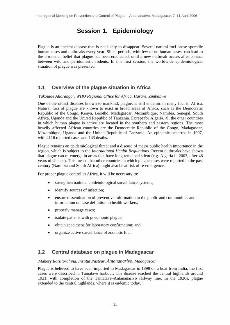
Session 1. Epidemiology
Plague is an ancient disease that is not likely to disappear. Several natural foci cause sporadic human cases and outbreaks every year. Silent periods, with few or no human cases, can lead to the erroneous belief that plague has been eradicated, until a new outbreak occurs after contact between wild and peridomestic rodents. In this first session, the worldwide epidemiological situation of plague was presented.
1.1 Overview of the plague situation in Africa Yakouidé Allarangar, WHO Regional Office for Africa, Harare, Zimbabwe
One of the oldest diseases known to mankind, plague, is still endemic in many foci in Africa. Natural foci of plague are known to exist in broad areas of Africa, such as the Democratic Republic of the Congo, Kenya, Lesotho, Madagascar, Mozambique, Namibia, Senegal, South Africa, Uganda and the United Republic of Tanzania. Except for Algeria, all the other countries in which human plague is active are located in the southern and eastern regions. The most heavily affected African countries are the Democratic Republic of the Congo, Madagascar, Mozambique, Uganda and the United Republic of Tanzania. An epidemic occurred in 1997, with 4116 reported cases and 143 deaths.
Plague remains an epidemiological threat and a disease of major public health importance in the region, which is subject to the International Health Regulations. Recent outbreaks have shown that plague can re-emerge in areas that have long remained silent (e.g. Algeria in 2003, after 48 years of silence). This means that other countries in which plague cases were reported in the past century (Namibia and South Africa) might also be at risk of re-emergence.
For proper plague control in Africa, it will be necessary to:
• strengthen national epidemiological surveillance systems;
• identify sources of infection;
• ensure dissemination of preventive information to the public and communities and information on case definition to health workers;
• properly manage cases;
• isolate patients with pneumonic plague;
• obtain specimens for laboratory confirmation; and
• organize active surveillance of zoonotic foci.
1.2 Central database on plague in Madagascar
Mahery Ratsitorahina, Institut Pasteur, Antananarivo, Madagascar
Plague is believed to have been imported to Madagascar in 1898 on a boat from India; the first cases were described in Tamatave harbour. The disease reached the central highlands around 1921, with completion of the Tamatave–Antananarivo railway line. In the 1920s, plague extended to the central highlands, where it is endemic today.

Interregional Meeting on Prevention and Control of Plague – Antananarivo, Madagascar, 7–11 April 2006
– 12 –
Plague cases have been recorded manually since 1955, and a national control programme was instituted in 1994 for surveillance and standardized notification. The mission of the central plague laboratory, based in the Institut Pasteur of Madagascar, is to collect and manage epidemiological and biological data, and a computerized database was set up for this purpose in 1995. Each suspected case or death is notified to the central plague laboratory on a standard form, recording patient identification, clinical data and the epidemiological and biological context. The central database makes it possible to follow key indicators of the programme and to provide feedback to health workers.
The trends for 2000–2005 were as follows (Figure 2): the male:female sex ratio was 1.4; the 5–14-year-old age group was overrepresented; 94% of plague cases were of the bubonic form and 3% of the pneumonic form; and 9.7% of notified cases and 19% of confirmed cases were lethal.
The central database represents a valuable tool for analysing plague trends in Madagascar and for improving the reliability of case and death notifications.
Figure 2 - Plague notification in Madagascar, 2000-2005
1.3 Plague situation in the Democratic Republic of the Congo Vital Mondonge, Ministry of Health, Kinshasa, Democratic Republic of the Congo
Plague has been described in the Democratic Republic of the Congo since 1928, with two known natural foci in the eastern region (North Kivu and Ituri). A national laboratory for the control of the plague was created in 1928. After independence, owing to lack of resources, the incidence of plague increased and the Ituri focus expanded. The setting up of local committees to act against epidemics tended to reduce the incidence up to 1999. During the civil war (2002–2003), however, massive displacements of population, degradation of habitats and a total breakdown of the health system brought about a drastic increase in the number of human cases in Ituri, although no data were reported during this period.
The current plague control system is integrated into surveillance of other diseases with epidemic potential. Plague cases reported for 2000–2005 are shown in Figure 3. The epizootic survey is, however, sparse. The country has a national plague laboratory, based in Bunia, with trained personnel, but there is a lack of resources specifically dedicated to plague control. In 2005, pulmonary plague cases were notified outside the known focus of Ituri, in a diamond mining population.
0
200
400
600
800
1000
1200
1400
2000 2001 2002 2003 2004 2005Year
Cas
es
No sample
Not confirmed
Confirmed

Interregional Meeting on Prevention and Control of Plague – Antananarivo, Madagascar, 7–11 April 2006
– 13 –
0
200
400
600800
1000
1200
1400
1600
2000 2001 2002 2003 2004 2005
Year
Cas
es a
nd d
eath
s
CasesDeaths
Plague is endemic in the Democratic Republic of the Congo and represents a heavy public health burden. Unfortunately, the lack of specific resources and appropriate means, the weakness of the surveillance system and delays in diagnosis increase the risk that the existing foci will be extended.
Figure 3 - Plague reporting in DRC, 2000-2005 (no data available for 2002–2003).
1.4 Plague situation in Central Asia B. Atshabar, Kazakh Scientific Centre for Quarantine and Zoonotic Diseases, Almaty, Kazakhstan
Central Asia consists of five countries: Kazakhstan, Kyrgyzstan, Tajikistan, Turkmenistan and Uzbekistan, which were part of the former Union of Soviet Socialist Republics. There are various types of active plague foci in this region, including desert, mountain and steppe (Figure 4), with a total area of 1.8 million km2. The largest, most active focus is in the Central Asian desert, which covers parts of three countries, Kazakhstan, Turkmenistan and Uzbekistan. Sporadic cases of plague are registered almost every year in this region. The main vectors are flea bites and direct contact (skinning and butchering infected camels). The only country in Central Asia that reports human plague cases to WHO is, however, Kazakhstan.
Plague control services in the Central Asian countries consist of a scientific coordinating centre (as in Kazakhstan) and a network of regional plague control stations or plague control divisions. The Kazakh Scientific Centre for Quarantine and Zoonotic Diseases is the only institution in Central Asia that undertakes field surveys for plague and produces diagnostic reagents and vaccine against plague.
The functions of the anti-plague institutions are:
− to determine the epidemic potential on the territory;
− to destroy insects in populated areas and in the field as part of public health assignments;
− to destroy rats in populated areas as part of public health assignments;
− to immunize humans at risk (e.g. livestock owners, geologists, oil workers);
− to train medical and veterinary staff and health volunteers; − to localize and control plague outbreaks to prevent epidemic spread of the disease; and
− to impose personal biosafety rules.

Interregional Meeting on Prevention and Control of Plague – Antananarivo, Madagascar, 7–11 April 2006
Figure 4 - Geographical distribution of plague natural foci in central Asia (Kazakhstan, Turkmenistan, Tajikistan, Uzbekistan and Kyrgyzstan)
Data Source: Kazakh Scientific Centre for Quarantine and Zoonotic Diseases, Almaty, Kazakhstan. Map production: Public Health Mapping & GIS; Communicable Diseases (CDS); World Health Organization.
– 14 –
1.5 Plague situation in the United Republic of Tanzania Rodes Makundi, Sokoine University of Agriculture, Dodoma, United Republic of Tanzania
Plague has been known in the United Republic of Tanzania for many years, the earliest authenticated cases being recorded over 100 years ago. Plague cases recorded between 1986 and 2004 are shown in Figure 5. Up to 1990, nine active and two quiescent foci were known. Most had been inactive since 1980, with the exception of the Lushoto district focus in the north-east, where human cases reappeared more than 20 years ago. This focus is very localized, in a mountainous rainforest area that was opened to agriculture some 30 years ago, with a few villages experiencing outbreaks persistently. Plague has infected more than 7000 persons, with a mortality of around 10%. The transmission is highly seasonal (December–February), with strong inter-annual variations. It is likely that an enzootic cycle existed in the forest before the first human cases were reported in 1980. Human activities have had a drastic effect on the ecology of reservoirs and vectors, with deforestation for agriculture, encroachment into the natural forest and fragmentation of reservoirs. Overlaps in the habitats of sylvatic and domestic or peridomestic rodent species have increased interactions among humans, rodents and fleas and have therefore facilitated plague epidemics.
More than 90% of plague patients have the bubonic form. The rare pneumonic cases are due to delayed treatment or inappropriate self-medication. The incidence profiles show a higher prevalence in women than in men in the age group 30–60 years. The incidence among children aged 5–14 years is twice that among adult women. Sociocultural and economic factors influence the incidence of plague in families and within the community. Belief in witchcraft has been blamed for delayed treatment and lack of proper treatment, and the social stigma associated with plague prevents families from seeking medical attention. They thus resort to self-medication.

Interregional Meeting on Prevention and Control of Plague – Antananarivo, Madagascar, 7–11 April 2006
– 15 –
Although no human plague cases were reported in the country in 2004–2006, there is no reason to conclude that the disease has disappeared, as surveillance of vectors and reservoirs shows an abundance comparable to that observed during plague outbreak years. Plague vectors and reservoirs in the plague foci are surveyed regularly, supported by the Ministry of Health and local district councils.
Figure 5 - Plague cases recorded in Tanzania, 1986–2004
0
100
200
300
400
500
600
700
1986 1987 1988 1989 1990 1991 1992 1993 1994 1995 1996 1997 1998 1999 2000 2001 2002 2003
Year
Cas
es
Number of cases
1.6 Plague situation in Mongolia Dashdawaa Otgonbaatar, Ministry of Health, Ulaanbaatar, Mongolia
Plague has been known in Mongolia since 1897, and the natural foci have been studied since 1911. In 1924, the Government organized the first structure for plague control. Today, close to 30% of the vast Mongolian territory (1.6 million km2) consists of natural foci.
The main plague reservoirs are the marmot (Marmota sibirica), the suslik or ground squirrel (Citellus undulatus), the pika (Ochotono pallasi) and the vole (Lasiopodomys brandti). The main vector is the flea Oropsylla silantiewi. Plague cases are registered every year; human transmission is seasonal, linked to the hunting period (May–October). As Mongolians traditionally hunt marmots for their fur and meat, the risk of human infection from aerosols released during skinning of marmots is very high. Flea bites also play a role in plague transmission from infected rodents to humans. Of the 160 human plague cases registered between 1971 and 2000 (Figure 6), 90% were the primary bubonic form, 4.2% the primary pneumonic form and 6% the septicaemic form. More than 40% of the cases of primary bubonic plague evolve into secondary pneumonic plague because of lack of treatment. The mortality rate is very high (up to 70%, five times the world average) because of the lack of treatment in remote areas and the low density of health structures in the huge territory of Mongolia.

Interregional Meeting on Prevention and Control of Plague – Antananarivo, Madagascar, 7–11 April 2006
– 16 –
Despite the severe logistic constraints to plague control in Mongolia, some measures have been implemented, including surveillance of active natural foci, data collection and processing, rat and insect eradication campaigns, public health education, immunization and genetic analysis of Y. pestis strains.
Collaboration has been established with plague control organizations in China and the Russian Federation, to share information above plague activity across national borders, such as the Altai Mountains, the Sailugem area, the Chitinsk foci next to the Russian border; Khangai, Khentii Mountain, the eastern steppe and Gobi Desert, and the Manjuur region close to China.
Figure 6 - Human plague distribution in Mongolia
1.7 Plague situation in China Rong Hai, Institute for Communicable Disease Control and Prevention, Beijing, China
Natural plague foci are widely distributed throughout China, and epizootics and human cases are described every year. The plague foci are distributed in 19 provinces and autonomous regions of China; new natural foci were identified in Sichuan and Guizhou provinces in 1999.
Since the 1990s, the incidence of plague in China has been increasing rapidly, with fewer than 10 human cases per year in the 1980s, nearly 100 cases in 1996 and 254 cases in 2000. A total of 631 human cases were reported between 1995 and 2004, with a fatality rate of 6.67%.
The surveillance network analyses host and vector populations and their spatial distribution and studies the genetic characteristics of the pathogen. Two main type of foci exist in China, corresponding to different geographical zones, reservoirs and infection mode. In the southern region, where more than half of the human cases are declared, R. flavipectus is the main reservoir, X. cheopis bites cause bubonic plague, and the lethality rate is low. In the western and northern provinces, the reservoirs are Spermophilus dauricus (north-east), Meriones unguiculatus (north) and Marmota himalayana (Qinghai–Tibet). Hunters are frequently
Data Source: Ministry of Health, Ulaanbaatar, Mongolia. Map Production: Public Health Mapping & GIS; Communicable Diseases (CDS); World Health Organization

Interregional Meeting on Prevention and Control of Plague – Antananarivo, Madagascar, 7–11 April 2006
– 17 –
contaminated while skinning infected animals, and most cases are septicaemic or primary pneumonic plague. Because of the remoteness of the north-western foci, the fatality rate is greater than 50%.
Y. pestis strains isolated from natural plague foci in China were analysed by ribotyping and showed clustering related to their geographical origin. Ribotype B covers a large area including most of the Qinghai–Tibet plateau and the western region of Yunnan. The pattern indicates that ribotype A and ribotype C are closely related.
Plague control measures include insecticide and rodenticide use and health personnel training to detect clinical manifestations and for diagnosis, treatment, control and reporting.
1.8 Plague situation in the Americas Kenneth Gage, Centers for Diseases Control and Prevention, Fort Collins, Colorado, USA
Following the introduction of plague in the Americas in the early 1900s, permanent foci of infection became established among native rodent and flea populations in a number of countries, including Brazil, Bolivia, Ecuador, Peru and the USA. Large epidemics have occurred recently only in Peru (1248 cases reported between 1992 and 1994). Ecuador experienced a small outbreak of pneumonic plague in 1998: the index case had skinned and cooked sick guinea-pigs, and other family members were infected via airborne transmission.
Although human plague in native rodent foci in the Americas often appears as isolated cases or small clusters of cases (Figure 7), the potential for more widespread outbreaks exists, particularly when the disease passes from native rodent hosts and their fleas to peridomestic rats and fleas. There is a risk that plague will spread from the Andes to other regions through trade and travel, as regional markets attract villagers and rural inhabitants move to cities.

Interregional Meeting on Prevention and Control of Plague – Antananarivo, Madagascar, 7–11 April 2006
– 18 –
Figure 7 - Human plague in the Americas
1.9 Contribution of molecular typing to the plague outbreak in Algeria, 2003
Viviane Chenal-Francisque, Alexandre Leclercq and Elisabeth Carniel, Institut Pasteur, Paris, France; Souad Bekhoucha, Oran University Hospital, Oran, Algeria; and Eric Bertherat, WHO, Geneva, Switzerland
Three major plague outbreaks were reported in Algeria during the first half of the twentieth century, and several sporadic cases were notified from sea harbours. These outbreaks are considered to have corresponded not to endemic foci but to repeated importations. The last human cases were reported in 1945 in Algiers and in 1950 in Oran.
After more than 50 years of silence, human plague cases were reported again in June 2003 in Algeria, in two villages in the southern part of the Oran area. The index case was an 11-year-old boy with severe septicaemic syndrome, and other cases of adenopathy and septicaemia were reported in the same village. The cases were diagnosed clinically and confirmed by F1 antigen testing with a rapid diagnostic test and isolation of the Y. pestis strain in the laboratory of the University Hospital of Oran. Bacteria with all the characteristics of Y. pestis biovar orientalis were isolated from the bubo aspirate and blood of several patients. Within 2 weeks of the onset of the outbreak, new cases appeared in another village 50 km away from the original focus (Figure 8). A total of 18 cases were recorded during this outbreak; 10 were confirmed, 3 were presumptive and 5 were suspected.
Molecular investigation of the isolated strains was conducted to establish whether the outbreak was due to an imported strain or to re-emergence of a quiescent focus and whether the two plague foci that appeared almost simultaneously were related. Ribotyping showed that all the
Countries reporting plague
Areas with past plague
Data Source and Map production: Centers for Disease Control and Prevention, Fort Collins, Colorado, USA. Reproduced with permission.

Interregional Meeting on Prevention and Control of Plague – Antananarivo, Madagascar, 7–11 April 2006
– 19 –
strains were of ribotype B, the ribotype most commonly associated with Y. pestis orientalis strains. All strains also had the same pulsed-field gel electrophoresis pattern, strengthening the hypothesis of a single outbreak. This pattern was different from those of strains isolated in 1944 and 1945 in Oran and of strains in other African countries. Grouping of the 2003 Algerian isolates by insertion sequence fingerprinting (restriction fragment length polymorphism) showed that they formed an independent cluster unrelated to any of the other orientalis strains studied. The origin of this outbreak remains unknown.
Figure 8 - Geographical repartition of human plague cases during the outbreak in Algeria, 2003
1.10 Plague situation in Peru Manuel Cespedes, National Institutes of Health, Lima, Peru
Plague was introduced into Peru early in the twentieth century via the main harbours. Human cases are reported every year, with a strong variation in number (Figure 9). They are possibly associated with an increase in the rodent population associated with ‘El Niño’, as, on three occasions, increases in the numbers of human cases have been noted the year after an ‘El Niño’ phenomenon (Figure 10).
Data source: Ministry of Health , Algeria. Map production: Public Health Mapping and GIS Communicable Diseases (CDS); World Health Organization.

Interregional Meeting on Prevention and Control of Plague – Antananarivo, Madagascar, 7–11 April 2006
– 20 –
Figure 9 - Plague in Peru, 1984–2004
Data Source: National Institutes of Health, Lima, Peru. Map Production: Public Health Mapping & GIS; Communicable Diseases (CDS); World Health Organization.
Several risk factors for plague have been identified: grain storage in the open air, which favours an abundance of rodents and fleas; promiscuity in housing; absence of rodent-proof devices; beds on the floor and infested with fleas; and the custom of raising guinea-pigs for their flesh.
Between 1992 and 2003, a control programme undertook searches for and treatment of contacts, destruction of insects in houses, vector and reservoir control (construction of cages for guinea-pigs outside houses, rodent capture), surveys and mapping, with the participation of regional laboratories. The zoonotic programme has, however, been discontinued, and the survey activities have decreased because of decentralization of economic resources and other national health priorities (malaria, dengue, yellow fever, HIV, tuberculosis).
Figure 10 - Relationship between “El Niño” phenomenon and human plague cases
in Peru, 1960–1999.
0
200
400
600
800
1000
1200
1960
1962
1964
1966
1968
1970
1972
1974
1976
1978
1980
1982
1984
1986
1988
1990
1992
1994
1996
1998
2000
year
case
s
"El Niño" phenomenon
0
200
400
600
800
1000
1200
1960
1962
1964
1966
1968
1970
1972
1974
1976
1978
1980
1982
1984
1986
1988
1990
1992
1994
1996
1998
2000
year
case
s
"El Niño" phenomenon"El Niño" phenomenon
Interregional Meeting on Prevention and Control of Plague – Antananarivo, Madagascar, 7–11 April 2006
– 21 –
1.11 Discussion Data for 2005 in Madagascar highlighted a notable decrease in the number of plague cases. Dr Ratsitorahina (Institut Pasteur, Madagascar) confirmed that the number of cases represented about one-third that in 2002. He said that sanitation efforts in urban zones were a possible explanation but emphasized that 1 year is too short a time to see the impact of such public health measures; observations over the coming years will determine whether the 2005 decrease was an artefact or a real trend.
In response to a question about the high lethality rate in Mongolia and current treatment in the country, Dr Otgonbaatar (Centre for Infectious Diseases with Natural Foci, Mongolia) replied that treatment is with streptomycin, and no resistance to this antibiotic has been documented so far. The high lethality might be explained by the presence of plague foci in 30% of the huge territory of Mongolia and by problems of access to health structures and the small number of health workers.
The issue of intra-family transmission and the role of fleas in the United Republic of Tanzania was raised. Dr Makundi (Sokoine University, United Republic of Tanzania) explained that several cases and deaths had been reported within families, and delayed treatment had resulted in secondary pneumonic plague. Pullex iritans is abundant and very likely to be involved in transmission when there are no cat or dog fleas.

Interregional Meeting on Prevention and Control of Plague – Antananarivo, Madagascar, 7–11 April 2006
– 22 –
Session 2. Clinical management and prevention in the human population
2.1 Treatment and prophylaxis Kevin Griffith, Centres for Diseases Control and Prevention, Fort Collins, Colorado, USA
With effective control, treatment and prevention, the overall incidence of plague has markedly decreased over the past 50 years. Nevertheless, plague remains a modern threat, due to both natural ecological occurrences and the possibility of intentional use of the infectious agent.
The widespread availability of antibiotics from the 1940s signalled the start of adequate treatment of plague. Aminoglycosides are effective against aerobic Gram-negative bacilli but are poorly absorbed after oral administration and have poor cerebrospinal fluid penetration; they are therefore not recommended for treating meningeal plague. The main treatment is with intravenous or intramuscular streptomycin. (There is no oral formulation.) This inexpensive molecule is effective for the bubonic, pneumonic and septicaemic forms of plague. Its disadvantages are potentially irreversible ototoxicity, reversible nephrotoxicity and the lack of an oral formulation. Gentamicin offers the advantage of a single daily administration.
Tetracyclines, chloramphenicol and sulfonamides can be used in patients in whom aminoglycosides are contraindicated. Tetracyclines are broad-spectrum bacteriostatic agents with few major side-effects, although they are contraindicated for pregnant woman and infants. They are effective against bubonic and septicaemic plague and can be administered orally or intravenously. Chloramphenicol is a broad-spectrum bacteriolytic agent with excellent cerebrospinal fluid penetration; however, it is no longer recommended because of severe adverse effects, including reversible bone marrow suppression, aplastic anaemia and ‘grey baby’ syndrome. Sulfonamides are indicated for the treatment of bubonic plague only. Fluoroquinolones have been shown to be effective in vitro and in studies in experimental animals, but no studies have been performed in humans. Although only a few antibiotic-resistant strains have been described in the literature, epidemiological and biological surveys of the susceptibility of Y. pestis to antimicrobial agents must be maintained.
The indications for chemoprophylaxis are close contact with a patient with pneumonic plague, exposure to Y. pestis-infected fleas or direct contact with Y. pestis. The current chemoprophylactic options are sulfonamides, tetracyclines and chloramphenicol. An expert committee reviewed and updated these recommendations (section 6.1).
Plague remains a clinical concern in modern times. While effective therapy exists, additional research is needed on safer, more efficient antibiotics for Gram-negative sepsis and means for early recognition and early treatment of plague.
2.2 Strategies for plague control in Madagascar Jean Randriambelosoa, Ministry of Health, Antananarivo, Madagascar
In Madagascar, about 1000 new cases of plague are notified every year, 300 out of which are biologically confirmed. The mortality rate is about 9%. The national plague control programme, instituted in 1991, coordinates plague control activities and provides support to districts during outbreaks. Morbidity reduction is the key objective of the programme. The main strategies are vector and reservoir control, epidemiological surveillance, early case management, outbreak control, community education for plague prevention and operational research. Rodent control

Interregional Meeting on Prevention and Control of Plague – Antananarivo, Madagascar, 7–11 April 2006
– 23 –
activities include periodic bush clearing, removal of potential rat habitats in houses, promotion of rat-proofing devices, incineration of dead rats, management of household waste and periodic rat capture for laboratory analysis. Before the start of the plague season, the 294 health centres in zones at risk are provided with insecticides, plague antibiotics, biological specimen collection kits and notification sheets. During outbreaks, the spread of plague is limited by early case detection and adequate treatment within 24 h of the onset of disease, as well as an environmental survey for 1 month after the last human case. Outbreaks of bubonic plague are relatively easy to contain, with active patient screening, destruction of insects within 24 h of human case identification and active search for and disposal of rat carcasses. Pneumonic plague outbreaks are more difficult to manage. The aim is to diagnose the disease during the invasion phase, before the onset of severe symptoms. Anti-plague treatment is prescribed immediately if any the following signs is present: influenza syndrome, rhinitis, nasal obstruction, pharyngeal tickle, mild hyperthermia (37.4–37.7 °C) or cough with slightly bloody sputum.
2.3 Field experience in Manjakandriana district, Madagascar
Régine Rakotosoa, Ministry of Health, Antananarivo, Madagascar
The district of Manjakandriana is located in a plague-endemic area of the central highlands, in a wet tropical climate. With 39 basic health centres and one district hospital for 222 000 inhabitants, it is among the best-equipped districts in the country. Eighteen of the 39 dispensaries regularly notify plague cases. Transport and communications are relatively well developed, with daily ‘bush taxis’, a tarmac road, radio and television channels and fixed and mobile phone connections. More than 80% of the children have access to a primary school. The main activities are agriculture and forest exploitation.
Between 1995 and 2005, 431 cases of plague were reported, of which 172 were confirmed. Of these 372 (85.2%) were the bubonic form and 28 (6%) the pulmonary form, with a high lethality rate of 35.2% (country average, 25%). Although a decrease in the number of plague cases has been noted since 2003, several risk factors are present in this district: the semi-nomadic life style of foresters and charcoal workers, who have poor hygiene and sanitation and contact with wild fauna in the forest; delay in case management, because the first consultation is often with a traditional healer; and performance of traditional death rituals, including population gatherings and body preparation. Efforts are being made to educate the community, with the active participation of the media, opinion leaders and district health agents. The involvement of traditional healers in plague education sessions and in the official health system is being encouraged.
2.4 Preliminary results of a multicentre study on the safety and efficacy of gentamicin versus doxycycline and streptomycin for treatment of naturally occurring human plague
Kevin Griffith, Centers for Disease Control and Prevention, Fort Collins, Colorado, USA
Streptomycin has been considered the treatment of choice for all three forms of plague since 1948 and is the current national standard in Madagascar. This drug can, however, have serious side-effects (ototoxicity and nephrotoxicity). While gentamicin has been used successfully to treat plague, no clinical trials of its efficacy have been conducted. In order to evaluate the efficacy and safety of gentamicin, the Ministry of Health of Madagascar and the Centers for Disease Control and Prevention in the USA are collaborating in a 3-year randomized controlled, non-blinded trial. The trial began on October 2004 and has continued for two seasons.

Interregional Meeting on Prevention and Control of Plague – Antananarivo, Madagascar, 7–11 April 2006
– 24 –
Plague patients are identified by clinic-based surveillance in high-incidence areas during the plague season (October–March), in one city hospital and at 10 rural sites. Patients who give informed consent are randomly assigned to receive either gentamicin or streptomycin, alone or with cotrimoxazole. A diagnosis of plague is confirmed biologically, either by strain isolation from a specimen or by acute and convalescent serological antibody testing. The main outcome evaluated is mortality at 14 days; the secondary outcomes include defervescence time, clinical recovery and oto- or renal toxicity.
Of a total of 23 patients enrolled so far, 14 were randomized to receive gentamicin and 9 to receive streptomycin. The results show no statistical difference in mortality or defervescence time. After 2 years of patient enrolment, the data are still limited. The interim results suggest, however, that gentamicin is as safe and efficient as streptomycin. The third season of this study will start in October 2006.
2.5 Clinical aspects of plague in Zobia, Democratic Republic of the Congo, 2005
Jeff Mutombo, Médecins sans Frontières, Kinshasa, Democratic Republic of the Congo
After several cases of a severe pulmonary syndrome, hyperthermia and haemoptysis were found, an alert was given by the Chief Medical Officer of Dingila, in eastern Democratic Republic of the Congo, on 7 February 2005. In the absence of treatment, the patients died within 2–3 days after onset of the disease. Most of the fatalities registered in January (more than 40 deaths) were among persons from the Damaseke diamond mining field, 25 km from the village of Zobia. The discovery of a new diamond field had induced a massive inflow of miners. The living conditions in the Damaske camp were very precarious and degraded, with a high level of promiscuity (10 000 people on 2 ha), and access to the area was difficult because of the lack of roads.
The differential diagnoses were haemorrhagic fever, pneumopathy due to common pneumopathogenic agents and pneumonic plague. This last diagnosis was confirmed by the clinical signs (sudden onset of disease), the therapeutic efficacy of gentamicin and the detection of F1 antigen in sputum samples with the Institut Pasteur rapid diagnostic test. Within 13 weeks, 134 cases and 57 deaths were recorded (fatality rate, 45%) (Figure 11). Serum and sputum samples were collected for laboratory confirmation, and treatment and protective and prophylactic measures were rapidly implemented.
For active cases, gentamicin was given at 3–6 mg/kg body weight. Patients responded well to the treatment if it was started within 72 h of disease onset. The most severely affected group was males aged 20–45. Of the cases, 63% were in miners, 15% in family members and 14% in traders running businesses at the mine. The protective measures included case isolation in four wards set up in the mining camp (the epicentre of the outbreak) and in three other locations. Protective masks and clothes were issued to health staff, patients and carers. The prophylactic measures comprised tracing of contacts and chemoprophylaxis with doxycycline and cotrimoxazole for 7 days after contact.

Interregional Meeting on Prevention and Control of Plague – Antananarivo, Madagascar, 7–11 April 2006
– 25 –
Figure 11 - Number of plague cases per week during the Damaske outbreak, December 2004- February 2005
2.6 Nosocomial risk: the Indian experience A.K. Harit, Chief Medical Officer (Public Health), New Delhi, India
India was badly affected by plague during the twentieth century. No plague cases had been reported, however, between 1967 and 1994, until a large outbreak hit the cities of Surat and Beed, with 876 cases and 54 deaths, resulting in an economic loss of US$ 3 billion. Massive panic ensued throughout the country. The response to this crisis situation was, unfortunately, inadequate and delayed owing to the 28 years of quiescence, deficient laboratory support and poor coordination and management.
The Indian health sector was strengthened subsequent to this episode, and a more efficient response was made to the outbreak of pneumonic plague in Shimla, Himachal Pradesh, in 2002. The outbreak lasted for 16 days and resulted in 16 cases and 4 deaths (fatality rate, 25%). The diagnosis was confirmed by Y. pestis strain isolation, PCR and serological analysis. Several nosocomial infections were reported at the hospital of the Postgraduate Institute of Medical Education and Research in Chandigarh among nurses and family members of the pneumonic plague patients. As no isolation ward was available on the hospital premises, the hospital identified a nodal officer and established an isolation ward with restricted entry of attendants. Contact tracing, staff rotation and supervised chemoprophylaxis of staff were enforced.
The salient features of the nosocomial aspect of the 2002 plague outbreak were early suspicion by the treating clinician and a prompt, coordinated response to the three nosocomial cases in two hospitals, with a secondary attack rate of 40%. The subsequent information, education and communications campaign focussed on awareness of symptoms and allaying apprehension. Treatment in the isolation ward and chemoprophylaxis for contacts were instituted, and an active door-to-door search was conducted for cases, with fumigation of residences and vehicles. Neighbouring states were alerted by the National Institute for Communicable Diseases, and intersectoral coordination was ensured. Long-term action was taken at the Plague Surveillance Unit in Shimla.
02468
1012141618202224
51 52 53 1 2 3 4 5 6 7 8 9 10 11
Week
Cas
es a
nd d
eath
s
CasesDeaths

Interregional Meeting on Prevention and Control of Plague – Antananarivo, Madagascar, 7–11 April 2006
– 26 –
2.7 Human case management and prevention in China Rong Hai, Institute for Communicable Disease Control and Prevention, Beijing, China
Epizootics occur every year in China, and 11 natural plague foci have been identified, which are distributed in 19 provinces. Plague surveillance at county, province and central levels has been enforced. The surveillance data indicate two main routes of human contamination. The southern focus harbours bubonic plague transmitted by the bites of X. cheopis infected after feeding on R. flavipectus. More than 50% of human cases derive from this focus. With adequate, early treatment, most of the cases recover. Marmot hunting and skinning are responsible for human transmission in the northern focus. Most of the cases are pneumonic or septicaemic plague, and, because of the remoteness of the northern areas, the fatality rate is more than 50%.
Streptomycin is the first-choice antibiotic treatment for human cases. Patient isolation, contact tracing and chemoprophylaxis are performed routinely. In some cases, emergency measures like road traffic quarantine are also enforced. Prevention measures include training in safety procedures in the laboratory, medical staff education, public health information with the participation of television, radio and newspapers, and reduction of rodent populations with chemical agents.
2.8 Vaccine development Christian Demeure, Institut Pasteur, Paris, France
Despite the availability of effective antibiotic treatments, the vaccine option remains valid because the fatality rate from plague is significant and immunization is cheaper than treatment. Vaccination with the live attenuated vaccine EV76 was used successfully by G. Girard and J. Robic to control plague in Madagascar and several other countries during the first half of the twentieth century. Vaccines made from killed bacteria were then developed and used until recently. None, however, conferred long-lasting protection against bubonic plague, none protected against pneumonic plague, and all sometimes had severe side-effects. With the advent of antibiotics, mass immunization against plague was abandoned in most endemic foci worldwide. During the past decade, the number of notified human cases of plague has increased steadily, and plague has been categorized as a re-emerging disease. The isolation of antibiotic-resistant strains and the threat of use of plague bacteria in bioterrorism have renewed interest in plague immunization.
Most of the vaccines under development are composed of a combination of two antigens: the F1 antigen encoded by the plasmid pFra, which is Y. pestis-specific, produced in large amounts and not essential to strain virulence; and the type III secretion system component LcrV, which is encoded by the pYV plasmid and common to the three pathogenic Yersinia species. The latter component is necessary for virulence. Various forms and galenic formulations of these vaccines have been tested successfully in mice, but the results in primates vary depending on the species. Human trials have passed phase I (toxicity testing).
Other vaccine approaches include naked DNA vaccines, Y. pestis strains attenuated by a known gene deletion and vaccines with other antigenic targets, like Yscf, and other surface virulence factors.
Since 2001, several vaccine programmes to prepare for potential bioterrorist attacks have been funded by large grants from the governments of the United Kingdom and the USA. The possibility that aerosolized Y. pestis could be deployed as a biological weapon dictates that vaccines should protect against pneumonic plague. Antigens trapped inside biodegradable polylactide microspheres constitute a suitable form for mucosal vaccination. Adjuvants (recombinant Salmonella flagellin, Neisseria meningitidis outer membrane proteins and

Interregional Meeting on Prevention and Control of Plague – Antananarivo, Madagascar, 7–11 April 2006
– 27 –
lipopolysaccharide) have also been proposed to trigger the innate immune response in the mucous membranes of the airways. Live avirulent Y. pseudotuberculosis also confers protection against bubonic plague in mice.
2.9 Discussion The issue of reservoir immunization was raised, and a report was made of a trial in the USA to immunize prairie dogs with vaccine inside meatballs. The results were encouraging.
Regarding immunization in China, Dr Rong (Instititute for Communicable Disease Control and Prevention, China) explained that there is a national immunization programme for high-risk personnel, such as laboratory technicians handling Y. pestis cultures. It is not a global approach but rather a case-by-case activity. China is producing its own F1-V recombinant vaccine.
The duration of the incubation phase of pneumonic plague was discussed. During the outbreak in Zobia, Democratic Republic of the Congo, an average of 6 days was noted, with a possible maximum of 10 days. The Indian delegates reported a minimum duration of 1 day and a maximum of 7 days.

Interregional Meeting on Prevention and Control of Plague – Antananarivo, Madagascar, 7–11 April 2006
– 28 –
Session 3. Laboratory diagnosis and strain analysis
Laboratory diagnosis is problematic in many plague-affected countries, where there is a short-term view and no integration into the national health system. Moreover, plague cases tend to occur in remote areas, where no laboratory confirmation is available. Simple tools that require limited resources and are available at the site of an outbreak are needed for confirmation of plague cases. Rapid field-compatible tests will not make bacteriological analysis redundant, and reference laboratories will remain of paramount importance for strain isolation and characterization and for retrospective confirmation by serology and molecular biology.
A videotape for training health workers in the use of the Institut Pasteur rapid diagnostic test was presented, which gave details of the procedure for sample aspiration from inflammatory bubos, specimen dilution and testing with an immunochromatographic strip. It gives field workers precise directions for appropriate use of the kit for specimen collection, testing and transport to a central plague laboratory.
3.1 Rapid diagnostic test of the Institut Pasteur, Madagascar Lila Rahalison, Institut Pasteur, Antananarivo, Madagascar
Biological diagnosis of plague is a challenge in situations of endemicity, emergence or re-emergence, because the disease is rare and might not be suspected on clinical signs; furthermore, human cases tend to appear in remote areas of developing countries, where the logistics, infrastructure and resources are limited. In many endemic countries, plague is a neglected disease, with little or no financial support for its control. It is nevertheless highly lethal, and, despite the promise of new immunization approaches, early diagnosis in the field is needed to save lives. The gold standard for confirmation of plague remains isolation of Y. pestis; however, the technique is not available in the field, and it is time-consuming, expensive and sensitive to the presence of contaminants and prior treatment and to delays in specimen transport.
The Institut Pasteur of Madagascar has developed a rapid diagnostic test based on immunochromatographic detection of the F1 antigen, which is specific to Y. pestis (Figure 12). The performance of this test has been documented extensively. The test is useful for alert and response to outbreaks, especially in developing countries. Its main advantages are a low detection limit (1–5 ng/ml), results within 15 min, specificity and sensitivity of ~100%, compatibility with samples from both humans and rodents, insensitivity to contaminants and prior treatment and low cost. Like other dipstick assays, it is a semi-quantitative test involving manual reading and a subjective threshold. The test must be performed by specific, trained health staff.
Field evaluation and pilot assays were conducted in 2001, and the tests have been used routinely in the Malagasy national plague control programme since 2002, in 40 plague-endemic districts throughout the country. The tests were also used during the 2003 outbreak in Algeria and during the 2005 outbreak of pulmonary plague in Zobia region, Democratic Republic of the Congo.
After 2 years of routine use in Madagascar, the test has been validated as a useful, simple tool that has allowed progress in plague diagnosis. Since use of the rapid diagnostic test, the confirmation rate has increased from less than 30% to nearly 60%, and a trend to a decrease in lethality and in pulmonary forms has been observed. A comparison of results obtained in the field and at the central laboratory showed 20–25% discordant results, mainly due to reading errors, underlining the importance of health worker training. The test will soon be available commercially and will be produced industrially by a company in India.

Interregional Meeting on Prevention and Control of Plague – Antananarivo, Madagascar, 7–11 April 2006
– 29 –
Figure 12 - F1 detection principle of the Institute Pasteur Rapid Diagnostic Test
3.2 Immunological diagnosis Philippe Thullier, Centre de recherche du service de santé des armées, La Tronche, France
The etiology of the outbreak of pneumonic plague in the Zobia region of the Democratic Republic of the Congo (see section 2.5) in December 2004 was first assessed from clinical and epidemiological data. The preliminary laboratory results, obtained on site, included direct microscopic observation of Gram-negative bacteria in sputum and blood smears and positive rapid diagnostic tests for F1 antigen in sputum. The cultures were negative, but transport difficulties and frequent self-medication with gentamycin might explain this finding. Confirmation of diagnosis was finally provided by laboratory-based serological evidence, with five cases of seroconversion among the six available pairs of sera, showing at least a fourfold increase in anti-F1 IgG titre. Anti-F1 IgM (presumptive cases) was found in 44 of 66 tested sera. Only two patients were shown to have anti-F1 IgA. The etiology of the outbreak was then proven, according to WHO criteria, by serodiagnosis.
The same diagnostic procedure was used to determine the nature of the outbreak in Uttar Kashi, India, in 2004, on the basis of three paired sera. These reports underline the importance of immunological diagnosis of infectious diseases, particularly plague, when the presence of pathogens is difficult to prove by strain isolation.
A rapid test based on protein A–gold conjugate has been developed for plague serodiagnosis by detection of anti-F1 IgG. It is compatible with human and rodent samples and perhaps those from the other animals that constitute the wild plague reservoir (marmots, gerbils, camels, carnivores). This test is designed for surveillance and control of plague foci; as it does not recognize IgM, it is not designed for diagnosis of human plague.
In the development of future rapid diagnosis tests, protein A might be replaced by antibodies directed against human IgG or human IgM, keeping F1 as the capture antigen. Such a test would allow on-site serodiagnosis of a human plague outbreak. More elaborate tests could be conceived, such as two capture lines and two colours of beads to test the presence of both human

Interregional Meeting on Prevention and Control of Plague – Antananarivo, Madagascar, 7–11 April 2006
– 30 –
IgG and IgM on the same strip. Such tests would be complementary to the current IgG immunochromatographic test with protein A developed in our laboratory and could be useful for seroepidemic surveillance of natural reservoirs of Y. pestis.
3.3 Current molecular diagnosis Herbert Tomaso, Bundeswehr Institute of Microbiology, Munich, Germany
Real-time PCR assays with fluorescent probes for the presumptive diagnosis of plague are highly sensitive and rapid. The time required and the detection limit are less than those of conventional PCR amplification, and the risk of cross-contamination between amplification products is also reduced because post-amplification procedures are not required. Real-time PCR equipment is, however, expensive and must be used by skilled personnel.
The causative agent of the 2004–2005 outbreak in the Democratic Republic of the Congo (section 2.5) was not isolated from the biological specimens collected in the field, probably because of the long delay in sample transport to the laboratory and widespread self-medication with gentamycin at the mine camp. Clinical symptoms, F1 antigen detection and serological diagnostic techniques (see section 3.2) were used. We used multiplex real-time PCR to prove the presence of Y. pestis in 38 clinical samples collected during this outbreak by amplification with hybridization probes targeting the 16S RNA gene and the pla plasmid, as well as a 5´ nuclease assay for pesticin. One bronchoalveolar wash and two sputum samples were positive for all three targets, demonstrating the presence of Y. pestis DNA. All other samples were negative in all PCRs. Typical melting curves were a great advantage for interpretation of the hybridization probe assays as compared with the 5´ nuclease assay, as little DNA was available, and it generated weak, late signals. Additionally, we attempted Y. pestis DNA amplification from strips of positive rapid diagnostic tests, but no specific amplification signal was generated.
Real-time PCR amplification with Y. pestis-specific probes was shown to be highly sensitive and reproducible. The suitability of real-time PCR must, however, be assessed in an endemic zone, with biological specimens.
3.4 Laboratory strategy during the 2005 outbreak in the Democratic Republic of the Congo
Jean-Christophe Shako, National Plague Laboratory, Kinshasa, Democratic Republic of the Congo
The WHO team organized an advance laboratory in Kisangani, which was able to culture Y. pestis and conduct Gram or Wayson staining, microscopic observation and rapid diagnostic tests. In Kisangani, located 1700 km from Zobia, the samples were packed and sent to the Institut Pasteur of Madagascar and to the National Health Laboratory Service in Johannesburg, South Africa, for further analysis. In Zobia, a small laboratory was set up, with three technicians, to conduct rapid diagnostic tests on sputum specimens, microscopic observation and specimen transport to Kisangani. Special safety procedures were enforced, with masks, eye protection, protective clothing and appropriate waste management. A total of 173 specimens (35 pharyngeal washes, 48 sputum samples and 90 sera) were collected and sent to reference laboratories.
Positive results were obtained with direct microscopic observation, the rapid diagnostic test for F1 antigen detection, a direct fluorescence assay for F1 antigen detection, real-time PCR (see

Interregional Meeting on Prevention and Control of Plague – Antananarivo, Madagascar, 7–11 April 2006
– 31 –
section 3.3) and specific serology (see section 3.2); however, culture and PCR gave negative results.
Several difficulties impaired bacteriological confirmation. Y. pestis is a slowly growing bacteria, which is difficult to cultivate and isolate. It is also difficult to obtain pure cultures of Y. pestis from non-sterile sputum samples. Transport of the specimens from Zobia to the reference laboratory in Johannesburg, via Kisangani and the Institut National de Recherche en Biologie in Kinshasa, significantly reduced the chances of isolating Y. pestis. Furthermore, self-medication, which was very frequent in this population, probably destroyed live Y. pestis bacilli. The outbreak occurred in a remote area devoid of laboratory structures adequate to confirm the diagnosis. The insufficient material conditions of the advance laboratory in Kisangani (no water, discontinuous power supply, out-of-date equipment) precluded optimum operation.
The situation in the Democratic Republic of the Congo since 1999 has interfered with data collection and plague surveillance, because of the inaccessibility of the plague-affected district, lack of morale and motivation of the staff involved in monitoring, and plundering of the equipment of the centre.
3.5 National strategy for diagnosis of plague in the USA Jeannine Petersen, Centers for Disease Control and Prevention, Fort Collins, Colorado, USA
Diagnostic testing for Y. pestis in the USA is categorized as either presumptive or confirmatory. All confirmed cases are notified to WHO. Presumptive tests are those with positive staining in the direct fluorescence antibody assay, positive PCR and positive titre in the passive haemagglutination inhibition assay of a single serum. Confirmatory tests are those in which Y. pestis is identified by a positive result in the bacteriophage lysis test at 28 °C and 37 °C, with a Y. pseudotuberculosis control (‘gold standard’ for plague diagnosis), and a fourfold difference in titre in paired serum specimens, one serum having a positive titre.
The laboratory structure comprises thousands of sentinel laboratories in hospitals and clinics that can recognize and rule out Y. pestis and 140 reference laboratories that provide presumptive and confirmatory testing. Standard reagents and training in plague diagnosis are provided to public health laboratories. During 1998–2005, 38 cases were confirmed, 85% from blood samples and 15% from bubo aspirates. The bacteriological confirmation rate was 70%, with an average specimen transport and processing time of 24 h.
Development and evaluation of improved diagnostic tests for Y. pestis include real-time multi-target PCR, commercially available rapid diagnostic tests in a dipstick format for F1 antigen detection, competitive ELISA and pulsed-field gel electrophoresis to identify epidemiologically linked strains.

Interregional Meeting on Prevention and Control of Plague – Antananarivo, Madagascar, 7–11 April 2006
– 32 –
3.6 Capacity-building for plague diagnosis in Africa Jean-Bosco Ndihokubwayo, WHO Regional Office for Africa, Harare, Zimbabwe
Laboratory-based information is critical for surveillance, early detection and control of plague outbreaks. In the African Region, however, laboratories are one of the weakest links in epidemiological surveillance of plague. In order to improve laboratory-based plague surveillance, the WHO Regional Office for Africa has implemented strategies for laboratory capacity-building and, in 2002, established a regional laboratory network for plague. The network is composed of national public health reference laboratories in 16 countries: Algeria, Angola, Botswana, Democratic Republic of the Congo, Kenya, Lesotho, Madagascar, Malawi, Mozambique, Namibia, South Africa, Swaziland, Uganda, United Republic of Tanzania, Zambia and Zimbabwe. All these countries have notified plague cases to WHO within the past 5 years or have identified wild plague foci. The laboratories are connected to the internet and are technically supported by the National Health Laboratory Service and the National Institute for Communicable Diseases in South Africa or by the WHO Collaborating Centre at the Institut Pasteur, Madagascar.
The activities for improving laboratory-based plague surveillance in Africa include establishing a regional external quality assessment programme involving 14 national reference plague laboratories (see section 3.7); organizing two international workshops (in 2002 and 2004) to update skills and knowledge on rodent surveillance, biosafety, bacteriological diagnosis and testing of Y. pestis strains for susceptibility to antimicrobial agents; and provide the necessary reagents to national plague reference laboratories for outbreak confirmation. Since 2001, all plague outbreaks that have occurred in the African Region have been biologically confirmed.
Plague remains an epidemiological threat and a disease of public health importance in the African Region, and laboratories must continue to be strengthened in order to improve epidemiological surveillance. The rate of laboratory confirmation of clinically reported cases is still insufficient, and there is probably under-notification because of the weak surveillance system.
3.7 External quality assessment: the experience of WHO and the National Institute for Communicable Diseases in Africa
Lorraine Arntzen, National Institute for Communicable Diseases, Johannesburg, South Africa
In 2002, the WHO Regional Office for Africa set up an African network of plague laboratories involving 16 countries (see section 3.6). A quality control programme has been conducted to identify potential problems and needs for training. The primary objectives of the programme are to conduct external quality assessment of specimens for laboratory confirmation of plague diagnosis, to identify and document national diagnosis capabilities, to determine the appropriate means for increasing laboratory expertise in a sustainable manner, and to advise WHO on the most significant needs for archiving and maintaining proficiency.
Since 2002, three surveys per year have been conducted and the results evaluated. As live samples of Y. pestis cannot be sent abroad, survey samples fixed on glass slides for Gram and Wayson staining have been prepared at the National Institute for Communicable Diseases for external quality assessment. The survey also includes identification of similar organisms (other Enterobacteriaceae) from cultures and rapid dipstick diagnostic tests for F1 antigen (manufactured by the Institut Pasteur, Madagascar). The samples from each survey are subjected to quality control for at least 3–4 weeks. They are stored under representative conditions and tested at defined intervals in order to assess specimen stability and quality over time. Feedback

Interregional Meeting on Prevention and Control of Plague – Antananarivo, Madagascar, 7–11 April 2006
– 33 –
to the participants is provided in English, French and Portuguese, as appropriate. The following trends have been noted: − The rates of response to the external quality assessment scheme are good, with a marked
improvement in proficiency in some laboratories.
− All laboratories are capable of simple staining, but none can perform serology.
− Most laboratories showed improvement in identifying organisms and were able to use the F1 antigen dipstick test correctly.
− Training is a continuing requirement, and refresher courses or in-house training of a small numbers of participants should be offered regularly.
This external quality assessment programme is a unique multi-national scheme in Africa. It helps in identifying areas for improvement and problems for each laboratory involved in the network. It is an essential part of efforts to strengthen public health laboratory services in Africa, and its long-term sustainability should be ensured.
3.8 Overview of current laboratory techniques Sylvie Cot, WHO, Geneva, Switzerland
Plague has some of the features of a re-emerging disease, as shown by the recent outbreaks in previously plague-free areas. Most natural human plague cases occur in remote zones, where the communication network is poor and access to health care difficult. Clinical diagnosis is often late, and biological confirmation is often not available.
The current (1999) WHO definitions of plague cases are as follows: (The definitions were updated by an expert committee, see section 6.2.)
Suspect case: compatible clinical and epidemiological features; microscopic observation of Gram-negative bipolar coccobacilli on a smear from a clinical sample.
Presumptive case: direct detection of Y. pestis F1 antigen or a single serum sample positive for anti-F1 antibody, without notification of former exposure to the disease or vaccination or an isolate from a clinical specimen with biochemical reactions compatible with Y. pestis or a positive PCR test.
Confirmed case: strain isolated from a clinical sample identified as Y. pestis by phage lysis of cultures at 20–25 °C and at 37 °C or a fourfold increase in anti-F1 antibody titre in paired sera.
The two confirmation techniques recommended currently are retrospective (minimum of 4 days for culture, 7 days for seroconversion over and above specimen transport delay), require transport of infectious samples to the laboratory and involve a culture procedure that is demanding in terms of equipment and staff expertise and presents a hazard for users. Obtaining a pair of sera from one patient remains a problem in many cases.
The main biological approaches for diagnosing human plague are:
• F1 antigen detection by direct fluorescence assay, ELISA, rapid diagnostic test or immunohistochemistry;
• serology (anti-F1 antibody detection) by ELISA, agglutination test, rapid diagnostic test or immunoblot;
• Y. pestis culture and identification by biochemistry profile compatibility or phage lysis, and antibiotic resistance screening; and
• microscope observation of a smear after Gram or Wayson staining.

Interregional Meeting on Prevention and Control of Plague – Antananarivo, Madagascar, 7–11 April 2006
– 34 –
The use of PCR and real-time PCR for diagnosing plague from clinical specimens must be assessed; molecular biology tools are commonly used in reference laboratories for strain analysis and genotyping.
Since the last conference on plague in Atlanta, Georgia, USA, in 2000, rapid diagnostic tests for F1 antigen detection have been produced and evaluated. These tests, which are compatible with field conditions, might be useful in helping clinicians to confirm a diagnosis, in conducting large-scale focus surveys, in reporting cases and in triggering early public health measures or notifying a plague case to WHO. Recommendations on the proper use of rapid diagnostic tests will be issued by an expert committee. The new International Health Regulations, to be implemented in June 2007, set the focus on early outbreak detection and the reporting of unusual health events related to high epidemic risk.
3.9 Discussion A question was asked about how to perform proper migration in rapid diagnostic test strips with small-volume specimens derived from puncture of a small lymph node. Dr Rahalison (Institut Pasteur, Madagascar) advised dilution of the sample up to the required volume in phosphate-buffered saline. A strong buffering solution is needed for dilution even at pH 7.2. Detection is less sensitive in blood than in sputum or bubo aspirate samples.
The question of the diagnostic value of the rapid diagnostic test was raised. Dr Chanteau (Centre de Recherche Médicale et Sanitaire, Niamey, Niger) explained that the high sensitivity and specificity of the test, assessed with known samples showing positive culture results, have been documented in the published literature. The diagnostic value under real field conditions with unknown specimens also depends, however, on other factors, like unsuitable bubo aspirate or reading errors, emphasizing the importance of user training. In Peru, blood samples in EDTA are used, as bubo aspiration is not allowed. F1 detection is less sensitive when performed on blood or urine samples than on bubo aspirates or sputum, and blood must be diluted 10 times to avoid erythrocyte interference during reading. A strongly positive sample should, however, generate a positive result with blood. A weakly positive sample might be seen as negative if blood is tested in a rapid diagnostic test.
Extension of the external quality assessment programme to regions other than Africa was requested. Dr Arntzen (National Institute for Communicable Diseases, Johannesburg, South Africa) said that the programme would soon be extended to the eastern Mediterranean.
With regard to post-mortem specimen collection for retrospective diagnosis, Dr Shako (National Plague Laboratory, Democratic Republic of the Congo) said that, in Zobia, it was difficult to obtain family consent to collect samples. The issue of post-mortem samples should be handled sensitively, in accordance with and respect for the local culture.
Interregional Meeting on Prevention and Control of Plague – Antananarivo, Madagascar, 7–11 April 2006
– 35 –
Session 4. Vector and reservoir
Plague is a zoonotic disease which is transmitted mainly by flea bites. The pattern of propogation of plague is enigmatic, with silence for years and then sudden explosions of epizootic and eventually human outbreaks. In order to gain a better understanding of the factors involved in plague transmission and to predict the conditions for human plague re-emergence, field work and studies of plague vectors and reservoirs are required, with the collaboration of ecologists, mammologists and entomologists.
4.1 Natural foci of plague: are they extending worldwide? Herwig Leirs, University of Antwerp, Belgium and Danish Pest Infestation Laboratory,
Copenhagen, Denmark
Extension of plague can be analysed worldwide or at country or focus level. The number of countries that notify plague cases has been increasing steadily since 1954, mainly because of more reporting from Africa. Once a country has notified plague cases, it remains at risk: plague has re-emerged in several countries where it had been silent for more than 20 years (e.g. India, Indonesia, Uganda and Zambia). Plague can re-emerge in a country after decades of presumed absence (Algeria, 2003) and in a new or reactivated focus in an endemic country (e.g. Mahajanga in Madagascar and Zobia in the Democratic Republic of the Congo). Identification of a new focus is always based on notification of human cases, and information about natural foci is sporadic or, most often, absent. Work in the United Republic of Tanzania showed the presence of plague-infected rodents in districts where human plague had never been reported. The origin of the outbreak in the Oran region in 2003 is still unknown: Was it a re-emergent enzootic focus or imported? This outbreak also directs attention to the need for urban rodent control, which is currently deteriorating in many potentially risky places, like large international sea harbours.
It is not always clear whether natural plague foci tend to extend or to shrink. Foci grow, disappear for a few years and are then active again. For instance, the Ituri focus in the Democratic Republic of the Congo was stable, with sharply defined borders, until the 1990s; it has since been expanding, as shown by the Damaske diamond mine outbreak in 2005. Evidence of an association between climatic factors and plague incidence has been documented in Kazakhstan (temperature increase and rainfall affect the gerbil population and increase plague risk), in Peru (see section 1.10 with respect to the ‘El Niño’ phenomenon) and in the USA. A mathematical model has been formulated to correlate gerbil density with epizootic and human plague risk prediction. Predictions for past centuries appear to concord with the historical occurrence of pandemics.
Thus, plague emergence, re-emergence and focus enlargement are related to various complex processes and underlying causes. Plague turns up in forgotten, new or extending foci. The area that needs to be under plague alert is increasing.
4.2 Rodent and vector surveillance in Madagascar Jocelyn Ratonvonjato, Institut Pasteur, Antananarivo, Madagascar
After the outbreaks of plague in Madagascar in the 1990s, vector and rodent surveillance was strengthened. As the epidemic risk of urban plague is high in insalubrious districts of Mahajanga

Interregional Meeting on Prevention and Control of Plague – Antananarivo, Madagascar, 7–11 April 2006
– 36 –
and Antananarivo, the national plague programme decided in 1996 to undertake routine rodent surveys in urban foci. The aims of rodent and vector surveillance are to detect plague circulation in urban reservoir populations, to assess entomological epidemic risk factors and to survey the susceptibility of fleas to insecticides. Between 2000 and 2004, the results of surveillance in nine districts of Antananarivo revealed the presence of three reservoir species: R. rattus, R. norvegicus and Mus musculus. R. norvegicus is the main plague reservoir, representing 99.8% of the small mammals captured. The flea species involved in transmission is X. cheopis, with an index of 3.54 in 2000 and a twofold decrease over the 4 next years. Rodent seroprevalence for anti-F1 antibody also decreased significantly between 2000 and 2004.
Insecticide susceptibility tests carried out against Propoxur (carbamate) and permethrin revealed the spatial heterogeneity of X. cheopis susceptibility. The fleas collected in Antananarivo were resistant to deltamethrin and to DDT but susceptible to malathion.
The survey of reservoirs and vectors in Antananarivo in 2000–2004 showed that plague is still circulating in the capital, despite a decrease in rodent anti-F1 antibody seroprevalence. Plague control activity therefore must still be reinforced to prevent disease re-emergence in the city. The resistance of fleas to insecticides indicates cautious and appropriate use of these chemicals for efficient campaigns.
4.3 Rodent and vector surveillance in Brazil Alzira de Almeida, Fundação Instituto Oswaldo Cruz, Recife, Brazil
Plague was introduced in Brazil through the port of Santos, in São Paulo State; the first human case was registered in 1899. Other ports then became infected, and control measures were taken, which resulted in elimination of infection from harbour cities. These control actions did not, however, prevent the spread of plague inland, and stable foci were established in most of the north-eastern states. Today, plague foci have been identified in eight north-eastern and one south-eastern state. The main rodent species naturally infected with Y. pestis are Bolomys lasiurus, Calomys callosus, R. rattus, Galea spp, Trichomys spp, Oligoryzomys nigripis and Oryzomys subflavus. The main vectors are the fleas Polygenis jordani bolhsi and P. tripus, while the flea species involved in transmission are Pulex irritans, Adoratopsylla spp, X. cheopis and Ctenocephalides spp. Polygenis spp do not present the phenomenon of proventricule blocking but can still transmit plague to their hosts.
Plague surveillance in Brazil is based on a national reference laboratory, a network of regional laboratories for diagnosis and field stations for rodent trapping. The plague bacillus is sought on rodents and fleas, and anti-F1 antibodies are identified in sentinel animals (rodents, domestic carnivores). Molecular biology analysis of strains is performed, as well as bacteriological and serological tests. Reagents for F1 detection are produced in the country.
After a period during which plague was active, with the occurrence of epizootics and human cases, the 1970s and 1980s showed a decrease in plague activity. Since 1986, Y. pestis has not been isolated from either rodents or fleas analysed by bacteriological tests. A few laboratory-confirmed human cases were reported in the focus on Ibiapa Mountain (State of Ceara) in 1994, 1996, 1997 and 2005, and serological surveys indicate that plague is still circulating in sentinel seropositive animals, mainly dogs. In this context, plague surveillance programmes are still justified and should be maintained.

Interregional Meeting on Prevention and Control of Plague – Antananarivo, Madagascar, 7–11 April 2006
– 37 –
4.4 Rodent and vector surveillance in the USA Kenneth Gage, Centers for Disease Control and Prevention, Fort Collins, Colorado, USA
Plague is a rare disease in the USA, with only 391 laboratory-confirmed human cases reported between 1970 and 2005 (average of 11 cases per year). These cases were acquired after the bite of a flea infected by a wild rodent or during handling of infected rodents, domestic cats and game (rabbits, wild carnivores, one documented case from pronghorn antelope). Peridomestic exposure accounts for 75–80% of human cases. The common hosts include rock squirrels, ground squirrels, prairie dogs, wood rats and deer mice. The number of reported human cases per year has fluctuated markedly since 1970, with large increases closely associated with the occurrence of widespread epizootics among wild rodents. For this reason, plague surveillance in the USA emphasizes rapid identification of plague epizootics among susceptible wild rodent populations, which allows local authorities to take appropriate preventive measures in a timely manner. The Centers for Disease Control and Prevention and public health programmes routinely analyse samples collected from wild rodents trapped in key locations, carcasses of rodents, rabbits or other animals from suspected epizootic sites, fleas collected from inactive rodent burrows, rodent-consuming carnivores (roaming dogs, coyotes), and domestic cats or dogs taken to veterinary clinics. Seven human cases have been documented during the past 20 years, which were due to handling infected domestic cats. All cases occur in the western states, with 80% of cases located in Arizona, Colorado and New Mexico (Figure 13).
In addition to routine surveillance, the Centers for Disease Control and Prevention investigate new strategies for better identifying areas and conditions associated with increased human risk for plague. These strategies include the use of geographical information systems and remote-sensing to clarify the spatial distribution of risk. Mathematical models of ecological niches are used to examine how risk can change from year to year, depending on climate variation (rainfall and temperature). They can be used to identify potential risk areas in the USA and in areas of Canada and Mexico where evidence of Y. pestis infection has been reported. The aim of this modelling tool is to obtain more effective spatial and temporal targeting with limited surveillance and prevention measures.
Figure 13 - Plague activity in the United Sates of America
Data Source and Map production: Centers for Disease Control and Prevention, Fort Collins, Colorado, USA. Reproduced with permission.

Interregional Meeting on Prevention and Control of Plague – Antananarivo, Madagascar, 7–11 April 2006
– 38 –
4.5 Rodent and vector surveillance in the northern Caucasus Georgy Grizhebovsky, Stavropol Research Antiplague Institute, Russian Federation
Six of the 11 known natural plague foci in the Russian Federation are located in the northern Caucasus: the north-west Caspian focus, the pre-Caspian sand focus, the Dagestan plain and foothill focus, the Dagestan high mountain focus, the Terek–Sunzha low mountain focus and the central Caucasian high mountain focus. The plague foci are located in the territories of Kabardino–Balkaria, Karachaevo–Circassia, Ingushetia, Dagestan, the Chechen Republic and the Stavropol Kray territory. Within a few years, the number of Y. pestis strains isolated in this region have made up to 60% of all strains isolated in the Russian Federation.
Epizootic surveillance of natural foci and epidemiological observation of the populations in these areas are the key measures. The units responsible for rodent and vector surveillance in the northern Caucasus are the Dagestan, Kabardino–Balkarian and Prichernomorskaya anti-plague stations and the anti-plague stations of the northern Caucasian railway. There are also several mobile teams working seasonally in the field. The activities of the anti-plague stations are coordinated by the Stavropol Antiplague Research Institute. The main aims of surveillance are to detect epizootics; to estimate the size and boundaries of the affected areas, the intensity and dynamics of the epizootic and the mammals involved; to determine the appropriate preventive and control measures; and to forecast the epizootic situation for the current and subsequent years.
During the past few years, local epizootic sites have been detected in Dagestan, the Chechen Republic and Stavropol Kray. In 2004, 60 Y. pestis strains were isolated in the central Caucasian focus located in the mountainous areas of Karachaevo–Circassia and Kabardino–Balkaria. This was the smallest number of strains isolated annually since the focus was identified in 1973.
4.6 Predictive thresholds for plague surveillance in Kazakhstan Herwig Leirs, University of Antwerp, Belgium, and Danish Pest Infestation Laboratory, Copenhagen, Denmark
Gerbils are the main plague reservoir in Kazakhstan. Human cases are reported every year, sometimes with devastating effect (Figure 14). Intervention teams monitor biological and entomological indexes: gerbils are trapped, and blood, spleen and fleas are collected for further bacteriological analysis. If plague is identified, campaigns are mounted to disinfect burrows located close to human settings. The plague control programme, initiated in 1947, has been successful, and the number of human cases has decreased significantly. It is, however, labour-intensive and expensive, and the question of its sustainability arises.
Analysis of historical data with an adequate statistical model can predict when and where plague epizootics are likely to occur. An abundance threshold model, based on estimates of burrow occupancy, correctly described the plague situation for 52 of the past 66 years; the incorrect predictions applied to years in which plague was expected but did not occur. No epizootics were ‘missed’. The abundance threshold model might therefore represent a cheap alternative for focused plague survey and control. The good level of event prediction provided by the model can indicate management solutions and field prevention strategies targeting high-risk foci in high-risk years.

Interregional Meeting on Prevention and Control of Plague – Antananarivo, Madagascar, 7–11 April 2006
– 39 –
Figure 14 - Natural plague foci in Kazakhstan
4.7 Rodent and vector surveillance in India Veena Mital, National Instiute for Communicable Diseases, New Delhi, India
More than 12 million people died of plague in India between 1898 and 1950. Human plague re-emerged in India after a gap of 28 years, in Beed (Maharashtra) and Surat (Gujarat) in 1994. The management of these outbreaks was inadequate, as no case definition was available and the survey was weak. Following this episode, action was taken at various levels to improve plague control, with surveillance and prevention guidelines, laboratory-based diagnostic confirmation, health staff training, information, education and communication activities and reagent production. The country is now self-sufficient for plague diagnosis reagents. In February 2002, an outbreak of pneumonic plague occurred in the district of Shimla (Himachal Pradesh). Adequate measures were implemented, allowing early detection and diagnostic confirmation, early antibiotic treatment, mass chemoprophylaxis and quarantine and vector control measures. The outbreak was confirmed and contained in the shortest possible time.
Today, plague is endemic in seven states of India (Figure 15). A surveillance network is active. The national central plague laboratory in Delhi coordinates plague surveillance in the country, maintains laboratory facilities and quality assurance procedures, performs outbreak investigations, maintains isolates, organizes inter-state plague coordination meetings and advises on control measures. The plague surveillance unit in Bangalore is in charge of trapping and collecting rodents, collecting dog sera, bacteriological, serological and entomological investigations, rodent ectoparasite surveys, insecticide susceptibility testing and investigation of rat falls and suspected outbreaks. It organizes field-based training and coordinates the surveillance activities of state plague control units. Seven state plague control units trap and collect rodents, collect sera and rodent organs, transport them to the respective laboratories, and conduct rodent ectoparasite surveys and rodent and flea control when required.
RUSSIAN FEDERATION
Data source and map production: the Kazakh Scientific Center for Quarantine and Zoonotic diseases (KSCQZD), WHO Collaborating Center for Plague. Reproduced with permission.

Interregional Meeting on Prevention and Control of Plague – Antananarivo, Madagascar, 7–11 April 2006
– 40 –
Laboratory analyses of biological specimens include:
• bacteriology, for detection, isolation and identification of Y. pestis;
• serology, for detection of antibodies against F1 antigen;
• molecular characterization of Y. pestis strains; and
• entomology, for identification of flea species and the flea index, insecticide susceptibility testing and identification of rodent species. (The predominant rodent species are Tetra indica, B. bengalensis and R. rattus; the main flea species are X. cheopis and X. astia.)
The effective surveillance system in place involves early detection and biological diagnosis. Timely action prevented human plague outbreaks in the southern India focus.
Figure 15 - Plague-endemic areas in India
4.8 Discussion The potential of geographical information systems for the detection of high-risk areas appeared to be acknowledged, but the question of the cost of access to this technique was raised. Dr Gage (Centers for Disease Control and Prevention, USA) pointed out that the software is available on the internet at a reasonably low price. Dr Bertherat (WHO, Switzerland) indicated that free software is available from WHO for surveillance purposes (Healthmap).
As a major earthquake occurred in India the year before the 1994 outbreak, the influence of climate and environmental variations on plague incidence was discussed. Dr Gage said that factors that cause rodents to move from one place to another (e.g. floods and volcanic eruptions)
Data Source: National Institute for Communicable Diseases, New Delhi, India. Map Production: Public Health Mapping & GIS; Communicable Diseases (CDS); World Health Organization

Interregional Meeting on Prevention and Control of Plague – Antananarivo, Madagascar, 7–11 April 2006
– 41 –
can trigger human outbreaks. After an earthquake, hygiene conditions deteriorate in camps where victims are temporarily lodged. Dr Harit (National Institute for Communicable Diseases, India) reported that, after the earthquake in 1993, affected people moved away from the devastated area to temporary shelters, where grain was stored. Infected wild rodents, attracted by the food, moved into close contact with the human population, and cases of human plague occurred subsequently.

Interregional Meeting on Prevention and Control of Plague – Antananarivo, Madagascar, 7–11 April 2006
– 42 –
Session 5. Risks of epidemics in urban settings
5.1 Plague surveillance in Odessa, Ukraine Lev Mogilevsky, Mechnikov Research Antiplague Institute, Odessa, Ukraine
Since its foundation in 1797, the City of Odessa has played a special role in the importation of plague into Ukraine: the first imported plague case was registered in Odessa within 3 years of its founding. During the nineteenth and twentieth centuries, the harbour was repeatedly ‘the gateway’ for plague, with spread of the disease inland. Rat epizootics always preceded human plague outbreaks.
Rodent control measures and rat-proofing of port buildings and ships led to a marked decrease in plague importation, and no cases of plague due to port rats have been reported since the end of the 1960s. The threat of plague did not, however, disappear. The ports of Odessa have stable sea connections with 133 countries in the world, including 17 countries where plague outbreaks have been reported during the past 20 years (eight countries in Africa, five in Asia and four in the Americas). Several international sea ports in Africa, Asia and the Americas are located close to wild natural plague foci. In 2003–2004, 1431 ships arrived in Odessa harbour from plague-endemic countries. Therefore, close epidemiological and epizootological surveillance is performed in all Odessa ports.
Data for the past 10 years show a strong increase in the number of rodents aboard ships. The main species of rodents trapped on ships are R. rattus (82%), R. norvegicus (5.5%) and M. domesticus (12.5%). R. rattus was not found between the end of the 1960s and the 1990s, so the re-appearance of this species, which is the main plague carrier, might indicate a risk of new port outbreaks.
Epidemiological surveillance for plague in Odessa consists of surveillance of international transport (directions and intensity); inspection of means of transport for rodents and ectoparasites in ports and in the City; sentinel control of objects in ports and their rat-proofing devices; epidemic preparedness; and the preparedness of the medical network to ensure early diagnosis, appropriate treatment and implementation of preventive and control measures.
5.2 Risk factors in Kinshasa, Democratic Republic of the Congo Anne Laudisoit, University of Antwerp, Belgium
Stretching along the Congo River for 30 km, Kinshasa Province covers a surface of more than 9900 km2. The estimated population of the town is more than 7 million, and the suburbs spread uncontrolled, exacerbating the pauperization due to years of civil war and political instability. The capital of the Democratic Republic of the Congo is characterized by a booming population, crawling suburbs and lack of basic infrastructure like roads and efficient sewage or garbage disposal. Small commensal rodents are present in high density in the streets and inside houses. Moreover, the city centre consists of a patchwork of built-up areas and small pieces of cultivated land, providing a variety of breeding sites for rodents.
In order to estimate the risk of disease transmission, an inventory of small mammals and their ectoparasites was carried out throughout the City of Kinshasa during spring 2003. The 429 small mammals trapped represented only five rodent species and one shrew species (Crocidura spp, 9.3%). M. musculus accounted for 64.3% of the total capture, R. rattus for 13.8%, R. norvegicus for 0.7%, Mastomys spp for 5.4% and Pelomys spp for 6.5%. Sherman traps were used, which

Interregional Meeting on Prevention and Control of Plague – Antananarivo, Madagascar, 7–11 April 2006
– 43 –
are too small for R. norvegicus and not attractive to R. rattus. A total of 815 lice, 494 acaridae and 119 fleas were collected on the hosts; only 2.5% of the ectoparasites were collected from M. musculus and Crocidura spp. The only two vector species identified were X. cheopis and X. brasiliensis. Forty per cent of the captured R. rattus were parasitized by fleas, predominantly by X. cheopis.
As the main actors in a potential urban plague cycle are present in the capital, the question arose of the health risk for Kinshasa inhabitants living in close contact with commensal rodents and their fleas. In densely populated areas connected by road with Kisangani and the war-torn, plague-endemic Kivu–Ituru province, a risk of plague importation into the capital cannot be ruled out. During the pneumonic plague outbreak that struck the Zobia area in 2005, at least one plague-infected person managed to reach Kisangani. Should a plague outbreak occur in Kisangani, the capacity of the health authorities to manage the situation and the ability of clinicians to diagnose the infection properly might be inadequate. Appropriate sanitary measures for rodent and flea control should be rapidly implemented in order to prevent plague importation along the Congo River to Kinshasa ports. Therefore, a plague survey should be carried out in the main docks of Kinshasa and in small ports, like Maluku and Kinkole, which receive goods from Mbandaka and Kisangani, bringing rats and fleas.
5.3 Plague, poverty and people: the socioeconomics of and human behaviour in plague outbreaks in urban settings
Steve Belmain, University of Greenwich, London, United Kingdom
Proximity to rodents is a key indicator for the transmission of any disease carried by these reservoirs. The access of rodents to human food, water and living quarters is highest among rural and urban poor people, who are the least able to manage the rodent pest problem because of lack of knowledge and tools. Socioeconomic conditions play an important role in facilitating human–rodent interactions, in view of the ways in which people change their environment (waste management) and their living standards (housing quality).
Although most modern plague outbreaks have tended to occur in rural settings, recent history in Africa and Asia shows that urban plague outbreaks are still possible, causing widespread panic and severe economic hardship beyond the outbreak zone. The Surat outbreak (India, 1994) is an example of crisis mismanagement. Closure of public places caused alarm, and one-fourth of the 2 million residents fled the town. The media amplified the panic by ‘sensationalism’, reporting unconfirmed cases and exaggerating the death toll. The Indian health authorities learnt from this episode, and the outbreak in Himachal Pradesh in 2002 was managed in a better way: the Government took rapid preventive action, and communication was improved; there was less panic and socioeconomic impact. The outbreak of pneumonic plague in Zobia (Democratic Republic of the Congo) in 2005 was somewhat different: people experienced fear, and the camp population of 7000 dropped to 2500 as residents fled to their home villages. There was, however, major disruption in the Democratic Republic of the Congo during the civil war, which is continuing (the Ituri area is still not fully secured), and the inhabitants have become used to severe hardship. In this socioeconomic context, an expert mission was deployed on site, and the outbreak was contained.
The risk for frequent, severe urban outbreaks can be mitigated by improving surveillance and response management. The responsibility of the media towards the community should be emphasized, so that sensible information is provided. Reducing people’s proximity to rodents is probably the most effective way of preventing urban plague outbreaks in the future.

Interregional Meeting on Prevention and Control of Plague – Antananarivo, Madagascar, 7–11 April 2006
– 44 –
5.4 Risk factors in urban foci of Mahajanga, Madagascar
Jean-Marc Duplantier, Institut de Recherches pour le Développement, Senegal
Plague was imported into Madagascar in 1898, and the first cases appeared in harbour towns and in the coastal area. During the 1920s, while plague became endemic in the central highlands, it disappeared from the coastal zone. In Mahajanga, a harbour city, plague re-emerged in 1991, with a strong seasonal effect: the plague season (July–November) starts when the temperatures are lowest (Figure 16). Y. pestis strains isolated in Mahajanga are related to those in the central highlands, indicating that the 1991 plague was not imported.
Between 1991 and 1999 (last human case), the Asian musk shrew Suncus murinus was the most abundant small mammal, while rats were scarce. Rodent trapping in 1995 revealed that X. cheopis was present on 70% of S. murinus, and that 43% of the shrews were seropositive for anti-F1 antibodies. For the first time in Madagascar, a Y. pestis strain was isolated from S. murinus. Between 1997 and 2001, a 4-year seasonal survey was conducted in two districts classified as high-risk areas (slaughterhouses near Marolaka market, the epicentre of the outbreak) and a medium-risk area (semi-rural district), in order to monitor the abundance of vectors and potential reservoirs and to study the dynamics of their infection with Y. pestis. During the first 2 years, the abundance of S. murinus and their X. cheopis fleas was clearly linked with the human plague season. Six strains of Y. pestis were isolated from S. murinus. During the past 2 years (2000–2001), when no human case occurred, the rat population was reconstituted, supplanting the shrew population; the flea abundance fluctuated irregularly.
Surveillance of reservoirs and vectors was reactivated in November 2005 and February 2006, and suspected human cases were reported. S. murinus is as abundant as rats, and one shrew was found to be seropositive for anti-F1 antibodies, indicating that plague is still present in the town. Vector and reservoir surveillance should therefore continue.
Figure 16 - Human plague in Mahajanga, 1991–2001
0
100
200
300
400
500
600
700
800
Suspected cases Confirmed cases
Year
Cas
es
1992 1993 1994 1995 1996 1997 1998 1999 2000 20011991

Interregional Meeting on Prevention and Control of Plague – Antananarivo, Madagascar, 7–11 April 2006
– 45 –
5.5 Plague situation and control in Antananarivo, Madagascar
Laurette Ralimanantsoa, Ministry of Health and Institut Pasteur, Antananarivo, Madagascar Plague was introduced into the highlands of Madagascar in the 1920s and reached the capital, Antananarivo, where it is endemic today, in 1921. Streptomycin treatment and sulfamide chemoprophylaxis were initiated in 1951, resulting in a decrease in plague in the capital. The disease re-emerged, however, in 1980, with a peak of human cases in 1997 and an outbreak in 2004 (Figure 17). During the past 5 years (2000–2005), 325 plague cases have been notified in the capital, 36 of which were confirmed; 26% were the pulmonary form and 74% the bubonic form. The lethality rate among notified cases was 1.8%. The reservoirs and vectors involved in the transmission include R. norvegicus and X. cheopis. The political crisis in 2002 perturbed case reporting, and the decrease observed during that year is likely to be an artefact due to underreporting.
An outbreak started in 2004, with misdiagnosed cases of bubonic plague. Three deaths occurred, and some cases evolved into a secondary pulmonary form. A total of 164 suspected cases were reported, and 40.8% were confirmed by culture or F1 antigen detection. Of the confirmed cases, 76% were the pulmonary form. The outbreak was managed by a steering committee involving the Ministry of Health, the municipality and several institutions, such as the Institut Pasteur, which decided on implementation of vector control measures, health community training and information campaigns. The main problems encountered during the outbreak were population fear, nosocomial contamination, uncontrolled self-medication and inappropriate use of rapid diagnostic tests (outside of the relevant clinical context).
Plague is still present in the capital, and the epidemic potential is high owing to the degraded standards of living in impoverished districts, with promiscuity and insalubrity. Information and education campaigns targeted at various levels of the population (health workers, authorities, communities) and rodent surveillance are efficient means for controlling disease outbreak and extension.
Figure 17 - Human plague cases in Antananarivo, 1980-2004
0
50
100
150
200
250
300
350
1980 1981 1982 1983 1984 1985 1986 1987 1988 1989 1990 1991 1992 1993 1994 1995 1996 1997 1998 1999 2000 2001 2002 2003 2004
year
case
s
Confirmed
Declared

Interregional Meeting on Prevention and Control of Plague – Antananarivo, Madagascar, 7–11 April 2006
– 46 –
5.6 Re-emergence of plague in Oran region, Algeria Dr Cassimi, Ministry of Health, Algiers, Algeria; Elisabeth Carniel, Institut Pasteur, Paris, France; Eric Bertherat, WHO, Geneva, Switzerland
After more than 50 years of silence, human plague cases were reported in June 2003 in two villages in the southern part of the Oran area (see Figure 8 and section 1.9). The index case was an 11-year-old boy with severe septicaemic syndrome. The diagnosis was not made rapidly, and the child died on 4 June. Other cases of adenopathy and septicaemia were reported in the same village. They were diagnosed clinically, and bacterial isolation of the Y. pestis strain was performed in the laboratory of the University Hospital of Oran.
Algerian health authorities officially notified the plague outbreak to WHO on 23 June and requested assistance. An expert mission was sent to Oran on 29 June to provide technical support in epidemiology, entomology, laboratory work and logistics. Strain characterization was performed at the Institut Pasteur in Paris (see section 1.9). A case definition adapted to the specific context of the outbreak was adopted:
• confirmed case: Y. pestis isolation or positive rapid diagnostic test in bacteriologically confirmed cases present within a 10-km radius;
• presumptive case: positive rapid diagnostic test without Y. pestis isolation; and
• suspected case: compatible clinical signs and epidemiological context.
The control measures included case management, laboratory diagnosis, epidemiological investigation, vector control and chemoprophylaxis. A total of 18 confirmed cases and one fatality were reported during the outbreak. The average age of the patients was 20 years; most of the patients were farmers or shepherds. The origin of the focus (re-activation of a silent wild focus or importation) is still not clear.
The outbreak was managed satisfactorily, and a panic reaction in the population was avoided. Clinicians were able to signal suspected diagnoses rapidly, and bacteriological confirmation was correctly performed locally, despite the fact that the health personnel were not familiar with plague. The proximity of Oran harbour, with regular trade with plague-endemic countries and with most large Mediterranean harbours, was a concern because of the potential risk of extension to urban areas. Other risk factors for urban plague are also present: an oued (dry river bed) winds through the town towards the harbour and constitutes an area where rural and urban rodents might come into contact. A railway that transports cereals connects Oran with Kahelia, where industrial mills produce flour. Rodent surveys are performed every 3 months; so far, the results have been negative, and it might be difficult to maintain adequate surveillance.
Plague might re-emerge again in the Mediterranean region and must be considered a potential threat. Potential new foci should be confirmed and investigated, with special attention to harbours with international trade. A national plan for plague control and regional collaboration are needed to maintain thorough epidemiological surveillance at international level.

Interregional Meeting on Prevention and Control of Plague – Antananarivo, Madagascar, 7–11 April 2006
– 47 –
5.7 Discussion Reducing the risk for urban plague is a challenge. As plague is likely to be present in the rodent populations of many cities, the risk for urban outbreaks still exists, even with improved sanitation.
In answer to a question about the results of laboratory analyses of the rodents trapped in Odessa harbour, Dr Mogilevsky replied that no Y. pestis strain had been isolated so far; some rodents were infected with Salmonella and Leptospira. It was pointed out, however, that one of the harbours in which boats returning to Odessa are repaired is Mahajanga in Madagascar.
As the rodent capture trial in Oran revealed no evidence of reservoir infection, a seroprevalence study in dogs was recommended to improve the chances of documenting plague in Oran harbour.
The question of the economic impact of plague was raised. No detailed evaluation of the economic loss generated by the Surat outbreak has been made, but the 3-week mission to Zobia cost up to US$ 100 000. This money was used to control the outbreak but was initially allocated to a plague control programme in Ituri. Dr Chanteau explained that the economic impact of a new focus is always higher than that in endemic areas.
The current rate of pneumonic plague in Antananarivo is 26% (data for 2000–2005). The promiscuity in impoverished districts does not appear to explain this high frequency. Dr Chanteau noted that, paradoxically, clinical management of plague in urban settings tends to be less efficient than in endemic rural areas, despite easier access to health structures and antibiotics. The clinicians have little experience with plague, and patients might delay a visit to a doctor.

Interregional Meeting on Prevention and Control of Plague – Antananarivo, Madagascar, 7–11 April 2006
– 48 –
6. Expert recommendations
6.1 Case management and chemoprophylaxis A group of experts was asked to review chapter 3 of the Plague Manual: Epidemiology, Distribution, Surveillance and Control 2 and sections 8.4 to 8.6 of the Operational Guidelines on Plague Epidemiology, Diagnosis, Case Management, Surveillance Prevention and Control3.
6.1.1 Patient management Initial management of a suspect plague patient
The group recommended a slight modification to the current wording (change in italics) of the first paragraph of chapter 3 of the WHO Plague Manual:
“When a diagnosis of human plague is suspected on clinical and epidemiological grounds, appropriate specimens for diagnosis should be obtained immediately and the patient should be started on specific antimicrobial therapy immediately after the specimens are obtained without waiting for a definitive answer from the lab.”
Antimicrobial treatment options: specific therapy
The group considered that there is not enough new scientific evidence to recommend any significant change to the antimicrobial treatment options, it simply underlined that:
“Appropriate antimicrobial therapy should be as recommended in the respective national guidelines or, if no national guidelines are available, should be chosen from the options listed in the WHO recommendations.”
Antimicrobial treatment of plague in pregnant women and children
The group considered that there had been no new information and recommended no change to the current recommendations.
Patients with suspected pneumonic plague: recommendations for isolation
The group endorsed the recommendations for precautions to be taken when treating a patient with suspected pneumonic plague (i.e. standard and respiratory droplet precautions) as described below2:
“In addition to standard precautions, use (respiratory) droplet precautions (box below), or the equivalent, for a pneumonic plague patient known (confirmed) or suspected to be infected with microorganisms (such as Yersinia pestis) transmitted by droplets (i.e. large-particle droplets > 5 µm) that can be generated by the patient during coughing, sneezing, talking, or the performance of procedures"
2 http://www.who.int/csr/resources/publications/plague/WHO_CDS_CSR_EDC_99_2_EN/en/index.html 3 "Operational Guidelines on Plague Epidemiology, Diagnosis, Case Management, Surveillance Prevention and Control" New Delhi, World Health Organization Regional Office for South-East Asia, 2004 (WHO Project No: ICP CSR 001)

Interregional Meeting on Prevention and Control of Plague – Antananarivo, Madagascar, 7–11 April 2006
– 49 –
The group provided guidance for the duration of maintaining precautions against exposure to respiratory droplets: "Respiratory droplet precautions may be stopped after 3 days of appropriate antimicrobial therapy and if the patient has defervesced.”
6.1.2 Chemoprophylaxis Post-exposure prophylaxis for persons exposed to a pneumonic plague patient
The group completed the definition of ‘exposure to a pneumonic plague patient’ and ‘close contact’:
“Persons who have come in contact within a closed space (e.g. room, vehicle, barrack, jail) with the suspected pneumonic plague patient during the 2 days prior to the development of symptoms in the suspected pneumonic plague patient until 2 days after the suspected pneumonic plague patient has started appropriate antimicrobial treatment should receive appropriate antimicrobial post-exposure prophylaxis if the exposure has occurred within the previous 7 days. Antimicrobial post-exposure prophylaxis should be given as recommended in the respective national guidelines or, if no national guidelines are available, should be chosen from the options listed in the WHO recommendations (see Table 3 in the WHO plague manual). The post-exposure prophylaxis should continue for 7 days after the last known contact with the suspected pneumonic plague patient.”
Post-exposure prophylaxis for persons exposed to Y. pestis-infected fleas, to a Y. pestis-infected mammal or to Y. pestis bacilli (e.g. during a laboratory accident)
The group recommended that:
“Persons who are likely to have been exposed to Y. pestis-infected fleas (e.g. closed community of a suspected case), to a Y. pestis-infected mammal or to Y. pestis bacilli (e.g. during a laboratory accident) should receive appropriate antimicrobial post-exposure prophylaxis if the exposure has occurred within the previous 7 days. Antimicrobial post-exposure prophylaxis
A. Patient Placement
Place the patient in a individual room. If an individual room is not available, place the patients of the same cluster cohorts together in a room (same symptoms, same date of onset ) When a separate individual room is not available and cohorting is not achievable, maintain spatial separation of at least two (2) metres between the infected patient and other patients and visitors. Special air handling and ventilation are not necessary, and the door may remain open.
B. Mask
In addition to wearing a mask as outlined under Standard Precautions, wear a mask when working within 2 metres of the patient. (Logistically, some hospitals may want to implement the wearing of a mask to enter the room.)
C. Patient Transport
Restrict the movement and transport of the patient. If transport or movement is necessary, minimize patient dispersal of droplets by masking the patient.
Droplet Precautions

Interregional Meeting on Prevention and Control of Plague – Antananarivo, Madagascar, 7–11 April 2006
– 50 –
should be given as recommended in the respective national guidelines or, if no national guidelines are available, should be chosen from the options listed in the WHO recommendations (see Table 3 in the WHO plague manual). The post-exposure prophylaxis should continue for 7 days after the last known at risk exposure.”
Chemoprophylaxis for persons who will be entering a plague risk area (e.g. area where a bubonic plague outbreak is occurring)
The group expanded on existing recommendations: "Persons who will be entering a plague risk area should receive appropriate antimicrobial chemoprophylaxis. Antimicrobial chemoprophylaxis should be given as recommended in the respective national guidelines or, if no national guidelines are available, should be chosen from the options listed in the WHO recommendations (see Table 3 in the WHO plague manual). The chemoprophylaxis should begin 1 day prior to entering the plague risk area and continue for the duration of exposure in the plague risk area until 7 days after leaving the plague risk area.”
6.1.3 Safe disposal of the bodies of the persons who have died due to plague The group endorsed the following recommendations from the operational guidelines4, which should be strictly adhered to at a minimum :
• In fatal cases occurring due to suspected plague, post-mortem should be discouraged.
• Cremation or burial of the dead body a per the local customs should be undertaken.
• Funeral ceremonies in the houses of plague victims may involve assembly of people and should be discouraged.
• The dead bodies of plague victims should not be handled and encoffined by the relatives or friends of the deceased. This should be done by professional undertakers well versed with safety procedures.
• The undertakers should use masks, protective clothing, boots and thick rubber gloves.
• Professionals handling the dead bodies should receive chemoprophylaxis in recommended dosages as per the advice of the doctor.
• A layer of lime as an absorbent material must be kept in the coffins before the dead bodies are placed in them.
• The dead body should be packed in an impervious body bag for transport form the place of death and should not be extracted from the bag, and should not be bathed before cremation/ burial.
In addition, special adaptations according to local culture, religion, customs or beliefs may be taken into account and considered by national authorities.
4 "Operational Guidelines on Plague Epidemiology, Diagnosis, Case Management, Surveillance Prevention and Control" New Delhi, World Health Organization Regional Office for South-East Asia, 2004 (WHO Project No: ICP CSR 001)

Interregional Meeting on Prevention and Control of Plague – Antananarivo, Madagascar, 7–11 April 2006
– 51 –
6.2 Laboratory strategy, standardization and case definition 6.2.1 Case definition WHO definitions, 1999
Suspected case: compatible clinical (fever, sepsis syndrome, lymphadenopathy and/or acute pneumonitis) and epidemiological features; microscopic observation of Gram-negative bipolar coccobacilli on a smear performed with a clinical sample (bubo aspirate, blood, sputum, pharyngeal swab)
Presumptive case: direct Y. pestis F1 antigen detection in clinical samples by a validated technique or a single serum positive for anti-F1 antibody without notion of former exposure to the disease or vaccination or an isolate from a clinical specimen demonstrates biochemical reactions compatible with Y. pestis or positive PCR test
Confirmed case: strain isolation from a clinical sample identified as Y. pestis by phage lysis of cultures at 20–25 °C and 37 °C or a fourfold rise in the anti-F1 antibody titer in paired serum samples.
Recommendations of expert consultation, 10 April 2006
(Case definitions assume all diagnostic tests have been validated for diagnosis of Y. pestis in clinical specimens.)
Suspected case: compatible clinical presentation; and consistent epidemiological features: exposure to infected animals or humans, and/or evidences of flea bites, and/or residence in or travel to a known endemic focus within the previous 10 days.
Presumptive case: meeting the definition of suspected case plus:
Putative new or re-emerging focus: at least two of the following tests positive:
• microscopy: material from bubo, blood or sputum contains Gram-negative coccobacilli, bipolar after Wayson or Giemsa staining;
• F1 antigen detected in bubo aspirate, blood or sputum;
• a single anti-F1 serology without evidence of previous Y. pestis infection or immunization; and
• PCR detection of Y. pestis in bubo aspirate, blood or sputum.
Known endemic focus: at least one of the following tests positive:
• microscopic evidence from bubo, blood or sputum sample of Gram-negative or Wayson or Giemsa bipolar coccobacilli; or
• a single anti-F1 serology without evidence of previous plague infection or immunization; or
• F1 antigen detected in bubo aspirate, blood or sputum; or
• PCR detection of Y. pestis in bubo aspirate, blood or sputum.

Interregional Meeting on Prevention and Control of Plague – Antananarivo, Madagascar, 7–11 April 2006
– 52 –
Confirmed case: meeting the definition of suspected case plus:
• an isolate from a clinical sample identified as Y. pestis (colonial morphology and two of the four following tests must be positive: phage lysis of cultures at 20–25 °C and 37 °C; F1 antigen detection; PCR; Y. pestis biochemical profile; or
• a fourfold rise in anti-F1 antibody titer in paired serum samples; or
• in endemic areas when no other confirmatory test can be performed, a positive rapid diagnostic test with immunochromatography to detect F1 antigen.
The occurrence of a suspected case in an area not known to be endemic for plague is to be notified to WHO in accordance with the revised International Health Regulations (which will come into force in June 2007). Notification triggers a verification process, which includes an expert committee consultation. The plague event is confirmed by the committee on the basis of the available evidence; additional laboratory investigations can be recommended.
6.2.2 Recommended disease algorithm The sequence of actions to be taken following a clinical suspicion of plague, in humans or animals, is shown in Figure 18.
6.2.3 Standardization and development Standardization
In order to standardize laboratory procedures, the following points should be addressed:
• external quality assessment to compare and standardize reagents for all diagnostic tests;
• multicentre evaluation to prove the robustness of assays recommended for diagnosis;
• specimen banks for test validation;
• improved, standardized specimen transport for laboratory diagnosis; and
• specimen transport methods for culture recovery, F1 antigen testing, PCR testing and serology (e.g. filter paper format).
The operational aspects of standardization will be defined on the basis of responses to a questionnaire to assess the respective contributions of the various partners.
Development
In order to strengthen laboratory confirmation, the following tools should be developed or improved:
• procedure to deploy a ‘mobile laboratory’ on site, with ‘field-geared high-tech’ equipment (e.g. real-time PCR);
• improved diagnostic tools (e.g. serology, media for isolating Y. pestis); and
• molecular screening tests for resistance of Y. pestis to antibiotics.

Interregional Meeting on Prevention and Control of Plague – Antananarivo, Madagascar, 7–11 April 2006
– 53 –
6.3 Vector and rodent control
6.3.1 Objectives The group commented on and highlighted those parts of WHO manuals that require revision:
6.3.2 Comments to pesticides manual Pesticides and their application for the control of vectors and pests of public health importance5: Chapter 5 on fleas and Chapter 14 on rodents. The information in this manual on pesticides is largely up-to-date, but more specific information is required for practical use of pesticides under conditions of plague. In particular, combined rodent and flea control is needed, especially application of insecticides before rodent control, preferred use of dust formulations of insecticides (except perhaps in houses infested with Pulex or Ctenocephalides fleas) and application of insecticides in rodent burrows or rodent bait boxes. Control strategies for acute epidemics and chronic infestations must be differentiated.
More generally, the manual should provide more practical information, preferably in an online version that can be updated regularly. The practical information should include:
• references to other sources with more complete information;
• a list of experts available for advice, including regional experts;
• lists of insecticides and rodenticides that are readily available in appropriate formulations;
• information about resistance; and
• reviewed safety information, potential hazards and instructions for use.
6.3.3 Comments to Chapter 5 of the manual on plague The current version of the Plague Manual: Epidemiology, distribution, surveillance and control 6, particularly Chapter 5 on control of plague transmission, is comprehensive but lacks straightforward, practical recommendations. References to regional guides and experts should be included. Online availability and regular updating are needed.
The overview of pesticides should be updated and should focus on specific use under plague conditions, including the appropriate formulations. The links with rodent and flea biology should be explained in greater detail. The use of acute rodenticides should be clearly rejected, and the use of second-generation anticoagulants should be stated as preferred. Personal protection, including repellents, should be discussed.
A section of the manual should cover the organization of control activities in chronic and acute situations, including how to inform communities and ensure their participation. An important issue is the area and duration of control actions. Exact areas depend on local conditions, but the manual should at least discuss the various aspects that must be taken into account in locating priority sites, determining a perimeter for rodent and flea control and determining the duration of control.
The manual should discuss reduction of the environment’s carrying capacity for rodents and fleas, as an important element in control strategies for chronic infestation. Monitoring methods should be presented, as well as information on when to start control.
5 Geneva, World Health Organization, 2006 (WHO/CDS/NTD/WHOPES/GCDPP/2006) 6 http://www.who.int/csr/resources/publications/plague/WHO_CDS_CSR_EDC_99_2_EN/en/index.html

Interregional Meeting on Prevention and Control of Plague – Antananarivo, Madagascar, 7–11 April 2006
– 54 –
Antibiotic treatment of cases
FIELD
LEVEL
CENTRAL
LABORATORY
Clinical suspicion of plague(human, animal)
Biological samples
Rapid diagnostic test F1 Ag detectionAny other test that can be performed
Presumptive or confirmed cases
Positive test Negative test Surveillance
Sample transport in Cary-Blair medium with notification form
Alert
Ministryof Health
Public health measures
Presumptive or confirmed cases
Positive test Negative test Other aetiology identified
Plague ruled out
All available diagnostic tests
No other aetiology identified
Suspect case
FIELD
LEVEL
Figure 18 - Actions to be taken following clinical suspicion of plague.

Interregional Meeting on Prevention and Control of Plague – Antananarivo, Madagascar, 7–11 April 2006
55
List of participants
Yakouidé Allarangar, WHO Regional Office for Africa, Harare, Zimbabwe Alzira de Almeida, Plague Laboratory, Departament of Microbiology, Centro de Pesquisas Aggeu Magalhães, Fundação Instituto Oswaldo Cruz, Recife, PE, Brazil Vololona Andriamamonjy, Ministry of Health, Betafo District, Madagascar Alison Andriamanarivo, Ministry of Health, Miarinarivo District, Madagascar Samuel Andrianalimanana, Ministry of Health, Ankazobe District, Madagascar Lorraine Arntzen, Microbiology Unit, National Institute for Communicable Diseases, Johannesburg, South Africa Pierre Aubry, Société de Pathologie Exotique, Institut Pasteur, Paris, France Charles Ben Beard, Bacterial Zoonoses Branch, Division of Vector-borne Infectious Diseases, Centers for Disease Control and Prevention, Fort Collins, USA Mike Begon, Ecology Department, University of Liverpool, Liverpool, United Kingdom Steve R. Belmain, Natural Resources Institute, University of Greenwich, Kent , United Kingdom Eric Bertherat, Epidemic and Pandemic Alert and Response, World Health Organization, Geneva, Switzerland Carine Brouat, Institut de Recherches pour le Développement, Montpellier, France Elisabeth Carniel, Centre National de Référence des Yersinia et du Centre Collaborateur de l’OMS, Institut Pasteur, Paris, France Manuel Cespedes, National Institute of Health, Laboratory Zoonosis Bacteriana, Lima, Peru Suzanne Chanteau, Centre de Recherche Médicale et Sanitaire, Réseau International des Instituts Pasteur, Niamey, Niger May Chu, Epidemic and Pandemic Alert and Response, World Health Organization, Geneva, Switzerland Sylvie Cot, Epidemic and Pandemic Alert and Response, World Health Organization, Geneva, Switzerland Christian Demeure, Unité des Yersinia, Institut Pasteur, Paris, France Jean Bernard Duchemin, Centre de Recherche Médicale et Sanitaire, Réseau International des Instituts Pasteur, Niamey, Niger Jean Marc Duplantier, Institut de Recherches pour le Développement, Institut de Recherches pour le Développement de Bel-Air, Dakar, Senegal Kenneth L. Gage, Flea-borne Diseases Laboratory, Bacterial Zoonoses Branch, Division of Vector-borne Infectious Diseases, Centers for Disease Control and Prevention, Fort Collins, Colorado, USA

Interregional Meeting on Prevention and Control of Plague – Antananarivo, Madagascar, 7–11 April 2006
56
Kevin Griffith, Bacterial Zoonoses Branch, Centers for Disease Control and Prevention, Fort Collins, Colorado, USA Georgy Grizhebovskiy, Stavropol Research Antiplague Institute, Stavropol, Russian Federation Rong Hai, Institute for Communicable Disease Control and Prevention, Beijing, China A.K. Harit, Ministry of Health, New Delhi, India B. Joseph Hinnebusch, Laboratory of Zoonotic Pathogens, National Institutes of Health, Rocky Mountain Laboratories, Montana, USA Marc Jouan, Institut Pasteur, Paris, France Kassimi El Hassani Afaf, External Relations Department, Ministry of Health, Algiers, Algeria Anne Laudisoit, Evolutionary Biology Group, Department of Biology, University of Antwerp, Antwerpen, Belgium Herwig Leirs, Evolutionary Biology Group, University of Antwerp, Antwerpen, Belgium and Danish Pest Infestation Laboratory, Danish Institute of Agricultural Sciences, Department of Integrated Pest Management, Kongens Lyngby, Denmark Anne Loiseau, Institut de Recherches pour le Développement, Montpellier, France Rhodes Makundi, Sokoine University of Agriculture, Morogoro, United Republic of Tanzania Mosa Milasoa, Direction des Urgences et de la Lutte contre le Maladies Transmissibles, Ministry of Health, Antananarivo, Madagascar Veena Mittal, Zoonotic Division, National Institute for Communicable Diseases, New Delhi, India Lev Mogilevsky, Ukrainian I.I. Mechnikov Research Antiplague Institute, Odessa, Ukraine Vital Mondonge Makuma, Division de la surveillance des épidémies à la direction de la lutte contre la maladie, Ministry of Health, Kinshasa, Democratic Republic of the Congo Marc Morillon, Service de Biologie Clinique, Hôpital d’Instruction des Armées Laveran, Marseilles, France Jeff Mutombo Madika, Pool d’Urgence Congo, Médecins Sans Frontières Belgique, Kinshasa, Democratic Republic of the Congo Rassul Nala, Ministry of Health, National Institute of Health, Maputo, Mozambique Jean-Bosco Ndihokubwayo, Communicable Disease Surveillance and Response, WHO Regional Office for Africa, Harare, Zimbabwe Bee Lee Ong, WHO Regional Office for the Western Pacific, Manila, Philippines Dashdawaa Otgonbaatar, Centre for Infectious Diseases with Natural Foci, Ministry of Health, Ulaanbaatar, Mongolia William Perea, Epidemic and Pandemic Alert and Response, World Health Organization, Geneva, Switzerland Jeannine Petersen, Diagnostic and Reference Laboratory, Bacterial Zoonoses Branch, Centers for Disease Control and Prevention, Fort Collins, Colorado, USA Lila Rahalison, Plague Unit, Central Plague Laboratory, Institut Pasteur, Antananarivo, Madagascar

Interregional Meeting on Prevention and Control of Plague – Antananarivo, Madagascar, 7–11 April 2006
57
Marcellin Rajaonarivo, Direction Régionale de Santé de la région Analamanga, Madagascar Minoarisoa Rajerison, Plague Unit, Central Plague Laboratory, Institut Pasteur, Antananarivo, Madagascar Jean Nirina Rakotoarimanana, Municipal Hygiene Office, Madagascar Philippe Berthin Rakotoarisoa, Health Service, District of Ambositra, Madagascar Lamina Arthur Rakotonjanabelo, Consultant, Lutte contre les Maladies, Antananarivo, Madagascar Bakolalao Brigitte Rakotosolofo, Service d’Epidémiologie du Bureau Municipal d’Hygiène, Antananarivo, Madagascar Régina Rakotosoa-Razafindrabesa, Service de Santé de District de Manjakandriana, Ampanotokana-Ambatomaro, Madagascar Huguette Mavoarisoa Ramiakajato, Plague Division, Ministry of Health, Antananarivo, Madagascar Marie Laurette Ralimanantsoa, Central Plague Laboratory, Institut Pasteur, Ministry of Health, Antananarivo, Madagascar Saina Israely Rambinintsoa, Chef du Service de Santé de District d’Arivonimamo, Madagascar Jean Randriambelosoa, Service de Lutte contre les Maladies Endémiques, Ministry of Health, Antananarivo, Madagascar Ruphin Paul Berthin Randrianirainy, Service de Santé de District d’Ambohimahasoa, Madagascar
Jocelyn Ratovonjato, Service d’Entomologie, Institut Pasteur, Antananarivo, Madagascar Mahery Ratsitorahina, Service d’Epidémiologie, Institut Pasteur, Antananarivo, Madagascar Anne-Marie Ravaomanarivo, Service de Santé de District de Mahajanga, Madagascar Jean Roux, Centre de Recherche Médicale et Sanitaire, Réseau International des Instituts Pasteur, Niamey, Niger Jean-Christophe Shako, National Plague Laboratory, Bunia, Democratic Republic of the Congo Jean Louis Soares, Institut Pasteur, Antananarivo, Madagascar
Wolf Splettstoesser, Bundeswehr Institute of Microbiology, Neuherbergstrasse München, Germany
Noromalala Sylvie Tidahy, Service de Santé de District de Bealanana, Madagascar Philippe Thullier, Centre de recherche du service de santé des armées, La Tronche, France Evgueni Tikhomirov, Epidemic and Pandemic Alert and Response, World Health Organization, Geneva, Switzerland Herbert Tomaso, Bundeswehr Institute of Microbiology, München, Germany Nadège Turquais, Epidemic and Pandemic Alert and Response, World Health Organization, Geneva, Switzerland

Interregional Meeting on Prevention and Control of Plague – Antananarivo, Madagascar, 7–11 April 2006
58
Experts who participated in working groups on recommendations Case management and chemoprophylaxis
Yakouidé Allarangar, WHO Regional Office for Africa; Harare, Zimbabwe
Kevin Griffith, Centers for Disease Control and Prevention, Bacterial Zoonotics Branch, Fort Collins, Colorado, USA
A-K. Harit, Ministry of Health, New Delhi, India
Marc Morillon, Hôpital d’Instruction des Armées, Marseilles, France
Jeff Mutombo Madika, Médecins sans Frontière Belgique, Congo
Jean Randriambelosoa, Service de Lutte contre les maladies endemique, MOH, Madagascar
Jean Roux, Centre de Recherche Médicale et Sanitaire, Reseau International des Instituts Pasteur, Niger
Jean Louis. Soares, Institut Pasteur, Madagascar
Evgueni Tikhomirov, Epidemic and Pandemic Alert and Response, WHO
Laboratory strategy, standardization and case definition
Lorraine Arntzen, National Instiute for Communicable Diseases, South Africa
Elisabeth Carniel, Institut Pasteur, Paris, France
Susanne Chanteau, Centre de Recherche Médicale et Sanitaire, Niger
May Chu, WHO, Switzerland
Christian Demeure, Institut Pasteur, Paris, France
Rong Hai, Institute for Communicable Disease Control and Prevention, China
Jean-Bosco Ndihokubwayo, WHO Regional Office for Africa, Zimbabwe
Jeannine Petersen, Centers for Disease Control and Prevention, Fort Collins, Colorado, USA
Lila Rahalison, Institut Pasteur, Madagascar
Jean-Christophe Shako, Plague Laboratory, Bunia, Democratic Republic of the Congo
Wolf Splettstoesser, Bundeswehr Institute of Microbiology, Munich, Germany
Philippe Thullier, Centre de recherche du service de santé des armées, Marseilles, France
Herbert Tomaso, Bundeswehr Institute of Microbiology, Munich, Germany
Compilation by Sylvie Cot, WHO, Switzerland
Vector and rodent control
Alzira de Almeida, Fundacão Instituto Oswaldo Cruz, Brazil
Mike Begon, University of Liverpool, United Kingdom
Steve Belmain, University of Greenwich, United Kingdom
Charles Ben Beard, Centers for Disease Control and Prevention, Fort Collins, Colorado, USA
Jean-Bernard Duchemin, Centre de Recherche Médicale et Sanitaire, Niger
Jean-Marc Duplantier, Institut de Recherches pour le Développement, Senegal
Kenneth Gage, Centers for Disease Control and Prevention, Fort Collins, Colorado, USA
Georgy Grizhebovskiy, Stavropol Research Antiplague Institute, Russian Federation

Interregional Meeting on Prevention and Control of Plague – Antananarivo, Madagascar, 7–11 April 2006
59
Joseph Hinnebusch, National Institutes of Health, USA
Herwig Leirs, University of Antwerp, Belgium
Rhodes Makundi, Sokoine University, United Republic of Tanzania
Veena Mittal, Ministry of Health, New Delhi, India
Bee Lee Ong, WHO Regional Office for the Western Pacific, Manila, Philippines
Jocelyn Ratovonjato, Institut Pasteur, Antananarivo, Madagascar

Interregional Meeting on Prevention and Control of Plague – Antananarivo, Madagascar, 7–11 April 2006
60

Interregional Meeting on Prevention and Control of Plague – Antananarivo, Madagascar, 7–11 April 2006
61
Annex 1 Plague Manual
Epidemiology, Distribution, Surveillance and Control7
Chapter 3 Treatment of Plague
Dr Jack D. Poland and Dr D. T. Dennis
Case management: therapy and prevention of spread When a diagnosis of human plague is suspected on clinical and epidemiological grounds, appropriate specimens for diagnosis should be obtained immediately and the patient should be started on specific antimicrobial therapy without waiting for a definitive answer from the laboratory (Table 2). Suspect plague patients with evidence of pneumonia should be placed in isolation, and managed under respiratory droplet precautions (1) . Specific therapy Aminoglycosides: streptomycin and gentamicin
Streptomycin is the most effective antibiotic against Y. pestis and the drug of choice for treatment of plague, particularly the pneumonic form (2–6). Therapeutic effect may be expected with 30 mg/kg/day (up to a total of 2 g/day) in divided doses given intramuscularly, to be continued for a full course of 10 days of therapy or until 3 days after the temperature has returned to normal. Gentamicin has been found to be effective in animal studies, and is used to treat human plague patients (7–10).
Chloramphenicol
Chloramphenicol is a suitable alternative to aminoglycosides in the treatment of bubonic or septicaemic plague and is the drug of choice for treatment of patients with Y. pestis invasion of tissue spaces into which other drugs pass poorly or not at all (such as plague meningitis, pleuritis, or endophthalmitis) (3,4,11,12). Dosage should be 50 mg/kg/day administered in divided doses either parenterally or, if tolerated, orally for 10 days. Chloramphenicol may be used adjunctively with aminoglycosides. Tetracyclines
This group of antibiotics is bacteriostatic but effective in the primary treatment of patients with uncomplicated plague (3–5). An oral loading dose of 15 mg/kg tetracycline (not to exceed 1 g total) should be followed by 25–50 mg/kg/day (up to a total of 2 g/day) for 10 days. Tetracyclines may also be used adjunctively with other antibiotics. Sulfonamides
Sulfonamides have been used extensively in plague treatment and prevention; however, some studies have shown higher mortality, increased complications, and longer duration of fever as compared with the use of streptomycin, chloramphenicol or tetracycline antibiotics (3–6,13). Sulfadiazine is given as a loading dose of 2–4 g followed by a dose of 1 g every 4–6 hours for a period of 10 days. In children, the oral loading dose is 75 mg/kg, followed by 150 mg/kg/day orally in six divided doses. The combination drug trimethoprim-sulfamethoxazole has been used both in treatment and prevention of plague (6,14,15).
7 http://www.who.int/csr/resources/publications/plague/WHO_CDS_CSR_EDC_99_2_EN/en/index.html

Interregional Meeting on Prevention and Control of Plague – Antananarivo, Madagascar, 7–11 April 2006
62
Fluoroquinolones
Fluoroquinolones, such as ciprofloxacin, have been shown to have good effect against Y. pestis in both in vitro and animal studies (16,17). Ciprofloxacin is bacteriocidal and has broad spectrum activity against most Gram-negative aerobic bacteria, including Enterobacteriaceae and Pseudomonas aeruginosa, as well as against many Gram-positive bacteria. Although it has been used successfully to treat humans with Francisella tularensis infection (18,19), no studies have been published on its use in treating human plague. . Other classes of antibiotics (penicillins, cephalosporins, macrolides)
These classes of antibiotics have been shown to be ineffective or of variable effect in treatment of plague and they should not be used for this purpose. Supportive therapy The clinician must prepare for intense supportive management of plague complications, utilizing the latest developments for dealing with Gram-negative sepsis (20). Aggressive monitoring and management of possible septic shock, multiple organ failure, adult respiratory distress syndrome (ARDS) and disseminated intravascular coagulopathy should be instituted. Treatment of plague during pregnancy and in children With correct and early therapy, complications of plague in pregnancy can be prevented. The choice of antibiotics during pregnancy is confounded by the potential adverse effects of three of the most effective drugs. Streptomycin may be ototoxic and nephrotoxic to the foetus. Tetracycline has an adverse effect on developing teeth and bones of the foetus. Chloramphenicol carries a low risk of "grey baby" syndrome or bone-marrow suppression. Experience has shown that an aminoglycoside judiciously administered is effective and safe for both mother and foetus, and in children. Because of its safety, intravenous or intramuscular administration, and ability to have blood concentrations monitored (21), gentamicin is the preferred antibiotic for treating plague in pregnancy (22).
Prophylactic therapy Persons in close contact with pneumonic plague patients, or persons likely to have been exposed to Y. pestis-infected fleas, to have had direct contact with body fluids or tissues of a Y. pestis-infected mammal, or exposed during a laboratory accident to known infectious materials should receive antibiotic preventive therapy, if the exposure was in the previous six days (23). The preferred antimicrobials for preventive or abortive therapy are the tetracyclines, chloramphenicol, or one of the effective sulfonamides (Table 3). True prophylaxis, i.e. the administration of an antibiotic prior to exposure, may be indicated when persons must be present for short periods in plague-active areas under circumstances in which exposure to plague sources (fleas, pneumonic cases) is difficult or impossible to prevent (23).
Hospital precautions Standard patient-care precautions should be applied to management of all suspected plague patients. These include prescribed procedures for handwashing, wearing of latex gloves, gowns, and protective devices to protect mucous membranes of the eye, nose and mouth during those procedures and patient-care activities likely to generate splashes or sprays of blood, body fluids, secretions and excretions (1). Additionally, a patient with suspected respiratory plague infection should be specifically managed under respiratory droplet precautions (1), including management in an individual room, restriction of movement of the patient outside the room, and masking of the patient as well as persons caring for the patient until the patient is no longer infectious.

Interregional Meeting on Prevention and Control of Plague – Antananarivo, Madagascar, 7–11 April 2006
63
Vaccination Worldwide, live attenuated and formalin-killed Y. pestis vaccines are variously available for human use. The vaccines are variably immunogenic and moderately to highly reactogenic. They do not protect against primary pneumonic plague. In general, vaccinating communities against epizootic and enzootic exposures is not feasible; further, vaccination is of little use during human plague outbreaks, since a month or more is required to develop a protective immune response. The vaccine is indicated for persons whose work routinely brings them into close contact with Y. pestis, such as laboratory technicians in plague reference and research laboratories and persons studying infected rodent colonies (23).
Table 2 Plague treatment guidelines
Drug Dosage Interval (hours) Route of administration
Streptomycin Adults Children
2 g/day 30 mg/kg/day
12 12
IM IM
Gentamicin Adults Children Infants/neonates
3 mg/kg/day 6.0–7.5 mg/kg/day 7.5 mg/kg/day
8 8 8
IM or IV IM or IV IM or IV
Tetracycline Adults Children ∃ 9 years
2 g/day 25–50 mg/kg/day
6 6
PO PO
Chloramphenicol Adults Children ∃ 1 year
50 mg/kg/day 50 mg/kg/day
6 6
PO or IV PO or IV
Doxycycline Adults
Children ∃ 9 years
200 mg/day 200 mg/day
12 or 24 12 or 24
PO PO
Oxytetracycline Adults Children ∃ 9 years
250–300 mg/day 250 mg/day
8,12 or 24 8,12, or 24
PO or IM PO or IM
IM=Intramuscular; IV=Intravascular; PO=Orally Adapted with permission from DT Dennis, Plague, in Conn’s current therapy 1996, RE Rakel (ed).
Philadelphia, WB Saunders, 1996, p 124

Interregional Meeting on Prevention and Control of Plague – Antananarivo, Madagascar, 7–11 April 2006
64
Table 3 Plague prophylaxis guidelines
Drug Dosage Interval (hours) Route of administation
Tetracycline Adult Children ∃ 9 years
1–2 g/day 25–50 mg/kg/day
6 or 12 6 or 12
PO PO
Doxycycline Adults Children ∃ 9 years
100–200 mg/day 100–200 mg/day
12 or 24 12 or 24
PO PO
Sulfamethoxazole/ trimethoprim Adults Children ∃ 2 months
1.6 g//day * 40 mg/kg/day *
12 12
PO PO
* Sulfamethoxazole component, PO=Orally Adapted with permission from DT Dennis, Plague, in Conn’s current therapy 1996, RE Rakel (ed). Philadelphia, WB Saunders, 1996, p 124.

Interregional Meeting on Prevention and Control of Plague – Antananarivo, Madagascar, 7–11 April 2006
65
References
1. Garner JS, Hospital Infection Control Practices Advisory Committee. Guideline for isolation precautions in hospitals. Infection Control Hospital Epidemiology, 1996, 17:53–80.
2. Campbell GL, Dennis DT. Plague and other Yersinia infections. Fauci AS, Braunwald E, Isselbacher KJ et al. (eds). Harrison’s principles of internal medicine. New York, McGraw-Hill, 1998:975–980.
3. Smadel JE, Woodward TE, Amies CR, Goodner K. Antibiotics in the treatment of bubonic and pneumonic plague in man. Annals of the New York Academy of Sciences, 1952, 55:1275–1285.
4. Meyer KF, Quan SF, McCrumb FR, Larson A. Effective treatment of plague. Annals of the New York Academy of Sciences, 1952, 55:1228–1274.
5. Pollitzer R. Plague. Geneva, World Health Organization, 1954 (Monograph series).
6. Butler T, Levin J, Linh NN, Chau DM, Adickman M, Arnold K. Yersinia pestis infection in Vietnam. II. Quantitative blood cultures and detection of endotoxin in the cerebrospinal fluid of patients with meningitis. Journal of Infectious Diseases, 1976, 133:493–499.
7. Byrne WR, Welkos SL, Pitt ML, Davis KJ et al. Antibiotic treatment of experimental pneumonic plague in mice. Antimicrobial Agents and Chemotherapy, 1998, 42:675–681.
8. Welty TK. Plague. In: Conn HF (ed.). Current therapy. Philadelphia, WB Saunders, 1984:44–45.
9. Hull HF, Montes JM, Mann JM. Plague masquerading as gastrointestinal illness. Western Journal of Medicine, 1986;145:485–487.
10. Wong TW. Plague in a pregnant patient. Tropical Doctor, 1986, 16:187–189.
11. McCrumb FR, Mercier S, Robic J, Bouillat M et al. Chloramphenicol and terramycin in the treatment of pneumonic plague. American Journal of Medicine, 1953, 14:284–293.
12. Becker TM, Poland JD, Quan TJ, White ME et al. Plague meningitis : a retrospective analysis of cases reported in the United States, 1970-1979. Western Journal of Medicine, 1987, 147:554–557.
13. Meyer KF. Modern therapy of plague. Journal of the American Medical Association, 1950, 144:982–985.
14. Ai NV, Hanh ND, Dien PV, Le NV. Co-trimoxazole in bubonic plague. British Medical Journal, 1973, 4:108–109.
15. Butler T, Bell WR, Linh NN, et al. Yersinia pestis infection in Vietnam. I. Clinical and hematological aspects. Journal of Infectious Diseases, 1974, 129(suppl):S78–S84.
16. Russell P, Eley SM, Green M, Stagg AJ et al. Efficacy of doxycycline and ciprofloxacin against experimental Yersinia pestis infection. Journal of Antimicrobial Chemotherapy, 1998, 301–305.
17. Frean JA, Arntzen L, Capper T, Bryskier A, Klugman KP. In vitro activities of 14 antibiotics against 100 human isolates of Yersinia pestis from a southern African plague focus. Antimicrobial Agents and Chemotherapy, 1996, 40:2646–2647.
18. Syrjala H, Schildt R, Raisainen. In vitro susceptibility of Francisella tularensis to fluoroquinolones and treatment of tularemia with norfloxacin and ciprofloxacin. European Journal of Clinical Microbiology and Infectious Diseases, 1991, 10:68–70.
19. Enderlin G, Morales L, Jacobs RF, Cross JT. Streptomycin and alternative agents for the treatment of tularemia: review of the literature. Clinical Infectious Diseases, 1994, 19:42–47.
20. Wheeler AP, Bernard GR. Treating patients with severe sepsis. New England Journal of Medicine, 1999, 340:207–214.
21. AHFS drug information 1999. Litvak K, Welsh OH, and Snow EK, (eds) American Society of Health System Pharmacists, Bethesda, MD, 1999, 64–71.
22. Inglesby TV, Henderson DA, Bartlett JG, Dennis DT et al. Plague as a biological weapon: medical and public health management. Journal of the American Medical Association, 1999; submitted for publication.
23. Centers for Disease Control and Prevention. Prevention of plague. Morbidity and Mortality Weekly Report, 1996, 45:1–15.