Embed Size (px)
Citation preview
1
Czech J. Anim. Sci., 63, 2018 (1): 1–10 Original Paper
doi: 10.17221/37/2016-CJAS
Supported by the Ministry of Education, Youth and Sports of the Czech Republic (Project “CENAKVA” No. CZ.1.05/2.1.00/01.0024 and Project “CENAKVA II” No. LO1205 under the NPU I program), and by the Czech Science Foundation (GA ČR) (Projects No. 14-28375P, No. 14-02940S, and No. 17-19714Y).
Interspecific Hybridization of Sturgeon Species Affects Differently Their Gonadal Development
Zuzana Linhartová1, Miloš Havelka1,2*, Martin Pšenička1, Martin Flajšhans1
1South Bohemian Research Center of Aquaculture and Biodiversity of Hydrocenoses, Faculty of Fisheries and Protection of Waters, University of South Bohemia in České Budějovice, Vodňany, Czech Republic2Faculty and Graduate School of Fisheries Sciences, Hokkaido University, Hakodate, Japan
*Corresponding author: [email protected]
ABSTRACT
Linhartová Z., Havelka M., Pšenička M., Flajšhans M. (2018): Interspecific hybridization of sturgeon species affects differently their gonadal development. Czech J. Anim. Sci., 63, 1–10.
Gonad development in fish is generally assumed to be negatively influenced by interspecific hybridization, resulting in sterility or sub-sterility. However, this is not the case in sturgeons (Acipenseridae), in which fertile hybrids are common. In the present study, we investigated gonad development in several sturgeon interspecific hybrids and purebred species. Six interspecific hybrid groups and three purebred groups were analyzed includ-ing 20 hybrid specimens with even ploidy, 40 specimens having odd ploidy levels, and 30 purebred specimens. Hybrids of species with the same ploidy (even ploidy – 2n, 4n) exhibited normally developed gonads similar to those seen in purebred specimens. In contrast, hybrids of species differing in ploidy (odd ploidy – 3n) did not display fully developed gonads. Ovaries were composed of oocytes or nests of differentiating oocytes that ceased development in early stages of meiosis (pachytene to zygotene) with a higher content of adipose and apoptotic tissue. Testes contained single spermatogonia along with Sertoli cells and spaces lacking germ cells. The obtained results showed that gonad development was influenced by genetic origin and ploidy of the stur-geon hybrids and were consistent with full fertility of hybrids with even ploidy. Sterility of females, but possibly limited fertility of males, is suggested for hybrids with odd ploidy.
Keywords: Acipenseriformes; gonad histology; hybrid fertility; hybrid gametogenesis
Hybridization is presumed to have evolution-ary significance in speciation of plants (Soltis and Soltis 2009) and animals (Dowling and Secor 1997), and has been widely utilized in plant and animal breeding (Allard 1999; Rosati et al. 2007). Hybridization may generate novel phenotypes, including advantages of hybrid vigour or positive heterosis and disadvantages mediated by intrinsic
or environmentally mediated incompatibilities. Among fish, interspecific hybridization is not rare. It occurs in natural populations (Fahy et al. 1988) and is used in fish breeding (Bartley et al. 2001). In general, the fertility of interspecific hybrids depends on the compatibility of karyotypes and their structure in the parent species. Hybridiza-tion between closely related species enables ap-
2
Original Paper Czech J. Anim. Sci., 63, 2018 (1): 1–10
doi: 10.17221/37/2016-CJAS
propriate chromosome pairing and segregation in meiosis. As a result, meiosis is not disrupted, and viable gametes are formed, and such interspecific hybrids with the same number of chromosomes as the parent species are fertile (Seehausen 2004). Hybridization of species differing in chromosome number causes meiotic mismatch of parent chro-mosomes or karyotypes and may result in sterility of the allopolyploid hybrids (Seehausen 2004).
Sturgeons (order Acipenseriformes) are one of the oldest groups of fish, having evolved more than 200 Mya (Bemis et al. 1997). Their evolution is inherently connected to several polyploidization and hybridization events (Fontana et al. 2007) and current species are distinguished by two scales of ploidy: (i) the evolutionary scale, which pre-sumes tetraploid (4n) – octaploid (8n) – dodeca-ploid – (12n) relationships (Birstein et al. 1997) and refers to ancient ploidy; and (ii) the functional scale, which presumes diploid (2n) – tetraploid (4n) – hexaploid (6n) relationships (Fontana et al. 2007) arising from significant functional genome re-diploidization in sturgeon evolution (Havelka et al. 2013).
Sturgeons are prone to interspecific hybridiza-tion under natural conditions (e.g. Ludwig et al. 2009) as well as in artificial propagation (Zhang et al. 2013). It is generally considered that stur-geon hybrids resulting from crosses of species of the same ploidy exhibit the ploidy of the parents and are fertile (Arefyjev 1997), while hybrids of species differing in ploidy levels exhibit a ploidy intermediate to those of parents (Flajshans and Vajcova 2000) and are sterile or partially sterile (Vasilev et al. 2014). Although this assumption is widely accepted, data on gonad development in sturgeon hybrids are scarce in the literature.
Determining the point at which gonad devel-opment breaks down in hybrids is important for understanding both the consequences of hybridi-zation on fitness and the extent of fertility. This requires characterization of gonad development in sturgeon hybrids of differing origins and ploidy. The goal of this study was to investigate whether genetic origin and ploidy of five sturgeon hybrids affect differently their gonadal development. Re-sults may have significance for sturgeon aquacul-ture, in which crossbreeds are utilized for their hybrid vigour (Bronzi et al. 2011), as well as for conservation of wild populations, as interspecific hybridization is considered to be a serious genetic
threat to endangered sturgeon populations (Ludwig et al. 2009), and fertile hybrids may contribute to the problem.
MATERIAL AND METHODS
Ethics. The study was conducted in the aqua-culture facility of the Genetic Fisheries Center at the Faculty of Fisheries and Protection of Waters (FFPW), University of South Bohemia in České Budějovice (USB), in Vodňany, Czech Republic. All experiments were carried out in accordance with the Animal Research Committee of the FFPW. Fish were maintained according to the principles based on the EU harmonized animal welfare act of the Czech Republic and principles of laboratory animal care in compliance with the national law (Act No. 246/1992 on the protection of animals against cruelty).
Fish and rearing conditions. The hybrid and purebred groups under study were established by factorial mating of sterlet Acipenser ruthenus L. 1758 (2n), beluga Huso huso L. 1758 (2n), Siberian sturgeon Acipenser baerii Brandt 1869 (4n), and Russian sturgeon Acipenser gueldenstaedtii Brandt & Ratzeburg 1833 (4n), all ploidy levels of the functional scale (Table 1; Supplementary Table S1 in Supplementary Online Material (SOM)). Ploidy and genome size of the species were described by Bytyutskyy et al. (2012, 2014).
Initial rearing of larvae of each mating was carried out in 0.3 m3 separate indoor tanks. The larvae were fed by Artemia nauplii and diced Sludge worms (Tubifex tubifex) ad libitum. After 100 days of initial rearing, progeny of each mating was moved to 3.5 m3 separate outdoor tanks with average tem-perature of 22°C, initial stocking density 7 kg/m3, and fed ad libitum a commercial diet (Coppens®
Start Premium; Coppens International B.V., the Netherlands) containing 54% of protein, 15% of fat, 1% of crude fibre, and 9.4% of ash). After the first summer, each fish was marked with a Visible Implant Elastomer tag (VIE) (Northwest Marine Technologies, USA) on the ventral side of the rostrum to indicate group origin and transferred for overwintering in 4 m3 indoor tanks at 4°C, without feeding. During the subsequent rearing season, fish were held in 3.5 m3 outdoor tanks with average temperature of 22°C and fed daily at 4% of fish biomass a commercial diet (Coppens®
3
Czech J. Anim. Sci., 63, 2018 (1): 1–10 Original Paper
doi: 10.17221/37/2016-CJAS
Supreme-10 containing 49% of protein, 10% of fat, 0.8% of crude fibre, and 7.9% of ash). At the age of two years, Individual Passive Integrated Transponder (PIT) tags (134.2 kHz; AEG Comp., Germany) were implanted subcutaneously. The fish were reared in outdoor earth ponds in the initial stocking density 25 kg/m3, and fed daily from April till October at 4% of fish biomass the abovementioned commercial diet until 4–6 years old (671.7 ± 438.4 g mean body mass (MB) and 565.8 ± 133.0 mm mean total length (LT)) and at that time histology was carried out. The pa-rameters specified above represented extensive rearing conditions. The groups comprised four interspecific hybrid groups: SR5 = Siberian stur-geon × Russian sturgeon (5 years), SSt4 = Siberian sturgeon × sterlet (4 years), RSt6 = Russian stur-geon × sterlet (6 years), StB5 = sterlet × beluga (5 years), and SS5 = Siberian sturgeon control group (5 years).
Separate ongrowing of a second group of sturgeon larvae and juveniles was conducted in 0.5 m3 recir-culating indoor tanks at mean water temperature 15°C, feeding as described above. Marking with VIE tags was done as described, and all tagged fish-of-the-year were stocked in density of 10 kg/m3
together for further indoor rearing in 3.2 m3 tanks at 16–18°C, at feeding rate of 4% of the fish biomass. These parameters represented intensive rearing conditions. Histological analysis was conducted at 1 year of age (328.0 ± 143.2 g mean MB and 390.3 ± 90.6 mm mean LT). Two hybrid groups (SSt1 = Si-berian sturgeon × sterlet, StS1 = sterlet × Siberian
sturgeon) and 2 purebred control groups (SS1 = Siberian sturgeon, StSt1 = sterlet) were included.
The examination of gonads was carried out in April 2014 in 4 to 6-year-old fish and in April 2015 in 1-year-old fish. Fish were separated according to individual PIT or VIE tags. Ten fish from each hybrid and control group were anaesthetized by immersion in 0.07 ml/l clove oil, sacrificed, gonads were dissected, and fat tissue was separated from gonads of the 4 to 6-year-old fish (not possible in gonads of 1-year-old fish due to size). The gonads were washed in phosphate buffered saline (PBS, 248 mOsm/kg, pH 8) (Sigma-Aldrich, USA), cut into small pieces, fixed in Bouin’s fixative (Sigma-Aldrich) overnight, and stored in 80% ethanol until further processing at 4°C.
Histology . Pieces of middle part of gonad (1–3 cm3) were cut, dehydrated in an ethanol-xylene series, embedded in paraffin blocks, cut into 5 µm sections using a rotary microtome Dia-path (Diapath Galileo, Italy), and placed on glass slides using a water bath (42°C). Three prepara-tions were made from each gonad sample. Paraffin slides were machine stained with haematoxylin and eosin (Tissue-Tek DRS 2000; Sakura, USA) according to standard procedures. The stage of development of the gonad was evaluated from histological sections under optical microscope Olympus BH2 (Olympus Corp., Japan) at ×200 and ×400 magnification, photographed with a Nikon 5100 camera (Nikon, Japan), and analyzed using Olympus MicroImage software (Version 4.0 for MS Windows 95/NT/98).
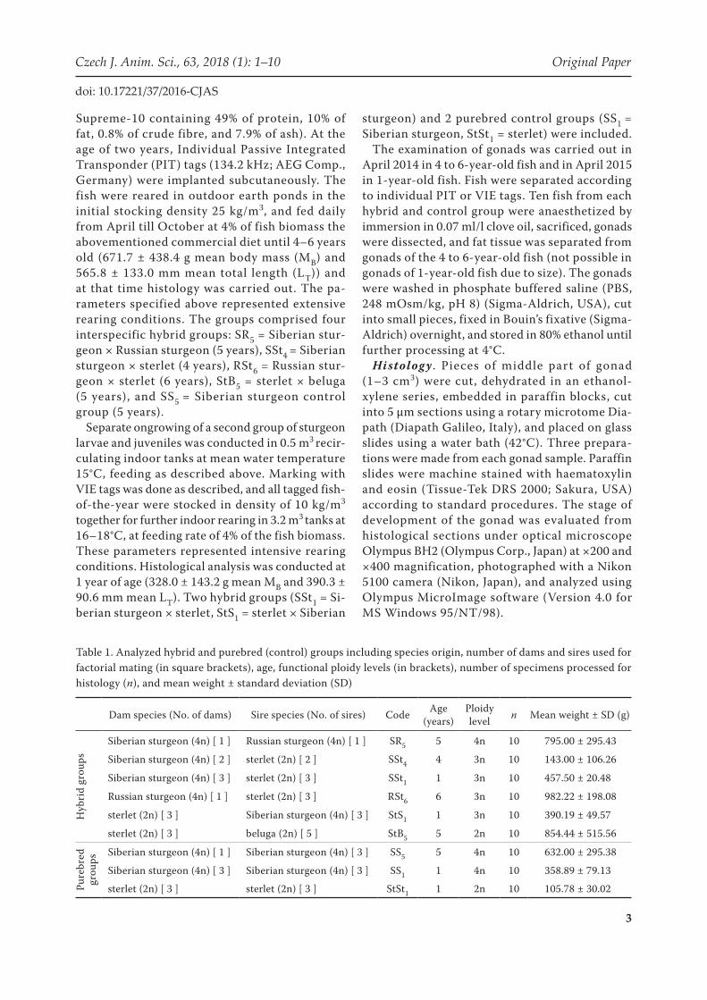
Table 1. Analyzed hybrid and purebred (control) groups including species origin, number of dams and sires used for factorial mating (in square brackets), age, functional ploidy levels (in brackets), number of specimens processed for histology (n), and mean weight ± standard deviation (SD)
Dam species (No. of dams) Sire species (No. of sires) Code Age (years)
Ploidy level n Mean weight ± SD (g)
Hyb
rid
grou
ps
Siberian sturgeon (4n) [ 1 ] Russian sturgeon (4n) [ 1 ] SR5 5 4n 10 795.00 ± 295.43
Siberian sturgeon (4n) [ 2 ] sterlet (2n) [ 2 ] SSt4 4 3n 10 143.00 ± 106.26
Siberian sturgeon (4n) [ 3 ] sterlet (2n) [ 3 ] SSt1 1 3n 10 457.50 ± 20.48
Russian sturgeon (4n) [ 1 ] sterlet (2n) [ 3 ] RSt6 6 3n 10 982.22 ± 198.08
sterlet (2n) [ 3 ] Siberian sturgeon (4n) [ 3 ] StS1 1 3n 10 390.19 ± 49.57
sterlet (2n) [ 3 ] beluga (2n) [ 5 ] StB5 5 2n 10 854.44 ± 515.56
Pure
bred
gr
oups
Siberian sturgeon (4n) [ 1 ] Siberian sturgeon (4n) [ 3 ] SS5 5 4n 10 632.00 ± 295.38
Siberian sturgeon (4n) [ 3 ] Siberian sturgeon (4n) [ 3 ] SS1 1 4n 10 358.89 ± 79.13
sterlet (2n) [ 3 ] sterlet (2n) [ 3 ] StSt1 1 2n 10 105.78 ± 30.02
4
Original Paper Czech J. Anim. Sci., 63, 2018 (1): 1–10
doi: 10.17221/37/2016-CJAS
Evaluation of cell apoptosis. A colorimetric non-isotopic FragEL™ DNA Fragmentation De-tection Kit (QIA33; Merck Millipore, USA) was used for the quantification and morphological characterization of normal and apoptotic gonad cells in paraffin-embedded tissue sections of all fish. The kit allows recognition of apoptotic nuclei in paraffin-embedded tissue sections by Frag-ment End Labeling (FragEL™) of DNA. Moreover, counterstaining with methyl green aids in the morphological evaluation of normal and apoptotic cells. Apoptosis was detected by dark labelling of DNA breaks in cell nuclei according to the manu-facturer’s protocol. Photographs were taken using an optical microscope Olympus BH2 (Olympus Corp.) at ×200 magnification with a Nikon 5100 camera (Nikon, Japan).
Molecular analyses. As suitable markers for direct identification of sturgeon species under study were not available, an alternative approach was used to investigate genetic makeup of par-ent fish based on the mtDNA control region and nuclear markers (microsatellites).
The genomic DNA was extracted from fin-clips of all parent fish using the NucleoSpin®tissue kit (MACHEREY-NAGEL, Germany). A standard poly-merase chain reaction (PCR) protocol was followed to amplify the mtDNA fragment of the control re-gion (Mugue et al. 2008) using forward primer AHR3 (CATACCATAATGTTTCATCTACC) and reverse primer DL651 (ATCTTAACATCTTCAGTG). The PCR reaction was carried out in 30 μl containing 0.25 μM of each primer, 75 mM Tris-HCl, pH 8.8, 20 mM (NH4)2SO4, 0.01% Tween 20, 2.5 mM MgCl2, 800 μM dNTP, 2.5 U Taq-Purple DNA polymerase, and 25 ng of DNA template. For PCR cycling, the following conditions were used: 95°C for 120 s, 5 cycles at 95°C for 60 s, 53°C for 60 s, 72°C for 60 s, followed by 30 cycles at 95°C for 30 s, 53°C for 45 s, 72°C for 60 s, and a final extension at 72°C for 12 min. The PCR products were pu-rified and sequenced by Macrogen Europe Inc. (the Netherlands) using primer AHR3. Partial sequences of the mtDNA control region were trimmed to 550 bp, aligned by GENEIOUS 6.1.8 software (http://www.geneious.com), and result-ing haplotypes were searched against the NCBI nucleotide database using Mega-BLAST.
The multidimensional factorial correspondence analysis (FCA) was used to reveal the origin of the parent fish based on nuclear markers. For
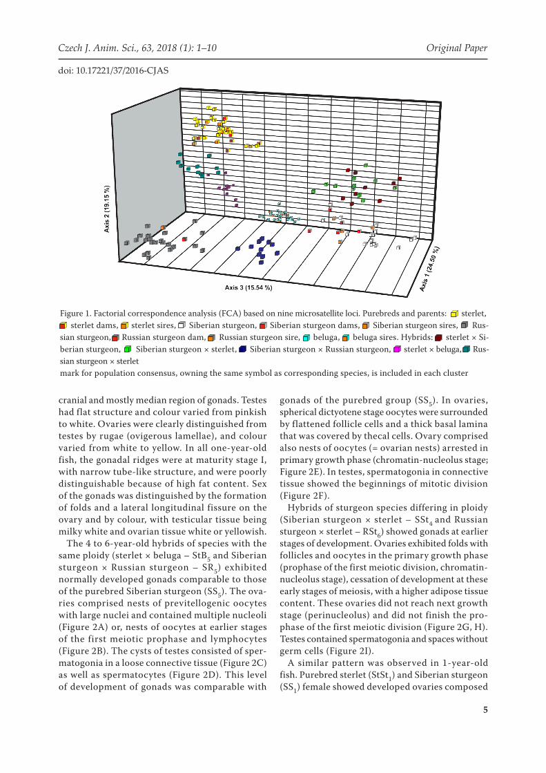
this purpose, the following DNA samples were additionally included in the analysis (number of samples in brackets): sterlet (19), beluga (19), Siberian sturgeon (15), Russian sturgeon (22), ster-let × beluga (10), Russian sturgeon × sterlet (10), Siberian sturgeon × Russian sturgeon (10), ster-let × Siberian sturgeon (10), Siberian sturgeon × sterlet (10). The microsatellite genotypes of these specimens were used to form reference clusters against which the genotypes of parental individuals were compared. Nine markers including Afu 19, Afu 68 (May et al. 1997); Aox 27, Aox 45 (King et al. 2001); and Spl 101, Spl 105, Spl 107, Spl 163, and Spl 173 (McQuown et al. 2000) were used for the analysis. Amplification was carried out according to the protocol described by Havelka et al. (2013). Microsatellite fragment analysis was performed on a 3500 ABI Genetic Analyzer (Ap-plied Biosystems, USA) using a GeneScan LIZ 600 size standard (Applied Biosystems), and genotypes were scored in GENEIOUS 6.1.8 (http://www.geneious.com), using Microsatellite Plugin 1.4. Genetic relationships among individuals based on the multilocus genotypes were visualized by FCA in GENETIX software (Version 4.05, 2004) for MS Windows. This enabled visualization of data in multidimensional space, with no a priori assumptions on grouping, using each allele as an independent variable.
RESULTS
All parent fish had mtDNA haplotypes (Sup-plementary Table S2 in SOM) corresponding to their species, showing no evidence of maternal gene introgression from other sturgeon species. The nuclear markers placed parent fish in clusters of A. ruthenus, A. baerii, A. gueldenstaedtii, and H. huso (Figure 1). Hence, they were assumed to be pure species based on nuclear markers, which, together with results of mtDNA analysis, showed parent fish to be pure specimens.
Gonad histology. Gonad development was as-sessed by their external appearance and histological examination of the inner structure and localization of germ cells. The sex ratio was 50 ± 5% for male and female, and only 3 intersexes were identified in purebred group of sterlet (StSt1). In all 4 to 6-year-old fish, the gonads were at maturity stage II, clearly discernible and surrounded by visceral fat in
5
Czech J. Anim. Sci., 63, 2018 (1): 1–10 Original Paper
doi: 10.17221/37/2016-CJAS
cranial and mostly median region of gonads. Testes had flat structure and colour varied from pinkish to white. Ovaries were clearly distinguished from testes by rugae (ovigerous lamellae), and colour varied from white to yellow. In all one-year-old fish, the gonadal ridges were at maturity stage I, with narrow tube-like structure, and were poorly distinguishable because of high fat content. Sex of the gonads was distinguished by the formation of folds and a lateral longitudinal fissure on the ovary and by colour, with testicular tissue being milky white and ovarian tissue white or yellowish.
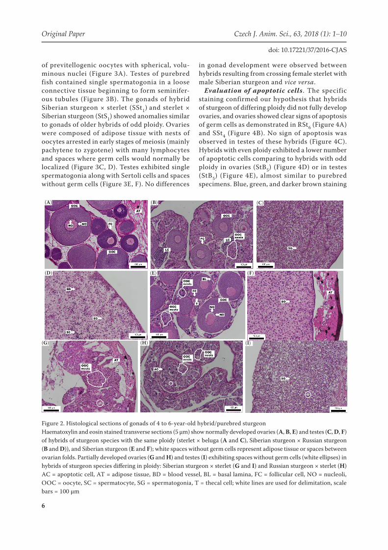
The 4 to 6-year-old hybrids of species with the same ploidy (sterlet × beluga – StB5 and Siberian sturgeon × Russian sturgeon – SR5) exhibited normally developed gonads comparable to those of the purebred Siberian sturgeon (SS5). The ova-ries comprised nests of previtellogenic oocytes with large nuclei and contained multiple nucleoli (Figure 2A) or, nests of oocytes at earlier stages of the first meiotic prophase and lymphocytes (Figure 2B). The cysts of testes consisted of sper-matogonia in a loose connective tissue (Figure 2C) as well as spermatocytes (Figure 2D). This level of development of gonads was comparable with
gonads of the purebred group (SS5). In ovaries, spherical dictyotene stage oocytes were surrounded by flattened follicle cells and a thick basal lamina that was covered by thecal cells. Ovary comprised also nests of oocytes (= ovarian nests) arrested in primary growth phase (chromatin-nucleolus stage; Figure 2E). In testes, spermatogonia in connective tissue showed the beginnings of mitotic division (Figure 2F).
Hybrids of sturgeon species differing in ploidy (Siberian sturgeon × sterlet – SSt4 and Russian sturgeon × sterlet – RSt6) showed gonads at earlier stages of development. Ovaries exhibited folds with follicles and oocytes in the primary growth phase (prophase of the first meiotic division, chromatin-nucleolus stage), cessation of development at these early stages of meiosis, with a higher adipose tissue content. These ovaries did not reach next growth stage (perinucleolus) and did not finish the pro-phase of the first meiotic division (Figure 2G, H). Testes contained spermatogonia and spaces without germ cells (Figure 2I).
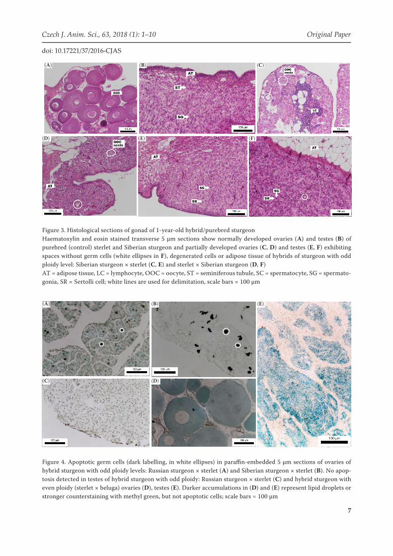
A similar pattern was observed in 1-year-old fish. Purebred sterlet (StSt1) and Siberian sturgeon (SS1) female showed developed ovaries composed
Figure 1. Factorial correspondence analysis (FCA) based on nine microsatellite loci. Purebreds and parents: sterlet, sterlet dams, sterlet sires, Siberian sturgeon, Siberian sturgeon dams, Siberian sturgeon sires, Rus-sian sturgeon, Russian sturgeon dam, Russian sturgeon sire, beluga, beluga sires. Hybrids: sterlet × Si- berian sturgeon, Siberian sturgeon × sterlet, Siberian sturgeon × Russian sturgeon, sterlet × beluga, Rus-sian sturgeon × sterletmark for population consensus, owning the same symbol as corresponding species, is included in each cluster
6
Original Paper Czech J. Anim. Sci., 63, 2018 (1): 1–10
doi: 10.17221/37/2016-CJAS
of previtellogenic oocytes with spherical, volu-minous nuclei (Figure 3A). Testes of purebred fish contained single spermatogonia in a loose connective tissue beginning to form seminifer-ous tubules (Figure 3B). The gonads of hybrid Siberian sturgeon × sterlet (SSt1) and sterlet × Siberian sturgeon (StS1) showed anomalies similar to gonads of older hybrids of odd ploidy. Ovaries were composed of adipose tissue with nests of oocytes arrested in early stages of meiosis (mainly pachytene to zygotene) with many lymphocytes and spaces where germ cells would normally be localized (Figure 3C, D). Testes exhibited single spermatogonia along with Sertoli cells and spaces without germ cells (Figure 3E, F). No differences
in gonad development were observed between hybrids resulting from crossing female sterlet with male Siberian sturgeon and vice versa.
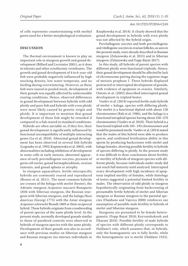
Evaluation of apoptotic cells . The specific staining confirmed our hypothesis that hybrids of sturgeon of differing ploidy did not fully develop ovaries, and ovaries showed clear signs of apoptosis of germ cells as demonstrated in RSt6 (Figure 4A) and SSt4 (Figure 4B). No sign of apoptosis was observed in testes of these hybrids (Figure 4C). Hybrids with even ploidy exhibited a lower number of apoptotic cells comparing to hybrids with odd ploidy in ovaries (StB5) (Figure 4D) or in testes (StB5) (Figure 4E), almost similar to purebred specimens. Blue, green, and darker brown staining
Figure 2. Histological sections of gonads of 4 to 6-year-old hybrid/purebred sturgeonHaematoxylin and eosin stained transverse sections (5 µm) show normally developed ovaries (A, B, E) and testes (C, D, F) of hybrids of sturgeon species with the same ploidy (sterlet × beluga (A and C), Siberian sturgeon × Russian sturgeon (B and D)), and Siberian sturgeon (E and F); white spaces without germ cells represent adipose tissue or spaces between ovarian folds. Partially developed ovaries (G and H) and testes (I) exhibiting spaces without germ cells (white ellipses) in hybrids of sturgeon species differing in ploidy: Siberian sturgeon × sterlet (G and I) and Russian sturgeon × sterlet (H)AC = apoptotic cell, AT = adipose tissue, BD = blood vessel, BL = basal lamina, FC = follicular cell, NO = nucleoli, OOC = oocyte, SC = spermatocyte, SG = spermatogonia, T = thecal cell; white lines are used for delimitation, scale bars = 100 µm
(A) (B) (C)
(D) (E) (F)
(G) (H) (I)
7
Czech J. Anim. Sci., 63, 2018 (1): 1–10 Original Paper
doi: 10.17221/37/2016-CJAS
Figure 3. Histological sections of gonad of 1-year-old hybrid/purebred sturgeonHaematoxylin and eosin stained transverse 5 µm sections show normally developed ovaries (A) and testes (B) of purebred (control) sterlet and Siberian sturgeon and partially developed ovaries (C, D) and testes (E, F) exhibiting spaces without germ cells (white ellipses in F), degenerated cells or adipose tissue of hybrids of sturgeon with odd ploidy level: Siberian sturgeon × sterlet (C, E) and sterlet × Siberian sturgeon (D, F)AT = adipose tissue, LC = lymphocyte, OOC = oocyte, ST = seminiferous tubule, SC = spermatocyte, SG = spermato-gonia, SR = Sertolli cell; white lines are used for delimitation, scale bars = 100 µm
Figure 4. Apoptotic germ cells (dark labelling, in white ellipses) in paraffin-embedded 5 µm sections of ovaries of hybrid sturgeon with odd ploidy levels: Russian sturgeon × sterlet (A) and Siberian sturgeon × sterlet (B). No apop-tosis detected in testes of hybrid sturgeon with odd ploidy: Russian sturgeon × sterlet (C) and hybrid sturgeon with even ploidy (sterlet × beluga) ovaries (D), testes (E). Darker accumulations in (D) and (E) represent lipid droplets or stronger counterstaining with methyl green, but not apoptotic cells; scale bars = 100 µm
(A) (B) (C)
(D) (E) (F)
(A) (B)
(C) (D)
(E)
8
Original Paper Czech J. Anim. Sci., 63, 2018 (1): 1–10
doi: 10.17221/37/2016-CJAS
of cells represents counterstaining with methyl green used for a better morphological evaluation.
DISCUSSION
The thermal environment is known to play an important role in sturgeon growth and gonad de-velopment (Billard and Lecointre 2001), as it does in teleosts and other ectothermic vertebrates. The growth and gonad development of 4 to 6-year-old fish were probably negatively influenced by high stocking density, low water temperate, and no feeding during overwintering. However, as these fish were reared in pooled stock, development of their gonads was equally affected by unfavourable rearing conditions. Hence, observed differences in gonad development between hybrids with odd ploidy and pure fish and hybrids with even ploidy were most likely caused by genetic origin and ploidy. It is important to mention that gonad development of these fish might be retarded if compared to a fish reared in standard conditions.
Hybrids are often unviable or, if they live, their gonad development is significantly influenced by functional incompatibility of multiple interacting genes (Lu et al. 2010). Abnormal gonad develop-ment has been observed in several fish hybrids (Legendre et al. 1992; Kopiejewska et al. 2003), with abnormalities including inhibition of development in some cells at early meiosis, abnormal appear-ance of early previtellogenic oocytes, pycnosis of germ cell nuclei, gonad hermaphroditism, ovarian tumours, and gonad aplasia or atrophy.
In sturgeon aquaculture, fertile interspecific hybrids are commonly reared and reproduced (Bronzi et al. 2011). The most common hybrids are crosses of the beluga with sterlet (bester), the Adriatic sturgeon Acipenser naccarii Bonaparte 1836 with Siberian sturgeon, the Russian stur-geon with Siberian sturgeon, and the kaluga Huso dauricus (Georgi 1775) with the Amur sturgeon Acipenser schrenckii Brandt 1869 or their reciprocal hybrid. These hybrids originate from crossbreeding of parent species of the same ploidy level. In the present study, normally developed gonads similar to those of purebred controls were observed in hybrids of sturgeon species with the same ploidy. Development of their gonads was also in accord-ance with previous studies on Siberian sturgeon and Russian sturgeon (no intersex individuals in
Rzepkowska et al. 2014). It clearly showed that the gonad development in hybrids with even ploidy was not affected by the hybrid origin.
Previtellogenic oocytes and both previtellogenic and vitellogenic oocytes in ovarian follicles, as seen in the present study, were already described in Russian sturgeon (Zelazowska et al. 2015) and in Siberian sturgeon (Zelazowska and Fopp-Bayat 2017).
In this study, all hybrids of parent species with different ploidy were functional triploids. Hence, their gonad development should be affected by lack of chromosome pairing during the zygotene stage of meiosis prophase I. These hybrids displayed protracted or interrupted development of gonads, with evidence of apoptosis in ovaries. Similarly, Omoto et al. (2002) described interrupted gonad development in triploid bester.
Vasilev et al. (2014) reported fertile male hybrids of sterlet × kaluga, species with differing ploidy. The sterlet is a functional diploid with about 120 chromosomes (Rab et al. 1986), and the kaluga is a functional tetraploid species having about 250–270 chromosomes (Vasilev et al. 2010). Their hybrid is a functional triploid with 185–195 chromosomes and would be presumed sterile. Vasilev et al. (2014) stated that the males of this hybrid were able to produce sperm, and confirmed fertilization ability in this sperm by producing backcrosses with sterlet and kaluga females, showing possible fertility in hybrids of species differing in ploidy. In the present study it was difficult to draw conclusions about fertility or sterility of hybrids of sturgeon species with dif-ferent ploidy, because individuals under study did not reach full maturity until analyzed. Interrupted ovary development with high incidence of apop-tosis implied sterility of females, while histology of testes suggested a potential limited fertility in males. The observation of odd ploidy in sturgeon hypothetically originating from backcrossing of presumably fertile hybrids of sterlet and Siberian sturgeon or Russian sturgeon to the parental spe-cies (Flajshans and Vajcova 2000) reinforces our assumption of possible male fertility in hybrids of sterlet and Siberian sturgeon.
Sturgeons are presumed to be female hetero-gametic (Fopp-Bayat 2010; Keyvanshokooh and Gharaei 2010). Possible fertility of male hybrids of species with different ploidy corresponds to Haldane’s rule, which assumes that, in hybrids, only the homogametic sex is fully fertile, while the heterogametic sex is sterile (Haldane 1922).
9
Czech J. Anim. Sci., 63, 2018 (1): 1–10 Original Paper
doi: 10.17221/37/2016-CJAS
Complete fertility of both sexes in hybrids of spe-cies with the same ploidy does not comply with this presumption, suggesting that sturgeon hybrid fertility is complex and may present unique char-acteristics, possibly due to the allopolyploid origin of the species (Fontana et al. 2007) and different levels of genome re-diploidization among sturgeon ploidy groups (Havelka et al. 2013).
CONCLUSION
Gonad mass as a proportion of the total body mass was not influenced by genetic origin or by ploidy in the analyzed specimens. Hybrids of species of the same ploidy showed gonad development similar to that of purebred controls. In contrast, hybrids of species with differing ploidy displayed inhibition of gonad development in some cells at early stages of meiosis. While ovarian development was inter-rupted and showed high incidence of apoptosis, testes continued to develop, and limited fertility of the hybrid males could not be excluded. In light of the evidence given in the present study and other studies to date, the general assumption of sterility of hybrid sturgeon with odd ploidy and therefore lack of concern with respect to their escape from farms should be seriously reconsidered. As a precaution, we suggest that all males of sturgeon hybrids should be assumed to be potentially fertile.
Acknowledgements. Granting agencies had no participation in the design of the study or inter-pretation of the results. The Lucidus Consultancy is gratefully acknowledged for English correction and suggestions. The authors declare that they have no competing interests.
REFERENCES
Allard R.W. (ed.) (1999): Principles of Plant Breeding. John Wiley and Sons, New York, USA.
Arefyev V.A. (1997): Sturgeon hybrids: natural reality and practical prospects. Aquaculture Magazine, 23, 53–58.
Bartley D.M., Rana K., Immink A.J. (2001): The use of inter-specific hybrids in aquaculture and fisheries. Reviews in Fish Biology and Fisheries, 10, 325–337.
Bemis W.E., Findeis E.K., Grande L. (1997): An overview of Acipenseriformes. Environmental Biology of Fishes, 48, 25–71.
Billard R., Lecointre G. (2001): Biology and conservation of sturgeon and paddlefish. Reviews in Fish Biology and Fisheries, 10, 355–392.
Birstein V.J., Hanner R., DeSalle R. (1997): Phylogeny of the Acipenseriformes: cytogenetic and molecular ap-proaches. Environmental Biology of Fishes, 48, 127–155.
Bronzi P., Rosenthal H., Gessner J. (2011): Global sturgeon aquaculture production: an overview. Journal of Applied Ichthyology, 27, 169–175.
Bytyutskyy D., Srp J., Flajshans M. (2012): Use of Feulgen image analysis densitometry to study the effect of genome size on nuclear size in polyploid sturgeons. Journal of Applied Ichthyology, 28, 704–708.
Bytyutskyy D., Kholodnyy V., Flajshans M. (2014): 3-D structure, volume, and DNA content of erythrocyte nuclei of polyploid fish. Cell Biology International, 38, 708–715.
Dowling T.E., Secor C.L. (1997): The role of hybridization and introgression in the diversification of animals. An-nual Review of Ecology and Systematics, 28, 593–619.
Fahy E., Martin S., Mulrooney M. (1988): Interactions of roach and bream in an Irish reservoir. Archiv für Hyd-robiologie, 114, 291–309.
Flajshans M., Vajcova V. (2000): Odd ploidy levels in stur-geon suggest a backcross of interspecific hexaploid stur-geon hybrids to evolutionary tetraploid and/or octaploid parental species. Folia Zoologica, 49, 133–138.
Fontana F., Zane L., Pepe A., Congiu L. (2007): Polyploidy in Acipenseriformes: cytogenetic and molecular approaches. In: Pisano E., Ozouf-Costaz C., Foresti F., Kapoor B.G. (eds): Fish Cytogenetics. Science Publisher, Enfield, USA, 385–403.
Fopp-Bayat D. (2010): Meiotic gynogenesis revealed not homogametic female sex determination system in Si-berian sturgeon (Acipenser baeri Brandt). Aquaculture, 305, 174–177.
Haldane J. (1922): Sex ration and unisexual sterility in ani-mal hybrids. Journal of Genetics, 12, 101–109.
Havelka M., Hulak M., Bailie D.A., Prodohl P.A., Flajs-hans M. (2013): Extensive genome duplications in stur-geons: new evidence from microsatellite data. Journal of Applied Ichthyology, 29, 704–708.
Keyvanshokooh S., Gharaei A. (2010): A review of sex determination and searches for sex-specific markers in sturgeon. Aquaculture Research, 41, e1–e7.
King T.L., Lubinski B.A., Spidle A.P. (2001): Microsatellite DNA variation in Atlantic sturgeon (Acipenser oxyrin-chus oxyrinchus) and cross-species amplification in the Acipenseridae. Conservation Genetics, 2, 103–119.
Kopiejewska W., Terlecki J., Chybowski L. (2003): Varied somatic growth and sex cell development in reciprocal hybrids of roach Rutilus rutilus (L.) and ide Leuciscus idus (L.). Archives of Polish Fisheries, 11, 33–44.
10
Original Paper Czech J. Anim. Sci., 63, 2018 (1): 1–10
doi: 10.17221/37/2016-CJAS
Legendre M., Teugels G.G., Cauty C., Jalabert B. (1992): A comparative study on morphology, growth rate and reproduction of Clarias gariepinus (Burchell, 1822), Heterobranchus longifilis Valenciennes, 1840, and their reciprocal hybrids (Pisces, Clariidae). Journal of Fish Biology, 40, 59–79.
Lu X., Shapiro J.A., Ting C.T., Li Y., Li C., Xu J., Huang H., Cheng Y.J., Greenberg A.J., Li S.H., Wu M.L., Shen Y., Wu C.I. (2010): Genome-wide misexpression of X-linked versus autosomal genes associated with hybrid male ste-rility. Genome Research, 20, 1097–1102.
Ludwig A., Lippold S., Debus L., Reinartz R. (2009): First evidence of hybridization between endangered sterlets (Acipenser ruthenus) and exotic Siberian sturgeons (Aci-penser baerii) in the Danube River. Biology Invasions, 11, 753–760.
May B., Krueger C.C., Kincaid H.L. (1997): Genetic varia-tion at microsatellite loci in sturgeon: primer sequence homology in Acipenser and Scaphirhynchus. Canadian Journal of Fisheries and Aquatic Sciences, 54, 1542–1547.
McQuown E.C., Sloze B.L., Sheehan R.J., Rodzen J., Tranah G.J., May B. (2000): Microsatellite analysis of genetic vari-ation in sturgeon (Acipenseridae): new primer sequences for Scaphirhynchus and Acipenser. Transactions of the American Fisheries Society, 129, 1380–1388.
Mugue N.S., Barmintseva A.E., Rastorguev S.M., Mugue V.N., Barminstev V.A. (2008): Polymorphism of the mi-tochondrial DNA control region in eight sturgeon species and development of a system for DNA-based species identification. Russian Journal of Genetics, 44, 793–798.
Omoto N., Maebayashi M., Adachi S., Arai K., Yamauchi K. (2002): Histological observations of gonadal development in gynogenetic diploids and triploids of a hybrid sturgeon, bester. Fisheries Science, 68 (Suppl. 2), 1271–1272.
Rab P. (1986): A note on the karyotype of the sterlet, Aci-penser ruthenus (Pisces, Acipenseridae). Folia Zoologica, 35, 73–78.
Rosati A., Tewolde A., Mosconi C. (eds) (2007): Animal Production and Animal Science Worldwide. Wageningen Academic Publishers, Wageningen, the Netherlands.
Rzepkowska M., Ostaszewska T., Gibala M., Roszko M.L. (2014): Intersex gonad differentiation in cultured Russian (Acipenser gueldenstaedtii) and Siberian (Acipenser ba-erii) sturgeon. Biology of Reproduction, 90, 1–10.
Seehausen O. (2004): Hybridization and adaptive radiation. Trends in Ecology and Evolution, 19, 198–207.
Soltis P.S., Soltis D.E. (2009): The role of hybridization in plant speciation. Annual Review in Plant Biology, 60, 561–588.
Vasilev V.P., Vasileva E.D., Shedko S.V., Novomodny G.V. (2010): How many times has polyploidization occurred during Acipenserid evolution? New data on the karyo-types of sturgeons (Acipenseridae, Actinopterygii) from the Russian Far East. Journal of Ichthyology, 50, 950–959.
Vasilev V.P., Rachek E.I., Lebedeva E.B., Vasileva E.D. (2014): Karyological study in backcross hybrids between the sterlet, Acipenser ruthenus, and kaluga, A. dauricus (Actinopterygii: Acipenseriformes: Acipenseridae): A. ru-thenus × (A. ruthenus × A. dauricus) and A. dauricus × (A. ruthenus × A. dauricus). Acta Ichthyologica et Pis-catoria, 44, 301–308.
Zelazowska M., Fopp-Bayat D. (2017): Previtellogenic and vitellogenic oocytes in ovarian follicles of cultured Sibe-rian sturgeon Acipenser baerii (Chondrostei, Acipenseri-formes). Journal of Morphology, 278, 50–61.
Zelazowska M., Jankowska W., Plewniak E., Rajek U. (2015): Ovarian nests in cultured Russian sturgeon Acipenser gueldenstaedtii and North American paddlefish Polyodon spathula comprised of previtellogenic oocytes. Journal of Fish Biology, 86, 1669–1679.
Zhang X., Wu W., Li L., Ma X., Chen J. (2013): Genetic variation and relationships of seven sturgeon species and ten interspecific hybrids. Genetics Selection Evolu-tion, 45, 21.
Received: 2016–03–23Accepted after corrections: 2017–10–06