Embed Size (px)
Citation preview
of September 27, 2016.This information is current as
Graft-versus-Tumor Effect in MiceGraft-versus-Host Disease and Preserve the Intestinal Helminths Regulate Lethal Acute
Elliott and M. Nedim InceB. Rothman, George J. Weiner, Bruce R. Blazar, David E.
PaulAdrian N. Holm, Ahmed Metwali, Joseph F. Urban, Jr., Yue Li, Hung-Lin Chen, Nadine Bannick, Michael Henry,
http://www.jimmunol.org/content/194/3/1011doi: 10.4049/jimmunol.1303099December 2014;
2015; 194:1011-1020; Prepublished online 19J Immunol
MaterialSupplementary
9.DCSupplemental.htmlhttp://www.jimmunol.org/content/suppl/2014/12/19/jimmunol.130309
Referenceshttp://www.jimmunol.org/content/194/3/1011.full#ref-list-1
, 30 of which you can access for free at: cites 63 articlesThis article
Subscriptionshttp://jimmunol.org/subscriptions
is online at: The Journal of ImmunologyInformation about subscribing to
Permissionshttp://www.aai.org/ji/copyright.htmlSubmit copyright permission requests at:
Email Alertshttp://jimmunol.org/cgi/alerts/etocReceive free email-alerts when new articles cite this article. Sign up at:
Print ISSN: 0022-1767 Online ISSN: 1550-6606. Immunologists, Inc. All rights reserved.Copyright © 2015 by The American Association of9650 Rockville Pike, Bethesda, MD 20814-3994.The American Association of Immunologists, Inc.,
is published twice each month byThe Journal of Immunology
by guest on September 27, 2016
http://ww
w.jim
munol.org/
Dow
nloaded from
by guest on September 27, 2016
http://ww
w.jim
munol.org/
Dow
nloaded from
The Journal of Immunology
Intestinal Helminths Regulate Lethal Acute Graft-versus-HostDisease and Preserve the Graft-versus-Tumor Effect in Mice
Yue Li,* Hung-Lin Chen,* Nadine Bannick,† Michael Henry,†,‡ Adrian N. Holm,*
Ahmed Metwali,* Joseph F. Urban, Jr.,x Paul B. Rothman,*,1 George J. Weiner,*,‡
Bruce R. Blazar,{ David E. Elliott,* and M. Nedim Ince*,‡
Donor T lymphocyte transfer with hematopoietic stem cells suppresses residual tumor growth (graft-versus-tumor [GVT]) in cancer patients
undergoing bone marrow transplantation (BMT). However, donor T cell reactivity to host organs causes severe and potentially lethal inflam-
mation called graft-versus-host disease (GVHD). High-dose steroids or other immunosuppressive drugs are used to treat GVHD that have
limited ability to control the inflammation while incurring long-term toxicity. Novel strategies are needed to modulate GVHD, preserve GVT,
and improve the outcome of BMT.Regulatory T cells (Tregs) control alloantigen-sensitized inflammation ofGVHD, sustainGVT, and prevent
mortality in BMT.Helminths colonizing the alimentary tract dramatically increase the Treg activity, therebymodulating intestinal or systemic
inflammatory responses.These observations ledus tohypothesize that helminths can regulateGVHDandmaintainGVTinmice.AcuteGVHD
was induced in helminth (Heligmosomoides polygyrus)–infected or uninfected BALB/c recipients of C57BL/6 donor grafts. Helminth infection
suppressed donor T cell inflammatory cytokine generation and reduced GVHD-related mortality, but maintained GVT. H. polygyrus
colonization promoted the survival of TGF-b–generating recipient Tregs after a conditioning regimen with total body irradiation
and led to a TGF-b–dependent in vivo expansion/maturation of donor Tregs after BMT. Helminths did not control GVHD when
T cells unresponsive to TGF-b–mediated immune regulation were used as donor T lymphocytes. These results suggest that helminths
suppress acute GVHD using Tregs and TGF-b–dependent pathways in mice. Helminthic regulation of GVHD and GVT through
intestinal immune conditioning may improve the outcome of BMT. The Journal of Immunology, 2015, 194: 1011–1020.
Graft-versus-host disease (GVHD) is a major and poten-tially severe complication of bone marrow transplanta-tion (BMT). The disease is mediated by alloreactivity of
donor T lymphocytes to recipient major or minor histocompati-bility Ags (1, 2). Although the acute form of GVHD affects theskin, intestine, and liver, chronic GVHD exhibits multiorgan in-filtration similar to various autoimmune diseases (3).
Intestinal inflammation in GVHD simulates inflammatory boweldiseases (IBDs), a group of immunological disorders that includesulcerative colitis and Crohn’s disease. Furthermore, allelic variants ofthe mammalian receptor protein for bacterial muramyldipeptide,CARD15/NOD2, influences the propensity to develop Crohn’s dis-ease, as well as GVHD (4, 5). Certain genetic variants of IL-23Rprotect individuals from these two disorders (6, 7). Inflammation inmouse models of IBD or GVHD is controlled by various immuno-modulatory mechanisms that include regulatory T cells (Tregs) thatsuppress inflammation driven by effector T lymphocytes (1, 4, 8, 9).Tregs express the transcription factor Foxp3 and contribute to intes-tinal immune regulation by cell contact–dependent mechanisms or bythe production of modulating cytokines, such as IL-10 and TGF-b(9–11). Helminthic regulation of intestinal immunity is associatedwith activation of Treg subsets and induction of regulatory cytokineproduction and depends on intact TGF-b circuitries (12–15).The immune modulatory murine nematode Heligmosomoides
polygyrus briefly resides in the submucosa of the mouse duodenumafter oral administration and then remains in intestinal lumenwithout causing systemic infection, until the adult worm is expelled.We demonstrated previously that helminths like H. polygyrus trig-ger intestinal Treg activity and regulatory cytokine generation withconsequent regulation of colitis in mice (13, 16). Other parasiticinfestations may have immunosuppressive properties and wereshown to reduce clinical activity in conditions such as multiplesclerosis, celiac disease, and IBD (17–21).Although GVHD can be prevented by depleting donor T cells from
the graft, recipients then are predisposed to severe infectious diseases.Engraftment, as well as the graft-versus-tumor (GVT) effect, may bediminished by donor T cell removal (1, 22, 23). In preclinical mousemodels, several laboratories showed that GVHD can be preventedwith preserved antitumor immunity (GVT) by coadministration ofdonor conventional T cells and Tregs given in equal numbers (22,
*Department of Internal Medicine, Carver College of Medicine, University of Iowa, IowaCity, IA 52242; †Department of Molecular Physiology and Biophysics, Carver College ofMedicine, University of Iowa, Iowa City, IA 52242; ‡Holden Comprehensive CancerCenter, Carver College of Medicine, University of Iowa, Iowa City, IA 52242; xDiet,Genomics, and Immunology Laboratory, Beltsville Human Nutrition Research Center,Agricultural Research Service, U.S. Department of Agriculture, Beltsville, MD 20705;and {Division of Blood and Marrow Transplantation, Department of Pediatrics, Universityof Minnesota, Minneapolis, MN 55455
1Current address: School of Medicine, Johns Hopkins University, Baltimore, MD.
Received for publication November 15, 2013. Accepted for publication November16, 2014.
This work was supported by Grants K08 DK082913 from the National Institute ofDiabetes and Digestive and Kidney Diseases, Lymphoma SPORE P50 CA097274from the National Cancer Institute, ACS-IRG-77-004-31 from the American CancerSociety and administered through the Holden Comprehensive Cancer Center at theUniversity of Iowa (to M.N.I.), R01 AI34495 from the National Institute of Allergyand Infectious Diseases, and R01 HL56067 and R01 HL11879 from the NationalHeart, Lung, and Blood Institute (to B.R.B.), as well as a VA Merit Award (to D.E.E.).
Address correspondence and reprint requests to Dr. M. Nedim Ince, Division ofGastroenterology and Hepatology, Department of Internal Medicine, University ofIowa Hospital and Clinics, 4546 John Colloton Pavilion, 200 Hawkins Drive, IowaCity, IA 52242-1009. E-mail address: [email protected]
The online version of this article contains supplemental material.
Abbreviations used in this article: BM, bone marrow; BMT, bone marrow transplan-tation; DN, dominant negative; GVHD, graft-versus-host disease; GVT, graft versustumor; IBD, inflammatory bowel disease; LAP, latency-associated protein; MFI,mean fluorescent intensity; MLN, mesenteric lymph node; TBI, total body irradia-tion; TCD, T cell depleted; Treg, regulatory T cell; WT, wild-type.
Copyright� 2015 by The American Association of Immunologists, Inc. 0022-1767/15/$25.00
www.jimmunol.org/cgi/doi/10.4049/jimmunol.1303099
by guest on September 27, 2016
http://ww
w.jim
munol.org/
Dow
nloaded from
24–26). However, the production of high numbers of human Tregssuitable for infusion remains a technically challenging goal.In this study, we show that H. polygyrus treatment of the recipient
protects mice from fatal acute GVHD and sustains GVT. Regulationof GVHD is associated with induction of Tregs that may regulateTh1 inflammation by means of TGF-b expression and secretion.Helminths reduce GVHD-related Th1 inflammation. Furthermore,in a TGF-b–dependent manner, H. polygyrus administrationdecreases GVHD-related mortality. Because the intestine is a pri-
mary organ for GVHD generation (5, 27), our results open thepossibility that intestinal immune conditioning of patients prior toBMT may be a useful strategy to reduce GVHD-related morbidityand mortality and preserve the donor graft’s antitumor immunity.
Materials and MethodsMice, H. polygyrus administration, and egg counting
We used wild-type (WT) C57BL/6 (H2b) and BALB/c (H2d) mice (JacksonLaboratory, Bar Harbor, ME), as well as a C57BL/6 mouse strain with
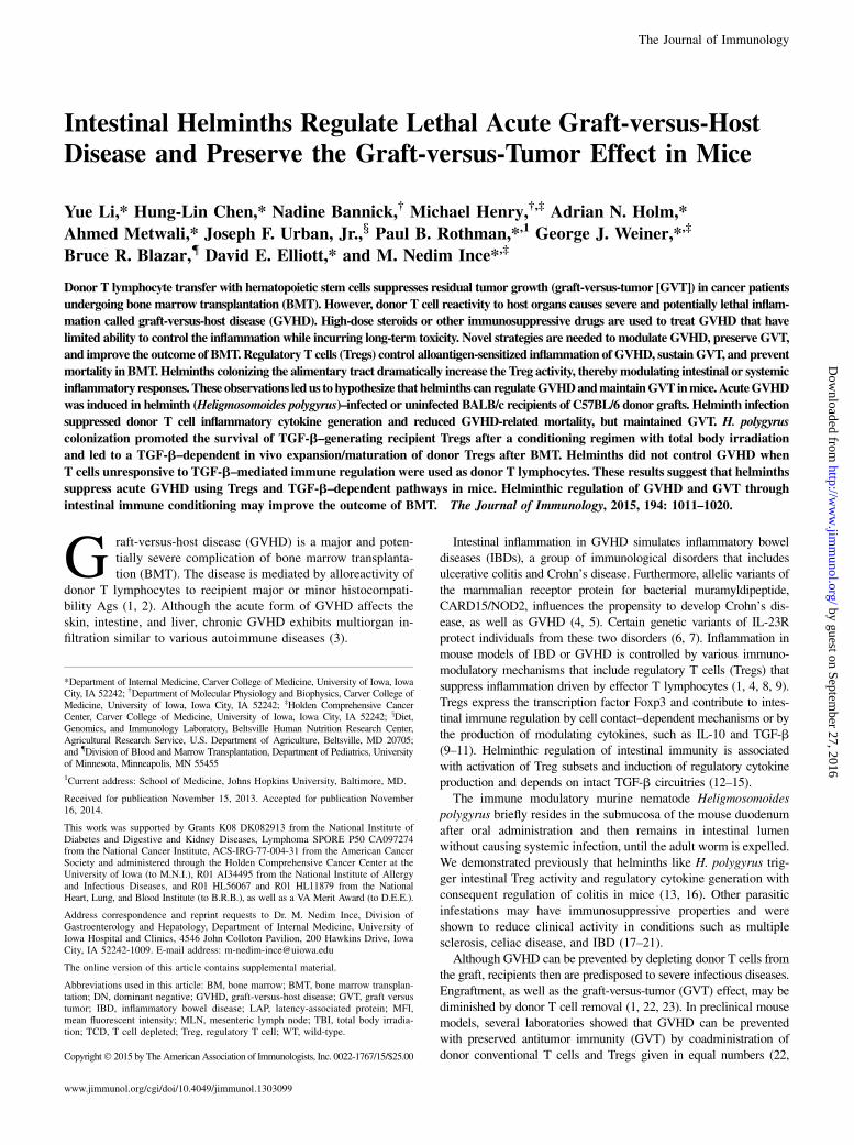
FIGURE 1. Helminths regulate acute GVHD in
mice. (A) H. polygyrus infection protects mice from the
severe inflammation during acute GVHD. Six days
after BMT, H. polygyrus–infected animals displayed
less skin discoloration or hunching body posture
compared with uninfected mice, with representative
examples shown. (B) Kaplan–Meier survival curves of
H. polygyrus–infected or uninfected BALB/c recipients
that received TCD BM and total splenic T cells from
C57BL/6 mice. Cumulative data from three indepen-
dent experiments (n = 9 uninfected, n = 10 infected).
(C) Weight change in the mice in (B) during the follow-
up period of the survival experiment. (D) Disease score
for uninfected (n = 9) and H polygyrus–infected (n =
10) mice during the entire course of the survival ex-
periment.
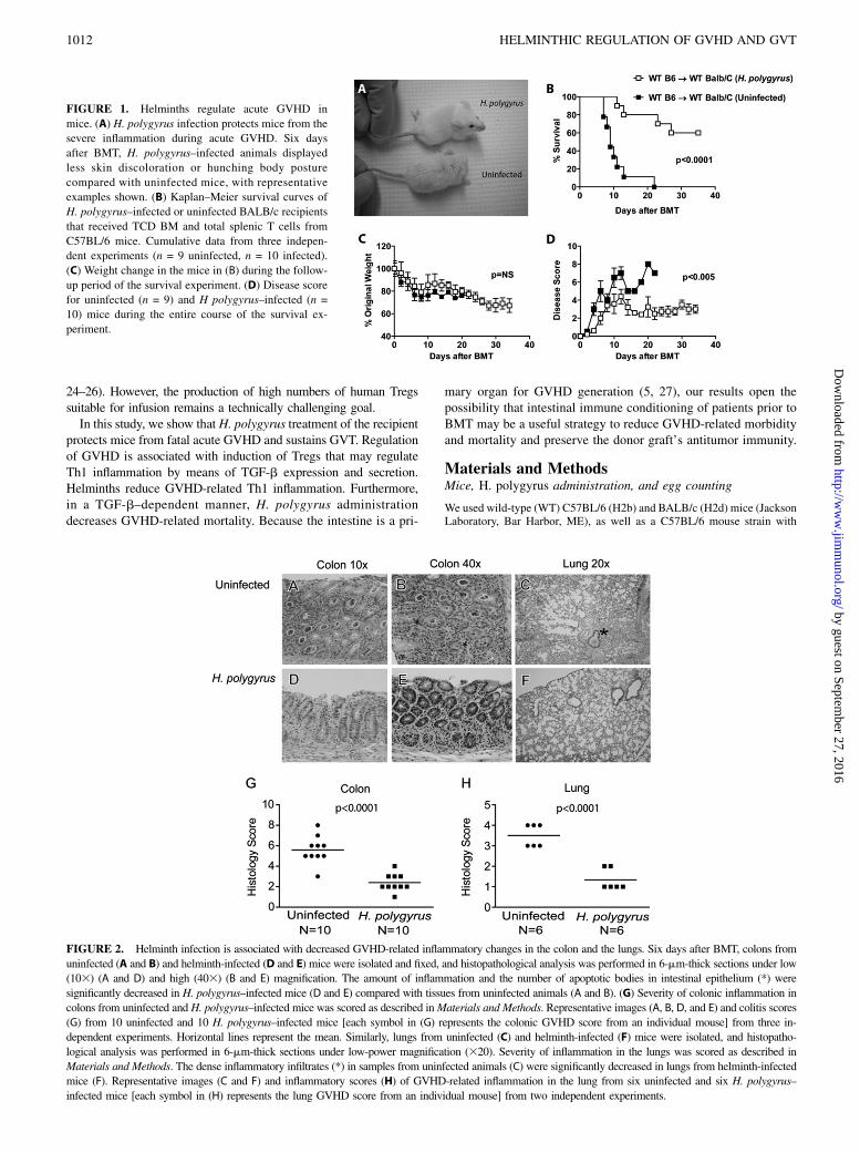
FIGURE 2. Helminth infection is associated with decreased GVHD-related inflammatory changes in the colon and the lungs. Six days after BMT, colons from
uninfected (A and B) and helminth-infected (D and E) mice were isolated and fixed, and histopathological analysis was performed in 6-mm-thick sections under low
(103) (A and D) and high (403) (B and E) magnification. The amount of inflammation and the number of apoptotic bodies in intestinal epithelium (*) were
significantly decreased in H. polygyrus–infected mice (D and E) compared with tissues from uninfected animals (A and B). (G) Severity of colonic inflammation in
colons from uninfected and H. polygyrus–infected mice was scored as described inMaterials and Methods. Representative images (A, B, D, and E) and colitis scores
(G) from 10 uninfected and 10 H. polygyrus–infected mice [each symbol in (G) represents the colonic GVHD score from an individual mouse] from three in-
dependent experiments. Horizontal lines represent the mean. Similarly, lungs from uninfected (C) and helminth-infected (F) mice were isolated, and histopatho-
logical analysis was performed in 6-mm-thick sections under low-power magnification (320). Severity of inflammation in the lungs was scored as described in
Materials and Methods. The dense inflammatory infiltrates (*) in samples from uninfected animals (C) were significantly decreased in lungs from helminth-infected
mice (F). Representative images (C and F) and inflammatory scores (H) of GVHD-related inflammation in the lung from six uninfected and six H. polygyrus–
infected mice [each symbol in (H) represents the lung GVHD score from an individual mouse] from two independent experiments.
1012 HELMINTHIC REGULATION OF GVHD AND GVT
by guest on September 27, 2016
http://ww
w.jim
munol.org/
Dow
nloaded from
a T cell–specific defect in TGF-b signaling (Cd4-TGFBR2, JacksonLaboratory #005551; also called TGF-bRII dominant negative [DN])(H2b) (28). Five- to six-week-old BALB/c mice were inoculated with 150H. polygyrus third-stage larvae by oral gavage. Infective H. polygyrusthird-stage larvae [original specimens archived at the U.S. National Hel-minthological Collection no. 81930; also named H. polygyrus (bakeri) orH. bakeri in some publications (29, 30)] were obtained from mouse fecalcultures of eggs by the modified Baermann method and stored at 4˚C untilused. The number of eggs in hydrated stool pellets was enumerated induplicate at the indicated time points for each mouse and are shown as eggnumber/stool weight. Mice were housed and handled following nationalguidelines and as approved by the Animal Review Committee of theUniversity of Iowa. Three weeks after initiation of helminth infection,mice underwent conditioning for BMT.
Cell purification for GVHD induction
Donor bone marrow (BM) cells were obtained from the femurs and tibias ofuninfected, 5–8-wk-old C57BL/6 mice, and T cells were depleted usingmouse pan-T cell beads (Dynal Biotech), according to the manufacturer’sinstructions. Donor T lymphocytes (CD3+) were magnetically enriched fromsplenic single-cell suspensions of uninfected, 5–8-wk-old C57BL/6 andTGF-bRII DN mice using the Pan T Cell Isolation Kit (Miltenyi Biotec).
Cell purification for in vitro cultures
To determine TGF-b cytokine generation of Treg-enriched and Treg-depleted cultures, CD4+ T cells were purified from splenic and mesen-teric lymph node (MLN) single-cell suspensions of H. polygyrus–infectedand uninfected BALB/c mice, using a CD4 T cell Isolation Kit (MiltenyiBiotec), and separated into CD25+ and CD252 fractions using anti-CD25PE labeling, followed by magnetic separation with anti-PE beads (MiltenyiBiotec). Enrichment or depletion efficiency was .98% with these tech-niques (data not shown). To determine helminthic regulation of donorT cell IFN-g, TNF-a, and IL-4 cytokine output during GVHD, donorCD3+ T cells were sorted from total anti-CD3 FITC–stained and anti-H2bPE–stained splenocytes from uninfected and H. polygyrus–infected BALB/crecipients 6 d after GVHD induction using a FACSVantage SE DiVa cellsorter (Becton Dickinson).
Total body irradiation and GVHD induction
Our studies used an MHC I/II mismatch, acute lethal GVHD model (26).Uninfected and helminth-infected BALB/c recipients (H2d) underwentlethal total body irradiation (TBI) from a [137Cs] source (8.5 Gy in twodivided doses given 4 h apart) and were administered 10 3 106 T cell–depleted (TCD) BM cells and 1.5 3 106 splenic T lymphocytes fromuninfected C57BL/6 WT donors. To determine the effect of helminth in-fection on the conditioning regimen (TBI) without BMT, mice underwentirradiation with total doses ranging from sublethal 4 Gy to lethal 15 Gy,according to the same protocol. Similar split irradiation doses were used,except that the 4-Gy group received a single dose. To characterize the roleof TGF-b signaling in helminth-induced regulation of donor T cell–me-diated GVHD, 1.5 3 106 donor splenic T cells from TGF-bRII DN micewere administered along with 10 3 106 TCD BM cells from C57BL/6 WTmice into uninfected and H. polygyrus–infected BALB/c recipients. Micewere monitored daily for survival for up to 112 d in different experiments.Disease severity was scored daily based on animal weight, posture, ac-tivity, fur texture, and skin integrity (31–33). In parallel experiments,uninfected and helminth-infected mice were sacrificed 6 d after GVHDinduction for cellular and histological analysis.
Quantification of tumor load and assessment of GVT bybioluminescent imaging
Luciferase-expressing A20 leukemia/lymphoma (A20-luc) cells syngeneicwith recipients (H2d) were used for these experiments (34). Each recipientmouse received 3 3 105 A20-luc tumor cells i.v. within 24 h after BMT.Tumor load was assessed regularly in BMT recipient mice using an Ami1000 Advanced Molecular Imager (Spectral Instruments, Tucson, AZ) liveanimal imaging system. Five minutes before bioluminescent imaging, micewere placed in an oxygenated isoflurane chamber and administeredD-luciferin (Promega, Madison, WI) i.p. BMT recipient animals wereimaged for 5 min, and tumor load was quantitated using Living Imagesoftware v2.50 (Caliper Life Sciences).
Flow cytometry
Six days after GVHD induction, uninfected andH. polygyrus–infected micewere sacrificed. Spleen and MLN cells were isolated for cellular analysis.
For surface staining, cells were suspended at 2 3 107 cells/ml in PBS with2% FCS, and FcRs were blocked with 2.4G2 mAb. Cells were stained withvarious combinations of anti-CD3 FITC, anti-CD3 PE-Cy7, anti-CD4FITC, anti-CD4 PE-Cy7 (eBioscience), anti-latent TGF-b (latency asso-ciated peptide [LAP]) PE (BioLegend), anti-H2b PE, anti-H2d PE, andanti-H2b allophycocyanin (BD Biosciences). For the intracellular Foxp3staining, cells were stained with anti-Foxp3 PE, Foxp3 PE-Cy7, or Foxp3allophycocyanin using Foxp3 staining buffer (eBioscience), according tothe manufacturer’s instructions.
In vitro cell culture and cytokine ELISA
Eight- to nine-week-old uninfected or H. polygyrus–infected BALB/c micewere sacrificed 3 wk after initiation of helminth infection. Magnetically pu-rified CD41CD251 (Treg-enriched) or CD41CD252 (Treg-depleted) splenicand MLN cells from uninfected or H. polygyrus–infected BALB/c mice werestimulated with plate-bound anti-CD3 and soluble anti-CD28 (each 1 mg/ml;
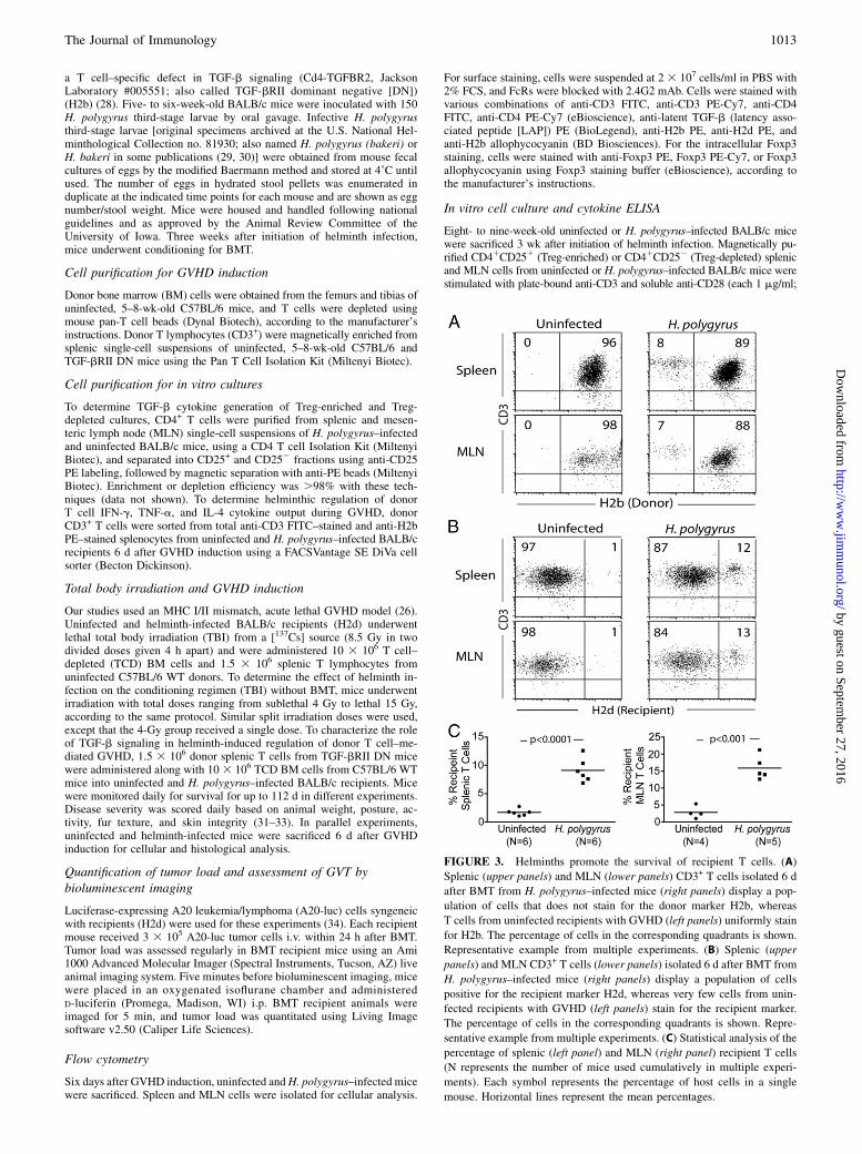
FIGURE 3. Helminths promote the survival of recipient T cells. (A)
Splenic (upper panels) and MLN (lower panels) CD3+ T cells isolated 6 d
after BMT from H. polygyrus–infected mice (right panels) display a pop-
ulation of cells that does not stain for the donor marker H2b, whereas
T cells from uninfected recipients with GVHD (left panels) uniformly stain
for H2b. The percentage of cells in the corresponding quadrants is shown.
Representative example from multiple experiments. (B) Splenic (upper
panels) and MLN CD3+ T cells (lower panels) isolated 6 d after BMT from
H. polygyrus–infected mice (right panels) display a population of cells
positive for the recipient marker H2d, whereas very few cells from unin-
fected recipients with GVHD (left panels) stain for the recipient marker.
The percentage of cells in the corresponding quadrants is shown. Repre-
sentative example from multiple experiments. (C) Statistical analysis of the
percentage of splenic (left panel) and MLN (right panel) recipient T cells
(N represents the number of mice used cumulatively in multiple experi-
ments). Each symbol represents the percentage of host cells in a single
mouse. Horizontal lines represent the mean percentages.
The Journal of Immunology 1013
by guest on September 27, 2016
http://ww
w.jim
munol.org/
Dow
nloaded from
eBioscience) for 48 h in cell culture medium with 1% FCS and 1 mg/mlBSA (13, 35). TGF-b cytokine concentration in acidified and realkalinizedsupernatants was determined using Ab pairs (R&D Systems), according to themanufacturer’s instructions. Results were calculated by subtracting the TGF-bconcentration of culture supernatants from the TGF-b concentrations of theculture media. To determine helminthic regulation of donor T cell IFN-g andTNF-a secretion, sorted donor splenic T cells (CD3+ and H2b+) from unin-fected and H. polygyrus–infected BALB/c mice with GVHD were stimulatedwith plate-bound anti-CD3 and soluble anti-CD28 (each 1 mg/ml) for 48 hin lymphocyte growth medium containing 10% FCS (15). Supernatants wereanalyzed for IFN-g, TNF-a, and IL-4 content using Ab pairs (R&D Systems).Similarly, sera isolated from uninfected and H. polygyrus–infected BALB/cmice 6 d after GVHD induction were analyzed for IFN-g and TNF-a.
Histopathology
Six days after GVHD induction, colons, lungs, and livers from uninfected orH. polygyrus–infected mice were fixed in 4% neutral buffered formalin andprocessed, and 6-mm sections were stained with H&E. Tissues were ana-lyzed for GVHD-related inflammation, and the severity of inflammation
was scored in a blinded fashion by A.N.H. (31, 36–39). GVHD-relatedcolitis was graded based on the degree of inflammation and the frequencyof crypt apoptosis. Inflammation was graded as none (0), mild (1), mod-erate (2), severe without ulcer (3), or severe with ulcer (4). Crypt apoptosiswas graded as rare (0), occasional per 10 crypts (1), few per 10 crypts (2),majority of crypts containing apoptotic bodies (3), or majority of cryptscontaining more than one apoptotic body (4). The minimal score in thisgrading system for colonic disease is 0, and the maximum score is 8.GVHD-related lung inflammation was graded based on the presenceof perivascular cuffing, vasculitis, peribronchiolar cuffing, and alveolarhemorrhage. The minimal score in this grading system for lung inflam-mation is 0, and the maximum is 4.
Statistical analysis
Differences in survival between groups were determined by the Kaplan–Meier log-rank test. Differences in cell number and composition, serumcytokine content, differences in splenic donor T cell cytokine generation,differences in TGF-b cytokine output of in vitro–stimulated cell cultures,and histopathological GVHD scores between two groups were determined
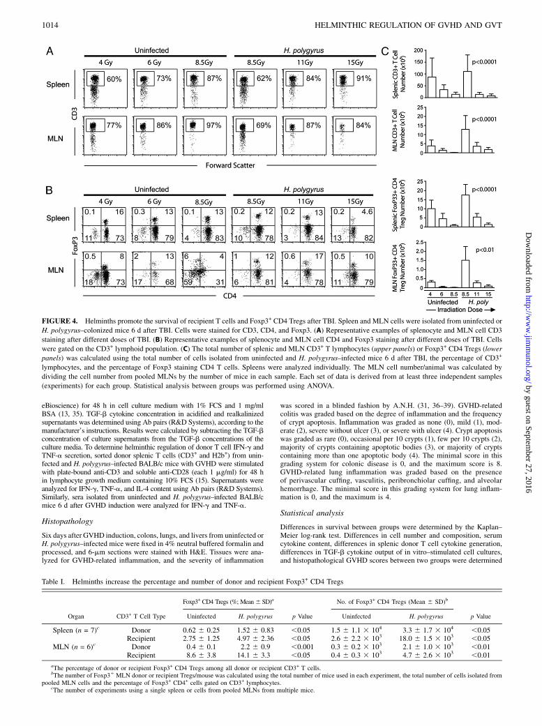
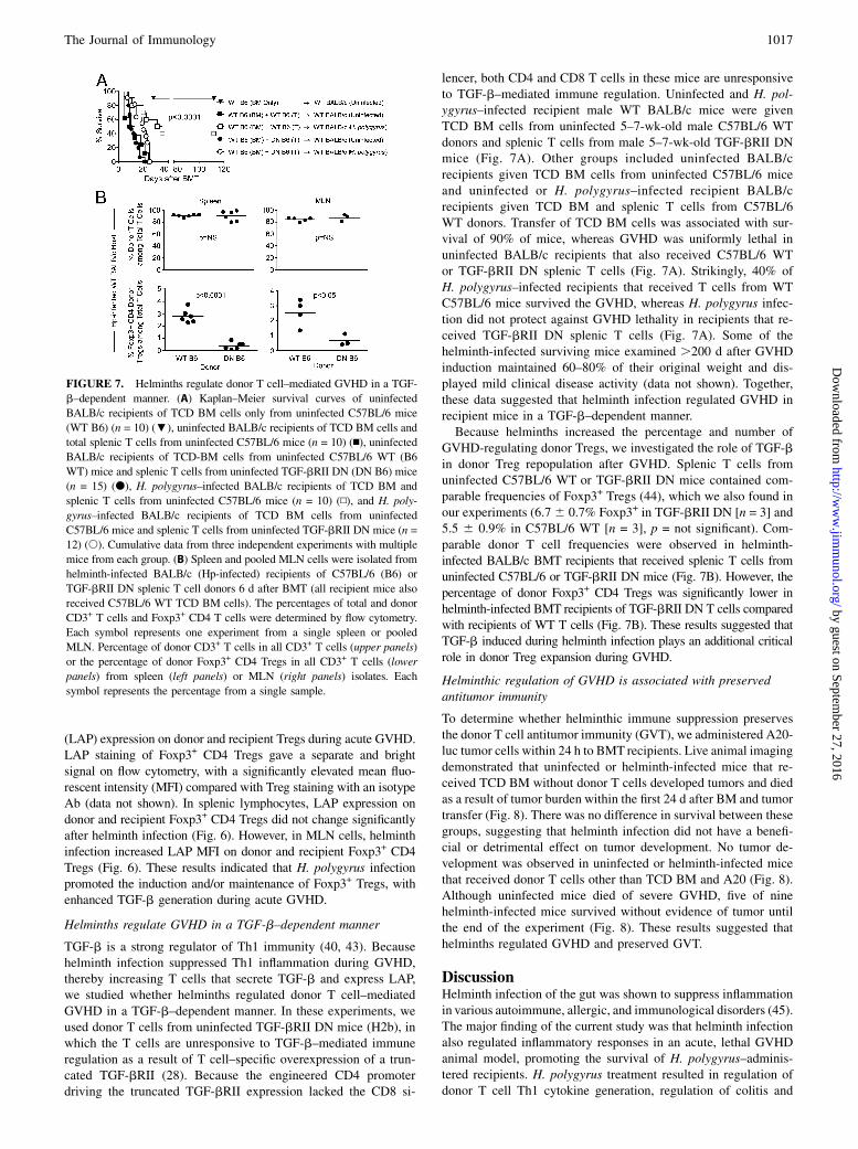
FIGURE 4. Helminths promote the survival of recipient T cells and Foxp3+ CD4 Tregs after TBI. Spleen and MLN cells were isolated from uninfected or
H. polygyrus–colonized mice 6 d after TBI. Cells were stained for CD3, CD4, and Foxp3. (A) Representative examples of splenocyte and MLN cell CD3
staining after different doses of TBI. (B) Representative examples of splenocyte and MLN cell CD4 and Foxp3 staining after different doses of TBI. Cells
were gated on the CD3+ lymphoid population. (C) The total number of splenic and MLN CD3+ T lymphocytes (upper panels) or Foxp3+ CD4 Tregs (lower
panels) was calculated using the total number of cells isolated from uninfected and H. polygyrus–infected mice 6 d after TBI, the percentage of CD3+
lymphocytes, and the percentage of Foxp3 staining CD4 T cells. Spleens were analyzed individually. The MLN cell number/animal was calculated by
dividing the cell number from pooled MLNs by the number of mice in each sample. Each set of data is derived from at least three independent samples
(experiments) for each group. Statistical analysis between groups was performed using ANOVA.
Table I. Helminths increase the percentage and number of donor and recipient Foxp3+ CD4 Tregs
Organ CD3+ T Cell Type
Foxp3+ CD4 Tregs (%; Mean6 SD)a
p Value
No. of Foxp3+ CD4 Tregs (Mean 6 SD)b
p ValueUninfected H. polygyrus Uninfected H. polygyrus
Spleen (n = 7)c Donor 0.62 6 0.25 1.52 6 0.83 ,0.05 1.5 6 1.1 3 104 3.3 6 1.7 3 104 ,0.05Recipient 2.75 6 1.25 4.97 6 2.36 ,0.05 2.6 6 2.2 3 103 18.0 6 1.5 3 103 ,0.05
MLN (n = 6)c Donor 0.4 6 0.1 2.2 6 0.9 ,0.001 0.3 6 0.2 3 103 2.1 6 1.0 3 103 ,0.01Recipient 8.6 6 3.8 14.1 6 3.3 ,0.05 0.4 6 0.3 3 103 4.7 6 2.6 3 103 ,0.01
aThe percentage of donor or recipient Foxp3+ CD4 Tregs among all donor or recipient CD3+ T cells.bThe number of Foxp31 MLN donor or recipient Tregs/mouse was calculated using the total number of mice used in each experiment, the total number of cells isolated from
pooled MLN cells and the percentage of Foxp3+ CD4+ cells gated on CD3+ lymphocytes.cThe number of experiments using a single spleen or cells from pooled MLNs from multiple mice.
1014 HELMINTHIC REGULATION OF GVHD AND GVT
by guest on September 27, 2016
http://ww
w.jim
munol.org/
Dow
nloaded from
using the Student t test. Differences in cell number and composition be-tween multiple groups were analyzed by ANOVA.
ResultsHelminth treatment of the recipient reduces GVHD
Acute GVHD was initiated in uninfected or H. polygyrus–infectedirradiated BALB/c recipients by transfer of total splenic T cellsand TCD BM cells from uninfected donor C57BL/6 mice. Micestarted to display signs of GVHD 4–5 d later with this regimen,and GVHD was characterized by loss of activity, skin discolor-ation, hunched body posture, and bloody diarrhea (31). Beginningat this time, uninfected mice displayed severe GVHD comparedwith the relatively normal appearance of H. polygyrus–infectedmice (Fig. 1A). H. polygyrus colonization of the recipient wasassociated with a significant increase in survival (p , 0.001)(Fig. 1B). Although weight loss associated with GVHD was notdifferent between uninfected and H. polygyrus–exposed recipients(Fig. 1C), helminth-infected mice exhibited significantly decreaseddisease activity (p , 0.005) (Fig. 1D). Weight loss or significantdisease activity were not seen in helminth-infected or uninfectedBALB/c recipients that were administered TCD BM without splenicT cells from uninfected C57BL/6 donors (Supplemental Fig. 1A).To determine whether irradiation alters the parasite fecundity, 8–9-wk-old male WT BALB/c mice underwent lethal TBI (8.5 Gy)without BMT 3 wk after helminth infection. Stool egg counts wereperformed in no-irradiation control and lethally irradiated miceprior to and 6 d after TBI. Stool egg counts were similar betweenirradiated mice and control animals that did not receive irradiation(Supplemental Fig. 2).We sacrificed parallel groups of uninfected and H. polygyrus–
administered mice at day six after GVHD induction and analyzedtissues by histopathology. Gut colonization with H. polygyrus wasassociated with reduced inflammatory infiltrates in the colon (meaninflammatory score, day 6 post-BMT, 2.4 6 0.8 in H. polygyrus–infected mice versus 5.66 1.3 in uninfected mice; n = 10/group foruninfected and helminth infected; p , 0.001) (Fig. 2). No inflam-mation was evident in the large intestine of helminth-infected oruninfected BALB/c mice without BMT (data not shown) or in micethat underwent TCD BM (Supplemental Fig. 1B). Numerous apo-ptotic bodies were evident in colonic samples from uninfectedanimals but not in samples from H. polygyrus–infected mice.Lung tissues from uninfected mice were characterized by densemononuclear cell infiltrates, as well as alveolar hemorrhages,whereas samples from H. polygyrus–administered animals showedfewer infiltrates, with preservation of the air sacs (mean inflam-matory score 1.3 6 0.5 in H. polygyrus–infected mice versus3.5 6 0.5 in uninfected mice; n = 6/group for uninfected and
helminth infected; p , 0.001) (Fig. 2). No inflammatory changeswere evident in the lungs of helminth-infected or uninfectedBALB/c mice without BMT (data not shown) or in mice that un-derwent TCD BM (Supplemental Fig. 1B). Liver tissues from un-infected or H. polygyrus–infected mice showed mild focal portalinfiltrates, with no difference between groups (data not shown).
Helminth infection is associated with the persistence ofrecipient T cells
At day six after GVHD induction, the spleen and MLN cells wereanalyzed for donor and recipient markers. Most splenic or MLNcells in uninfected or H. polygyrus–infected BMT mice were CD3+
T lymphocytes (Fig. 3). No B cells were seen by CD19 staining(Supplemental Fig. 3). Although .97% of splenic and 95% ofMLN T cells were donor derived in uninfected mice, 9 6 2% ofsplenic and 16 6 2% of MLN cells were H2d+ recipient cells inH. polygyrus–infected mice (Fig. 3). These data suggested thathelminths stimulated the survival of recipient T lymphocytes.Recipient T cell survival during GVHD may be due to helminth-
induced protection from the TBI or due to suppression of donorT cell attack. To distinguish between these possibilities, wemeasured splenic and MLN T cell number and composition afterTBI (conditioning regimen) without BMT. Eradication of T cellsthrough TBI in uninfected mice was dose dependent, with,20,000 splenic and ,1,000 MLN T cells surviving 8.5-Gy TBI,the dose used in BMT experiments (Fig. 4). Similarly, ,2000splenic and ,100 MLN Foxp3+ Tregs survived 8.5-Gy lethal TBIwithout BMT. In contrast, T cells from helminth-infected micesurvived TBI doses of 8.5 Gy and higher (11 and 15 Gy), althoughthe number of surviving T cells and Tregs gradually decreasedwith the increase in radiation dose (Fig. 4). Thus, helminthspromoted the survival of recipient T cells and recipient Foxp3+
CD4 Tregs to the conditioning regimen, making the recipientTregs a dominant Treg pool in the early period after BMT (seealso Table I).
Helminths regulate donor T cell cytokine generation but do notinterfere with the engraftment or early in vivo expansion ofdonor CD3+ T cells
Regulation of GVHD may involve suppression of early donor T cellproliferation (36). The number of splenic or MLN donor T cells wasnot different in helminth-infected recipients compared with unin-fected mice (Table II). To determine the effect of helminth infectionon donor T cell cytokine production, equal numbers of FACS-sortedsplenic donor T cells from uninfected and H. polygyrus–infectedrecipients were isolated 6 d after GVHD induction and studied forin vitro cytokine output. Helminth infection stimulated donor T cell
Table II. Donor T cell numbers in spleens and MLN of uninfected or H. polygyrus–colonized BMT recipients
Organ Uninfected (Mean 6 SD) H. polygyrus (Mean 6 SD) p Value
Spleen (3106/mouse) 3.7 6 1.9 (n = 10) 4.8 6 2.6 (n = 10) NSMLN (3105/mouse) 0.8 6 0.4 (n = 4) 1.7 6 0.9 (n = 5) NS
Table III. Helminthic regulation of cytokine production during GVHD
Sample Source and Cytokine Uninfected (Mean 6 SEM) H. polygyrus (Mean 6 SEM) p Value
Donor T cell IFN-g (ng/ml) 153.4 6 8.4 (n = 3) 54.7 6 33.9 (n = 3) ,0.05Donor T cell TNF-a (ng/ml) 8.0 6 0.3 (n = 3) 5.1 6 0.6 (n = 3) ,0.05Donor T cell IL-4 (ng/ml) 0.5 6 0.1 (n = 3) 2.8 6 0.4 (n = 3) ,0.05Serum IFN-g (ng/ml) 2.0 6 0.12 (n = 5) 0.37 6 0.07 (n = 5) ,0.001Serum TNF-a (ng/ml) 1.9 6 0.3 (n = 5) 0.7 6 0.1 (n = 5) ,0.01
The Journal of Immunology 1015
by guest on September 27, 2016
http://ww
w.jim
munol.org/
Dow
nloaded from
IL-4 output and led to reduced anti-CD3/28–stimulated donor T cellinflammatory cytokine (IFN-g, TNF-a) production (Table III). Aparallel decrease in serum IFN-g and TNF-a was observed inhelminth-infected mice (Table III). Thus, H. polygyrus infectionregulated donor T cells with suppression of inflammatory cytokineand stimulation of Th2 or regulatory cytokine production. Hel-minthic regulation of donor T cells did not suppress the engraftmentand early expansion of donor T cells.
Helminths increase the percentage and number of donor andrecipient Foxp3+ Tregs
Helminths promote the survival of Foxp3+ Tregs that were shown toregulate GVHD (22). To determine whether helminthic regulationof GVHD is associated with the induction of Tregs, we analyzed thepercentage and total numbers of donor- or recipient-derived Tregsin the spleen and MLN cells of uninfected and H. polygyrus–infected mice 6 d after BMT. Helminth infection led to a significantincrease in the percentage and number of donor, as well as recip-ient, Foxp3+ CD4 Tregs in the spleen and MLNs (Table I). These
data suggested that helminth-induced protection from GVHD wasassociated with increased donor and recipient Tregs in lymphoidcompartments. Induction of Tregs may be one of the mechanisms ofhelminthic regulation of acute GVHD, because donor CD25+CD4+
T cells enriched for Foxp3+ Tregs regulated acute GVHD whencotransferred with conventional T cells (Supplemental Fig. 4),confirming previous observations that studied regulation of GVHDby Tregs in adoptive-transfer models (22).
CD4 T lymphocytes enriched for Foxp3+ Tregs generate moreTGF-b than do other peripheral CD4 T cells
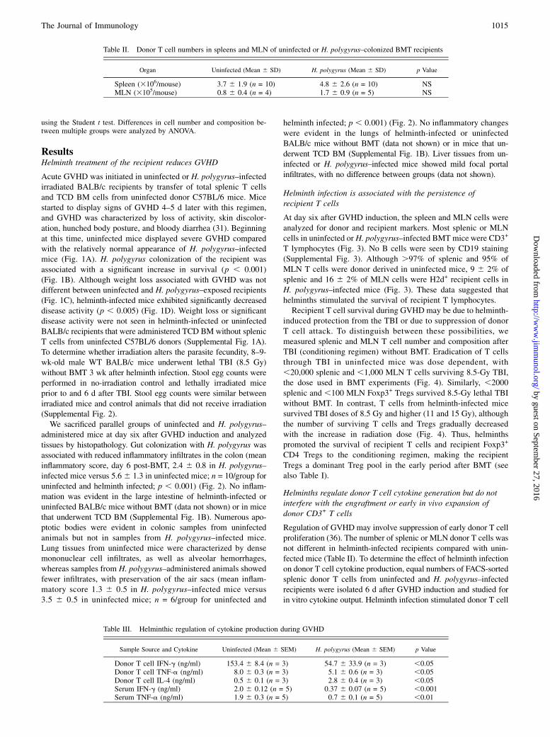
Immune regulatory pathways involving TGF-b lead to peripheralinduction and maintenance of Tregs (40) and may be essential inhelminth-induced immune modulation (13, 14). We showed pre-viously that H. polygyrus colonization stimulates T cell TGF-bgeneration that is essential for Treg functions, such as IL-10production (13). We studied whether Tregs that are increasedduring GVHD in helminth-infected mice generated more TGF-bon a per-cell basis. Most Foxp3+ CD4 T cells are found in theCD25+CD4+ T cell compartment. Plate-bound anti-CD3– andsoluble anti-CD28–stimulated splenic and MLN CD4+CD25+
T cells from H. polygyrus–infected or uninfected BALB/c micegenerated ∼2-fold more TGF-b compared with the CD4+CD252
T cell fraction, as shown by ELISA from supernatants harvested48 h after stimulation (Fig. 5). MLN T cell isolates from helminth-infected mice generated significantly more TGF-b compared withMLN T cell isolates from uninfected mice (Fig. 5). TGF-b cyto-kine content in anti-CD3/28–stimulated TCD parallel cultureswas ,20 pg/ml and did not increase with anti-CD3/28 stimulation(data not shown). These data suggested that, during GVHD, hel-minths induced the proliferation or generation or promoted thesurvival of TGF-b–producing Foxp3+ CD4 Tregs.
Helminth infection is associated with an increase in MLNdonor and recipient Treg latent TGF-b expression duringGVHD
Pre–pro–TGF-b peptide is cleaved into N-terminal LAP andC-terminal TGF-b protein after transcription and translation (41).LAP is expressed on T cells that may regulate immune responses ina TGF-b–dependent manner (42). Therefore, we studied latent TGF-b
FIGURE 5. Helminths enhance TGF-b generation from Treg-enriched
and Treg-depleted MLN CD4 T cells. TGF-b output of plate-bound anti-
CD3–stimulated and soluble anti-CD28–stimulated splenic and MLN
CD25-enriched or -depleted CD4+ T cells from H. polygyrus–colonized
(Hp) or uninfected (Uninf) BALB/c mice without BMT was measured by
ELISA from the cell culture supernatants. Data are representative exam-
ples from at least three independent experiments. *p , 0.05, CD252
versus CD25+, **p , 0.01 and ***p , 0.001, corresponding MLN cell
groups in uninfected versus H. polygyrus–infected mice.
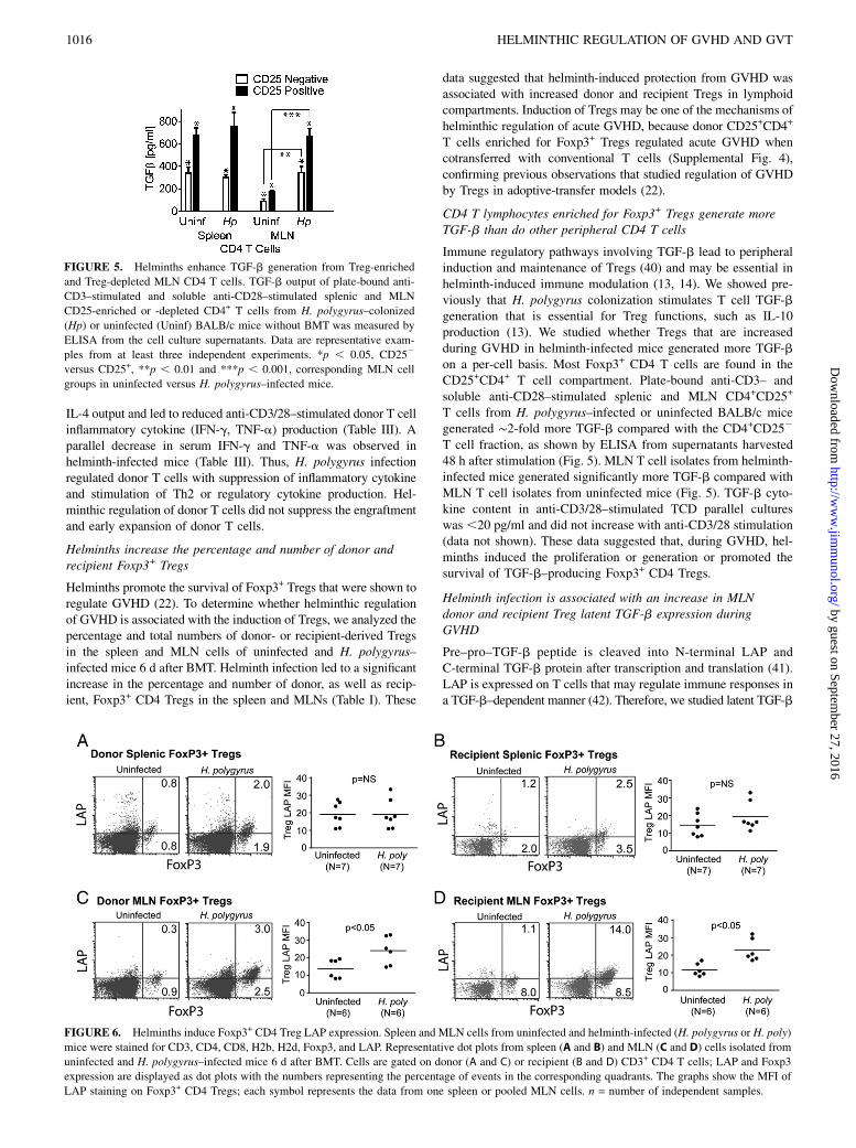
FIGURE 6. Helminths induce Foxp3+ CD4 Treg LAP expression. Spleen and MLN cells from uninfected and helminth-infected (H. polygyrus or H. poly)
mice were stained for CD3, CD4, CD8, H2b, H2d, Foxp3, and LAP. Representative dot plots from spleen (A and B) and MLN (C and D) cells isolated from
uninfected and H. polygyrus–infected mice 6 d after BMT. Cells are gated on donor (A and C) or recipient (B and D) CD3+ CD4 T cells; LAP and Foxp3
expression are displayed as dot plots with the numbers representing the percentage of events in the corresponding quadrants. The graphs show the MFI of
LAP staining on Foxp3+ CD4 Tregs; each symbol represents the data from one spleen or pooled MLN cells. n = number of independent samples.
1016 HELMINTHIC REGULATION OF GVHD AND GVT
by guest on September 27, 2016
http://ww
w.jim
munol.org/
Dow
nloaded from
(LAP) expression on donor and recipient Tregs during acute GVHD.LAP staining of Foxp3+ CD4 Tregs gave a separate and brightsignal on flow cytometry, with a significantly elevated mean fluo-rescent intensity (MFI) compared with Treg staining with an isotypeAb (data not shown). In splenic lymphocytes, LAP expression ondonor and recipient Foxp3+ CD4 Tregs did not change significantlyafter helminth infection (Fig. 6). However, in MLN cells, helminthinfection increased LAP MFI on donor and recipient Foxp3+ CD4Tregs (Fig. 6). These results indicated that H. polygyrus infectionpromoted the induction and/or maintenance of Foxp3+ Tregs, withenhanced TGF-b generation during acute GVHD.
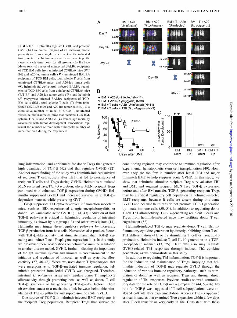
Helminths regulate GVHD in a TGF-b–dependent manner
TGF-b is a strong regulator of Th1 immunity (40, 43). Becausehelminth infection suppressed Th1 inflammation during GVHD,thereby increasing T cells that secrete TGF-b and express LAP,we studied whether helminths regulated donor T cell–mediatedGVHD in a TGF-b–dependent manner. In these experiments, weused donor T cells from uninfected TGF-bRII DN mice (H2b), inwhich the T cells are unresponsive to TGF-b–mediated immuneregulation as a result of T cell–specific overexpression of a trun-cated TGF-bRII (28). Because the engineered CD4 promoterdriving the truncated TGF-bRII expression lacked the CD8 si-
lencer, both CD4 and CD8 T cells in these mice are unresponsiveto TGF-b–mediated immune regulation. Uninfected and H. pol-ygyrus–infected recipient male WT BALB/c mice were givenTCD BM cells from uninfected 5–7-wk-old male C57BL/6 WTdonors and splenic T cells from male 5–7-wk-old TGF-bRII DNmice (Fig. 7A). Other groups included uninfected BALB/crecipients given TCD BM cells from uninfected C57BL/6 miceand uninfected or H. polygyrus–infected recipient BALB/crecipients given TCD BM and splenic T cells from C57BL/6WT donors. Transfer of TCD BM cells was associated with sur-vival of 90% of mice, whereas GVHD was uniformly lethal inuninfected BALB/c recipients that also received C57BL/6 WTor TGF-bRII DN splenic T cells (Fig. 7A). Strikingly, 40% ofH. polygyrus–infected recipients that received T cells from WTC57BL/6 mice survived the GVHD, whereas H. polygyrus infec-tion did not protect against GVHD lethality in recipients that re-ceived TGF-bRII DN splenic T cells (Fig. 7A). Some of thehelminth-infected surviving mice examined .200 d after GVHDinduction maintained 60–80% of their original weight and dis-played mild clinical disease activity (data not shown). Together,these data suggested that helminth infection regulated GVHD inrecipient mice in a TGF-b–dependent manner.Because helminths increased the percentage and number of
GVHD-regulating donor Tregs, we investigated the role of TGF-bin donor Treg repopulation after GVHD. Splenic T cells fromuninfected C57BL/6 WT or TGF-bRII DN mice contained com-parable frequencies of Foxp3+ Tregs (44), which we also found inour experiments (6.76 0.7% Foxp3+ in TGF-bRII DN [n = 3] and5.5 6 0.9% in C57BL/6 WT [n = 3], p = not significant). Com-parable donor T cell frequencies were observed in helminth-infected BALB/c BMT recipients that received splenic T cells fromuninfected C57BL/6 or TGF-bRII DN mice (Fig. 7B). However, thepercentage of donor Foxp3+ CD4 Tregs was significantly lower inhelminth-infected BMT recipients of TGF-bRII DN T cells comparedwith recipients of WT T cells (Fig. 7B). These results suggested thatTGF-b induced during helminth infection plays an additional criticalrole in donor Treg expansion during GVHD.
Helminthic regulation of GVHD is associated with preservedantitumor immunity
To determine whether helminthic immune suppression preservesthe donor T cell antitumor immunity (GVT), we administered A20-luc tumor cells within 24 h to BMT recipients. Live animal imagingdemonstrated that uninfected or helminth-infected mice that re-ceived TCD BM without donor T cells developed tumors and diedas a result of tumor burden within the first 24 d after BM and tumortransfer (Fig. 8). There was no difference in survival between thesegroups, suggesting that helminth infection did not have a benefi-cial or detrimental effect on tumor development. No tumor de-velopment was observed in uninfected or helminth-infected micethat received donor T cells other than TCD BM and A20 (Fig. 8).Although uninfected mice died of severe GVHD, five of ninehelminth-infected mice survived without evidence of tumor untilthe end of the experiment (Fig. 8). These results suggested thathelminths regulated GVHD and preserved GVT.
DiscussionHelminth infection of the gut was shown to suppress inflammationin various autoimmune, allergic, and immunological disorders (45).The major finding of the current study was that helminth infectionalso regulated inflammatory responses in an acute, lethal GVHDanimal model, promoting the survival of H. polygyrus–adminis-tered recipients. H. polygyrus treatment resulted in regulation ofdonor T cell Th1 cytokine generation, regulation of colitis and
FIGURE 7. Helminths regulate donor T cell–mediated GVHD in a TGF-
b–dependent manner. (A) Kaplan–Meier survival curves of uninfected
BALB/c recipients of TCD BM cells only from uninfected C57BL/6 mice
(WT B6) (n = 10) (;), uninfected BALB/c recipients of TCD BM cells and
total splenic T cells from uninfected C57BL/6 mice (n = 10) (n), uninfected
BALB/c recipients of TCD-BM cells from uninfected C57BL/6 WT (B6
WT) mice and splenic T cells from uninfected TGF-bRII DN (DN B6) mice
(n = 15) (d), H. polygyrus–infected BALB/c recipients of TCD BM and
splenic T cells from uninfected C57BL/6 mice (n = 10) (N), and H. poly-
gyrus–infected BALB/c recipients of TCD BM cells from uninfected
C57BL/6 mice and splenic T cells from uninfected TGF-bRII DN mice (n =
12) (s). Cumulative data from three independent experiments with multiple
mice from each group. (B) Spleen and pooled MLN cells were isolated from
helminth-infected BALB/c (Hp-infected) recipients of C57BL/6 (B6) or
TGF-bRII DN splenic T cell donors 6 d after BMT (all recipient mice also
received C57BL/6 WT TCD BM cells). The percentages of total and donor
CD3+ T cells and Foxp3+ CD4 T cells were determined by flow cytometry.
Each symbol represents one experiment from a single spleen or pooled
MLN. Percentage of donor CD3+ T cells in all CD3+ T cells (upper panels)
or the percentage of donor Foxp3+ CD4 Tregs in all CD3+ T cells (lower
panels) from spleen (left panels) or MLN (right panels) isolates. Each
symbol represents the percentage from a single sample.
The Journal of Immunology 1017
by guest on September 27, 2016
http://ww
w.jim
munol.org/
Dow
nloaded from
lung inflammation, and enrichment for donor Tregs that generatehigh quantities of TGF-b (42) and that regulate GVHD (22).Another novel finding of the study was helminth-induced survivalof recipient T cell subsets after TBI that led to persistence ofrecipient T cells and Tregs during GVHD. Helminths stimulatedMLN recipient Treg TGF-b secretion, where MLN recipient Tregscontinued with enhanced TGF-b expression during GVHD. Hel-minths suppressed GVHD and increased survival in a TGF-b–dependent manner, while preserving GVT.TGF-b suppresses Th1 cytokine–driven inflammation models in
mice, such as IBD, experimental allergic encephalomyelitis, ordonor T cell–mediated acute GVHD (1, 41, 43). Induction of hostTGF-b pathways is critical in helminthic regulation of intestinalimmunity, as shown by our group (13) and other investigators (14).Helminths may trigger these regulatory pathways by increasingTGF-b production from host cells. Nematodes also produce factorswith TGF-b–like activity that stimulate mammalian TGF-b sig-naling and induce T cell Foxp3 gene expression (14). In this study,we broadened these observations on helminthic immune regulationto another disease model, GVHD, further indicating the importanceof the gut immune system and luminal microenvironment in theinitiation and regulation of mucosal, as well as systemic, allor-eactivity (27, 46–48). When we used donor T lymphocytes thatwere unresponsive to TGF-b–mediated immune regulation, hel-minthic protection from lethal GVHD was abrogated. Therefore,intestinal H. polygyrus larvae may regulate donor T lymphocytealloreactivity through promoting host, as well as donor, T cellTGF-b synthesis or by generating TGF-b–like factors. Theseobservations attest to a mechanistic link between helminthic stim-ulation of TGF-b pathways and suppression of acute GVHD.One source of TGF-b in helminth-infected BMT recipients is
the recipient Treg population. Recipient Tregs that survive the
conditioning regimen may contribute to immune regulation afterexperimental hematopoietic stem cell transplantation (49). How-ever, they are too few in number after lethal TBI and majormismatch BMT to help suppress acute GVHD. In this study, weshow that helminths stimulate recipient Treg survival after TBIand BMT and augment recipient MLN Treg TGF-b expressionbefore and after BM transfer. TGF-b–generating recipient Tregsmay be a critical regulatory cell population in helminth-infectedBMT recipients, because B cells are absent during this acuteGVHD and because helminths do not promote TGF-b generationby innate immune cells (50, 51). In addition to regulating donorT cell Th1 alloreactivity, TGF-b–generating recipient T cells andTregs from helminth-infected mice may facilitate donor T cellengraftment (52).Helminth-induced TGF-b may regulate donor T cell Th1 in-
flammatory cytokine generation by directly inhibiting donor T cellTh1 differentiation (41) or by stimulating T cell or Treg IL-10production. Helminths induce T cell IL-10 generation in a TGF-b–dependent manner (13, 25). Helminths also may regulateGVHD-related Th1 responses through induced Th2 cytokinegeneration, as we demonstrate in this study.In addition to regulating Th1 inflammation, TGF-b is important
for the induction and maintenance of Tregs, implying that hel-minthic induction of TGF-b may regulate GVHD through theinduction of various immune-regulatory pathways, such as stim-ulation of donor as well as recipient Tregs and through directregulation of Th1 responses. Previous studies showed contradic-tory data for the role of TGF-b in Treg expansion (44, 53–56). Norole for TGF-b was suggested if T cell subpopulations were an-alyzed 6–8 wk after experimentation, whereas TGF-b appearedcritical in studies that examined Treg expansion within a few daysafter T cell transfer or very early in life. Consistent with these
FIGURE 8. Helminths regulate GVHD and preserve
GVT. (A) Live animal imaging of all surviving mouse
populations from a single experiment at the indicated
time points; the bioluminescence scale was kept the
same at each time point for all groups. (B) Kaplan–
Meier survival curves of uninfected BALB/c recipients
of TCD BM cells from uninfected C57BL/6 mice (WT
B6) and A20-luc tumor cells (;), uninfected BALB/c
recipients of TCD BM cells, total splenic T cells from
uninfected C57BL/6 mice, and A20-luc tumor cells
(n), helminth (H. polygyrus)–infected BALB/c recipi-
ents of TCD BM cells from uninfected C57BL/6 mice
(WT B6) and A20-luc tumor cells (P), and helminth
(H. polygyrus)–infected BALB/c recipients of TCD-
BM cells (BM), total splenic T cells (T) from unin-
fected C57BL/6 mice and A20-luc tumor cells (N). N =
cumulative number of mice. p , 0.001, uninfected
versus helminth-infected mice that received TCD BM,
splenic T cells, and A20-luc. (C) Percentage mortality
associated with tumor development. Proportions rep-
resent the number of mice with tumor/total number of
mice that died during the experiment.
1018 HELMINTHIC REGULATION OF GVHD AND GVT
by guest on September 27, 2016
http://ww
w.jim
munol.org/
Dow
nloaded from
early-response experiments, we demonstrated that TGF-b is crit-ical for rapid and robust donor Treg expansion and is crucial inregulating acute lethal GVHD.TGF-b is produced as a pre–pro-peptide where the N-terminal
cleaved portion of the protein, LAP, is secreted from the cell andnoncovalently attached to the cleaved C-terminal part of theoriginal pre–pro-peptide (41). The C-terminal protein is the TGF-bcytokine. Separation of the noncovalently attached N-terminalLAP leads to activation of TGF-b, permitting the binding of TGF-bto the TGF-bR complex and triggering signal transduction. Inaddition to the secreted TGF-b cytokine, noncovalently attachedLAP and TGF-b proteins are present in membrane-bound formson regulatory cell subsets that dampen immune responses in a cellcontact– and TGF-b–dependent manner (42). We found thatH. polygyrus infection was associated with an increase in MLNdonor and recipient Foxp3+ Tregs expressing LAP and that pro-tection from acute GVHD requires donor T cell TGF-b signaling.This confirms the importance of TGF-b in regulating intestinalimmunity (57) and the importance of the gut immune system inregulating GVHD.GVHD has remained a challenge of clinical practice, with in-
creasing cases of hematopoietic stem cell transplantation to treatvarious hematological or nonhematological diseases (23). Variousstudies showed the importance of Tregs in regulating effectordonor T cells and suppressing acute or chronic GVHD (22, 26,58–61), with ex vivo Tregs being a new area of clinical investi-gation in BMT. Purification and in vitro expansion of Tregs toa sufficient dose to manage GVHD is a challenge in clinicalpractice. Therefore, new clinical strategies to expand Tregsin vivo are being investigated (62). Our results suggest thatin vivo induction of Tregs by self-limited colonization of the gutwith helminths and use of the TGF-b pathway may suppressdonor T cell Th1 immune reactivity and enable regulated donorT cell engraftment.Helminths have been used in patients to successfully treat in-
flammation (17). Helminths may regulate immunity directly orthrough enriching the intestinal microbiome for beneficial or pro-biotic strains (63), because GVHD is associated with major shifts inthe composition of intestinal flora in animal models or patients (46).So helminths may also regulate GVHD through modulating the gutflora. With recent evidence showing that helminth products mayregulate inflammatory responses similar to helminth infections (64),exposure to helminths or helminth products may become a noveland safe therapy for GVHD with preserved antitumor immunity(GVT), allowing the broader use of BMT.
DisclosuresThe authors have no financial conflicts of interest.
References1. Shlomchik, W. D. 2007. Graft-versus-host disease. Nat. Rev. Immunol. 7: 340–352.2. Socie, G., and B. R. Blazar. 2009. Acute graft-versus-host disease: from the
bench to the bedside. Blood 114: 4327–4336.3. Ferrara, J. L., J. E. Levine, P. Reddy, and E. Holler. 2009. Graft-versus-host
disease. Lancet 373: 1550–1561.4. Abraham, C., and J. H. Cho. 2009. Inflammatory bowel disease. N. Engl. J. Med.
361: 2066–2078.5. Penack, O., E. Holler, and M. R. van den Brink. 2010. Graft-versus-host disease:
regulation by microbe-associated molecules and innate immune receptors. Blood115: 1865–1872.
6. Duerr, R. H., K. D. Taylor, S. R. Brant, J. D. Rioux, M. S. Silverberg, M. J. Daly,A. H. Steinhart, C. Abraham, M. Regueiro, A. Griffiths, et al. 2006. A genome-wide association study identifies IL23R as an inflammatory bowel disease gene.Science 314: 1461–1463.
7. Elmaagacli, A. H., M. Koldehoff, O. Landt, and D. W. Beelen. 2008. Relation ofan interleukin-23 receptor gene polymorphism to graft-versus-host disease afterhematopoietic-cell transplantation. Bone Marrow Transplant. 41: 821–826.
8. Johnson, B. D., E. E. Becker, J. L. LaBelle, and R. L. Truitt. 1999. Role ofimmunoregulatory donor T cells in suppression of graft-versus-host diseasefollowing donor leukocyte infusion therapy. J. Immunol. 163: 6479–6487.
9. Josefowicz, S. Z., L. F. Lu, and A. Y. Rudensky. 2012. Regulatory T cells:mechanisms of differentiation and function. Annu. Rev. Immunol. 30: 531–564.
10. Beres, A. J., D. Haribhai, A. C. Chadwick, P. J. Gonyo, C. B. Williams, andW. R. Drobyski. 2012. CD8+ Foxp3+ regulatory T cells are induced during graft-versus-host disease and mitigate disease severity. J. Immunol. 189: 464–474.
11. Shevach, E. M. 2009. Mechanisms of foxp3+ T regulatory cell-mediated sup-pression. Immunity 30: 636–645.
12. Redpath, S. A., N. van der Werf, A. M. Cervera, A. S. MacDonald, D. Gray,R. M. Maizels, and M. D. Taylor. 2013. ICOS controls Foxp3(+) regulatoryT-cell expansion, maintenance and IL-10 production during helminth infection.Eur. J. Immunol. 43: 705–715.
13. Ince, M. N., D. E. Elliott, T. Setiawan, A. Metwali, A. Blum, H. L. Chen,J. F. Urban, R. A. Flavell, and J. V. Weinstock. 2009. Role of T cell TGF-betasignaling in intestinal cytokine responses and helminthic immune modulation.Eur. J. Immunol. 39: 1870–1878.
14. Grainger, J. R., K. A. Smith, J. P. Hewitson, H. J. McSorley, Y. Harcus,K. J. Filbey, C. A. Finney, E. J. Greenwood, D. P. Knox, M. S. Wilson, et al.2010. Helminth secretions induce de novo T cell Foxp3 expression and regu-latory function through the TGF-b pathway. J. Exp. Med. 207: 2331–2341.
15. Setiawan, T., A. Metwali, A. M. Blum, M. N. Ince, J. F. Urban, Jr., D. E. Elliott,and J. V. Weinstock. 2007. Heligmosomoides polygyrus promotes regulatoryT-cell cytokine production in the murine normal distal intestine. Infect. Immun.75: 4655–4663.
16. Elliott, D. E., T. Setiawan, A. Metwali, A. Blum, J. F. Urban, Jr., andJ. V. Weinstock. 2004. Heligmosomoides polygyrus inhibits established colitis inIL-10-deficient mice. Eur. J. Immunol. 34: 2690–2698.
17. Elliott, D. E., and J. V. Weinstock. 2012. Helminth-host immunological inter-actions: prevention and control of immune-mediated diseases. Ann. N. Y. Acad.Sci. 1247: 83–96.
18. Fleming, J. O., A. Isaak, J. E. Lee, C. C. Luzzio, M. D. Carrithers, T. D. Cook,A. S. Field, J. Boland, and Z. Fabry. 2011. Probiotic helminth administration inrelapsing-remitting multiple sclerosis: a phase 1 study. Mult. Scler. 17: 743–754.
19. Croese, J., S. T. Gaze, and A. Loukas. 2013. Changed gluten immunity in celiacdisease by Necator americanus provides new insights into autoimmunity. Int. J.Parasitol. 43: 275–282.
20. Daveson, A. J., D. M. Jones, S. Gaze, H. McSorley, A. Clouston, A. Pascoe,S. Cooke, R. Speare, G. A. Macdonald, R. Anderson, et al. 2011. Effect ofhookworm infection on wheat challenge in celiac disease—a randomiseddouble-blinded placebo controlled trial. PLoS ONE 6: e17366.
21. McSorley, H. J., S. Gaze, J. Daveson, D. Jones, R. P. Anderson, A. Clouston,N. E. Ruyssers, R. Speare, J. S. McCarthy, C. R. Engwerda, et al. 2011. Sup-pression of inflammatory immune responses in celiac disease by experimentalhookworm infection. PLoS ONE 6: e24092.
22. Kohrt, H. E., A. B. Pillai, R. Lowsky, and S. Strober. 2010. NKT cells, Treg, andtheir interactions in bone marrow transplantation. Eur. J. Immunol. 40: 1862–1869.
23. Gooley, T. A., J. W. Chien, S. A. Pergam, S. Hingorani, M. L. Sorror, M. Boeckh,P. J. Martin, B. M. Sandmaier, K. A. Marr, F. R. Appelbaum, et al. 2010. Re-duced mortality after allogeneic hematopoietic-cell transplantation. N. Engl.J. Med. 363: 2091–2101.
24. Nguyen, V. H., R. Zeiser, and R. S. Negrin. 2006. Role of naturally arisingregulatory T cells in hematopoietic cell transplantation. Biol. Blood MarrowTransplant. 12: 995–1009.
25. Hoffmann, P., J. Ermann, M. Edinger, C. G. Fathman, and S. Strober. 2002.Donor-type CD4(+)CD25(+) regulatory T cells suppress lethal acute graft-versus-host disease after allogeneic bone marrow transplantation. J. Exp. Med.196: 389–399.
26. Taylor, P. A., C. J. Lees, and B. R. Blazar. 2002. The infusion of ex vivo acti-vated and expanded CD4(+)CD25(+) immune regulatory cells inhibits graft-versus-host disease lethality. Blood 99: 3493–3499.
27. Murai, M., H. Yoneyama, T. Ezaki, M. Suematsu, Y. Terashima, A. Harada,H. Hamada, H. Asakura, H. Ishikawa, and K. Matsushima. 2003. Peyer’s patch isthe essential site in initiating murine acute and lethal graft-versus-host reaction.Nat. Immunol. 4: 154–160.
28. Gorelik, L., and R. A. Flavell. 2000. Abrogation of TGFbeta signaling in T cellsleads to spontaneous T cell differentiation and autoimmune disease. Immunity12: 171–181.
29. Maizels, R. M., J. P. Hewitson, and W. C. Gause. 2011. Heligmosomoides pol-ygyrus: one species still. Trends Parasitol. 27: 100–101.
30. Behnke, J., and P. D. Harris. 2010. Heligmosomoides bakeri: a new name for anold worm? Trends Parasitol. 26: 524–529.
31. Cooke, K. R., L. Kobzik, T. R. Martin, J. Brewer, J. Delmonte, Jr.,J. M. Crawford, and J. L. Ferrara. 1996. An experimental model of idiopathicpneumonia syndrome after bone marrow transplantation: I. The roles of minor Hantigens and endotoxin. Blood 88: 3230–3239.
32. Tran, I. T., A. R. Sandy, A. J. Carulli, C. Ebens, J. Chung, G. T. Shan,V. Radojcic, A. Friedman, T. Gridley, A. Shelton, et al. 2013. Blockade of in-dividual Notch ligands and receptors controls graft-versus-host disease. J. Clin.Invest. 123: 1590–1604.
33. Brennan, T. V., L. Lin, X. Huang, D. M. Cardona, Z. Li, K. Dredge, N. J. Chao,and Y. Yang. 2012. Heparan sulfate, an endogenous TLR4 agonist, promotesacute GVHD after allogeneic stem cell transplantation. Blood 120: 2899–2908.
34. Highfill, S. L., P. C. Rodriguez, Q. Zhou, C. A. Goetz, B. H. Koehn, R. Veenstra,P. A. Taylor, A. Panoskaltsis-Mortari, J. S. Serody, D. H. Munn, et al. 2010. Bonemarrow myeloid-derived suppressor cells (MDSCs) inhibit graft-versus-host
The Journal of Immunology 1019
by guest on September 27, 2016
http://ww
w.jim
munol.org/
Dow
nloaded from
disease (GVHD) via an arginase-1-dependent mechanism that is up-regulated byinterleukin-13. Blood 116: 5738–5747.
35. Ince, M. N., D. E. Elliott, T. Setiawan, A. Blum, A. Metwali, Y. Wang,J. F. Urban, Jr., and J. V. Weinstock. 2006. Heligmosomoides polygyrus inducesTLR4 on murine mucosal T cells that produce TGFbeta after lipopolysaccharidestimulation. J. Immunol. 176: 726–729.
36. Pillai, A. B., T. I. George, S. Dutt, and S. Strober. 2009. Host natural killerT cells induce an interleukin-4-dependent expansion of donor CD4+CD25+Foxp3+ T regulatory cells that protects against graft-versus-host disease. Blood113: 4458–4467.
37. Pillai, A. B., T. I. George, S. Dutt, P. Teo, and S. Strober. 2007. Host NKT cellscan prevent graft-versus-host disease and permit graft antitumor activity afterbone marrow transplantation. J. Immunol. 178: 6242–6251.
38. Kaplan, D. H., B. E. Anderson, J. M. McNiff, D. Jain, M. J. Shlomchik, andW. D. Shlomchik. 2004. Target antigens determine graft-versus-host diseasephenotype. J. Immunol. 173: 5467–5475.
39. Carlson, M. J., M. L. West, J. M. Coghill, A. Panoskaltsis-Mortari, B. R. Blazar,and J. S. Serody. 2009. In vitro-differentiated TH17 cells mediate lethal acutegraft-versus-host disease with severe cutaneous and pulmonary pathologicmanifestations. Blood 113: 1365–1374.
40. Li, M. O., and R. A. Flavell. 2008. TGF-beta: a master of all T cell trades. Cell134: 392–404.
41. Li, M. O., Y. Y. Wan, S. Sanjabi, A. K. Robertson, and R. A. Flavell. 2006.Transforming growth factor-beta regulation of immune responses. Annu. Rev.Immunol. 24: 99–146.
42. Chen, M. L., B. S. Yan, Y. Bando, V. K. Kuchroo, and H. L. Weiner. 2008.Latency-associated peptide identifies a novel CD4+CD25+ regulatory T cellsubset with TGFbeta-mediated function and enhanced suppression of experi-mental autoimmune encephalomyelitis. J. Immunol. 180: 7327–7337.
43. Banovic, T., K. P. MacDonald, E. S. Morris, V. Rowe, R. Kuns, A. Don, J. Kelly,S. Ledbetter, A. D. Clouston, and G. R. Hill. 2005. TGF-beta in allogeneic stemcell transplantation: friend or foe? Blood 106: 2206–2214.
44. Fahlen, L., S. Read, L. Gorelik, S. D. Hurst, R. L. Coffman, R. A. Flavell, andF. Powrie. 2005. T cells that cannot respond to TGF-beta escape control byCD4(+)CD25(+) regulatory T cells. J. Exp. Med. 201: 737–746.
45. van Riet, E., F. C. Hartgers, and M. Yazdanbakhsh. 2007. Chronic helminthinfections induce immunomodulation: consequences and mechanisms. Immu-nobiology 212: 475–490.
46. Jenq, R. R., C. Ubeda, Y. Taur, C. C. Menezes, R. Khanin, J. A. Dudakov, C. Liu,M. L. West, N. V. Singer, M. J. Equinda, et al. 2012. Regulation of intestinalinflammation by microbiota following allogeneic bone marrow transplantation.J. Exp. Med. 209: 903–911.
47. Vossen, J. M., H. F. Guiot, A. C. Lankester, A. C. Vossen, R. G. Bredius,R. Wolterbeek, H. D. Bakker, and P. J. Heidt. 2014. Complete suppression of thegut microbiome prevents acute graft-versus-host disease following allogeneicbone marrow transplantation. PLoS ONE 9: e105706.
48. Holler, E., P. Butzhammer, K. Schmid, C. Hundsrucker, J. Koestler, K. Peter, W.Zhu, D. Sporrer, T. Hehlgans, M. Kreutz, et al. 2014. Metagenomic analysis ofthe stool microbiome in patients receiving allogeneic stem cell transplantation:loss of diversity is associated with use of systemic antibiotics and more pro-nounced in gastrointestinal graft-versus-host disease. Biol. Blood MarrowTransplant. 20: 640–645.
49. Bayer, A. L., M. Jones, J. Chirinos, L. de Armas, T. H. Schreiber, T. R. Malek, andR. B. Levy. 2009. Host CD4+CD25+ T cells can expand and comprise a majorcomponent of the Treg compartment after experimental HCT. Blood 113: 733–743.
50. Blum, A. M., L. Hang, T. Setiawan, J. P. Urban, Jr., K. M. Stoyanoff, J. Leung,and J. V. Weinstock. 2012. Heligmosomoides polygyrus bakeri induces tolero-genic dendritic cells that block colitis and prevent antigen-specific gut T cellresponses. J. Immunol. 189: 2512–2520.
51. Hang, L., T. Setiawan, A. M. Blum, J. Urban, K. Stoyanoff, S. Arihiro,H. C. Reinecker, and J. V. Weinstock. 2010. Heligmosomoides polygyrus in-fection can inhibit colitis through direct interaction with innate immunity.J. Immunol. 185: 3184–3189.
52. Nador, R. G., D. Hongo, J. Baker, Z. Yao, and S. Strober. 2010. The changedbalance of regulatory and naive T cells promotes tolerance after TLI and anti-T-cell antibody conditioning. Am. J. Transplant. 10: 262–272.
53. Marie, J. C., J. J. Letterio, M. Gavin, and A. Y. Rudensky. 2005. TGF-beta1maintains suppressor function and Foxp3 expression in CD4+CD25+ regulatoryT cells. J. Exp. Med. 201: 1061–1067.
54. Schramm, C. M., L. Puddington, C. Wu, L. Guernsey, M. Gharaee-Kermani,S. H. Phan, and R. S. Thrall. 2004. Chronic inhaled ovalbumin exposure inducesantigen-dependent but not antigen-specific inhalational tolerance in a murinemodel of allergic airway disease. Am. J. Pathol. 164: 295–304.
55. Huber, S., C. Schramm, H. A. Lehr, A. Mann, S. Schmitt, C. Becker,M. Protschka, P. R. Galle, M. F. Neurath, and M. Blessing. 2004. Cutting edge:TGF-beta signaling is required for the in vivo expansion and immunosuppressivecapacity of regulatory CD4+CD25+ T cells. J. Immunol. 173: 6526–6531.
56. Mamura, M., W. Lee, T. J. Sullivan, A. Felici, A. L. Sowers, J. P. Allison, andJ. J. Letterio. 2004. CD28 disruption exacerbates inflammation in Tgf-beta12/2
mice: in vivo suppression by CD4+CD25+ regulatory T cells independent ofautocrine TGF-beta1. Blood 103: 4594–4601.
57. Li, M. O., Y. Y. Wan, and R. A. Flavell. 2007. T cell-produced transforminggrowth factor-beta1 controls T cell tolerance and regulates Th1- and Th17-celldifferentiation. Immunity 26: 579–591.
58. Anderson, B. E., J. M. McNiff, C. Matte, I. Athanasiadis, W. D. Shlomchik, andM. J. Shlomchik. 2004. Recipient CD4+ T cells that survive irradiation regulatechronic graft-versus-host disease. Blood 104: 1565–1573.
59. Brunstein, C. G., J. S. Miller, Q. Cao, D. H. McKenna, K. L. Hippen,J. Curtsinger, T. Defor, B. L. Levine, C. H. June, P. Rubinstein, et al. 2011. In-fusion of ex vivo expanded T regulatory cells in adults transplanted with umbilicalcord blood: safety profile and detection kinetics. Blood 117: 1061–1070.
60. Hippen, K. L., S. C. Merkel, D. K. Schirm, C. Nelson, N. C. Tennis, J. L. Riley,C. H. June, J. S. Miller, J. E. Wagner, and B. R. Blazar. 2011. Generation andlarge-scale expansion of human inducible regulatory T cells that suppress graft-versus-host disease. Am. J. Transplant. 11: 1148–1157.
61. Edinger, M., P. Hoffmann, J. Ermann, K. Drago, C. G. Fathman, S. Strober, andR. S. Negrin. 2003. CD4+CD25+ regulatory T cells preserve graft-versus-tumoractivity while inhibiting graft-versus-host disease after bone marrow transplan-tation. Nat. Med. 9: 1144–1150.
62. Koreth, J., K. Matsuoka, H. T. Kim, S. M. McDonough, B. Bindra, E. P. Alyea,III, P. Armand, C. Cutler, V. T. Ho, N. S. Treister, et al. 2011. Interleukin-2 andregulatory T cells in graft-versus-host disease. N. Engl. J. Med. 365: 2055–2066.
63. Walk, S. T., A. M. Blum, S. A. Ewing, J. V. Weinstock, and V. B. Young. 2010.Alteration of the murine gut microbiota during infection with the parasitic hel-minth Heligmosomoides polygyrus. Inflamm. Bowel Dis. 16: 1841–1849.
64. Ruyssers, N. E., B. Y. De Winter, J. G. De Man, A. Loukas, M. S. Pearson,J. V. Weinstock, R. M. Van den Bossche, W. Martinet, P. A. Pelckmans, andT. G. Moreels. 2009. Therapeutic potential of helminth soluble proteins inTNBS-induced colitis in mice. Inflamm. Bowel Dis. 15: 491–500.
1020 HELMINTHIC REGULATION OF GVHD AND GVT
by guest on September 27, 2016
http://ww
w.jim
munol.org/
Dow
nloaded from


![RESEARCH ARTICLE Establishment of a Murine Graft-versus ... · of the curative potential of allografts is attributed to the ‘‘graft-versus-tumor’’ (GvT) effect [4]. In MM,](https://img.pdfslide.net/doc/110x75/5f3590abebab9b13db2308bc/research-article-establishment-of-a-murine-graft-versus-of-the-curative-potential.jpg)