Embed Size (px)
Citation preview
Intestinal Microbiota around Colorectal Cancer Genesis
Giovanni Brandi, Francesco De Rosa, Giuseppina Liguori Valentina Agostini, Stefania Di Girolamo
L. and A. Seràgnoli Department of Hematology and Oncological Sciences Sant’Orsola-Malpighi Hospital, University of Bologna, Italy
Valerie Gaboriau-Routhiau
INSERM, U989 Universite´ Paris Descartes, Paris, France
Pierre Raibaud
Unité d’Écologie et Physiologie du Systéme Digestif, INRA, France
Guido Biasco L. and A. Seràgnoli Department of Hematology and Oncological Sciences
Sant’Orsola-Malpighi Hospital, University of Bologna, Italy
1 Introduction
Colorectal cancer (CRC) pathogenesis is well known from a molecular perspective, but how endoluminal colonic factors interact with mucosal genome remains to be determined. Moreover, when referring to them, often only diet components reaching the large intestine are considered, forgetting about a silent, important player: intestinal microflora, or microbiota.
In fact, the gut in newborns is considered sterile, but bacterial colonization occurs quickly and the adult human intestinal tract hosts a complex microbial system, the number of which overcomes by a log the entire number of host eukaryotic cells, playing a crucial role in the regulation of both enteric and sys-temic homeostasis. Even though the beneficial relationship between the host and the microbiota is largely demonstrated, and in certain conditions the intestinal microflora can increase the risk of carcinogenesis and promote the tumoral growth.
In fact, intestinal autochthonous bacteria are involved in the catabolism of several elements de-rived from diet or from endogenous secretions, they can modulate the expression of host genes participat-ing in several pathological functions and can interfere with the immune system and the inflammation mechanisms. Furthermore, the gut microbiota is involved in redox stress damages, motility, angiogenesis, proliferation, differentiation, and fat storage regulation (Huycke & Gaskins, 2004).
The application of DNA-based molecular methods has helped to reduce many of the logistical problems associated with the identification of autochthonous microorganisms by cultural-based methods, but at the moment a significant part of the intestinal bacteria cannot be assigned to known genera or spe-cies (Bäckhed et al., 2005).
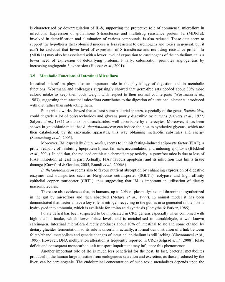
In this review, we want to summarize the mechanisms thought to be involved in the bacterial car-cinogenesis of CRC. In particular, we will focus on the difference in the role of intestinal microbiota (IM) in at-risk population with generic or familial/inherited risk factors (chromosomal instability path-way) and in subjects with chronic intestinal inflammatory disease (IBD-related pathway) (Figure 1).
In the first case, IM produces itself metabolites directly damaging DNA or affecting the expression of genes regulating cell cycle and proliferation; in the second, IM likely increases the level of oxidative stress of the mucosa, inducing a chronic inflammatory state, which over time can result in tissue hy-perproliferation and dysplasia. An improved knowledge of the fundamental differences in pathogenetic mechanisms of the potential bacterial carcinogenesis could also influence prophylactic strategies for co-lon cancer.
2 Putative Role of Intestinal Microflora in the Development of Colorec-tal Cancer Related to Chromosomal Instability Pathway
2.1 Genetic Bases and Pathological Changes in Sporadic Colorectal Cancer
The pathologic mechanism underlying both sporadic and familial colorectal cancer is still in part refera-ble to the model proposed by Fearon and Vogelstein in 1990. According to this model, progression from normal to dysplastic epithelium and finally to invasive carcinoma (the so-called adenoma-carcinoma se-quence) is associated with the accumulation of multiple clonally selected genetic alterations (Beggs &Hodgson, 2008). Among these, allelic loss or loss of heterozygosity (LOH) in the APC tumour sup-pressor gene represents an early event in colorectal carcinogenesis, determining the precocious clonal ex-pansion of the mutated cell and subsequent adenoma formation. Chromosome instability (characteristic
of up
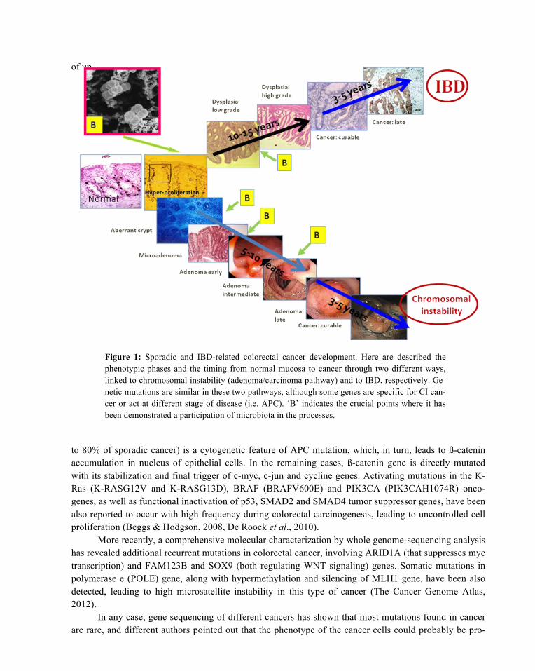
Figure 1: Sporadic and IBD-related colorectal cancer development. Here are described the phenotypic phases and the timing from normal mucosa to cancer through two different ways, linked to chromosomal instability (adenoma/carcinoma pathway) and to IBD, respectively. Ge-netic mutations are similar in these two pathways, although some genes are specific for CI can-cer or act at different stage of disease (i.e. APC). ‘B’ indicates the crucial points where it has been demonstrated a participation of microbiota in the processes.
to 80% of sporadic cancer) is a cytogenetic feature of APC mutation, which, in turn, leads to ß-catenin accumulation in nucleus of epithelial cells. In the remaining cases, ß-catenin gene is directly mutated with its stabilization and final trigger of c-myc, c-jun and cycline genes. Activating mutations in the K-Ras (K-RASG12V and K-RASG13D), BRAF (BRAFV600E) and PIK3CA (PIK3CAH1074R) onco-genes, as well as functional inactivation of p53, SMAD2 and SMAD4 tumor suppressor genes, have been also reported to occur with high frequency during colorectal carcinogenesis, leading to uncontrolled cell proliferation (Beggs & Hodgson, 2008, De Roock et al., 2010).
More recently, a comprehensive molecular characterization by whole genome-sequencing analysis has revealed additional recurrent mutations in colorectal cancer, involving ARID1A (that suppresses myc transcription) and FAM123B and SOX9 (both regulating WNT signaling) genes. Somatic mutations in polymerase e (POLE) gene, along with hypermethylation and silencing of MLH1 gene, have been also detected, leading to high microsatellite instability in this type of cancer (The Cancer Genome Atlas, 2012).
In any case, gene sequencing of different cancers has shown that most mutations found in cancer are rare, and different authors pointed out that the phenotype of the cancer cells could probably be pro-
duced by the typical aneuploid karyotype of the cancer cells itself, because an altered number of chromo-somes unbalances at once the expression of thousands of genes and proteins (Duesberget al., 2011). In fact, this suggestion is confirmed by a whole genome-sequencing analysis showing a recurrent copy-number amplifications of ERBB2 and IGF2 genes, as well as recurrent chromosomal translocation, due to the fusion between NAV2 and the WNT pathway member TCF7L1 (The Cancer Genome Atlas Net-work. 2012).
Bacteria can play a role in this molecular dynamic process. Some sporadic data suggest the role of different bacteria not only in mutation rate but also in chromosome aberrations and in downregulation of DNA mismatch repair protein (Cuevas-Ramos et al., 2010; Maddocks et al., 2009).
Human adenocarcinoma would phenotypically evolve trough aberrant crypt foci (ACF) both pre-ceded by unchecked cell proliferation. The fast enterocytes turnover suggests that stem cells on crypt fundus, and not the mature ones, are the target of oncogenic mutations. In particular, the CD133 cell, barely detectable in the normal colon but more frequent in cancer (2,5% of the population), is responsible for cancer initiation and propagation. Two models try to explain tumoral formation: the so-called bottom-up model states that the initially mutated cell is on the bottom of the crypt, thus proliferating in the lu-minal direction. On the contrary, by the top-down model the first mutation hits a cell on the apex between two crypts (i.e. on the luminal surface) which then proliferates towards crypt fundus. Interestingly, some putative “oncogenic bacteria” are found in the bottom of the crypts, near the stem cell regions (Maddocks et al., 2009).
One of the first events in colonic carcinogenesis is crypt fission, i.e. the division of a hyperprolif-erating crypt in two “daughter” crypts. This is responsible of aberrant crypt foci formation. Aberrant crypt foci (ACF) density is variable in relation to the age of the patient and pathology: it is low in patients with benign pathologies of the large bowel; vice versa, it is high in patients with adenomatous polyposis and colorectal neoplasia. The presence of ACF seems to be a good marker of colon cancer risk, since there is a strict correlation between the number of ACF and the prevalence of adenomatous polypoid le-sions and colon cancer.
2.2 Endoluminal Factors
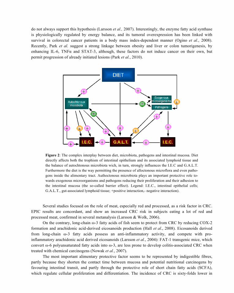
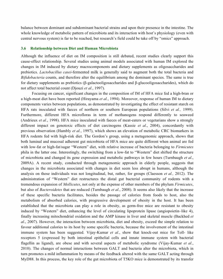
Endoluminal factors have a great impact on various local and distant parameters, and probably they also influence individual CRC risk together with the genetic background. The most important endoluminal risk factors are the diet and IM; their interplay is summarized in Figure 2.
2.3 Diet
The geographic differences in CRC incidence, investigated in several migrant and dynamic studies performed since early Seventies, is largely due to environmental factors, especially diet (Flood et al., 2000). With the word “diet”, we mean nutrition in its different sides, such as food composition and variety, global energetic balance, body weight and other anthropometric characteristics. Various observational and randomised, controlled studies evaluated the relationship between these different aspects and CRC, but results has not been conclusive (Martínez et al., 2008).
Colorectal cancer risk seems to increase with energy intake (Franceschi et al., 1997). The EPIC study, a large epidemiologic survey on nutritional risk factors, showed that people with large waist circumference or waist/hip ratio have an increased CRC risk (Pischon et al., 2006). Chronic hyperinsulinaemia is likely one of the most important risk factor, as many nutritional factor implicated in CRC genesis seem to elevate insulinaemia (Giovannucci, 2002). However, epidemiologic studies’ results
do not always support this hypothesis (Larsson et al., 2007). Interestingly, the enzyme fatty acid synthase is physiologically regulated by energy balance, and its tumoral overexpression has been linked with survival in colorectal cancer patients in a body mass index-dependent manner (Ogino et al., 2008). Recently, Park et al. suggest a strong linkage between obesity and liver or colon tumorigenesis, by enhancing IL-6, TNFα and STAT-3, although, these factors do not induce cancer on their own, but permit progression of already initiated lesions (Park et al., 2010).
Figure 2: The complex interplay between diet, microbiota, pathogens and intestinal mucosa. Diet directly affects both the trophism of intestinal epithelium and its associated lymphoid tissue and the balance of autochtonous microbiota wich, in turn, strongly influences the I.E.C and G.A.L.T. Furthermore the diet is the way permitting the presence of alloctonous microflora and even patho-gens inside the alimentary tract. Authoctonous microbiota plays an important protective role to-wards exogenous microorganisms and pathogens reducing their proliferation and their adhesion to the intestinal mucosa (the so-called barrier effect). Legend: I.E.C., intestinal epithelial cells; G.A.L.T., gut-associated lymphoid tissue; +positive interaction,- negative interaction).
Several studies focused on the role of meat, especially red and processed, as a risk factor in CRC. EPIC results are concordant, and show an increased CRC risk in subjects eating a lot of red and processed meat, confirmed in several metanalysis (Larsson & Wolk, 2006).
On the contrary, long-chain ω-3 fatty acids of fish seem to protect from CRC by reducing COX-2 formation and arachidonic acid-derived eicosanoids production (Hall et al., 2008). Eicosanoids derived from long-chain ω-3 fatty acids possess an anti-inflammatory activity, and compete with pro-inflammatory arachidonic acid derived eicosanoids (Larsson et al., 2004): FAT-1 transgenic mice, which convert ω-6 polyunsaturated fatty acids into ω-3, are less prone to develop colitis-associated CRC when treated with chemical carcinogens (Nowak et al., 2007).
The most important alimentary protective factor seems to be represented by indigestible fibres, partly because they shorten the contact time between mucosa and potential nutritional carcinogens by favouring intestinal transit, and partly through the protective role of short chain fatty acids (SCFA), which regulate cellular proliferation and differentiation. The incidence of CRC is sixty-folds lower in
native Africans than in Asian or Caucasian Americans. The former consume less protein and fat, and show a tenfold lower colonic crypt cell proliferation rate (O’Keefe et al., 2007). Various observational studies, including EPIC, reported a reduced incidence of CRC in populations with a high fibre diet (Peters et al., 2003); anyway, recent randomised trials on augmented fibre intake yielded conflicting results. In fact, both Wheat Bran Fibre Trial and Polyp prevention Trial did not find any significant difference in colorectal adenomas recurrence rate between the control and the study group with high fibre intake (Alberts et al., 2000, Schatzkin et al., 2000), while Toronto Polyp Prevention Trial showed a reduction of polyps in subjects with low fat, high fibre diet with respect to people keeping their usual diet (Asano & McLeod, 2002).
There are many hypotheses on the role of dietary calcium in the prevention of CRC. A recent systematic review by WCRF/AICR showed a significant inverse relation between total calcium intake and CRC risk, but modest in entity (World Cancer Research Fund/American Institute for Cancer Research, 2007), depending on the administered dose, on basal calcium reserves of the single subject and on the type of supplemental form (Martínez et al., 2008).
3 Intestinal Microbiota
3.1 Description of Human Microbiota
In human colon are harboured up to 1013 bacteria (Savage, 1977), and this huge population may either reside within and colonize the gastrointestinal tract (i.e. autochthonous bacteria), or pass transiently through the gastrointestinal tract (i.e. allochthonous bacteria). Autochthonous bacteria can be classified into dominant or subdominant depending on their concentration. In the colon, anaerobic-aerobic ratio varies, being lower on mucosal surface and higher in the lumen (Eckburg et al., 2005).
This heterogeneous population is traditionally studied with cultural methods, which allow ex vivo isolation of only a limited portion (40 – 60%) of our IM strains. Recently, molecular techniques permit a different approach to microbiota with identification of new species, which, however, cannot be further characterised. These molecular analyses are based on amplification of the 16S rRNA, a component of 30S small subunit of prokaryote ribosomes. The most utilized technique is polymerase chain reaction (PCR) employing an enzymatic reaction that allows in vitro amplification of a specific region of DNA providing extensive information about human microbial diversity and taxonomy (Kuczynski et al., 2011).
In the last years, the increased interest about the relationship among bacteria has prompted to ex-amine the microbiota by animal models and culture-independent genomic methods. Sequenc-ing/metagenomics approaches (by 454 pyrosequencing and Illumina) provided greater information about the potential functional role of the microbes and their complex genome. More recently, to better under-stand the interactions between human microbiome and host, novel functional metagenomic approaches were developed. Transcriptomics and proteomics, including MS-based shotgun proteomics, identify a large spectrum of proteins produced by microbial genes, giving an important contribution to understand the interactions between microbiome and the human host (Kolmeder et al., 2012).However, in certain contexts and, in particular, in the study of the role of individual bacterial species using gnotobiotic animal models, traditional methods are still necessary and not substitutable.
For long time, the organism is thought to be sterile before birth, although some recent findings suggest reconsidering the sterility of utero environment (Jiménez et al., 2008).The newborn is quickly colonised by microbes coming from the environment and the mother, called pioneer bacteria. Such
population is constituted by facultative anaerobes, which burn out all the oxygen in the colonic lumen and create the environmental conditions needed by strict anaerobes (Nicholson et al., 2012). These ones will then become the vast majority, the other being only mere spectator and metabolically negligible (Fanaro et al., 2003).
Only a restricted number of bacterial types colonise the gut. The dominant flora belongs to at least five bacterial phyla: Firmicutes, Bacteroidetes, Actinobacteria, Proteobacteria and Fusobacteria. There are six genera of strict anaerobes: Bacteroides, Eubacteria, Bifidobacteria, Clostridia, Peptostreptococci and Ruminococci, while most represented aerobic bacteria being of the genera Escherichia, Enterococcus, Streptococcus and Klebsiella (O’Hara & Shanahan, 2006). The number of bacterial species present in the human intestine is high, and 57 species are common to > 90% of subjects (Qin et al., 2010).
A member of IM has to fulfil several features: a metabolic apparatus fit for available nutrients, the ability to escape host immune response and to replicate quickly enough to avoid expulsion through the anal canal. Mechanisms underlying bacterial homing have been extensively described for pathogens (migration and adhesion to mucus, other bacteria-expressed receptors), but are still obscure for autochthonous IM. In particular, epithelium-adherent bacteria are numerically irrelevant in human colon; the opposite is true in the case of rodents (Thompson-Chagoyán et al., 2007).
Many studies have shown that significant inter-individually variability exists. A recent study of faecal 16S rRNA gene sequences collected from 14 unrelated adults over the course of a year showed large differences in microbial-community structure between individuals, while the community membership in each host was generally stable during this period. Conversely, the variability of IM composition is reduced in individuals living within the same family, but the relative influence of genetic and environmental factors, including diet, remains to be elucidated (Zoetendal et al., 2001). Nonetheless, a recent study concluded that host genotype is probably a key factor (Khachatryan et al., 2008).
Recently, by metagenomic approach, Arumugam et al. (Arumugam et al., 2011) showed that intestinal microbiota, notwithstanding its interindividual variability, is not built in a random fashion, but is stratified along three main clusters (so called enterotypes) based on corresponding Bacteroides, Prevotella and Ruminococcus genera. Around these three main contributors, there are other bacteria, both dominant and subdominant. It is interesting to note that functional profiles are supported also by subdominant bacteria, assessing that defined functions are shared among different bacteria, indifferently by their numerousness. In this context, few numerous bacterial populations can regain, in the alimentary tract, a role so far neglected. Linked to it, is notable that these three enterotypes utilize different routes to extract energy from fermentable colonic substrates.
Most studies indicate that the intraindividual human flora of adult subjects is quite stable over prolonged periods of time with relative abundance of Bifidobacteria and Clostridia in adolescent (Zoetendal et al., 1998). Interestingly, microbiota in old age is different to young age, and it is stable over limited time, although there is an imbalance of the main phyla with a decrease of Firmicutes and, in particular in the centenarians, of Faecalibacterium prausnitzii, which has anti-inflammatory properties.
Some temporal variability in relation to diet changes has been suggested; it happens during the first few weeks after birth, while in the adult life the diet-detected microflora fluctuations can be fully defined and may reflect changes in bacterial metabolic activity rather than changes in microbial composition. Population studies, conducted with metagenomic approaches, showed that changes in microbiota induced by diet are slow and that exists a stable metabolic core between individuals, despite changes in bacterial communities (Claesson et al., 2012, Human microbiome project consortium, 2012).
As well as diet, there are some stresses that can influence the balance of microbiota; in particular, antibiotics modify microbiota, which is, after therapy, characterized by a different equilibrium respect to pre-treatment (Dethlefsen & Relman, 2011).
In each individual, intestinal microbiota is in a state of floating balance, thanks to an interconnecting network allowing minimal variations (Hughes & Sperandio, 2008). In such a state dominating bacterial strains are in steady growth phase, the exponential one being characteristic only of the post-implant period. Due to the intraindividual stability of IM in opposition to its extraordinary interindividual variability, every subject has a unique and distinct microbial pattern, like an adjunctive fingerprint. In fact, in every individual different genome are present and interact, one inherited from the parents and thousands of others from their microflora, but these latter ones are quite fortuitous, resulting from the uncontrolled entry of viable bacteria in his ecosystem.
3.2 Mucosa-Associated Bacteria
The majority of research has focused on microflora recovered from faecal samples or intestinal content, even in studying the aspects of host-microflora relation that imply mucosal proximity. In other sites, such as stomach, this is not a concern, since mucosa-associated bacteria and luminal flora are quite similar in number and typology as assessed by culture methods. Recent works have shown that this is not the case in the intestine.
In fact, in this enclave of the outside environment limited by a living wall, the microbial population reaches its own equilibrium thanks to interactions between biotic (intestinal secretions, bacteriocines etc.) and abiotic components (food, fibres, fermentation metabolites). Employing techniques of capture dissection laser and molecular methods, it has been realized that the distribution of the bacteria inside of the large intestine of rodents is not uniform and that some phyla are differently distributed in relation to the lumen or in the vicinity of the epithelium (Nava et al., 2011). The mucus layer and the innate immune system, at least in mice, actively contains microbiota mainly in the lumen, limiting penetration into the mucosa and avoiding excessive proinflammatory signaling (Artis, 2008). In particular, Paneth cells via MyD88/NF-kB pathway actively hamper bacterial penetration through antimicrobial peptide secretion (Vaishnava et al., 2008).
Zoetendal and colleagues analysed, through denaturing-gradient gel electrophoresis (DGGE), the 16S rRNA gene on faecal and bioptic samples from ten subjects, and reported that the number of bacteria in mucosal sample is quite uniform along the colon. Interestingly, the profiles at different location in the same individual are similar, indicating that such population is uniform also qualitatively. Finally, DGGE profiles from mucosal and faecal samples of the same individual were in most cases different, indicating that the two populations are not completely interchangeable (Zoetendal et al., 2002).
Another study by Green and colleagues (Green et al., 2006) focused on the characterisation of mucosa-adherent bacteria. Using the same method, they examined mucosal bioptic specimens from 33 healthy individuals and, by comparing DGGE profiles, they showed that samples from different sites of the same patient harboured very similar bacterial communities, confirming previous data, while all subjects had different profiles. According to a previous work that outlines the importance of genetic factors in this context (Zoetendal et al., 1998), this study suggests that host factors are important in modulating microflora composition. They also matched gene sequences of 16S rRNA DGGE bands with entries in the GeneBank data base, attributing most of them to uncultured species in the genera Bacteroides, Clostridium, Ruminococcus and Faecalibacterium.
Surprisingly, terminal ileum harbours a number of mucosa-associated bacteria higher than the
colon (Ahmed et al., 2007). This may be linked to the higher number of unidentified helical bacteria not found in the large bowel. Overall, bacterial number is quite similar in the whole colon length, but Lactobacilli are more prominent in the distal large intestine. Bacteroides and Enterobacteriaceae are uniformly distributed in ileal and colonic mucosa, while Bifidobacteria are more prominent in the colon. Moreover, the mucus plays a crucial role in regulating the relationships between bacteria and the colonic mucosa. Recently, it was found that the epithelium of the colon is protected by an inner mucus layer formed by Muc-2 mucin impervious for bacteria that, vice versa, can be found in the outer loose non-attached mucus layer (Johansson et al., 2008). In case of Muc-2 mucin deficient mice, the bacteria are in close contact with epithelial cells and are even found in deep of crypt (i.e. near the stem cells of colon epithelium). The normal segregation of bacteria away from epithelium appears to play an important role in the genesis, or better, in the prevention of colon cancer, because it has been observed that mice lacking Muc-2 are prone to develop colon cancer (Velcich et al., 2002).
Moreover, different strains of the same bacterial species can have different tendency to establish an association with the mucosa. It is not known if this characteristic found in pathogenic strains can also be present in autochthonous microflora. The use of FISH for the study of the microbiota in humans has shown that bacteria are localized (albeit, in a limited number of colonies) in the side of the intraluminal mucus layer with a composition similar to that of the faecal contents (Van der Waaij et al., 2005). More refined molecular methods have then definitively established that bacterial populations related to the mucosa are different from those faecal (Eckburg et al., 2005).
In normal human intestine, such mucosa-associated bacterial population is relatively small. Schultsz and colleagues (Schultsz et al., 1999) performed bacterial rRNA in situ hybridization on bioptic specimens of inflammatory bowel disease (IBD) and non-IBD patients, mostly with irritable bowel syndrome. Interestingly, in normal individuals the number of bacteria in the mucus layer is very small: in the vast majority of sections, there were no bacteria at all. Swidsinski and colleagues, using FISH technique, have confirmed that the number of bacteria on the mucosa is low (<107 cfu) and that the mucus layer is often free from bacteria in over 80% of biopsies of normal subjects (Swidsinski et al., 2007). We performed a similar study using scanning electron microscope and had analogous results (Brandi et al., 1997). Moreover, our data showed that in mice there are many mucosa-associated bacteria. On the contrary, in human large bowel, bacteria are not in close contact with epithelium, and they are rarely found even in mucus layer. When present, they are clustered in small groups separated by wide areas with no bacteria at all. Studies performing quantitative evaluation with various techniques of mucosa-associated bacteria reported a concentration (105 – 107 colony forming units) lower than the faecal one, in subdominant position (Zoetendal et al., 2002, Ahmed et al., 2007). In conclusion, if the mucus of the human colon has a variable amount of bacteria, besides not fully corresponding to faecal microbiota, human colonic epithelium remains strictly germ-free under normal conditions.
3.3 Animal Models and Intestinal Microflora
3.3.1 Differences BetweenHuman and Rodent Microflora
Rodents are occasionally employed to study several characteristics of IM but some concerns exist in translating these data into humans. Differences can be identified both with classical and molecular approach; bacterial species likely belong to the same classes, while familiae and genera are host-specific. For example, in rodents, the number of endoluminal bacterial along the alimentary tract is substantially constant, ranging between 108 to 109 colony forming units (CFU)/ml. Conversely, in humans, the number
of bacteria detectable in the small bowel is negligible (~104 – 105 CFU/ml), increasing from the jejunum to the ileocecal valve and reaching the highest concentration in the cecum. Furthermore, the relationship between the bacterial flora and the intestinal epithelium could be substantially different between rodents and humans. In fact, in rodents there is an intimate relationship between the intestinal mucosa and a large amount of bacteria, often found to cluster over the mucus gel or in direct contact with epithelial cells, whereas in humans such correlation is lacking. These data and the difference in host-microbiota relationship between humans and mice constitute the major limitations of the murine model.
3.4 Molecular and Morpho-Functional Characteristics of Gastrointestinal Tract Induced by Mi-croflora
The use of animal models without bacteria (germ-free) compared to those with normal microflora (holoxenic) has fostered the study of morpho-functional changes induced by the presence of microflora in the digestive tract and, therefore, of the main functions of this complex ecosystem. Some studies also focused on gnotobiotic rodents, i.e. animals with gut colonised by known, definite bacterial strains. Human flora-associated animals (HFA), belonging to this group, can be obtained by inoculating germfree animals (e.g. mice) with human faeces (Raibaud et al., 1980). Human flora-associated mice and rats had and will undoubtedly have great importance in elucidating IM role in pathogenesis of intestinal diseases, but are also limited by various issues. Microflora obtained from faeces may not completely overlap with the intestinal one, and some bacterial strains may not colonise the murine gut; in particular, Bifidobacteria and Lactobacilli seem to be spontaneously eliminated (Raibaud et al., 1980). On the other hand, a recent study has demonstrated that most constituents of IM are able to colonize rodents and are stable in time (Hirayama & Itoh, 2005).
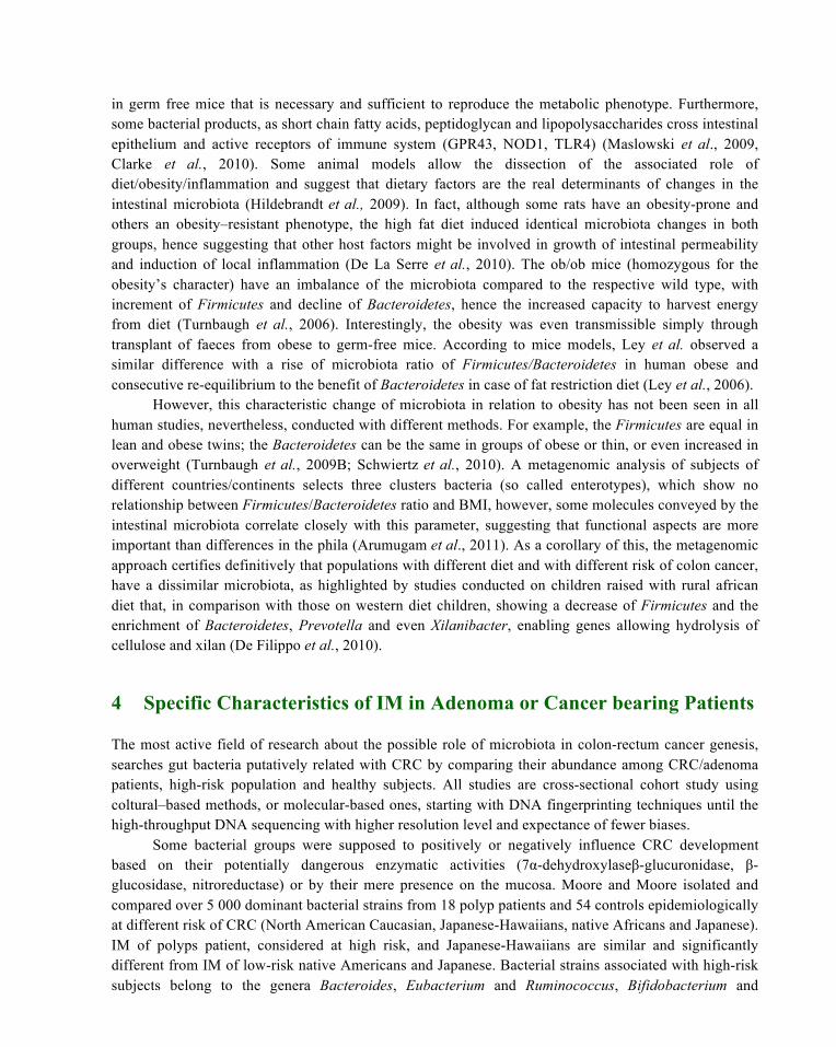
Intestinal microflora inoculated in animal models should be standardized in order to obtain reproducible animal models, which are often associated to microflora coming from only one subject and thus are not directly comparable. Some studies are therefore focusing on definition of a reproducible average human flora to standardise bacterial strains employed in HFA models (Hirayama & Itoh, 2005). The difference of the relationship between bacteria and host mucosa in holoxenic mice, HFA ones and humans are showed in Figure3. However, animal models are our best chance to investigate the role of IM, in particular for practical and ethical limits of research on humans. Germ-free animals, when compared to holoxenic counterparts, present defects in the development of intestinal immune system, in the nutrient absorption and in the intestinal morphology and motility (Lee & Mazmanian, 2010). Germfree mice are characterised by a reduced thickness of the colonic wall, inadequate differentiation of the small intestine and inferior epithelial proliferation compared with controls. In particular, they show a defective development in GALT (gut-associated lymphoid tissues), fewer and smaller Peyer’s patches and reduced expression of toll-like receptors (TLRs) and of CD4+ T cells in the lamina propria (Macpherson & Harris, 2004). In the absence of IM enterocyte cell cycle is prolonged and crypt cell proliferation rate is reduced (Alam et al., 1994). The presence of bacteria in the intestinal lumen and mucosal surface can modify some cell kinetic parameters producing a condition of hyperproliferation compared to germfree life, but the type of IM is also important. This is evident in large bowel, where mucosal proliferation rate (evaluated by bromodeoxyuridine intraperitoneal injetion 1 h before sacrifice) is significantly higher in holoxenic and HFA mice compared to germfree ones. Interestingly, human flora drives also higher mucosal proliferation rate than mice one (Brandi et al., unpublished data).
The renewal of intestinal epithelium and even the building of aberrant crypts and adenomas follow a horizontal pattern, characterised by the production of new crypts through a phenomenon of fission (Li
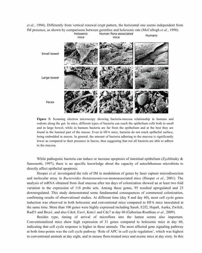
et al., 1994). Differently from vertical renewal crypt pattern, the horizontal one seems independent from IM presence, as shown by comparisons between germfree and holoxenic rats (McCullogh et al., 1998).
Figure 3: Scanning electron microscopy showing bacteria-mucosa relationship in humans and rodents along the gut. In mice, different types of bacteria can reach the epithelium cells both in small and in large bowel; while in humans bacteria are far from the epithelium and at the best they are found in the luminal part of the mucus. Even in HFA mice, bacteria do not reach epithelial surface, being embedded in mucus. In general, the amount of bacteria adhering to the mucosa is significantly lower as compared to their presence in faeces, thus suggesting that not all bacteria are able to adhere to the mucosa. While pathogenic bacteria can induce or increase apoptosis of intestinal epithelium (Zychlinsky &
Sansonetti, 1997), there is no specific knowledge about the capacity of autochthonous microbiota to directly affect epithelial apoptosis.
Hooper et al. investigated the role of IM in modulation of genes by laser capture microdissection and molecular array in Bacteroides thetaiotaomicron-monoassociated mice (Hooper et al., 2001). The analysis of mRNA obtained from ileal mucosa after ten days of colonization showed an at least two fold variation in the expression of 118 probe sets. Among these genes, 95 resulted upregulated and 23 downregulated. This study demonstrated some fundamental consequences of commensal colonization, confirming results of observational studies. At different time (day 8 and day 60), most cell cycle genes induction was observed in both holoxenic and conventional mice compared to HFA mice inoculated at the same time. More than 100 genes were highly expressed including Sass6, E2f2, Hspa8, Aurka, Zwilch, Rad51 and Brca1, and also Cdc6, Exo1, Kntc1 and Cdc7 at day 60 (Gaboriau-Routhiau et al., 2009).
Besides type, timing of arrival of microflora into the lumen seems also important. Conventionalized mice show high expression of 31 genes compared to holoxenic mice at day 60, indicating that cell cycle response is higher in these animals. The most affected gene signaling pathway at both time-points was the cell cycle pathway ‘Role of APC in cell cycle regulation’, which was highest in conventional animals at day eight, and in mouse flora-treated mice and axenic mice at day sixty. In this
pathway, a polyubiquitin chain gets attached to a protein substrate by an ubiquitin-ligase, which targets it for degradation by the 26S proteasome. This is an important step in the cell cycle, as cell division progression is governed by degradation of different regulatory proteins in the ubiquitin-dependent pathway. Anaphase-promoting complex (APC) is an ubiquitin ligase that plays a key role in the cell cycle.
Around 50% of genes elicited in the ileal mucosa in response to bacterial colonization are linked to immune pathways. Transcriptomic analysis of terminal ileum mucosa from GF, holoxenic and HFA mice shows that cell cycles-specific genes are tenfold higher in HFA mice compared to holoxenic (Gaboriau-Routhiau et al., 2009).
Bacterial presence and subsequent GALT TLRs activation are indeed fundamental for intestinal epithelium to achieve its normal trophism and gut-associated lymphoid tissue (GALT) to mature (Round & Mazmanian, 2009), but the type of IM is very important and only a restricted number of normal microbiota is able to stimulate the mucosal T-cell response. In particular human IM seems quite unable to stimulate the immune system in mice, and transcriptome analysis of immmune genes in HFA mice clustered with GF rather than holoxenic ones, supporting the impact of host-specific microbiota for immune stimulation (Gaboriau-Routhiau et al., 2009). This observation was recently confirmed by Chung et al. that analysed immune maturation and gut microbiota composition of GF mice colonized at birth with rodents gut microbiota (MMb) and human gut microbiota (HMb), showing that HMb-colonized mice have a poorly developed small intestinal immune system, quite similar to that in GF mice, demonstrating that there is an essential interaction between specific microbe-host and the maturation of the intestinal immune system. Inducing infection of Salmonella enteric, these authors showed that HMb-colonized mice presented intestinal inflammation respect to MMb mouse, assessing that exists a host-specific microbiota that plays a critical role in modulation of immune system and GUT immune maturation (Chung et al., 2012).
Members of microbiota as Bacteroides fragilis are important for the mucosal immune system stimulation of mammals (Mazmanian & Kasper, 2006). In particular, Bacteroides fragilis is able to prevent colitis in two different experimental models (Round & Mazmanian, 2010) and its capsular molecule Polysaccharide A (PSA) directs the differentiation of Interleukin-10 (IL-10)-secreting TReg cells (Mazmanian&Kasper, 2006). Furthermore, it has been demonstrated that oral treatment with purified PSA could reduce the expression of cytokine and the infiltration of lymphocyte, due to increased production of IL-10 and Foxp3 expression (Round & Mazmanian, 2010).
Another bacterium, responsible to modulate the nature of the intestinal immune responses, is the Segmented Filamentous bacteria (SFB). This unculturable species detected in rodent intestine adheres to intestinal mucosa and stimulates a large spectrum of innate and adaptive immune responses, which notably mediate the abundance of lamina propria Th17 cells and the secretion of antimicrobial peptides (Gaboriau-Routhiau et al., 2009, Ivanov et al., 2009, Lee & Mazmanian, 2010). Furthermore, SFBs colonization plays a protective role against Citrobacter rodentium, an enteropathogenic bacterium that produces in rodent’s intestinal inflammation similar to E. Coli (EPEC) in humans (Ivanov et al., 2009).
Beyond the relationship with immune system, microbiota drives several other functions. Some experiments also demonstrated that inoculation of a single dominant bacterial strain (e.g.
Bacteroides thetaiotaomicron) in germfree mice causes complete epithelial differentiation and resumption of normal cellular proliferation (Umesaki et al., 1995).
Monocolonization with this microorganism does not induce inflammation, contrary to Salmonella enteritidis that upregulates IL-8. The contemporary association of B. thetaiotaomicron and S. enteritidis
is characterized by downregulation of IL-8, supporting the protective role of commensal microflora in infections. Expression of glutathione S-transferase and multidrug resistance protein 1a (MDR1a), involved in detoxification and elimination of various compounds, is also reduced. These data seem to support the hypothesis that colonised mucosa is less resistant to carcinogens and toxics in general, but it can’t be excluded that lower level of expression of S-transferase and multidrug resistance protein 1a (MDR1a) may also be associated with a lower level of exposition to carcinogens of the epithelium, thus a lower need of expression of detoxifying proteins. Finally, colonization promotes angiogenesis by increasing angiogenin-3 expression (Hooper et al., 2001).
3.5 Metabolic Functions of Intestinal Microflora
Intestinal microflora plays also an important role in the physiology of digestion and in metabolic functions. Wostmann and colleagues surprisingly showed that germ-free rats needed about 30% more caloric intake to keep their body weight with respect to their normal counterparts (Wostmann et al., 1983), suggesting that intestinal microflora contributes to the digestion of nutritional elements introduced with diet rather than subtracting them.
Pioneeristic works showed that at least some bacterial species, especially of the genus Bacteroides, could degrade a lot of polysaccharides and glycans poorly digestible by humans (Salyers et al., 1977, Salyers et al., 1981) to mono- or disaccharides, well absorbable by enterocytes. Moreover, it has been shown in gnotobiotic mice that B. thetaiotaomicron can induce the host to synthetize glycans, which are then catabolized, by its enzymatic apparatus, this way obtaining metabolic substrates and energy (Sonnenburg et al., 2005).
Moreover, IM, especially Bacterioides, seems to inhibit fasting-induced adipocyte factor (FIAF), a protein capable of inhibiting lipoprotein lipase, fat mass accumulation and inducing apoptosis (Bäckhed et al., 2004). In addition, the reduced antiblastic chemotherapy toxicity in germfree mice is due to loss of FIAF inhibition, at least in part. Actually, FIAF favours apoptosis, and its inhibition thus limits tissue damage (Crawford & Gordon, 2005, Brandi et al., 2006A).
B. thetaiotaomicron seems also to favour nutrient absorption by enhancing expression of digestive enzymes and transporters such as Na-glucose cotransporter (SGLT1), colypase and high affinity epithelial copper transporter (CRT1), thus suggesting that IM is important in utilisation of dietary macromolecules.
There are also evidences that, in humans, up to 20% of plasma lysine and threonine is synthetized in the gut by microflora and then absorbed (Metges et al., 1999). In animal model it has been demonstrated that bacteria have a key role in nitrogen recycling in the gut, as urea generated in the host is hydrolysed into ammonia, which is available for amino acid synthesis (Forsythe & Parker, 1985).
Folate deficit has been suspected to be implicated in CRC genesis especially when combined with high alcohol intake, which lower folate levels and is metabolised to acetaldehyde, a well-known carcinogen. Intestinal microflora directly produces about 10% of intestinal folate and some ethanol by dietary glucides fermentation, so its role is uncertain: actually, a formal demonstration of a link between folate/ethanol metabolism and genetic changes of intestinal epithelium is still lacking (Giovannucci et al., 1995). However, DNA methylation alteration is frequently reported in CRC (Selgrad et al., 2008); folate deficit and consequent monocarbon unit transport impairment may influence this phenomenon.
Another important role of IM is much less beneficial for the host. In fact, bacterial metabolites produced in the human large intestine from endogenous secretion and excretion, as those produced by the liver, can be carcinogenic. The endoluminal concentration of such toxic metabolites depends upon the
balance between dominant and subdominant bacterial strains and upon their presence in the intestine. The whole knowledge of metabolic pattern of microbiota and its interaction with host’s physiology (even with central nervous system) is far to be reached, but research’s field could be take off by “omics” approach.
3.6 Relationship between Diet and Human Microbiota
Although the influence of diet on IM composition is still debated, recent studies clearly support this cause-effect relationship. Several studies using animal models associated with human IM explored the changes in IM induced by dietary macrocomponents and dietary supplements as oligosaccharides and prebiotics. Lactobacillus casei-fermented milk is generally said to augment both the total bacteria and Bifidobacteria counts, and therefore alter the equilibrium among the dominant species. The same is true for dietary supplements as prebiotics (β-galactooligosaccharides and β-glucooligosaccharides), which do not affect total bacterial count (Djouzi et al., 1997).
Focusing on cancer, significant changes in the composition of IM of HFA mice fed a high-bran or a high-meat diet have been reported (Hirayama et al., 1994). Moreover, response of human IM to dietary components varies between populations, as demonstrated by investigating the effect of resistant starch on HFA rats inoculated with faeces of northern or southern European populations (Silvi et al., 1999). Furthermore, different HFA microfloras in term of methanogens respond differently to seaweed (Andrieux et al., 1998). HFA mice inoculated with faeces of meat-eaters or vegetarians show a strongly different impact on genotoxic effects of diet carcinogens (Kassie et al., 2004), concordantly with previous observation (Hambly et al., 1997), which shows an elevation of metabolic CRC biomarkers in HFA rodents fed with high-risk diet. The Gordon’s group, using a metagenomic approach, shows that both luminal and mucosal adherent gut microbiota of HFA mice are quite different when animal are fed with low-fat or high-fat/sugar “Western” diet, with relative increase of bacteria belonging to Firmicutes phila in the latter one. Interestingly, the switching from a low-fat to “Western” diet shifted the structure of microbiota and changed its gene expression and metabolic pathways in few hours (Turnbaugh et al., 2009A). A recent study, conducted through metagenomic approach in elderly people, suggests that changes in the microbiota associated with changes in diet seem less abrupt in humans, although the analysis on those individuals was not longitudinal, but, rather, for groups (Claesson et al., 2012). The administration of “Western” diet restructures the distal gut bacterial community of rodents with a tremendous expansion of Mollicutes, not only at the expense of other members of the phylum Firmicutes, but also of Bacteroidetes that are reduced (Turnbaugh et al., 2008). It seems also likely that the increase of these specific bacteria facilitates, besides the passage of calories from foods to host, also the metabolism of absorbed calories, with progressive development of obesity in the host. It has been established that the microbiota can play a role in obesity, as germ-free mice are resistant to obesity induced by “Western” diet, enhancing the level of circulating lipoprotein lipase (angiopoietin–like 4), finally increasing mitochondrial oxidation and the AMP kinase in liver and skeletal muscle (Backhed et al., 2007). However, the interaction between microbiota, diet and obesity, exceed the simple relation to favour additional calories to its host by some specific bacteria, because the involvement of the intestinal immune system has been suggested. Vijay-Kumar et al., show that knock-out mice for Toll- like receptors 5 (expressed by both intestinal epithelial cells and innate immune system with bacterial flagellin as ligand), are obese and with several aspects of metabolic syndrome (Vijay-Kumar et al., 2010). The changes of normal interactions between GALT and bacteria alter the microbiota, which in turn promotes a mild inflammation by means of the feedback altered with the same GALT acting through MyD88. In this process, the key role of the gut microbiota of T5KO mice is demonstrated by its transfer
in germ free mice that is necessary and sufficient to reproduce the metabolic phenotype. Furthermore, some bacterial products, as short chain fatty acids, peptidoglycan and lipopolysaccharides cross intestinal epithelium and active receptors of immune system (GPR43, NOD1, TLR4) (Maslowski et al., 2009, Clarke et al., 2010). Some animal models allow the dissection of the associated role of diet/obesity/inflammation and suggest that dietary factors are the real determinants of changes in the intestinal microbiota (Hildebrandt et al., 2009). In fact, although some rats have an obesity-prone and others an obesity–resistant phenotype, the high fat diet induced identical microbiota changes in both groups, hence suggesting that other host factors might be involved in growth of intestinal permeability and induction of local inflammation (De La Serre et al., 2010). The ob/ob mice (homozygous for the obesity’s character) have an imbalance of the microbiota compared to the respective wild type, with increment of Firmicutes and decline of Bacteroidetes, hence the increased capacity to harvest energy from diet (Turnbaugh et al., 2006). Interestingly, the obesity was even transmissible simply through transplant of faeces from obese to germ-free mice. According to mice models, Ley et al. observed a similar difference with a rise of microbiota ratio of Firmicutes/Bacteroidetes in human obese and consecutive re-equilibrium to the benefit of Bacteroidetes in case of fat restriction diet (Ley et al., 2006).
However, this characteristic change of microbiota in relation to obesity has not been seen in all human studies, nevertheless, conducted with different methods. For example, the Firmicutes are equal in lean and obese twins; the Bacteroidetes can be the same in groups of obese or thin, or even increased in overweight (Turnbaugh et al., 2009B; Schwiertz et al., 2010). A metagenomic analysis of subjects of different countries/continents selects three clusters bacteria (so called enterotypes), which show no relationship between Firmicutes/Bacteroidetes ratio and BMI, however, some molecules conveyed by the intestinal microbiota correlate closely with this parameter, suggesting that functional aspects are more important than differences in the phila (Arumugam et al., 2011). As a corollary of this, the metagenomic approach certifies definitively that populations with different diet and with different risk of colon cancer, have a dissimilar microbiota, as highlighted by studies conducted on children raised with rural african diet that, in comparison with those on western diet children, showing a decrease of Firmicutes and the enrichment of Bacteroidetes, Prevotella and even Xilanibacter, enabling genes allowing hydrolysis of cellulose and xilan (De Filippo et al., 2010).
4 Specific Characteristics of IM in Adenoma or Cancer bearing Patients
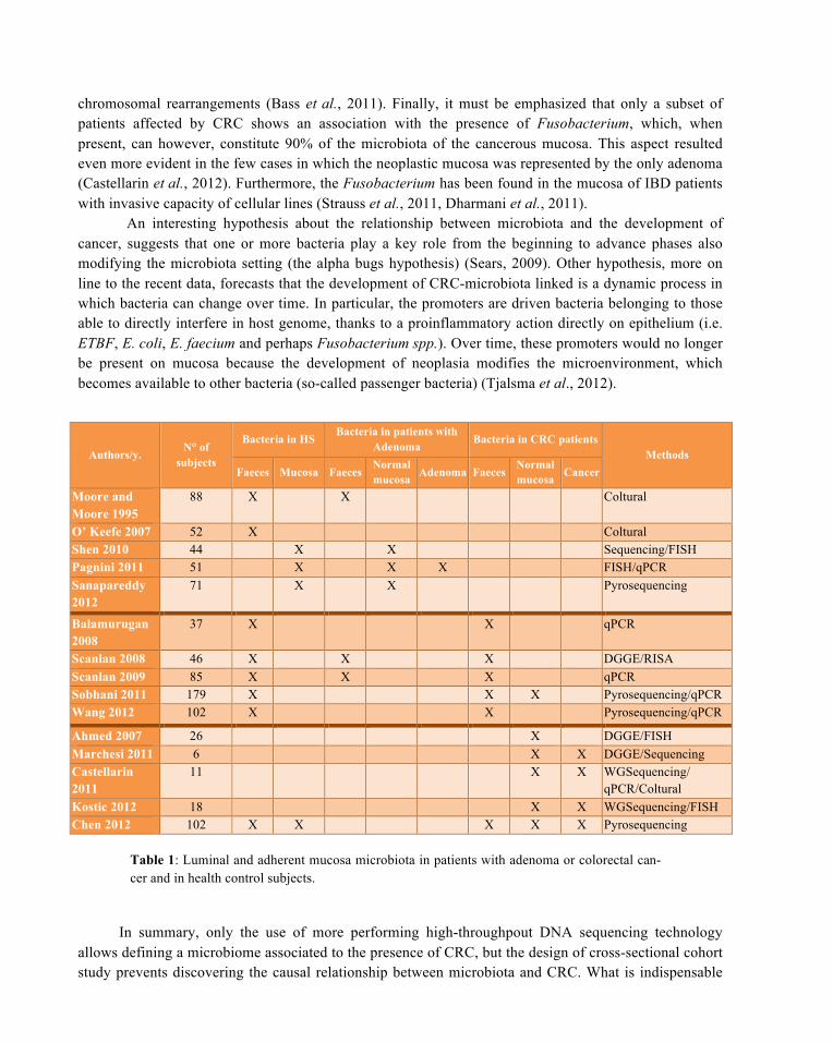
The most active field of research about the possible role of microbiota in colon-rectum cancer genesis, searches gut bacteria putatively related with CRC by comparing their abundance among CRC/adenoma patients, high-risk population and healthy subjects. All studies are cross-sectional cohort study using coltural–based methods, or molecular-based ones, starting with DNA fingerprinting techniques until the high-throughput DNA sequencing with higher resolution level and expectance of fewer biases.
Some bacterial groups were supposed to positively or negatively influence CRC development based on their potentially dangerous enzymatic activities (7α-dehydroxylaseβ-glucuronidase, β-glucosidase, nitroreductase) or by their mere presence on the mucosa. Moore and Moore isolated and compared over 5 000 dominant bacterial strains from 18 polyp patients and 54 controls epidemiologically at different risk of CRC (North American Caucasian, Japanese-Hawaiians, native Africans and Japanese). IM of polyps patient, considered at high risk, and Japanese-Hawaiians are similar and significantly different from IM of low-risk native Americans and Japanese. Bacterial strains associated with high-risk subjects belong to the genera Bacteroides, Eubacterium and Ruminococcus, Bifidobacterium and
Faecalibacterium prausnitzii (Moore & Moore, 1995). O’ Keefe et al. linked 7α-dehydroxylase bacteria to high CRC risk population, while Lactobacillus plantarum to low risk population (O’Keefe et al., 2007).
The use of molecular methods (q-PCR for bacterial DNA and RNA), then confirmed by classical coltural methods, demonstrates that on the adenoma mucosa there is a lower number of bacteria compared to the normal colonic mucosa, while there is no difference between the concentration of bacterial DNA on the normal mucosa of patients with or without adenoma (Pagnini et al., 2011). It is possible that the reduction of bacteria on the adenoma mucosa is linked to the activation of specific a-defensin antibacterial. Two consecutive case-control studies performed by the same group and using different molecular analysis (terminal restriction fragment length polymorphism, clonal sequencing and FISH, or more advanced sequencing technology and q-PCR) to investigate the bacterial communities of normal rectal mucosa in patients with polyps or controls, suggest differences in bacterial composition with a higher bacteria richness (i.e. the number of taxa in the sample) in cases, (87 more abundant taxa was found), without differences for evenness (i.e. taxa distribution within the sample) (Shen et al., 2010, Sanapareddy et al., 2012). Interestingly, the differences in richness are entirely due to low- abundance taxa and seem unrelated to diet. A bacterial profile of adenoma subjects is characterized by Proteobacteria increasing and Bacteroidetes decreasing without differences for Firmicutes, the most represented phylum. At genus level, polyps’ subjects showed higher abundance of Faecalibacterium, Shigella and Dorea spp and reduction of Bacteroides spp and Coprococcus spp. The FISH analysis confirms that the outer mucus layer is the unique ecosystem of mucosa adherent-bacteria, even in normal mucosa of patients with adenoma. In a perspective of cause and effect, it can be assumed that changes in the bacterial population may have preceded the onset of adenoma formation.
Scanlan et al., in two related studies using DNA fingerprinting techniques (DGGE, RIS, qPCR) and metabonomic tools, analysed interindividual and intraindividual variability of faecal microflora in healthy, colorectal cancer and polypectomyzed subjects (Scanlan et al., 2008, Scanlan et al., 2009). Only the polyp group shows significantly different interindividual DGGE profiles, in CRC patients significantly higher number of Clostridiumcoccoides and Desulfovibriosp (producer of hydrogen sulphide, a well know genotoxic agent) were found. No diversity has been detected concerning Bacteroides in the three groups. Using high-throughput DNA sequencing technology, faecal microbiota of CRC Caucasic (Sobhani et al., 2011) and Asiatic patients (Wang et al., 2012) was compared to normal subjects, respectively, in a retrospective or prospective manner. Although the total number of bacteria was similar in CRC and controls (Sobhani et al., 2011), both studies detect a differing faecal microbiota structure in cancer patients compared with controls. According to Sobhani, the Bacteroides/Prevotella are the only bacteria group higher in cancer patients, while other dominant or subdominant bacteria as Bifidobacterium genus, Lactobacillus/Leuconostoc group, Clostridiumcoccoides/C leptum group and Faecalibacterium prausnitzii did not show any differences. It is believed that the main changes of microbiota in CRC patients refer to depletion of butyrate-producing bacteria and to increase of opportunistic pathogens. Both Clostridium coccoides/ C leptum group and Faecalibacterium prausnitzii are strong producer of butyrate but their depletion are not constantly detected in different studies. Wang detects a reduction of Clostridium coccoides but not of Faecalibacterium prausnitzii. Vice versa, Balamurugan et al. (Balamurugan et al., 2008) shows an important decrease of Faecalibacterium prausnitzii in patients with cancer. The Bacteroides, whose occurrence turned out to be unrelated to diet, are 1000 times more present in the faeces than in the mucosa but, above all, they correlate with constant increase in pro-inflammatory cytokine IL-17 in the mucosa (Sobhani et al., 2011).
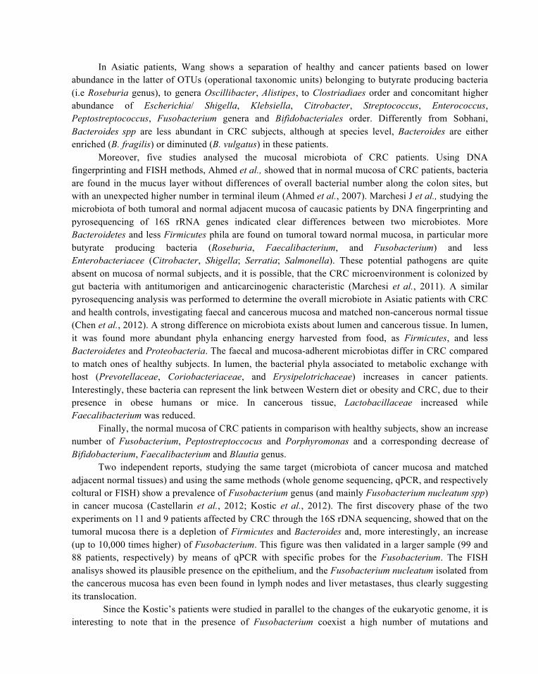
In Asiatic patients, Wang shows a separation of healthy and cancer patients based on lower abundance in the latter of OTUs (operational taxonomic units) belonging to butyrate producing bacteria (i.e Roseburia genus), to genera Oscillibacter, Alistipes, to Clostriadiaes order and concomitant higher abundance of Escherichia/ Shigella, Klebsiella, Citrobacter, Streptococcus, Enterococcus, Peptostreptococcus, Fusobacterium genera and Bifidobacteriales order. Differently from Sobhani, Bacteroides spp are less abundant in CRC subjects, although at species level, Bacteroides are either enriched (B. fragilis) or diminuted (B. vulgatus) in these patients.
Moreover, five studies analysed the mucosal microbiota of CRC patients. Using DNA fingerprinting and FISH methods, Ahmed et al., showed that in normal mucosa of CRC patients, bacteria are found in the mucus layer without differences of overall bacterial number along the colon sites, but with an unexpected higher number in terminal ileum (Ahmed et al., 2007). Marchesi J et al., studying the microbiota of both tumoral and normal adjacent mucosa of caucasic patients by DNA fingerprinting and pyrosequencing of 16S rRNA genes indicated clear differences between two microbiotes. More Bacteroidetes and less Firmicutes phila are found on tumoral toward normal mucosa, in particular more butyrate producing bacteria (Roseburia, Faecalibacterium, and Fusobacterium) and less Enterobacteriacee (Citrobacter, Shigella; Serratia; Salmonella). These potential pathogens are quite absent on mucosa of normal subjects, and it is possible, that the CRC microenvironment is colonized by gut bacteria with antitumorigen and anticarcinogenic characteristic (Marchesi et al., 2011). A similar pyrosequencing analysis was performed to determine the overall microbiote in Asiatic patients with CRC and health controls, investigating faecal and cancerous mucosa and matched non-cancerous normal tissue (Chen et al., 2012). A strong difference on microbiota exists about lumen and cancerous tissue. In lumen, it was found more abundant phyla enhancing energy harvested from food, as Firmicutes, and less Bacteroidetes and Proteobacteria. The faecal and mucosa-adherent microbiotas differ in CRC compared to match ones of healthy subjects. In lumen, the bacterial phyla associated to metabolic exchange with host (Prevotellaceae, Coriobacteriaceae, and Erysipelotrichaceae) increases in cancer patients. Interestingly, these bacteria can represent the link between Western diet or obesity and CRC, due to their presence in obese humans or mice. In cancerous tissue, Lactobacillaceae increased while Faecalibacterium was reduced.
Finally, the normal mucosa of CRC patients in comparison with healthy subjects, show an increase number of Fusobacterium, Peptostreptoccocus and Porphyromonas and a corresponding decrease of Bifidobacterium, Faecalibacterium and Blautia genus.
Two independent reports, studying the same target (microbiota of cancer mucosa and matched adjacent normal tissues) and using the same methods (whole genome sequencing, qPCR, and respectively coltural or FISH) show a prevalence of Fusobacterium genus (and mainly Fusobacterium nucleatum spp) in cancer mucosa (Castellarin et al., 2012; Kostic et al., 2012). The first discovery phase of the two experiments on 11 and 9 patients affected by CRC through the 16S rDNA sequencing, showed that on the tumoral mucosa there is a depletion of Firmicutes and Bacteroides and, more interestingly, an increase (up to 10,000 times higher) of Fusobacterium. This figure was then validated in a larger sample (99 and 88 patients, respectively) by means of qPCR with specific probes for the Fusobacterium. The FISH analisys showed its plausible presence on the epithelium, and the Fusobacterium nucleatum isolated from the cancerous mucosa has even been found in lymph nodes and liver metastases, thus clearly suggesting its translocation.
Since the Kostic’s patients were studied in parallel to the changes of the eukaryotic genome, it is interesting to note that in the presence of Fusobacterium coexist a high number of mutations and
chromosomal rearrangements (Bass et al., 2011). Finally, it must be emphasized that only a subset of patients affected by CRC shows an association with the presence of Fusobacterium, which, when present, can however, constitute 90% of the microbiota of the cancerous mucosa. This aspect resulted even more evident in the few cases in which the neoplastic mucosa was represented by the only adenoma (Castellarin et al., 2012). Furthermore, the Fusobacterium has been found in the mucosa of IBD patients with invasive capacity of cellular lines (Strauss et al., 2011, Dharmani et al., 2011).
An interesting hypothesis about the relationship between microbiota and the development of cancer, suggests that one or more bacteria play a key role from the beginning to advance phases also modifying the microbiota setting (the alpha bugs hypothesis) (Sears, 2009). Other hypothesis, more on line to the recent data, forecasts that the development of CRC-microbiota linked is a dynamic process in which bacteria can change over time. In particular, the promoters are driven bacteria belonging to those able to directly interfere in host genome, thanks to a proinflammatory action directly on epithelium (i.e. ETBF, E. coli, E. faecium and perhaps Fusobacterium spp.). Over time, these promoters would no longer be present on mucosa because the development of neoplasia modifies the microenvironment, which becomes available to other bacteria (so-called passenger bacteria) (Tjalsma et al., 2012).
Authors/y. N° of
subjects
Bacteria in HS Bacteria in patients with
Adenoma Bacteria in CRC patients
Methods Faeces Mucosa Faeces
Normal mucosa Adenoma Faeces
Normal mucosa Cancer
Moore and Moore 1995
88 X X Coltural
O’ Keefe 2007 52 X Coltural Shen 2010 44 X X Sequencing/FISH Pagnini 2011 51 X X X FISH/qPCR Sanapareddy 2012
71 X X Pyrosequencing
Balamurugan 2008
37 X X qPCR
Scanlan 2008 46 X X X DGGE/RISA Scanlan 2009 85 X X X qPCR Sobhani 2011 179 X X X Pyrosequencing/qPCR Wang 2012 102 X X Pyrosequencing/qPCR
Ahmed 2007 26 X DGGE/FISH Marchesi 2011 6 X X DGGE/Sequencing Castellarin 2011
11 X X WGSequencing/ qPCR/Coltural
Kostic 2012 18 X X WGSequencing/FISH Chen 2012 102 X X X X X Pyrosequencing
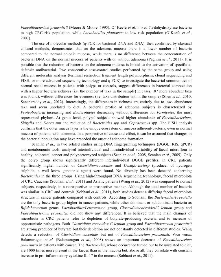
Table 1: Luminal and adherent mucosa microbiota in patients with adenoma or colorectal can-cer and in health control subjects.
In summary, only the use of more performing high-throughpout DNA sequencing technology allows defining a microbiome associated to the presence of CRC, but the design of cross-sectional cohort study prevents discovering the causal relationship between microbiota and CRC. What is indispensable
and we are still missing in order to define a cause-effect relationship, is to understand the changes in the intestinal microbiota in time and space, starting from the condition of normality and arriving to full blown CRC.
5 Animal Models, Intestinal Microflora and Sporadic Cancer Risk
Most studies on the relationship between IM and CRC have been conducted on rodent models, and are therefore biased by differences in the host-microbiota relationship between humans and animal. As a whole colorectal cancer tumor in rodent share many genetic and phenotypic features with human tumour (Corpet & Pierre, 2005). Studies often follow these schemes:
• Comparison among germfree, holoxenic and gnotobiotic rodents APC or carrier of other cancer-prone mutations;
• Comparison among germfree, holoxenic and gnotobiotic rodents treated with chemical carcinogens;
• Comparison among germfree, holoxenic and gnotobiotic rodents in capability of activating or inhibiting endogenous pro/co-carcinogens.
5.1 The Genetic Cancer-Prone Model (APC)
Since the serendipitous discover of APC mice in 1990 (Moser et al., 1990), many animals genetically predisposed to gastrointestinal cancer have been studied to understand the pathogenetic bases of these tumours. In particular, the APC mouse has been the first and most studied model in investigating the putative role of IM in genetically prone subjects. Considering that mutations in Apc are not only responsibly for familial adenomatous polyposis syndrome (FAP) but frequently occur in the sporadic CRC, the Min mice provide an interesting in vivo model to study human colorectal cancer, although mice develop mainly adenomas in small bowel and human only in large bowel. In these APC Min/+mice usually Wnt/βcatenin, together with Cox2 and NOS hyper expression, plays a major role in tumorigenesis. However, in these mice tumors occur mainly in small bowel. Several mutant of genetically modified APC Min/+ exist with, like humans, different number of adenomas. In particular, the variant of APC MinIN/+mice with deletion of exon 14 shows a severe colon polyposis, thus better simulating the human FAP’s condition.
As suggested by Dove’s study, the microbial state in APC mice seems not to remarkably influence the development of multiple adenomas in small and large bowel, neither in number nor in quality, with just a higher trend to develop adenomas in jejunum in presence of microflora (Dove et al., 1997). However, recently, Li et al. showed that a tumor load, either in small and large bowel of APC Min/+ mice, is strictly regulated by the presence of commensal microflora, which works, at least in part, by triggering the c-JUN/JNK and STAT3 signaling pathways (Li et al., 2012). Thus, further studies supports the key role of My D88 dependent activation of NF-KB in myeloid cells for tumorigenesis in APC Min/+ mice (Rakoff-Nahoume et al., 2007).
It is also true that some strains of bacteria seem to play a more critical role in CRC genesis of APC Min/+mice. For example, Newman and colleagues (Newman et al., 2001) have demonstrated that APC mice infected with C. Rodentium, a murine pathogen strongly adherent to the epithelium trough a type III secretion system (a molecular syringe-like mechanism), develops a fourfold increased number of colic adenomas than uninfected APC mice. Furthermore, this study has shown that medium highness of
dysplastic crypts is comparable with infected APC mice and infected wild type mice, demonstrating that even strong genetic background becomes negligible in case of C. Rodentium infection. The increased number of adenomas depends on the capability of this microorganism to induce hyperproliferation of epithelium (Barthold & Jonas, 1977), but its role in human gut is controversial. It is probable that the mechanism is similar to EHEC and EPEC pathogens, based on attaching and effacing lesions (AE). A comparison between germ-free and conventional mice infected with C. Rodentium shows that intestinal colonization does not require the type III secretion system in germ-free animals, and commensal bacteria are necessary to clear this pathogen from the mammalian intestine during infection, that occurs trough bacterial competition, by decreasing the number of anaerobes and increasing the number of Proteobacteria which compete with C. Rodentium for carbon sources (Kamada et al., 2012).
Furthermore, if, a human colonic bacterium as enterotoxigenic Bacteroides fragilis (ETBF) (responsible of large amount of infective human diarrhoea but also asymptomatic, carried up to 35% of population) colonizes APC Min/+mic, triggers colitis and strongly induce colonic tumors. This is strictly due to its toxin: a protease able to bind colin epithelial cells and stimulate the E-cadherin cleavage, actually nontoxigenic B. Fragilis doesn’t induce colonic tumor. Interestingly, ETBF induces adenoma or microadenoma early or very early after colonization, via both activation of Th17 in the lamina propria with IL-17 release and γδ-T cell with STAT3 pathway (Wu et al., 2009). Indirectly, these data can explain the prevalence of adenoma in small bowel of APC Min/+mice, because the SFB housing this part of bowel induces a strong Th17/IL17 reaction (Gaboriau-Routhiau et al., 2009).
Unfortunately, no sufficient data exist on intestinal microflora composition and relationship with mucosa of familial adenomatous polyposis patients. The unique, recent, exception suggests an unexpected characteristic: the presence of APC-like sequences in microbiota of FAP patients, thus suggesting a putative horizontal transfer of genetic information between eukaryotic and prokaryotic word (Holec et al., 2012). In conclusion, although neither a very strong genetic pattern seems to be sufficient to develop adenomas in absence of commensal bacteria, this process is emphasized in presence of proinflammatory bacteria.
5.2 The Chemical Carcinogenesis Route
The most frequently used chemical cancerogenous in experimental models are cycasin and 1,2-dimethylhydrazine (DMH), both procarcinogens transformed in azoxymethane in the presence of, respectively, bacterial beta-glucosidase and bacterial or mucosal beta-glucuronidase.
As expected, cycasine is ineffective in inducing CRC in germfree rats, while DMH can induce colon neoplasia also in this population (Reddy et al., 1975, Onoue et al., 1977, Horie et al., 1999A). Like humans, in several carcinogen- induced rodent tumors (in particular who’s due to DMH/AOM) the Wnt/ β-catenin pathway plays a back bone role, although the APC mutation is rare (Corpet & Pierre, 2005). Horie and Kanazawa evaluated the effect of intestinal microflora in the development of colonic neoplasia experimentally induced by DMH by comparing germ-free, holoxenic and gnotobiotic mice. In germfree rodents treated with DMH via subcutaneous route, the proliferation of crypts is higher than in holoxenic mice, but both the large/dysplastic adenoma and large/multiple ACF are significantly more represented in holoxenic than in germfree animals. The autochthonous microflora seems to have a suppressant effect on initiation of carcinogenesis induced by DMH. However, the size and the histopathological characteristics of adenomas developed in holoxenic animals suggest that bacterial flora may have an effect in promoting dysplastic transformation and tumoral growth (Horie et al., 1999A).
Furthermore, every single bacterial species might be differently involved in the various phases of
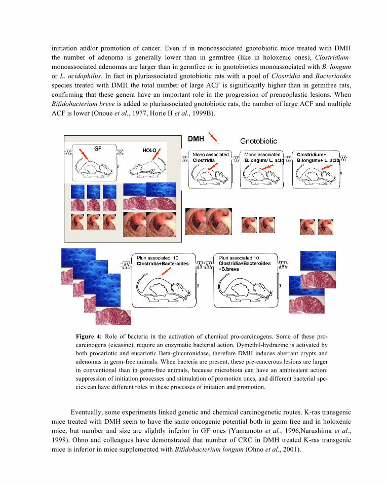
initiation and/or promotion of cancer. Even if in monoassociated gnotobiotic mice treated with DMH the number of adenoma is generally lower than in germfree (like in holoxenic ones), Clostridium-monoassociated adenomas are larger than in germfree or in gnotobiotics monoassociated with B. longum or L. acidophilus. In fact in pluriassociated gnotobiotic rats with a pool of Clostridia and Bacterioides species treated with DMH the total number of large ACF is significantly higher than in germfree rats, confirming that these genera have an important role in the progression of preneoplastic lesions. When Bifidobacterium breve is added to pluriassociated gnotobiotic rats, the number of large ACF and multiple ACF is lower (Onoue et al., 1977, Horie H et al., 1999B).
Figure 4: Role of bacteria in the activation of chemical pro-carcinogens. Some of these pro-carcinogens (cicasine), require an enzymatic bacterial action. Dymethil-hydrazine is activated by both procariotic and eucariotic Beta-glucuronidase, therefore DMH induces aberrant crypts and adenomas in germ-free animals. When bacteria are present, these pre-cancerous lesions are larger in conventional than in germ-free animals, because microbiota can have an ambivalent action: suppression of initiation processes and stimulation of promotion ones, and different bacterial spe-cies can have different roles in these processes of initation and promotion.
Eventually, some experiments linked genetic and chemical carcinogenetic routes. K-ras transgenic mice treated with DMH seem to have the same oncogenic potential both in germ free and in holoxenic mice, but number and size are slightly inferior in GF ones (Yamamoto et al., 1996,Narushima et al., 1998). Ohno and colleagues have demonstrated that number of CRC in DMH treated K-ras transgenic mice is inferior in mice supplemented with Bifidobacterium longum (Ohno et al., 2001).
This data show that IM as a whole interacts with chemical carcinogens, while suggesting a different role for each bacterial strain, since some favour carcinogenesis and other do not or hamper it. However, these results are limited by differences between humans and rodents and by inadequate representation of multi-step adenoma-carcinoma sequence.
5.3 Intestinal Microflora and Endogenous Carcinogens
Intestinal bacteria has been involved in the tumoral process since it has hydrolytic and reductasic enzymatic activities (such as nitroreductase, azoreductase, beta-glucuronidase, beta-glucosidase, arylsulfatases and alcohol dehydrogenases) having the capacity to produce or activate cancerogenous metabolites from digestion products (McBain & Macfarlane, 1998). Some of these metabolites require an enzymatic action conduced by bacteria only and are not able to induce tumors in germ-free rodents.
A biunivocal relation seems to exist between bacterial enzymes and dietary carcinogenic metabolites: in fact, if it is demonstrated that metabolites are activated by IM, it is also true that diet can influence enzymatic activity. Hambly et al. evaluated the influence of high- and low-risk dietary regimens on enzymatic activity markers in HFA mice: high-risk diet increased 2.5 fold β-glucuronidase activity and halfed beta-glucosydasic activity (Hambly et al., 1997). Concomitantly ACF, preneoplastic precursors of CRC, also increase.
In the last years, many compounds modulated or metabolised by IM have been identified, investigated and seem to be involved in colorectal carcinogenesis: in the next sections, the main ones will be briefly outlined.
5.3.1 Heterocyclic Amines and other Products of Pyrolysis
The heterocyclic amines (HCA), which originate from fried or broiled proteinaceous foods, seem to be carcinogenic in mice, rats, and monkeys producing hepatic, intestinal, and mammary tumors (Schoeffner & Thorgeirsson, 2000). For example, one HCA, 2-amino-3-methyl-3H-imidazol [4,5-f]quinoline (IQ), produced through the pyrolysis of creatinine, can be converted into 2-amino-3-methyl-3H-imidazo[4,5-f]quinoline-7-one (HOIQ, a direct-acting mutagen) by bacterial β-glucuronidase (Carman et al., 1988). In fact, after absorption in the upper part of the gastrointestinal tract, IQ is mainly metabolized in the liver. Here UDP-glucuronosyl transferases lead to the formation of harmless glucuronidated derivatives. These metabolites are partly excreted via the bile into the digestive lumen, where they come into contact with the resident microflora (Kassie et al., 2001). In GF rats treated with a single dose of IQ the DNA damage in form of strand breaks (Comet Tail Test) is significantly lower than in conventional and human flora associated animals (Knasmüller et al., 2001). The Comet assay performed on colonocytes and hepatocytes showed that the presence of bacterical β-glucuronidase in the digestive lumen dramatically increased (3-fold) the genotoxicity of IQ in the colon (Humblot et al., 2007). When the DNA damage is measured by alkaline single-cell gel electrophoresis assay DNA, the test exhibits significantly fewer alkaline-labile breaks in GF rats than in rats colonized with conventional murine or human bacteria, and this happens not only in colon cells but also in hepatocites (Kassie et al., 2001). The supplementation of the feed with Lactobacilli or Bifidobacteriumlongum seems to attenuate the induction of colon cancer by this same amine in a still unknown manner (Reddy & Rivenson, 1993, Knasmüller et al., 2001).
Other products of pyrolisis (such as benzopyrene), whose derivatives are inactive when joined to glucuronic acid, can be reactivated by the action of bacterial beta-glucuronidase, with successive damage to DNA (Renwick & Drasar, 1976). A very few bacterial strains bearing the ability to produce such metabolites in the intestinal lumen have been identified. This results from the fact that in vitro
experiments using culture media frequently give different results than in vivo experiments using HFA rodents. For instance, our unpublished experience showed that human strains of Clostridium might express their β-glucuronidase activity in vitro, in vivo or both. This can be due to genetic organization of the β-glucoronidase gene, which differs according to the analized strain (E. coli, L. gasseri, R. gnavus) (Beaud et al., 2005).
Benzopyrene originates from pyrolysis of organic material or food preparation at high temperature and is mutagenic and carcinogenic. This metabolite can be excreted as glucuronide (40%) and sulfate (9%) (Boroujerdi et al., 1981) or can be oxidated in the liver to epoxides, which are conjugated to glutathione and excreted in the bile. In the gut biliary metabolites of benzopyrene are hydrolysed by IM (Renwick & Drasar, 1976). In fact fecal excretion of benzopyrene glucuronide is higher in germ-free rats than in conventional ones (Rafter et al., 1987). Furthermore the DNA-benzopyrene adducts in colonic tissue seem to be produced only by bacterial β-glucuronidase hydrolysis of benzopyrene glucuronide (Kinoshita & Gelboin, 1978).
In summary, a western meat-rich diet may, apart the obesity risk, increase the risk of CRS, affecting the microbiota composition towards a profile with more effective metabolites of heterocyclic amines.
5.3.2 Secondary Biliary Acids and Diacylglycerol
Ileal bile salt transport is highly efficient (95%) but up to 800 mg of bile salts can escape the enterohepatic circulation daily and, in the colon, 7α-hydoxylating bacteria, such as Clostridia, convert primary biliary acids into secondary, e.g. deoxicolic (DCA) and lithocolic (LCA). Several observational studies suggest the role of faecal bile acids in CRC development (Tong et al., 2008).
Secondary biliary acids can induce cell necrosis, hyperplasia, and alteration of the DNA synthesis and increase of genotoxic activity of several mutagens in vitro. Actually, most animal studies conclude that DCA is a promoter of the carcinogenesis process (Pereira et al., 2004) and high concentration of secondary bile acids in faeces, blood and bile has been linked to the pathogenesis of CRC (McGarr et al., 2005). Some researchers argue that bile acids may cause DNA damage and act as carcinogens in humans (Bernstein et al., 2009) and it has been demonstrated that cell signaling pathways activated by DCA in mammalian epithelial cells include antiapoptotic cell signaling pathways involved in carcinogenesis, like protein kinase C (Zhu et al., 2002), ERK 1/2 via the epidermal growth factor receptor (Rao et al., 2002); β-catenin (Pai et al., 2004) and JNK 1/2 pathway (Gupta et al., 2004). According to Pereira and colleagues, the fact that secondary bile acids cause apoptosis in colonic epithelial cells could exert selective pressure for emergence of epithelial cell mutants which are resistant to apoptosis (for example, via loss of p53) (Pereira et al., 2004).
It is true also that a diet high in fat stimulates higher secretion rates of bile acids from the gallbladder, and the hypothesis that individuals who consume a high fat diet have higher levels of secondary bile acids in faeces has been confirmed in several studies (Hardison, 1978). An increased LCA/DCA ratio is also associated with a shift of the proliferative compartment towards the apex of colorectal crypts, an anomaly associated with cancer (Biasco et al., 1991).
Presence of high deoxycolate’s concentration enhances production of diacylglicerol (DAG), an activator of protein kinase C and, therefore, a promoter of carcinogenesis. DAG seems to be produced from phosphatidylcholine degradation through the action of bacterial enzymes. DAG formation in an acidic environment is negligible, therefore it has been hypothesized that Lactobacilli and Bifidobacteria exert a beneficial action on intestinal mucosa by reducing luminal pH (Vulevic et al., 2004).
5.3.3 Sulphide
Sulphidogenic bacteria are commensals microrganism utilizing sulphate (SO4\s\up5(-)-) as energy source and oxidant agent with consequent production of sulphide (S\s\up5(-)-) (Huycke & Gaskins, 2004); most of them belong to Proteobacteria phylum. Hydrogen sulphide concentration in the intestinal lumen is regulated on one hand by its bacteria production and on the other hand by the level of epithelial enzyme activity RHOD (thiolmethyltransferase and rhodanese), which degrades H2S.
Sulphide is a genotoxic agent, at 1 mM strongly increases crypt proliferation, and expands the proliferative zone to the upper crypt (Christl et al., 1996). Furthermore, it stimulates cell cycle entry and can induce hypoxia-triggered proliferation in intestinal epithelial cells through an MAPK, Akt, ERK and p21-dependent mechanisms (Cai et al., 2010). Furthermore, COX-2 is up-regulated in the epithelium after H2S challenge (Attene-Ramos et al., 2010).
A recent study has showed that animals fed dextran sodium sulphate (DSS), a chemical toxic to the intestinal epithelium, develop low to high-grade dysplasia, as well as adenoma and carcinoma, while a control group fed dextran sodium sulphate and metronidazole displayed no dysplasia. This demonstrates that cancer development in animals fed a sulphate source is dependent on bacterial metabolism (Deplancke et al., 2003).
Between sulphidogenic and metanogenic bacteria seems to be a substrate competition: the more is represented by the production of methane, the less is intestinal colonization of sulphidogenic bacteria (Strocchi et al., 1994). The role IM plays in the production of sulphide and methane, considering possible interactions with diet and genetic background, remains to be defined.
5.3.4 Fecapentaenes
The fecapentaenes (FPs) are conjugated either lipids produced in the large bowel by Bacteroides spp. from polyunsaturated ether phospholipids (plasmalogens) whose natural origin and function are unknown. Their production is greatly enhanced by bile in an unknown manner. Fecapentaene-12 causes direct oxidative DNA damage via production of the reactive oxygen species O2-, O2\s\up5(-)-, and OH\s\up5(.) (Szekely & Gates, 2006). FPs are strong mutagens (900 times more potent than N-methyl-N-nitrosurea), but there is no evidence for FPs as initiators and are considered promoters (Hinzman et al., 1987). The potential of fecapentaene-12 (FP-12) to promote tumor development was tested in a rat colon carcinogenesis model using N-methyl-N-nitrosourea (MNU) as the initiating agent (Zarkovic et al., 1993). The number of carcinoma-bearing rats as well as the average number of carcinomas per rat was significantly higher in the MNU + FP-12 group as compared to the MNU-alone values. Aberrant crypt foci (ACF) were found in all carcinogen-treated rats, including those that did not contain tumors, whereas none were observed in the FP-12 and control groups. The average number of ACF containing >10 aberrant crypts per focus was significantly higher in the MNU FP-12 group. These findings suggested that FP-12 could express promoting activity in chemical induced colon carcinogenesis.
Data reporting fecapentaenes excretion in man are apparently contradictory, since the excretion is higher in low risk subjects and lower in cancer patients: The FPs faecal excretion in groups at different risk of CRC is higher in vegetarian than in omnivores and lower in colon cancer patients than in controls (De Kok & Van Maanen, 2000). This apparent contradiction has been correlated to the lower exposition of intestinal mucosa to fecapentaenes in subjects with high excretion.
Due to these conflicting results, only few studies have been recently conducted on fecapentaenes role in colorectal carcinogensis, which currently seems negligible, but the existence of an interindividually variable mutagenic potential in the faeces seems reasonable.
5.3.5 Butyrate
The greatest part of intestinal microflora is strictly anaerobic, so the final products of its fermentative metabolism are short-chain fatty acids: in human colon, acetate, propionate and butyrate are the most represented (Høverstad et al., 1984). In humans, short-chain fatty acids are absorbed and used as energy substrates, and butyrate represents the 60-70% of enterocytes’ energy sources (Roediger, 1980). The main producers of butyrate are eubacteria, clostridia and roseburia (Nicholson et al., 2012). Diet with high levels of non-digestible carbohydrates stimulates the growth of specific butyrate-producing bacteria, hence increased plasma levels of butyrate.
Butyrate regulates cellular proliferation and differentiation through various supposed mechanisms: basically it inhibits NF-kB and histone deacetylase (Gibson, 2000, Hamer et al., 2008) and stimulates the detoxifying enzyme glutathione S-transferase (Ebert et al., 2003). That is why it is supposed that butyrate is one of the principle mediators in the protective role of fibre (Bingham et al., 2003). Some studies have supported the role of butyrate in colon carcinogenesis observing the down-regulation of butyrate transporters, like MCT1 and SMCT1, in neoplastic colonic tissue: this down-regulation could be responsible for reduced activity of butyrate in colonic mucosa and, consequently, for the increase in dysplastic alterations (Lambert et al., 2003).
The suppression of NF-kB has an important anti-inflammatory effect and, consequently, has a role in the prevention of inflammation related cancer. In fact, NF-kB regulates the expression of cytokines, inflammatory enzymes, immune receptors and acute phase proteins and has a responsibility in colon cancerogenesis (Hamer et al., 2008). The anti-inflammatory effect of butyrate could be due also to inhibition of interferon α production (Klampfer et al., 2003) or to upregulatin of PPAR-γ (peroxisome proliferator-activated receptor γ) (Kinoshita et al., 2002).
Histone deacetylase inhibition is responsible for the enhancement of the accessibility of transcription factor to DNA and for the modulation of fundamental apoptosis and cell cycle genes. Cancer cells seem to be more sensitive to this effect than normal cells, although there are no explanations for this different response (Dashwood et al., 2006).
Other effects of butyrate are inhibition of tumor cell migration by decreasing DAF (decay accelerating factor) (Andoh et al., 2002), inhibition of angiogenesis (Zgouras et al., 2003) and inactivation of metalloproteinase. More in general butyrate seems to decrease proliferation on the upper side of the crypt, increasing contemporary proliferation on the basal compartment of the crypt. This peculiar activity, called the butyrate paradox, could support the protective role on dysplastic/neoplastic tissue and, at the same time, could introduce to the procancerogenic role of butyrate on in vitro nonneoplastic cells (Comalada et al., 2006). The explanation of this double activity is still unknown.
It is important to note that fermentation of indigestible carbohydrates with consequent SCFA production takes place mostly in proximal colon, while protein fermentation occurs in its distal portion. This metabolic difference might be responsible for the prevalent distal localisation of most colic diseases. Human studies are needed to confirm the role of butyrate on cancer progression and/or prevention.
6 Bacteria and Inflammatory Bowel Disease – Associated Colorectal Cancer
Inflammatory bowel disease (IBD) is characterized by chronic, relapsing inflammation of the gastrointestinal tract. The two main types of IBD are Crohn’s disease (CD) and ulcerative colitis (UC).
The first one can affect any portion of the gut, but usually the terminal ileum and the colon. Inflammation, often with granulomas, involves the whole thickness of the gut wall and can be destroying, leading to stenosis and fistulas. It is usually discontinuous, with areas affected separated by apparently normal mucosa. Ulcerative colitis, on the contrary, always affects the rectum, and inflammation can spread cranially in a continuous fashion up to the caecum. Only mucosa and submucosa are affected, and inflammation is characteristically non-granulomatous (Podolsky, 2002). Patients affecting to severe UC, refractory to medical therapy, often undergo to total colectomy with anal-ileal pouch anastomosis (IPAA). This surgical procedure is commonly followed by pouchitis, a nonspecific inflammation of the ileal pouch (Meagher et al., 1998).
IBD is a strong risk factor for CRC development, with a prevalence of 2-3% at 10 years (Canavan et al., 2006). In the following section, the role of bacteria in IBD pathogenesis and their implication in malignant transformation will be discussed. Actually many data, albeit often based on animal models, support these hypothesis.
6.1 Pathogenesis of IBD
The aetiology of the disease is largely unknown, although various factors have been implicated in its pathogenesis, including the influence of genetic, environmental and microbial factors (Xavier & Podolsky, 2007). Among others, there is increasing evidence showing an important role for bacteria, in particular defects in both immune response and microbial recognition genes are pivotal for IBD onset in genetically predisposed patients (Bouma & Strober, 2003). Four main mechanisms, not mutually exclusive, have been suggested in the pathogenesis of IBD in humans: microbial pathogens, alteration of commensal microflora, defect of host mucosal barrier function, defect of host immune regulation (Farrell & LaMont, 2002).
6.2 Microbial Pathogens
Over the years various microbial pathogens have been suggested as aetiologic factors in IBD: Listeria monocitogenes, Helicobacter hepaticus, Chlamydia, Enterobacteriaceae, reoviruses and paramyxovirus (Liu et al., 1995). Mycobacterium avium subspecies paratuberculosis(MAP), which has been isolated in surgical specimens of Crohn’s disease,was strongly suspected as an etiologic factor of IBD onset. Actually, MAP is an obligate intracellular pathogen that causes Johne’s disease, a granulomatous inflammatory condition of the ruminants which affects the distal intestine, characterized by diarrhea and wasting and resembling human Crohn’s disease (Chiodini, 1989).
The detection of this organism in those individuals with defective innate immunological defenses, such as CD patients by various techniques has been reported, including culture, PCR, FISH, or serology (Behr & Schurr, 2006); others have shown immune response against mycobacterial antigens (Ibbotson et al., 1992). However, epidemiologic studies do not show increased prevalence of Crohn’s disease in spouses of patients, physicians treating patients, or farmers and veterinarians working with infected animals (Farrell & LaMont, 2002). Moreover, anti-TNF therapy and corticosteroids, risk factors for disseminated mycobacterial infections (Wallis et al., 2004), are effective in Crohn’s disease, while clinical studies failed to demonstrate the efficacy of antimycobacterium triple antibiotic therapy in CD patients (Thomas et al., 1998).
The most commonly IBD related bacterium is E. Coli belonging to the Enterobacteriaceae. In IBD patients, an increase of E. Coli was observed (Kotlowski et al., 2007). The adherent invasive E. coli was associated with ileal mucosal lesions in CD patients, with increased number and capability to adhere
to the intestinal epithelial cells, disrupting the intestinal barrier (Rolhion & Darfeuille-Michaud, 2007). This bacterium is more invasive in CD patients compared to UC patients (Sasaki et al., 2007).
Further studies demonstrated that the pathotype adhesive and invasive E. coli stimulates the production of IL-8, an important proinflammatory cytokine produced by macrophages and other cell types (Martin et al., 2004).
The different strains of Fusobacterium nucleatum, a gram-negative bacterial species of human mouth and gut, could have a pathogen role in IBD (Strauss et al., 2011). Two pathogen strains of F. nucleatum are capable to adhere to the colonic mucosa and to up-regulate the expression of TNF-α mRNA (Ohkusa et al., 2009). Furthermore, these pathogen strains have a role in regulating the expression of TNF-α, IL-10β mRNA and in the up-regulation MUC1 (4-fold) and MUC2 (12- to 15-fold) (Dharmani et al., 2011). As a while, due to these conflicting data no single microbial agent has been proven so far to be the cause of IBD in humans.
6.3 Changes in Commensal Microflora
Notwithstanding the difficulties to assess the normal microflora per group of subjects due to the personal pattern of IM, many studies have reported changes in microflora in patients with IBD, especially Crohn’s disease (Qin et al., 2010). In particular, IBD patients are characterized by a reduction of dominant members of the gut microbiota.
Recent studie suggest a decreased in microbial diversity in the active phase of the disease, describing an increase relative number of Enterobacteriaceae, especially of the enteroinvasive strains, and a decrease of Clostridium and Bacteroides species, with no substantial differences between ulcerative colitis and Crohn’s disease (Frank et al., 2007, Baumgart et al., 2007). While many studies agree with decreased Clostridia concentration (Gophna et al., 2006, Manichanh et al., 2006), for Bacteroides results are less clear, since some studies report an opposite pattern (Swidsinski et al., 2005, Kleessen et al., 2002).
Since Bacteroides and Clostridia produce butyrate and other short-chain fatty acids (Høverstad et al., 1984), the major energetic substrates of colonocytes (Roediger, 1980), their reduction could explain the reduced short-chain fatty acids concentration in the feces of patients with IBD (Marchesi et al., 2007). This, coupled with increased hydrogen sulphide production by other species – which inhibits short-chain fatty acid utilization – suggests the possibility of nutritional deficiency of colonocytes of IBD patients, which could lead to a loss of function. Actually, Roediger and colleagues hypothesized that ulcerative colitis can be the consequence of this mechanism (Roediger et al., 1993).
The disequilibrium between bacteria with anti or proinflammatory properties (due to their own characteristics and/or relationship with intestinal epithelium) can be involved in IBD onset. A recent study confirmed these data and also showed that Faecalibacterium prausnitzii, a member of the Clostridium leptum phylogenic group, has interesting anti-inflammatory properties both in vitro and in vivo on murine models. Moreover, its reduction on ileal mucosa is associated with higher risk of postoperative recurrence of Crohn’s disease (Sokol et al., 2008). Therefore, the disequilibrium between bacteria showing anti or proinflammatory features (due to their own characteristics and/or their relationship with intestinal epithelium) can be involved in IBD onset.
Furthermore, Escherichia coli concentration is increased in both faeces and mucosa of IBD patients (Frank et al., 2007, Baumgart et al., 2007). It is also present in granulomas (Ryan et al., 2004) and near ulcers and fistulae (Liu et al., 1995). These invasive strains express virulence factors and can replicate within macrophages which, in turn, secrete large quantities of TNF (Glasser et al., 2001),
contributing to inflammation. Whether these changes are primary or secondary is still controversial. The relationship amongst bacteria, mucus, mucose (epithelium and gut-associated lymphoid tissue
[GALT]) seems to be very important: in IBD patients, significantly more bacteria harbors in the intestinal mucus layer. Actually in a light microscope study on bioptic specimens were not seen bacteria at all in most control sections, while in 42% of IBD patients more than 50 bacteria per mucosal surface area examined have been observed, and the type of bowel preparation before undergoing the endoscopic procedure does not significantly affect this result (Schultsz et al., 1999).
6.4 Defect of Host Mucosal Barrier Function and Immune Regulation
Under physiologic conditions, the complex interaction between host and intestinal microflora is finely regulated. This ultimately leads to a tolerogenic response, while retaining the ability to mount an immune response against bacterial detrimental to the host (e.g. invasive pathogens). Any defect in this homeostatic system could result in an enhanced and inappropriate inflammatory response, which could itself damage the host.
An increase of bacterial translocation through the lamina propria triggers pattern recognition receptor (PRR), TRL stimulation and pro-inflammatory chemokine and cytokine secretion, which induce NF-kB pathway activation (Cario, 2010).
It has been demonstrated in vitro that proinflammatory cytokines induce a defect in epithelial barrier function via an apoptosis-independent mechanism (Bruewer et al., 2003); in particular interferon-γ can induce internalization of tight junction proteins (occludin, JAM-A, claudin-1) (Bruewer et al., 2005). This could allow non-invasive bacteria and gut antigens to cross epithelium and stimulate an immune response (Clark et al., 2005).In fact, IBD is characterized by enhanced mucosal permeability, but it is not clear whether the defect is primary or secondary: actually TNF upregulates claudin 2 expression, which is involved in pore formation. Moreover, altered regulation of apoptosis and tight junction components, have been reported in active Crohn’s disease (Zeissig et al., 2007). Some studies suggest that the defect is primary: Hollander and colleagues reported enhanced intestinal permeability both in patients with Crohn’s disease and their relatives, hypothesising an aetiologic role (Hollander et al., 1986).
The pathogenetic mechanisms previously described can ultimately lead to tolerance rupture and chronic intestinal inflammation, as suggested by efficacy of fecal diversion in Crohn’s disease relapse (Rutgeerts et al., 1991), but some evidence supports also the hypothesis of a primary defect in the immune system. Crohn’s disease patients have impaired microbial killing, which leads to overexposure of the microflora to the immune system and consequent activation of adaptive immunity (Korzenik, 2007).
This may be due to defective production of antimicrobial peptides such as α-defensin. Alpha-Defensins are peptides produced by Paneth cells in response to microbial products or proinflammatory cytokines. Their antibactericidial property is significantly efficacious against Enterobacteriaceae and BacteroidesVulgatus. Studies reported a significantly reduction of these peptides in association with ileal CD, in particular in patients with NOD-2 mutations (Nuding et al., 2007).More generally, there is evidence for reduced mucosal antimicrobial activity in Crohn’s disease. Genetic polymorphisms characterized by reduced synthesis of these proteins have been associated with Crohn’s disease, such as reduced copy number of α-defensin 2 (Fellermann et al., 2006) and NOD2/CARD15 variants (Hisamatsu et al., 2003). Moreover, the tolerogenic molecule gp96 is underexpressed in Crohn’s disease patients (Schreiter et al., 2005).
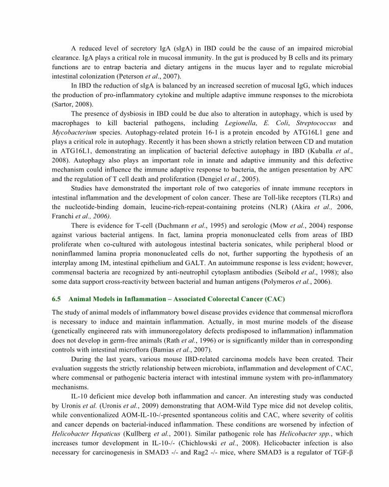
A reduced level of secretory IgA (sIgA) in IBD could be the cause of an impaired microbial clearance. IgA plays a critical role in mucosal immunity. In the gut is produced by B cells and its primary functions are to entrap bacteria and dietary antigens in the mucus layer and to regulate microbial intestinal colonization (Peterson et al., 2007).
In IBD the reduction of sIgA is balanced by an increased secretion of mucosal IgG, which induces the production of pro-inflammatory cytokine and multiple adaptive immune responses to the microbiota (Sartor, 2008).
The presence of dysbiosis in IBD could be due also to alteration in autophagy, which is used by macrophages to kill bacterial pathogens, including Legionella, E. Coli, Streptococcus and Mycobacterium species. Autophagy-related protein 16-1 is a protein encoded by ATG16L1 gene and plays a critical role in autophagy. Recently it has been shown a strictly relation between CD and mutation in ATG16L1, demonstrating an implication of bacterial defective autophagy in IBD (Kuballa et al., 2008). Autophagy also plays an important role in innate and adaptive immunity and this defective mechanism could influence the immune adaptive response to bacteria, the antigen presentation by APC and the regulation of T cell death and proliferation (Dengjel et al., 2005).
Studies have demonstrated the important role of two categories of innate immune receptors in intestinal inflammation and the development of colon cancer. These are Toll-like receptors (TLRs) and the nucleotide-binding domain, leucine-rich-repeat-containing proteins (NLR) (Akira et al., 2006, Franchi et al., 2006).
There is evidence for T-cell (Duchmann et al., 1995) and serologic (Mow et al., 2004) response against various bacterial antigens. In fact, lamina propria mononucleated cells from areas of IBD proliferate when co-cultured with autologous intestinal bacteria sonicates, while peripheral blood or noninflammed lamina propria mononucleated cells do not, further supporting the hypothesis of an interplay among IM, intestinal epithelium and GALT. An autoimmune response is less evident; however, commensal bacteria are recognized by anti-neutrophil cytoplasm antibodies (Seibold et al., 1998); also some data support cross-reactivity between bacterial and human antigens (Polymeros et al., 2006).
6.5 Animal Models in Inflammation – Associated Colorectal Cancer (CAC)
The study of animal models of inflammatory bowel disease provides evidence that commensal microflora is necessary to induce and maintain inflammation. Actually, in most murine models of the disease (genetically engineered rats with immunoregolatory defects predisposed to inflammation) inflammation does not develop in germ-free animals (Rath et al., 1996) or is significantly milder than in corresponding controls with intestinal microflora (Bamias et al., 2007).
During the last years, various mouse IBD-related carcinoma models have been created. Their evaluation suggests the strictly relationship between microbiota, inflammation and development of CAC, where commensal or pathogenic bacteria interact with intestinal immune system with pro-inflammatory mechanisms.
IL-10 deficient mice develop both inflammation and cancer. An interesting study was conducted by Uronis et al. (Uronis et al., 2009) demonstrating that AOM-Wild Type mice did not develop colitis, while conventionalized AOM-IL-10-/-presented spontaneous colitis and CAC, where severity of colitis and cancer depends on bacterial-induced inflammation. These conditions are worsened by infection of Helicobacter Hepaticus (Kullberg et al., 2001). Similar pathogenic role has Helicobacter spp., which increases tumor development in IL-10-/- (Chichlowski et al., 2008). Helicobacter infection is also necessary for carcinogenesis in SMAD3 -/- and Rag2 -/- mice, where SMAD3 is a regulator of TGF-β
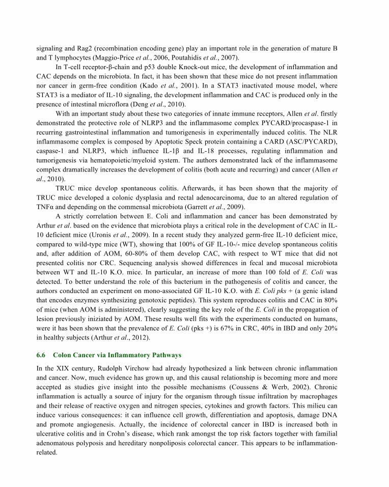
signaling and Rag2 (recombination encoding gene) play an important role in the generation of mature B and T lymphocytes (Maggio-Price et al., 2006, Poutahidis et al., 2007).
In T-cell receptor-β-chain and p53 double Knock-out mice, the development of inflammation and CAC depends on the microbiota. In fact, it has been shown that these mice do not present inflammation nor cancer in germ-free condition (Kado et al., 2001). In a STAT3 inactivated mouse model, where STAT3 is a mediator of IL-10 signaling, the development inflammation and CAC is produced only in the presence of intestinal microflora (Deng et al., 2010).
With an important study about these two categories of innate immune receptors, Allen et al. firstly demonstrated the protective role of NLRP3 and the inflammasome complex PYCARD/procaspase-1 in recurring gastrointestinal inflammation and tumorigenesis in experimentally induced colitis. The NLR inflammasome complex is composed by Apoptotic Speck protein containing a CARD (ASC/PYCARD), caspase-1 and NLRP3, which influence IL-1β and IL-18 processes, regulating inflammation and tumorigenesis via hematopoietic/myeloid system. The authors demonstrated lack of the inflammasome complex dramatically increases the development of colitis (both acute and recurring) and cancer (Allen et al., 2010).
TRUC mice develop spontaneous colitis. Afterwards, it has been shown that the majority of TRUC mice developed a colonic dysplasia and rectal adenocarcinoma, due to an altered regulation of TNFα and depending on the commensal microbiota (Garrett et al., 2009).
A strictly correlation between E. Coli and inflammation and cancer has been demonstrated by Arthur et al. based on the evidence that microbiota plays a critical role in the development of CAC in IL-10 deficient mice (Uronis et al., 2009). In a recent study they analyzed germ-free IL-10 deficient mice, compared to wild-type mice (WT), showing that 100% of GF IL-10-/- mice develop spontaneous colitis and, after addition of AOM, 60-80% of them develop CAC, with respect to WT mice that did not presented colitis nor CRC. Sequencing analysis showed differences in fecal and mucosal microbiota between WT and IL-10 K.O. mice. In particular, an increase of more than 100 fold of E. Coli was detected. To better understand the role of this bacterium in the pathogenesis of colitis and cancer, the authors conducted an experiment on mono-associated GF IL-10 K.O. with E. Coli pks + (a genic island that encodes enzymes synthesizing genotoxic peptides). This system reproduces colitis and CAC in 80% of mice (when AOM is administered), clearly suggesting the key role of the E. Coli in the propagation of lesion previously iniziated by AOM. These results well fits with the experiments conducted on humans, were it has been shown that the prevalence of E. Coli (pks +) is 67% in CRC, 40% in IBD and only 20% in healthy subjects (Arthur et al., 2012).
6.6 Colon Cancer via Inflammatory Pathways
In the XIX century, Rudolph Virchow had already hypothesized a link between chronic inflammation and cancer. Now, much evidence has grown up, and this causal relationship is becoming more and more accepted as studies give insight into the possible mechanisms (Coussens & Werb, 2002). Chronic inflammation is actually a source of injury for the organism through tissue infiltration by macrophages and their release of reactive oxygen and nitrogen species, cytokines and growth factors. This milieu can induce various consequences: it can influence cell growth, differentiation and apoptosis, damage DNA and promote angiogenesis. Actually, the incidence of colorectal cancer in IBD is increased both in ulcerative colitis and in Crohn’s disease, which rank amongst the top risk factors together with familial adenomatous polyposis and hereditary nonpoliposis colorectal cancer. This appears to be inflammation-related.
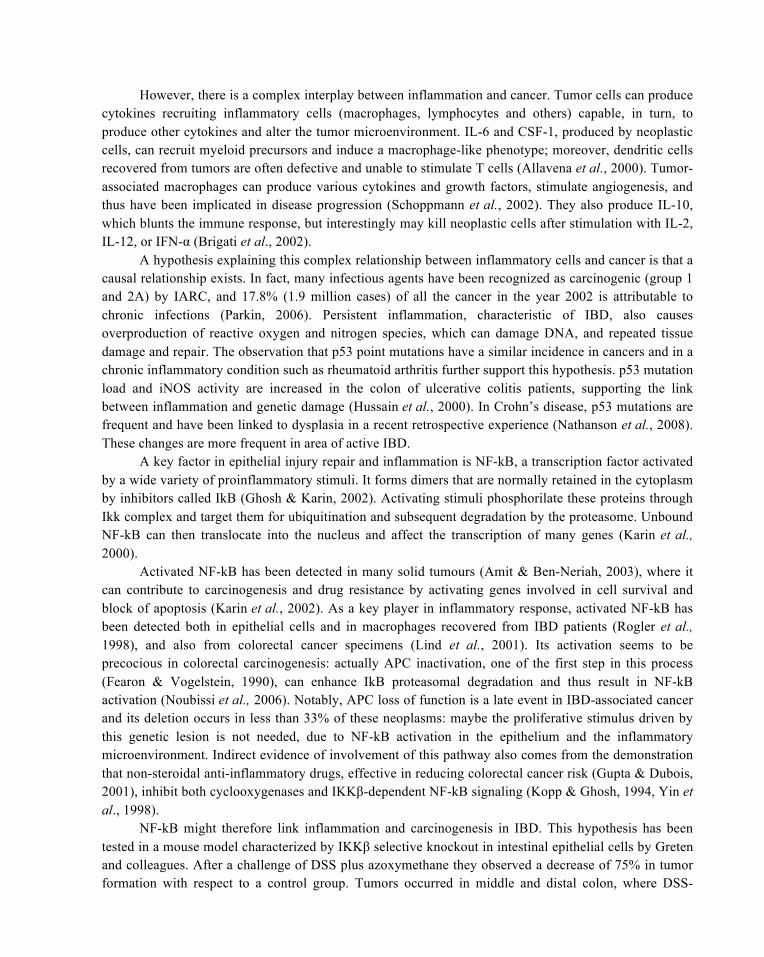
However, there is a complex interplay between inflammation and cancer. Tumor cells can produce cytokines recruiting inflammatory cells (macrophages, lymphocytes and others) capable, in turn, to produce other cytokines and alter the tumor microenvironment. IL-6 and CSF-1, produced by neoplastic cells, can recruit myeloid precursors and induce a macrophage-like phenotype; moreover, dendritic cells recovered from tumors are often defective and unable to stimulate T cells (Allavena et al., 2000). Tumor-associated macrophages can produce various cytokines and growth factors, stimulate angiogenesis, and thus have been implicated in disease progression (Schoppmann et al., 2002). They also produce IL-10, which blunts the immune response, but interestingly may kill neoplastic cells after stimulation with IL-2, IL-12, or IFN-α (Brigati et al., 2002).
A hypothesis explaining this complex relationship between inflammatory cells and cancer is that a causal relationship exists. In fact, many infectious agents have been recognized as carcinogenic (group 1 and 2A) by IARC, and 17.8% (1.9 million cases) of all the cancer in the year 2002 is attributable to chronic infections (Parkin, 2006). Persistent inflammation, characteristic of IBD, also causes overproduction of reactive oxygen and nitrogen species, which can damage DNA, and repeated tissue damage and repair. The observation that p53 point mutations have a similar incidence in cancers and in a chronic inflammatory condition such as rheumatoid arthritis further support this hypothesis. p53 mutation load and iNOS activity are increased in the colon of ulcerative colitis patients, supporting the link between inflammation and genetic damage (Hussain et al., 2000). In Crohn’s disease, p53 mutations are frequent and have been linked to dysplasia in a recent retrospective experience (Nathanson et al., 2008). These changes are more frequent in area of active IBD.
A key factor in epithelial injury repair and inflammation is NF-kB, a transcription factor activated by a wide variety of proinflammatory stimuli. It forms dimers that are normally retained in the cytoplasm by inhibitors called IkB (Ghosh & Karin, 2002). Activating stimuli phosphorilate these proteins through Ikk complex and target them for ubiquitination and subsequent degradation by the proteasome. Unbound NF-kB can then translocate into the nucleus and affect the transcription of many genes (Karin et al., 2000).
Activated NF-kB has been detected in many solid tumours (Amit & Ben-Neriah, 2003), where it can contribute to carcinogenesis and drug resistance by activating genes involved in cell survival and block of apoptosis (Karin et al., 2002). As a key player in inflammatory response, activated NF-kB has been detected both in epithelial cells and in macrophages recovered from IBD patients (Rogler et al., 1998), and also from colorectal cancer specimens (Lind et al., 2001). Its activation seems to be precocious in colorectal carcinogenesis: actually APC inactivation, one of the first step in this process (Fearon & Vogelstein, 1990), can enhance IkB proteasomal degradation and thus result in NF-kB activation (Noubissi et al., 2006). Notably, APC loss of function is a late event in IBD-associated cancer and its deletion occurs in less than 33% of these neoplasms: maybe the proliferative stimulus driven by this genetic lesion is not needed, due to NF-kB activation in the epithelium and the inflammatory microenvironment. Indirect evidence of involvement of this pathway also comes from the demonstration that non-steroidal anti-inflammatory drugs, effective in reducing colorectal cancer risk (Gupta & Dubois, 2001), inhibit both cyclooxygenases and IKKβ-dependent NF-kB signaling (Kopp & Ghosh, 1994, Yin et al., 1998).
NF-kB might therefore link inflammation and carcinogenesis in IBD. This hypothesis has been tested in a mouse model characterized by IKKβ selective knockout in intestinal epithelial cells by Greten and colleagues. After a challenge of DSS plus azoxymethane they observed a decrease of 75% in tumor formation with respect to a control group. Tumors occurred in middle and distal colon, where DSS-
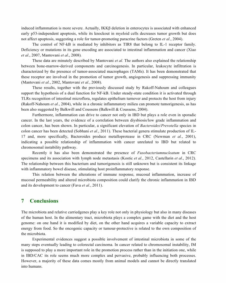
induced inflammation is more severe. Actually, IKKβ deletion in enterocytes is associated with enhanced early p53-independent apoptosis, while its knockout in myeloid cells decreases tumor growth but does not affect apoptosis, suggesting a role for tumor-promoting paracrine factors (Greten et al., 2004).
The control of NF-kB is mediated by inhibitors as TIR8 that belong to IL-1 receptor family. Deficiency or mutations in its gene encoding are associated to intestinal inflammation and cancer (Xiao et al., 2007, Mantovani et al., 2008).
These data are minutely described by Mantovani et al. The authors also explained the relationship between bone-marrow-derived components and carcinogenesis. In particular, leukocyte infiltration is characterized by the presence of tumor-associated macrophages (TAMs). It has been demonstrated that these receptor are involved in the promotion of tumor growth, angiogenesis and suppressing immunity (Mantovani et al., 2002, Mantovani et al., 2008).
These results, together with the previously discussed study by Rakoff-Nahoum and colleagues support the hypothesis of a dual function for NF-kB. Under steady-state condition it is activated through TLRs recognition of intestinal microflora, regulates epithelium turnover and protects the host from injury (Rakoff-Nahoum et al., 2004), while in a chronic inflammatory milieu can promote tumorigenesis, as has been also suggested by Balkwill and Coussens (Balkwill & Coussens, 2004).
Furthermore, inflammation can drive to cancer not only in IBD but plays a role even in sporadic cancer. In the last years, the evidence of a correlation between diysbiosis/low grade inflammation and colon cancer, has been shown. In particular, a significant elevation of Bacteroides/Prevotella species in colon cancer has been detected (Sobhani et al., 2011). These bacterial genera stimulate production of IL-17 and, more specifically, Bacteroides produce metalloprotease in CRC (Newman et al., 2001), indicating a possible relationship of inflammation with cancer unrelated to IBD but related to chromosomal instability pathway.
Recently it has also been demonstrated the presence of Fusobacteriumnucleatum in CRC specimens and its association with lymph node metastasis (Kostic et al., 2012, Castellarin et al., 2012). The relationship between this bacterium and tumorigenesis is still unknown but is consistent its linkage with inflammatory bowel disease, stimulating host proinflammatory response.
This relation between the alterations of immune response, mucosal inflammation, increase of mucosal permeability and altered microbiota composition could clarify the chronic inflammation in IBD and its development to cancer (Fava et al., 2011).
7 Conclusions
The microbiota and relative carriedgenes play a key role not only in physiology but also in many diseases of the human host. In the alimentary tract, microbiota plays a complex game with the diet and the host genome: on one hand it is modified by diet, on the other hand acquires a variable capacity to extract energy from food. So the oncogenic capacity or tumour-protective is related to the own composition of the microbiota.
Experimental evidences suggest a possible involvement of intestinal microbiota in some of the many steps eventually leading to colorectal carcinoma. In cancer related to chromosomal instability, IM is supposed to play a more important role in the promotion process rather than in the initiation one, while in IBD/CAC its role seems much more complex and pervasive, probably influencing both processes. However, a majority of these data comes mostly from animal models and cannot be directly translated into humans.
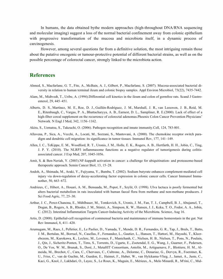
In humans, the data obtained bythe modern approaches (high-throughout DNA/RNA sequencing and molecular imaging) suggest a loss of the normal bacterial confinement away from colonic epithelium with progressive transformation of the mucosa and microbiota itself, in a dynamic process of carcinogenesis.
However, among several questions far from a definitive solution, the most intriguing remain those about the putative oncogenic or tumour-protective potential of different bacterial strains, as well as on the possible percentage of colorectal cancer, strongly linked to the microbiota action.
References
Ahmed, S., Macfarlane, G. T., Fite, A., McBain, A. J., Gilbert, P., Macfarlane, S. (2007). Mucosa-associated bacterial di-versity in relation to human terminal ileum and colonic biopsy samples. Appl Environ Microbiol, 73(22), 7435-7442.
Alam, M., Midtvedt, T., Uribe, A. (1994).Differential cell kinetics in the ileum and colon of germfree rats. Scand J Gastro-enterol, 29, 445–451.
Alberts, D. S., Martínez, M. E., Roe, D. J., Guillén-Rodríguez, J. M., Marshall, J. R., van Leeuwen, J. B., Reid, M. E., Ritenbaugh, C., Vargas, P. A., Bhattacharyya, A. B., Earnest, D. L., Sampliner, R. E.(2000). Lack of effect of a high-fiber cereal supplement on the recurrence of colorectal adenomas.Phoenix Colon Cancer Prevention Physicians’ Network. N Engl J Med, 342, 1156–1162.
Akira, S., Uematsu, S., Takeuchi, O. (2006). Pathogen recognition and innate immunity.Cell, 124, 783-801.
Allavena, P., Sica, A., Vecchi, A., Locati, M., Sozzani, S., Mantovani, A. (2000). The chemokine receptor switch para-digm and dendritic cell migration: its significance in tumor tissues. Immunol Rev, 177, 141–149.
Allen, I. C., TeKippe, E. M., Woodford, R. T., Uronis, J. M., Holle, E. K., Rogers, A. B., Hertfarth, H. H., Jobin, C., Ting, J. P. Y. (2010). The NLRP3 inflammasome functions as a negative regulator of tumorigenesis during colitis-associated cancer. J Exp Med, 207, 1045-1056.
Amit, S. & Ben-Neriah, Y. (2003).NF-kappaB activation in cancer: a challenge for ubiquitination- and proteasome-based therapeutic approach. Semin Cancer Biol, 13, 15–28.
Andoh, A., Shimada, M., Araki, Y., Fujiyama, Y., Bamba, T. (2002). Sodium butyrate enhances complement-mediated cell injury via down-regulation of decay-accelerating factor expression in colonic cancer cells. Cancer Immunol Immu-nother, 50, 663–672.
Andrieux, C., Hibert, A., Houari, A. M., Bensaada, M., Popot, F., Szylit, O. (1998). Ulva lactuca is poorly fermented but alters bacterial metabolism in rats inoculated with human faecal flora from methane and non-methane producers. J Sci Food Agric, 77, 25–30.
Arthur, J. C., Perez-Chanona, E., Mühlbauer, M., Tomkovich, S., Uronis, J. M., Fan, T. J., Campbell, B. J., Abujamel, T., Dogan, B., Rogers, A. B., Rhodes, J. M., Stintzi, A., Simpson, K. W., Hansen, J. J., Keku, T. O., Fodor, A. A., Jobin, C. (2012). Intestinal Inflammation Targets Cancer-Inducing Activity of the Microbiota. Science, Aug 16.
Artis, D. (2008). Epithelial-cell recognition of commensal bacteria and maintenance of immune homeostasis in the gut. Nat Rev Immunol, 8, 411–420.
Arumugam, M., Raes, J., Pelletier, E., Le Paslier, D., Yamada, T., Mende, D. R., Fernandes, G. R., Tap, J., Bruls, T., Batto, J. M., Bertalan, M., Borruel, N., Casellas, F., Fernandez, L., Gautier, L., Hansen, T., Hattori, M., Hayashi, T., Kleer-ebezem, M., Kurokawa, K., Leclerc, M., Levenez, F., Manichanh, C., Nielsen, H. B., Nielsen, T., Pons, N., Poulain, J., Qin, J., Sicheritz-Ponten, T., Tims, S., Torrents, D., Ugarte, E., Zoetendal, E. G., Wang, J., Guarner, F., Pedersen, O., De Vos, W. M., Brunak, S., Doré, J., MetaHIT Consortium, Antolín, M., Artiguenave, F., Blottiere, H. M., Al-meida, M., Brechot, C., Cara, C., Chervaux, C., Cultrone, A., Delorme, C., Denariaz, G., Dervyn, R., Foerstner, K. U., Friss, C., van de Guchte, M., Guedon, E., Haimet, F., Huber, W., van Hylckama-Vlieg, J., Jamet, A., Juste, C., Kaci, G., Knol, J., Lakhdari, O., Layec, S., Le Roux, K., Maguin, E., Mérieux, A., Melo Minardi, R., M'rini, C., Mul-
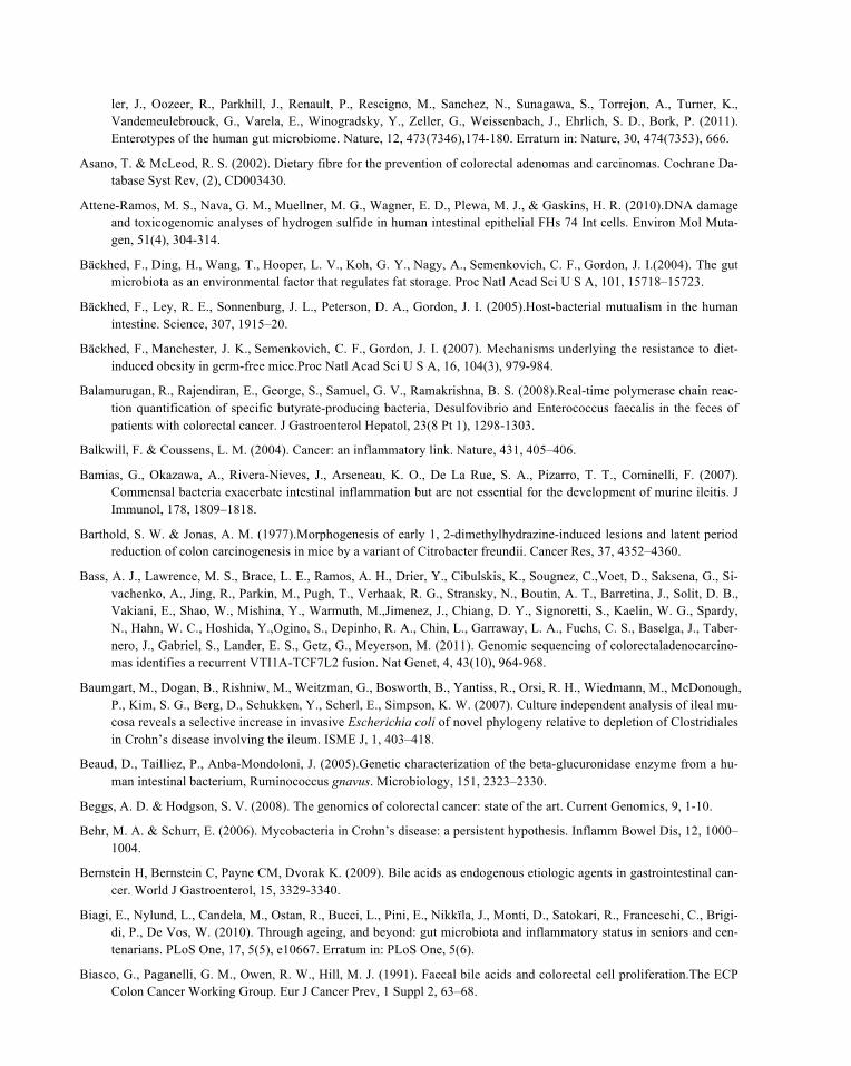
ler, J., Oozeer, R., Parkhill, J., Renault, P., Rescigno, M., Sanchez, N., Sunagawa, S., Torrejon, A., Turner, K., Vandemeulebrouck, G., Varela, E., Winogradsky, Y., Zeller, G., Weissenbach, J., Ehrlich, S. D., Bork, P. (2011). Enterotypes of the human gut microbiome. Nature, 12, 473(7346),174-180. Erratum in: Nature, 30, 474(7353), 666.
Asano, T. & McLeod, R. S. (2002). Dietary fibre for the prevention of colorectal adenomas and carcinomas. Cochrane Da-tabase Syst Rev, (2), CD003430.
Attene-Ramos, M. S., Nava, G. M., Muellner, M. G., Wagner, E. D., Plewa, M. J., & Gaskins, H. R. (2010).DNA damage and toxicogenomic analyses of hydrogen sulfide in human intestinal epithelial FHs 74 Int cells. Environ Mol Muta-gen, 51(4), 304-314.
Bäckhed, F., Ding, H., Wang, T., Hooper, L. V., Koh, G. Y., Nagy, A., Semenkovich, C. F., Gordon, J. I.(2004). The gut microbiota as an environmental factor that regulates fat storage. Proc Natl Acad Sci U S A, 101, 15718–15723.
Bäckhed, F., Ley, R. E., Sonnenburg, J. L., Peterson, D. A., Gordon, J. I. (2005).Host-bacterial mutualism in the human intestine. Science, 307, 1915–20.
Bäckhed, F., Manchester, J. K., Semenkovich, C. F., Gordon, J. I. (2007). Mechanisms underlying the resistance to diet-induced obesity in germ-free mice.Proc Natl Acad Sci U S A, 16, 104(3), 979-984.
Balamurugan, R., Rajendiran, E., George, S., Samuel, G. V., Ramakrishna, B. S. (2008).Real-time polymerase chain reac-tion quantification of specific butyrate-producing bacteria, Desulfovibrio and Enterococcus faecalis in the feces of patients with colorectal cancer. J Gastroenterol Hepatol, 23(8 Pt 1), 1298-1303.
Balkwill, F. & Coussens, L. M. (2004). Cancer: an inflammatory link. Nature, 431, 405–406.
Bamias, G., Okazawa, A., Rivera-Nieves, J., Arseneau, K. O., De La Rue, S. A., Pizarro, T. T., Cominelli, F. (2007). Commensal bacteria exacerbate intestinal inflammation but are not essential for the development of murine ileitis. J Immunol, 178, 1809–1818.
Barthold, S. W. & Jonas, A. M. (1977).Morphogenesis of early 1, 2-dimethylhydrazine-induced lesions and latent period reduction of colon carcinogenesis in mice by a variant of Citrobacter freundii. Cancer Res, 37, 4352–4360.
Bass, A. J., Lawrence, M. S., Brace, L. E., Ramos, A. H., Drier, Y., Cibulskis, K., Sougnez, C.,Voet, D., Saksena, G., Si-vachenko, A., Jing, R., Parkin, M., Pugh, T., Verhaak, R. G., Stransky, N., Boutin, A. T., Barretina, J., Solit, D. B., Vakiani, E., Shao, W., Mishina, Y., Warmuth, M.,Jimenez, J., Chiang, D. Y., Signoretti, S., Kaelin, W. G., Spardy, N., Hahn, W. C., Hoshida, Y.,Ogino, S., Depinho, R. A., Chin, L., Garraway, L. A., Fuchs, C. S., Baselga, J., Taber-nero, J., Gabriel, S., Lander, E. S., Getz, G., Meyerson, M. (2011). Genomic sequencing of colorectaladenocarcino-mas identifies a recurrent VTI1A-TCF7L2 fusion. Nat Genet, 4, 43(10), 964-968.
Baumgart, M., Dogan, B., Rishniw, M., Weitzman, G., Bosworth, B., Yantiss, R., Orsi, R. H., Wiedmann, M., McDonough, P., Kim, S. G., Berg, D., Schukken, Y., Scherl, E., Simpson, K. W. (2007). Culture independent analysis of ileal mu-cosa reveals a selective increase in invasive Escherichia coli of novel phylogeny relative to depletion of Clostridiales in Crohn’s disease involving the ileum. ISME J, 1, 403–418.
Beaud, D., Tailliez, P., Anba-Mondoloni, J. (2005).Genetic characterization of the beta-glucuronidase enzyme from a hu-man intestinal bacterium, Ruminococcus gnavus. Microbiology, 151, 2323–2330.
Beggs, A. D. & Hodgson, S. V. (2008). The genomics of colorectal cancer: state of the art. Current Genomics, 9, 1-10.
Behr, M. A. & Schurr, E. (2006). Mycobacteria in Crohn’s disease: a persistent hypothesis. Inflamm Bowel Dis, 12, 1000–1004.
Bernstein H, Bernstein C, Payne CM, Dvorak K. (2009). Bile acids as endogenous etiologic agents in gastrointestinal can-cer. World J Gastroenterol, 15, 3329-3340.
Biagi, E., Nylund, L., Candela, M., Ostan, R., Bucci, L., Pini, E., Nikkïla, J., Monti, D., Satokari, R., Franceschi, C., Brigi-di, P., De Vos, W. (2010). Through ageing, and beyond: gut microbiota and inflammatory status in seniors and cen-tenarians. PLoS One, 17, 5(5), e10667. Erratum in: PLoS One, 5(6).
Biasco, G., Paganelli, G. M., Owen, R. W., Hill, M. J. (1991). Faecal bile acids and colorectal cell proliferation.The ECP Colon Cancer Working Group. Eur J Cancer Prev, 1 Suppl 2, 63–68.
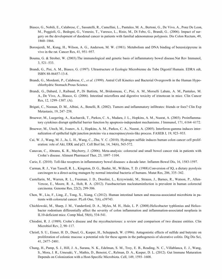
Biasco, G., Nobili, E., Calabrese, C., Sassatelli, R., Camellini, L., Pantaleo, M. A., Bertoni, G., De Vivo, A., Ponz De Leon, M., Poggioli, G., Bedogni, G., Venesio, T., Varesco, L., Risio, M., Di Febo, G., Brandi, G.. (2006). Impact of sur-gery on the development of duodenal cancer in patients with familial adenomatous polyposis. Dis Colon Rectum, 49, 1860–1866.
Boroujerdi, M., Kung, H., Wilson, A. G., Anderson, M. W. (1981). Metabolism and DNA binding of benzo(a)pyrene in vivo in the rat. Cancer Res, 41, 951–957.
Bouma, G. & Strober, W. (2003).The immunological and genetic basis of inflammatory bowel disease.Nat Rev Immunol, 3, 521–533.
Brandi, G., Pisi, A. M., Biasco, G. (1997). Ultrastructure et Ecologie Microbienne du Tube Digestif Humain. EDRA edt, ISBN 88-86457-13-8.
Brandi, G., Mordenti, P., Calabrese, C., et al. (1999). Antral Cell Kinetics and Bacterial Overgrowth in the Human Hypo-chlorhydric Stomach.Prous Science.
Brandi, G., Dabard, J., Raibaud, P., Di Battista, M., Bridonneau, C., Pisi, A. M., Morselli Labate, A. M., Pantaleo, M. A., De Vivo, A., Biasco, G. (2006). Intestinal microflora and digestive toxicity of irinotecan in mice. Clin Cancer Res, 12, 1299–1307. (A).
Brigati, C., Noonan, D. M., Albini, A., Benelli, R. (2002). Tumors and inflammatory infiltrates: friends or foes? Clin Exp Metastasis, 19, 247–258.
Bruewer, M., Luegering, A., Kucharzik, T., Parkos, C. A., Madara, J. L., Hopkins, A. M., Nusrat, A. (2003). Proinflamma-tory cytokines disrupt epithelial barrier function by apoptosis-independent mechanisms. J Immunol, 171, 6164–6172.
Bruewer, M., Utech, M., Ivanov, A. I., Hopkins, A. M., Parkos, C. A., Nusrat, A. (2005). Interferon-gamma induces inter-nalization of epithelial tight junction proteins via a macropinocytosis-like process. FASEB J, 19, 923–933.
Cai, W. J., Wang, M. J., Ju, L. H., Wang, C., Zhu, Y. C. (2010). Hydrogen sulfide induces human colon cancer cell prolif-eration: role of Akt, ERK and p21. Cell Biol Int, 14, 34(6), 565-572.
Canavan, C., Abrams, K. R., Mayberry, J. (2006). Meta-analysis: colorectal and small bowel cancer risk in patients with Crohn’s disease. Aliment Pharmacol Ther, 23, 1097–1104.
Cario, E. (2010). Toll-like receptors in inflammatory bowel diseases: a decade later. Inflamm Bowel Dis, 16, 1583-1597.
Carman, R. J., Van Tassell, R. L., Kingston, D. G., Bashir, M., Wilkins, T. D. (1988).Conversion of IQ, a dietary pyrolysis carcinogen to a direct-acting mutagen by normal intestinal bacteria of humans. Mutat Res, 206, 335–342.
Castellarin, M., Warren, R. L., Freeman, J. D., Dreolini, L., Krzywinski, M., Strauss, J., Barnes, R., Watson, P., Allen-Vercoe, E., Moore, R. A., Holt, R. A. (2012). Fusobacterium nucleatuminfection is prevalent in human colorectal carcinoma. Genome Res, 22(2), 299-306.
Chen, W., Liu, F., Ling, Z., Tong, X., Xiang, C.(2012). Human intestinal lumen and mucosa-associated microbiota in pa-tients with colorectal cancer. PLoS One, 7(6), e39743.
Chichlowski, M., Sharp, J. M., Vanderford, D. A., Myles, M. H., Hale, L. P. (2008).Helicobacter typhlonius and Helico-bacter rodentium differentially affect the severity of colon inflammation and inflammation-associated neoplasia in IL10-deficient mice. Comp Med, 58(6), 534-541.
Chiodini, R. J. (1989). Crohn’s disease and the mycobacterioses: a review and comparison of two disease entities. Clin Microbiol Rev, 2, 90–117.
Christl, S. U., Eisner, H. D., Dusel, G., Kasper, H., Scheppach, W. (1996). Antagonistic effects of sulfide and butyrate on proliferation of colonic mucosa: a potential role for these agents in the pathogenesis of ulcerative colitis. Dig Dis Sci, 41, 2477–2481.
Chung, H., Pamp, S. J., Hill, J. A., Surana, N. K., Edelman, S. M., Troy, E. B., Reading, N. C., Villablanca, E. J., Wang, S., Mora, J. R., Umesaki, Y., Mathis, D., Benoist, C., Relman, D. A., Kasper, D. L. (2012). Gut Immune Maturation Depends on Colonization with a Host-Specific Microbiota. Cell, 149, 1593–1608.
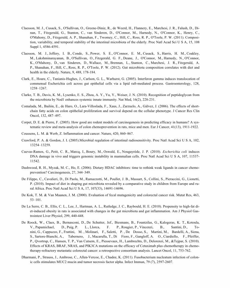
Claesson, M. J., Cusack, S., O'Sullivan, O., Greene-Diniz, R., de Weerd, H., Flannery, E., Marchesi, J. R., Falush, D., Di-nan, T., Fitzgerald, G., Stanton, C., van Sinderen, D., O'Connor, M., Harnedy, N., O'Connor, K., Henry, C., O'Mahony, D., Fitzgerald, A. P., Shanahan, F., Twomey, C., Hill, C., Ross, R. P., O'Toole, P. W. (2011). Composi-tion, variability, and temporal stability of the intestinal microbiota of the elderly. Proc Natl Acad Sci U S A, 15, 108 Suppl 1, 4586-4591.
Claesson, M. J., Jeffery, I. B., Conde, S., Power, S. E., O'Connor, E. M., Cusack, S., Harris, H. M., Coakley, M., Lakshminarayanan, B., O'Sullivan, O., Fitzgerald, G. F., Deane, J., O'Connor, M., Harnedy, N., O'Connor, K., O'Mahony, D., van Sinderen, D., Wallace, M., Brennan, L., Stanton, C., Marchesi, J. R., Fitzgerald, A. P., Shanahan, F., Hill, C., Ross, R. P., O'Toole, P. W. (2012). Gut microbiota composition correlates with diet and health in the elderly. Nature, 9, 488, 178-184.
Clark, E., Hoare, C., Tanianis-Hughes, J., Carlson, G. L., Warhurst, G. (2005). Interferon gamma induces translocation of commensal Escherichia coli across gut epithelial cells via a lipid raft-mediated process. Gastroenterology, 128, 1258–1267.
Clarke, T. B., Davis, K. M., Lysenko, E. S., Zhou, A. Y., Yu, Y., Weiser, J. N. (2010). Recognition of peptidoglycan from the microbiota by Nod1 enhances systemic innate immunity. Nat Med, 16(2), 228-231.
Comalada, M., Bailón, E., de Haro, O., Lara-Villoslada, F., Xaus, J., Zarzuelo, A., Gálvez, J. (2006). The effects of short-chain fatty acids on colon epithelial proliferation and survival depend on the cellular phenotype. J Cancer Res Clin Oncol, 132, 487–497.
Corpet, D. E. & Pierre, F. (2005). How good are rodent models of carcinogenesis in predicting efficacy in humans? A sys-tematic review and meta-analysis of colon chemoprevention in rats, mice and men. Eur J Cancer, 41(13), 1911-1922.
Coussens, L. M. & Werb, Z. Inflammation and cancer. Nature, 420, 860–867.
Crawford, P. A. & Gordon, J. I. (2005).Microbial regulation of intestinal radiosensitivity. Proc Natl Acad Sci U S A, 102, 13254–13259.
Cuevas-Ramos, G., Petit, C. R., Marcq, I., Boury, M., Oswald, E., Nougayrède, J. P. (2010). Escherichia coli induces DNA damage in vivo and triggers genomic instability in mammalian cells. Proc Natl Acad Sci U S A, 107, 11537-11542.
Dashwood, R. H., Myzak, M. C., Ho, E. (2006). Dietary HDAC inhibitors: time to rethink weak ligands in cancer chemo-prevention? Carcinogenesis, 27, 344–349.
De Filippo, C., Cavalieri, D., Di Paola, M., Ramazzotti, M., Poullet, J. B., Massart, S., Collini, S., Pieraccini, G., Lionetti, P. (2010). Impact of diet in shaping gut microbiota revealed by a comparative study in children from Europe and ru-ral Africa. Proc Natl Acad Sci U S A, 17, 107(33), 14691-14696.
De Kok, T. M. & Van Maanen, J. M. (2000). Evaluation of fecal mutagenicity and colorectal cancer risk. Mutat Res, 463, 53–101.
De La Serre, C. B., Ellis, C. L., Lee, J., Hartman, A. L., Rutledge, J. C., Raybould, H. E. (2010). Propensity to high-fat di-et-induced obesity in rats is associated with changes in the gut microbiota and gut inflammation. Am J Physiol Gas-trointest Liver Physiol, 299, 440-448.
De Roock, W., Claes, B., Bernasconi, D., De Schutter, Jef., Biesmans, B., Fountzilas, G., Kalogeras, K. T., Kotoula, V., Papamichael, D., Puig, P. L., Llorca, F. P., Rougier, P., Vincenzi, B., Santini, D., To-nini, G., Cappuzzo, F., Frattini, M. , Molinari, F., Saletti, P. , De Dosso, S., Martini, M., Bardelli, A., Siena, S., Sartore-Bianchi, A., Tabernero, J., Macarulla, T., Di Fiore, F., Gangloff, A. O., Ciardiello, F., Pfeiffer, P., Qvortrup, C., Hansen, T. P., Van Cutsem, E., Piessevaux, H., Lambrechts, D., Delorenzi, M., &Tejpar, S. (2010). Effects of KRAS, BRAF, NRAS, and PIK3CA mutations on the efficacy of Cetuximab plus chemotherapy in chemo-therapy-refractory metastatic colorectal cancer: a retrospective consortium analysis. Lancet Oncol, 11, 753-762.
Dharmani, P., Strauss, J., Ambrose, C., Allen-Vercoe, E., Chadee, K. (2011). Fusobacterium nucleatum infection of colon-ic cells stimulates MUC2 mucin and tumor necrosis factor alpha. Infect Immun, 79 (7), 2597-2607.
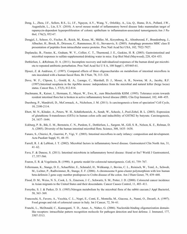
Deng, L., Zhou, J.F., Sellers, R.S., Li, J.F., Nguyen, A.V., Wang, Y., Orlofsky, A., Liu, Q., Hume, D.A., Pollard, J.W., Augenlicht, L., Lin, E.Y. (2010). A novel mouse model of inflammatory bowel disease links mammalian target of rapamycin-dependent hyperproliferation of colonic epithelium to inflammation-associated tumorigenesis.Am J Pa-thol, 176(2), 952-67.
Dengjel, J., Schoor, O., Fischer, R., Reich, M., Kraus, M., Müller, M., Kreymborg, K., Altenberend, F., Brandenburg, J., Albacher, H., Brock, R., Driessen, C., Rammensee, H. G., Stevanovic, S. (2005). Autophagy promotes MHC class II presentation of peptides from intracellular source proteins. Proc Natl Acad Sci USA, 102, 7922-7927.
Deplancke, B., Finster, K., Graham, W. V., Collier, C. T., Thurmond, J. E., Gaskins, H. R. (2003). Gastrointestinal and microbial responses to sulfate-supplemented drinking water in mice. Exp Biol Med (Maywood), 228, 424–433.
Dethlefsen, L. &Relman, D. A. (2011). Incomplete recovery and individualized responses of the human distal gut microbi-ota to repeated antibiotic perturbation. Proc Natl Acad Sci U S A, 108 Suppl 1, 455445-61.
Djouzi, Z. & Andrieux, C. (1997). Compared effects of three oligosaccharides on metabolism of intestinal microflora in rats inoculated with a human faecal flora. Br J Nutr, 78, 313–324.
Dove, W. F., Clipson, L., Gould, K. A., Luongo, C., Marshall, D. J., Moser, A. R., Newton, M. A., Jacoby, R.F. (1997).Intestinal neoplasia in the ApcMin mouse: independence from the microbial and natural killer (beige locus) status. Cancer Res, 1, 57(5), 812-814.
Duchmann, R., Kaiser, I., Hermann, E., Mayet, W., Ewe, K., zum Büschenfelde KHM. (1995). Tolerance exists towards resident intestinal flora but is broken in active inflammatory bowel disease (IBD). Clin Exp Immunol, 102, 448–455.
Duesberg, P., Mandrioli, D., McCormack, A., Nicholson, J. M. (2011). Is carcinogenesis a form of speciation? Cell Cycle, 10, 2100-2114.
Ebert, M. N., Klinder, A., Peters, W. H., Schäferhenrich, A., Sendt, W., Scheele, J., Pool-Zobel, B. L. (2003). Expression of glutathione S-transferases (GSTs) in human colon cells and inducibility of GSTM2 by butyrate. Carcinogenesis, 24, 1637–1644.
Eckburg, P. B., Bik, E. M., Bernstein, C. N., Purdom, E., Dethlefsen, L., Sargent, M., Gill, S. R., Nelson, K. E., Relman, D. A. (2005). Diversity of the human intestinal microbial flora. Science, 308, 1635–1638.
Fanaro, S., Chierici, R., Guerrini, P., Vigi, V. (2003). Intestinal microflora in early infancy: composition and development. Acta Paediatr Suppl, 91, 48–55.
Farrell, R. J. & LaMont, J. T. (2002). Microbial factors in inflammatory bowel disease. Gastroenterol Clin North Am, 31, 41–62.
Fava, F. & Danese, S. (2011). Intestinal microbiota in inflammatory bowel disease: friend or foe? World J Gastroenterol, 17, 557-566.
Fearon, E. R. & Vogelstein, B. (1990). A genetic model for colorectal tumorigenesis. Cell, 61, 759–767.
Fellermann, K., Stange, D. E., Schaeffeler, E., Schmalzl, H., Wehkamp, J., Bevins, C. L., Reinisch, W., Teml, A., Schwab, M., Lichter, P., Radlwimmer, B., Stange, E. F. (2006). A chromosome 8 gene-cluster polymorphism with low human beta-defensin 2 gene copy number predisposes to Crohn disease of the colon. Am J Hum Genet, 79, 439–448.
Flood, D. M., Weiss, N. S., Cook, L. S., Emerson, J. C., Schwartz, S. M., Potter, J. D. (2000). Colorectal cancer incidence in Asian migrants to the United States and their descendants. Cancer Causes Control, 11, 403–411.
Forsythe, S. J. & Parker, D. S. (1985).Nitrogen metabolism by the microbial flora of the rabbit caecum.J Appl Bacteriol, 58, 363–369.
Franceschi, S., Favero, A., Vecchia, C. L., Negri, E., Conti, E., Montella, M., Giacosa, A., Nanni, O., Decarli, A. (1997). Food groups and risk of colorectal cancer in Italy. Int J Cancer, 72, 56–61.
Franchi, L., McDonald, C., Kanneganti, T. D., Amer, A., Núñez, G. (2006). Nucleotide-binding oligomerization domain-like receptors: intracellular pattern recognition molecole for pathogen detection and host defense. J. Immunol, 177, 3507-3513.
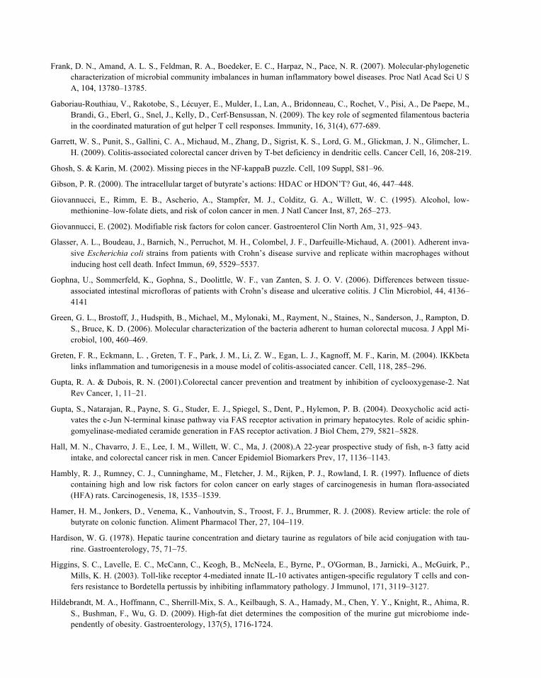
Frank, D. N., Amand, A. L. S., Feldman, R. A., Boedeker, E. C., Harpaz, N., Pace, N. R. (2007). Molecular-phylogenetic characterization of microbial community imbalances in human inflammatory bowel diseases. Proc Natl Acad Sci U S A, 104, 13780–13785.
Gaboriau-Routhiau, V., Rakotobe, S., Lécuyer, E., Mulder, I., Lan, A., Bridonneau, C., Rochet, V., Pisi, A., De Paepe, M., Brandi, G., Eberl, G., Snel, J., Kelly, D., Cerf-Bensussan, N. (2009). The key role of segmented filamentous bacteria in the coordinated maturation of gut helper T cell responses. Immunity, 16, 31(4), 677-689.
Garrett, W. S., Punit, S., Gallini, C. A., Michaud, M., Zhang, D., Sigrist, K. S., Lord, G. M., Glickman, J. N., Glimcher, L. H. (2009). Colitis-associated colorectal cancer driven by T-bet deficiency in dendritic cells. Cancer Cell, 16, 208-219.
Ghosh, S. & Karin, M. (2002). Missing pieces in the NF-kappaB puzzle. Cell, 109 Suppl, S81–96.
Gibson, P. R. (2000). The intracellular target of butyrate’s actions: HDAC or HDON’T? Gut, 46, 447–448.
Giovannucci, E., Rimm, E. B., Ascherio, A., Stampfer, M. J., Colditz, G. A., Willett, W. C. (1995). Alcohol, low-methionine–low-folate diets, and risk of colon cancer in men. J Natl Cancer Inst, 87, 265–273.
Giovannucci, E. (2002). Modifiable risk factors for colon cancer. Gastroenterol Clin North Am, 31, 925–943.
Glasser, A. L., Boudeau, J., Barnich, N., Perruchot, M. H., Colombel, J. F., Darfeuille-Michaud, A. (2001). Adherent inva-sive Escherichia coli strains from patients with Crohn’s disease survive and replicate within macrophages without inducing host cell death. Infect Immun, 69, 5529–5537.
Gophna, U., Sommerfeld, K., Gophna, S., Doolittle, W. F., van Zanten, S. J. O. V. (2006). Differences between tissue-associated intestinal microfloras of patients with Crohn’s disease and ulcerative colitis. J Clin Microbiol, 44, 4136–4141
Green, G. L., Brostoff, J., Hudspith, B., Michael, M., Mylonaki, M., Rayment, N., Staines, N., Sanderson, J., Rampton, D. S., Bruce, K. D. (2006). Molecular characterization of the bacteria adherent to human colorectal mucosa. J Appl Mi-crobiol, 100, 460–469.
Greten, F. R., Eckmann, L. , Greten, T. F., Park, J. M., Li, Z. W., Egan, L. J., Kagnoff, M. F., Karin, M. (2004). IKKbeta links inflammation and tumorigenesis in a mouse model of colitis-associated cancer. Cell, 118, 285–296.
Gupta, R. A. & Dubois, R. N. (2001).Colorectal cancer prevention and treatment by inhibition of cyclooxygenase-2. Nat Rev Cancer, 1, 11–21.
Gupta, S., Natarajan, R., Payne, S. G., Studer, E. J., Spiegel, S., Dent, P., Hylemon, P. B. (2004). Deoxycholic acid acti-vates the c-Jun N-terminal kinase pathway via FAS receptor activation in primary hepatocytes. Role of acidic sphin-gomyelinase-mediated ceramide generation in FAS receptor activation. J Biol Chem, 279, 5821–5828.
Hall, M. N., Chavarro, J. E., Lee, I. M., Willett, W. C., Ma, J. (2008).A 22-year prospective study of fish, n-3 fatty acid intake, and colorectal cancer risk in men. Cancer Epidemiol Biomarkers Prev, 17, 1136–1143.
Hambly, R. J., Rumney, C. J., Cunninghame, M., Fletcher, J. M., Rijken, P. J., Rowland, I. R. (1997). Influence of diets containing high and low risk factors for colon cancer on early stages of carcinogenesis in human flora-associated (HFA) rats. Carcinogenesis, 18, 1535–1539.
Hamer, H. M., Jonkers, D., Venema, K., Vanhoutvin, S., Troost, F. J., Brummer, R. J. (2008). Review article: the role of butyrate on colonic function. Aliment Pharmacol Ther, 27, 104–119.
Hardison, W. G. (1978). Hepatic taurine concentration and dietary taurine as regulators of bile acid conjugation with tau-rine. Gastroenterology, 75, 71–75.
Higgins, S. C., Lavelle, E. C., McCann, C., Keogh, B., McNeela, E., Byrne, P., O'Gorman, B., Jarnicki, A., McGuirk, P., Mills, K. H. (2003). Toll-like receptor 4-mediated innate IL-10 activates antigen-specific regulatory T cells and con-fers resistance to Bordetella pertussis by inhibiting inflammatory pathology. J Immunol, 171, 3119–3127.
Hildebrandt, M. A., Hoffmann, C., Sherrill-Mix, S. A., Keilbaugh, S. A., Hamady, M., Chen, Y. Y., Knight, R., Ahima, R. S., Bushman, F., Wu, G. D. (2009). High-fat diet determines the composition of the murine gut microbiome inde-pendently of obesity. Gastroenterology, 137(5), 1716-1724.
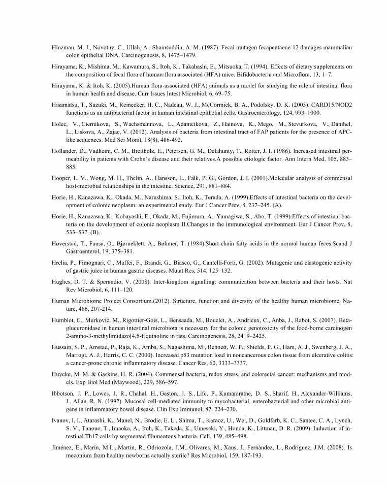
Hinzman, M. J., Novotny, C., Ullah, A., Shamsuddin, A. M. (1987). Fecal mutagen fecapentaene-12 damages mammalian colon epithelial DNA. Carcinogenesis, 8, 1475–1479.
Hirayama, K., Mishima, M., Kawamura, S., Itoh, K., Takahashi, E., Mitsuoka, T. (1994). Effects of dietary supplements on the composition of fecal flora of human-flora associated (HFA) mice. Bifidobacteria and Microflora, 13, 1–7.
Hirayama, K. & Itoh, K. (2005).Human flora-associated (HFA) animals as a model for studying the role of intestinal flora in human health and disease. Curr Issues Intest Microbiol, 6, 69–75.
Hisamatsu, T., Suzuki, M., Reinecker, H. C., Nadeau, W. J., McCormick, B. A., Podolsky, D. K. (2003). CARD15/NOD2 functions as an antibacterial factor in human intestinal epithelial cells. Gastroenterology, 124, 993–1000.
Holec, V., Ciernikova, S., Wachsmannova, L., Adamcikova, Z., Hainova, K., Mego, M., Stevurkova, V., Danihel, L., Liskova, A., Zajac, V. (2012). Analysis of bacteria from intestinal tract of FAP patients for the presence of APC-like sequences. Med Sci Monit, 18(8), 486-492.
Hollander, D., Vadheim, C. M., Brettholz, E., Petersen, G. M., Delahunty, T., Rotter, J. I. (1986). Increased intestinal per-meability in patients with Crohn’s disease and their relatives.A possible etiologic factor. Ann Intern Med, 105, 883–885.
Hooper, L. V., Wong, M. H., Thelin, A., Hansson, L., Falk, P. G., Gordon, J. I. (2001).Molecular analysis of commensal host-microbial relationships in the intestine. Science, 291, 881–884.
Horie, H., Kanazawa, K., Okada, M., Narushima, S., Itoh, K., Terada, A. (1999).Effects of intestinal bacteria on the devel-opment of colonic neoplasm: an experimental study. Eur J Cancer Prev, 8, 237–245. (A).
Horie, H., Kanazawa, K., Kobayashi, E., Okada, M., Fujimura, A., Yamagiwa, S., Abo, T. (1999).Effects of intestinal bac-teria on the development of colonic neoplasm II.Changes in the immunological environment. Eur J Cancer Prev, 8, 533–537. (B).
Høverstad, T., Fausa, O., Bjørneklett, A., Bøhmer, T. (1984).Short-chain fatty acids in the normal human feces.Scand J Gastroenterol, 19, 375–381.
Hrelia, P., Fimognari, C., Maffei, F., Brandi, G., Biasco, G., Cantelli-Forti, G. (2002). Mutagenic and clastogenic activity of gastric juice in human gastric diseases. Mutat Res, 514, 125–132.
Hughes, D. T. & Sperandio, V. (2008). Inter-kingdom signalling: communication between bacteria and their hosts. Nat Rev Microbiol, 6, 111–120.
Human Microbiome Project Consortium.(2012). Structure, function and diversity of the healthy human microbiome. Na-ture, 486, 207-214.
Humblot, C., Murkovic, M., Rigottier-Gois, L., Bensaada, M., Bouclet, A., Andrieux, C., Anba, J., Rabot, S. (2007). Beta-glucuronidase in human intestinal microbiota is necessary for the colonic genotoxicity of the food-borne carcinogen 2-amino-3-methylimidazo[4,5-f]quinoline in rats. Carcinogenesis, 28, 2419–2425.
Hussain, S. P., Amstad, P., Raja, K., Ambs, S., Nagashima, M., Bennett, W. P., Shields, P. G., Ham, A. J., Swenberg, J. A., Marrogi, A. J., Harris, C. C. (2000). Increased p53 mutation load in noncancerous colon tissue from ulcerative colitis: a cancer-prone chronic inflammatory disease. Cancer Res, 60, 3333–3337.
Huycke, M. M. & Gaskins, H. R. (2004). Commensal bacteria, redox stress, and colorectal cancer: mechanisms and mod-els. Exp Biol Med (Maywood), 229, 586–597.
Ibbotson, J. P., Lowes, J. R., Chahal, H., Gaston, J. S., Life, P., Kumararatne, D. S., Sharif, H., Alexander-Williams, J., Allan, R. N. (1992). Mucosal cell-mediated immunity to mycobacterial, enterobacterial and other microbial anti-gens in inflammatory bowel disease. Clin Exp Immunol, 87. 224–230.
Ivanov, I. I., Atarashi, K., Manel, N., Brodie, E. L., Shima, T., Karaoz, U., Wei, D., Goldfarb, K. C., Santee, C. A., Lynch, S. V., Tanoue, T., Imaoka, A., Itoh, K., Takeda, K., Umesaki, Y., Honda, K., Littman, D. R. (2009). Induction of in-testinal Th17 cells by segmented filamentous bacteria. Cell, 139, 485–498.
Jiménez, E., Marín, M.L., Martín, R., Odriozola, J.M., Olivares, M., Xaus, J., Fernández, L., Rodríguez, J.M. (2008). Is meconium from healthy newborns actually sterile? Res Microbiol, 159, 187-193.
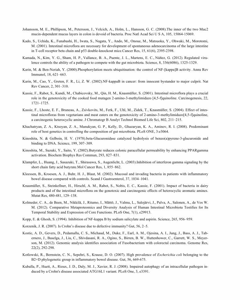
Johansson, M. E., Phillipson, M., Petersson, J., Velcich, A., Holm, L., Hansson, G. C. (2008).The inner of the two Muc2 mucin-dependent mucus layers in colon is devoid of bacteria. Proc Natl Acad Sci U S A, 105, 15064-15069.
Kado, S., Uchida, K., Funabashi, H., Iwata, S., Nagata, Y., Ando, M., Onoue, M., Matsuoka, Y., Ohwaki, M., Morotomi, M. (2001). Intestinal microflora are necessary for development of spontaneous adenocarcinoma of the large intestine in T-cell receptor beta chain and p53 double-knockout mice.Cancer Res, 15, 61(6), 2395-2398.
Kamada, N., Kim, Y. G., Sham, H. P., Vallance, B. A., Puente, J. L., Martens, E. C., Núñez, G. (2012). Regulated viru-lence controls the ability of a pathogen to compete with the gut microbiota. Science, 8, 336(6086), 1325-1329.
Karin, M. & Ben-Neriah, Y. (2000).Phosphorylation meets ubiquitination: the control of NF-[kappa]B activity. Annu Rev Immunol, 18, 621–663.
Karin, M., Cao, Y., Greten, F. R., Li, Z. W. (2002).NF-kappaB in cancer: from innocent bystander to major culprit. Nat Rev Cancer, 2, 301–310.
Kassie, F., Rabot, S., Kundi, M., Chabicovsky, M., Qin, H. M., Knasmüller, S. (2001). Intestinal microflora plays a crucial role in the genotoxicity of the cooked food mutagen 2-amino-3-methylimidazo [4,5-f]quinoline. Carcinogenesis, 22, 1721–1725.
Kassie, F., Lhoste, E. F., Bruneau, A., Zsivkovits, M., Ferk, F., Uhl, M., Zidek, T., Knasmüller, S. (2004). Effect of intes-tinal microfloras from vegetarians and meat eaters on the genotoxicity of 2-amino-3-methylimidazo[4,5-f]quinoline, a carcinogenic heterocyclic amine. J Chromatogr B Analyt Technol Biomed Life Sci, 802, 211–215.
Khachatryan, Z. A., Ktsoyan, Z. A., Manukyan, G. P., Kelly, D., Ghazaryan, K. A., Aminov, R. I. (2008). Predominant role of host genetics in controlling the composition of gut microbiota. PLoS ONE, 3:e3064.
Kinoshita, N. & Gelboin, H. V. (1978).beta-Glucuronidase catalyzed hydrolysis of benzo(a)pyrene-3-glucuronide and binding to DNA. Science, 199, 307–309.
Kinoshita, M., Suzuki, Y., Saito, Y. (2002).Butyrate reduces colonic paracellular permeability by enhancing PPARgamma activation. Biochem Biophys Res Commun, 293, 827–831.
Klampfer, L., Huang, J., Sasazuki, T., Shirasawa, S., Augenlicht, L. (2003).Inhibition of interferon gamma signaling by the short chain fatty acid butyrate.Mol Cancer Res, 1, 855–862.
Kleessen, B., Kroesen, A. J., Buhr, H. J., Blaut, M. (2002). Mucosal and invading bacteria in patients with inflammatory bowel disease compared with controls. Scand J Gastroenterol, 37, 1034–1041.
Knasmüller, S., Steinkellner, H., Hirschl, A. M., Rabot, S., Nobis, E. C., Kassie, F. (2001). Impact of bacteria in dairy products and of the intestinal microflora on the genotoxic and carcinogenic effects of heterocyclic aromatic amines. Mutat Res, 480-481, 129–138.
Kolmeder, C. A., de Been, M., Nikkilä, J., Ritamo, I., Mättö, J., Valmu, L., Salojärvi, J., Palva, A., Salonen, A., de Vos W. M. (2012). Comparative Metaproteomics and Diversity Analysis of Human Intestinal Microbiota Testifies for Its Temporal Stability and Expression of Core Functions. PLoS One, 7(1), e29913.
Kopp, E. & Ghosh, S. (1994). Inhibition of NF-kappa B by sodium salicylate and aspirin. Science, 265, 956–959.
Korzenik, J. R. (2007). Is Crohn’s disease due to defective immunity? Gut, 56, 2–5.
Kostic, A. D., Gevers, D., Pedamallu, C. S., Michaud, M., Duke, F., Earl, A. M., Ojesina, A. I., Jung, J., Bass, A. J., Tab-ernero, J., Baselga, J., Liu, C., Shivdasani, R. A., Ogino, S., Birren, B. W., Huttenhower, C., Garrett, W. S., Meyer-son, M. (2012). Genomic analysis identifies association of Fusobacterium with colorectal carcinoma. Genome Res, 22(2), 292-298.
Kotlowski, R., Bernstein, C. N., Sepehri, S., Krause, D. O. (2007). High prevalence of Escherichia coli belonging to the B2+D phylogenetic group in inflammatory bowel disease. Gut, 56, 669-675.
Kuballa, P., Huett, A., Rioux, J. D., Daly, M. J., Xavier, R. J. (2008). Impaired autophagy of an intracellular pathogen in-duced by a Crohn's disease associated ATG16L1 variant. PLoS One, 3, e3391.
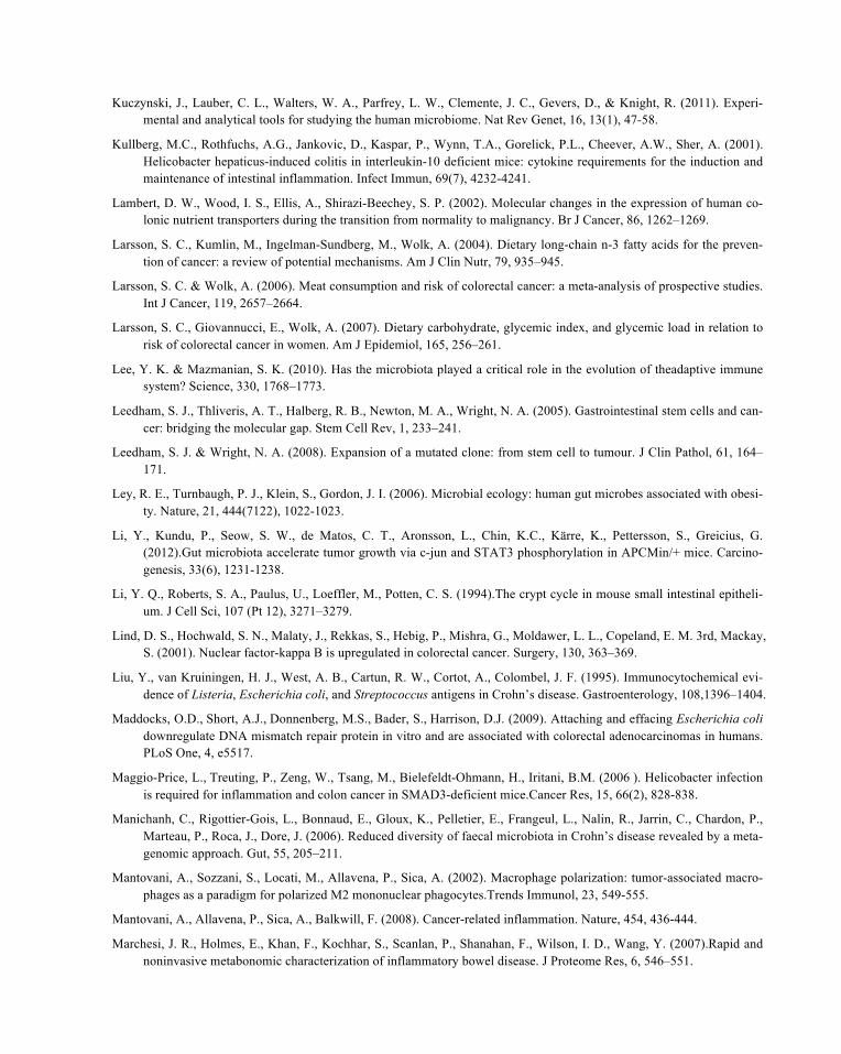
Kuczynski, J., Lauber, C. L., Walters, W. A., Parfrey, L. W., Clemente, J. C., Gevers, D., & Knight, R. (2011). Experi-mental and analytical tools for studying the human microbiome. Nat Rev Genet, 16, 13(1), 47-58.
Kullberg, M.C., Rothfuchs, A.G., Jankovic, D., Kaspar, P., Wynn, T.A., Gorelick, P.L., Cheever, A.W., Sher, A. (2001). Helicobacter hepaticus-induced colitis in interleukin-10 deficient mice: cytokine requirements for the induction and maintenance of intestinal inflammation. Infect Immun, 69(7), 4232-4241.
Lambert, D. W., Wood, I. S., Ellis, A., Shirazi-Beechey, S. P. (2002). Molecular changes in the expression of human co-lonic nutrient transporters during the transition from normality to malignancy. Br J Cancer, 86, 1262–1269.
Larsson, S. C., Kumlin, M., Ingelman-Sundberg, M., Wolk, A. (2004). Dietary long-chain n-3 fatty acids for the preven-tion of cancer: a review of potential mechanisms. Am J Clin Nutr, 79, 935–945.
Larsson, S. C. & Wolk, A. (2006). Meat consumption and risk of colorectal cancer: a meta-analysis of prospective studies. Int J Cancer, 119, 2657–2664.
Larsson, S. C., Giovannucci, E., Wolk, A. (2007). Dietary carbohydrate, glycemic index, and glycemic load in relation to risk of colorectal cancer in women. Am J Epidemiol, 165, 256–261.
Lee, Y. K. & Mazmanian, S. K. (2010). Has the microbiota played a critical role in the evolution of theadaptive immune system? Science, 330, 1768–1773.
Leedham, S. J., Thliveris, A. T., Halberg, R. B., Newton, M. A., Wright, N. A. (2005). Gastrointestinal stem cells and can-cer: bridging the molecular gap. Stem Cell Rev, 1, 233–241.
Leedham, S. J. & Wright, N. A. (2008). Expansion of a mutated clone: from stem cell to tumour. J Clin Pathol, 61, 164–171.
Ley, R. E., Turnbaugh, P. J., Klein, S., Gordon, J. I. (2006). Microbial ecology: human gut microbes associated with obesi-ty. Nature, 21, 444(7122), 1022-1023.
Li, Y., Kundu, P., Seow, S. W., de Matos, C. T., Aronsson, L., Chin, K.C., Kärre, K., Pettersson, S., Greicius, G. (2012).Gut microbiota accelerate tumor growth via c-jun and STAT3 phosphorylation in APCMin/+ mice. Carcino-genesis, 33(6), 1231-1238.
Li, Y. Q., Roberts, S. A., Paulus, U., Loeffler, M., Potten, C. S. (1994).The crypt cycle in mouse small intestinal epitheli-um. J Cell Sci, 107 (Pt 12), 3271–3279.
Lind, D. S., Hochwald, S. N., Malaty, J., Rekkas, S., Hebig, P., Mishra, G., Moldawer, L. L., Copeland, E. M. 3rd, Mackay, S. (2001). Nuclear factor-kappa B is upregulated in colorectal cancer. Surgery, 130, 363–369.
Liu, Y., van Kruiningen, H. J., West, A. B., Cartun, R. W., Cortot, A., Colombel, J. F. (1995). Immunocytochemical evi-dence of Listeria, Escherichia coli, and Streptococcus antigens in Crohn’s disease. Gastroenterology, 108,1396–1404.
Maddocks, O.D., Short, A.J., Donnenberg, M.S., Bader, S., Harrison, D.J. (2009). Attaching and effacing Escherichia coli downregulate DNA mismatch repair protein in vitro and are associated with colorectal adenocarcinomas in humans. PLoS One, 4, e5517.
Maggio-Price, L., Treuting, P., Zeng, W., Tsang, M., Bielefeldt-Ohmann, H., Iritani, B.M. (2006 ). Helicobacter infection is required for inflammation and colon cancer in SMAD3-deficient mice.Cancer Res, 15, 66(2), 828-838.
Manichanh, C., Rigottier-Gois, L., Bonnaud, E., Gloux, K., Pelletier, E., Frangeul, L., Nalin, R., Jarrin, C., Chardon, P., Marteau, P., Roca, J., Dore, J. (2006). Reduced diversity of faecal microbiota in Crohn’s disease revealed by a meta-genomic approach. Gut, 55, 205–211.
Mantovani, A., Sozzani, S., Locati, M., Allavena, P., Sica, A. (2002). Macrophage polarization: tumor-associated macro-phages as a paradigm for polarized M2 mononuclear phagocytes.Trends Immunol, 23, 549-555.
Mantovani, A., Allavena, P., Sica, A., Balkwill, F. (2008). Cancer-related inflammation. Nature, 454, 436-444.
Marchesi, J. R., Holmes, E., Khan, F., Kochhar, S., Scanlan, P., Shanahan, F., Wilson, I. D., Wang, Y. (2007).Rapid and noninvasive metabonomic characterization of inflammatory bowel disease. J Proteome Res, 6, 546–551.
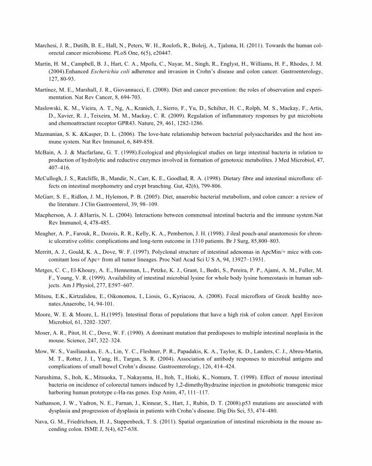
Marchesi, J. R., Dutilh, B. E., Hall, N., Peters, W. H., Roelofs, R., Boleij, A., Tjalsma, H. (2011). Towards the human col-orectal cancer microbiome. PLoS One, 6(5), e20447.
Martin, H. M., Campbell, B. J., Hart, C. A., Mpofu, C., Nayar, M., Singh, R., Englyst, H., Williams, H. F., Rhodes, J. M. (2004).Enhanced Escherichia coli adherence and invasion in Crohn’s disease and colon cancer. Gastroenterology, 127, 80-93.
Martínez, M. E., Marshall, J. R., Giovannucci, E. (2008). Diet and cancer prevention: the roles of observation and experi-mentation. Nat Rev Cancer, 8, 694-703.
Maslowski, K. M., Vieira, A. T., Ng, A., Kranich, J., Sierro, F., Yu, D., Schilter, H. C., Rolph, M. S., Mackay, F., Artis, D., Xavier, R. J., Teixeira, M. M., Mackay, C. R. (2009). Regulation of inflammatory responses by gut microbiota and chemoattractant receptor GPR43. Nature, 29, 461, 1282-1286.
Mazmanian, S. K. &Kasper, D. L. (2006). The love-hate relationship between bacterial polysaccharides and the host im-mune system. Nat Rev Immunol, 6, 849-858.
McBain, A. J. & Macfarlane, G. T. (1998).Ecological and physiological studies on large intestinal bacteria in relation to production of hydrolytic and reductive enzymes involved in formation of genotoxic metabolites. J Med Microbiol, 47, 407–416.
McCullogh, J. S., Ratcliffe, B., Mandir, N., Carr, K. E., Goodlad, R. A. (1998). Dietary fibre and intestinal microflora: ef-fects on intestinal morphometry and crypt branching. Gut, 42(6), 799-806.
McGarr, S. E., Ridlon, J. M., Hylemon, P. B. (2005). Diet, anaerobic bacterial metabolism, and colon cancer: a review of the literature. J Clin Gastroenterol, 39, 98–109.
Macpherson, A. J. &Harris, N. L. (2004). Interactions between commensal intestinal bacteria and the immune system.Nat Rev Immunol, 4, 478-485.
Meagher, A. P., Farouk, R., Dozois, R. R., Kelly, K. A., Pemberton, J. H. (1998). J ileal pouch-anal anastomosis for chron-ic ulcerative colitis: complications and long-term outcome in 1310 patients. Br J Surg, 85,800–803.
Merritt, A. J., Gould, K. A., Dove, W. F. (1997). Polyclonal structure of intestinal adenomas in ApcMin/+ mice with con-comitant loss of Apc+ from all tumor lineages. Proc Natl Acad Sci U S A, 94, 13927–13931.
Metges, C. C., El-Khoury, A. E., Henneman, L., Petzke, K. J., Grant, I., Bedri, S., Pereira, P. P., Ajami, A. M., Fuller, M. F., Young, V. R. (1999). Availability of intestinal microbial lysine for whole body lysine homeostasis in human sub-jects. Am J Physiol, 277, E597–607.
Mitsou, E.K., Kirtzalidou, E., Oikonomou, I., Liosis, G., Kyriacou, A. (2008). Fecal microflora of Greek healthy neo-nates.Anaerobe, 14, 94-101.
Moore, W. E. & Moore, L. H.(1995). Intestinal floras of populations that have a high risk of colon cancer. Appl Environ Microbiol, 61, 3202–3207.
Moser, A. R., Pitot, H. C., Dove, W. F. (1990). A dominant mutation that predisposes to multiple intestinal neoplasia in the mouse. Science, 247, 322–324.
Mow, W. S., Vasiliauskas, E. A., Lin, Y. C., Fleshner, P. R., Papadakis, K. A., Taylor, K. D., Landers, C. J., Abreu-Martin, M. T., Rotter, J. I., Yang, H., Targan, S. R. (2004). Association of antibody responses to microbial antigens and complications of small bowel Crohn’s disease. Gastroenterology, 126, 414–424.
Narushima, S., Itoh, K., Mitsuoka, T., Nakayama, H., Itoh, T., Hioki, K., Nomura, T. (1998). Effect of mouse intestinal bacteria on incidence of colorectal tumors induced by 1,2-dimethylhydrazine injection in gnotobiotic transgenic mice harboring human prototype c-Ha-ras genes. Exp Anim, 47, 111–117.
Nathanson, J. W., Yadron, N. E., Farnan, J., Kinnear, S., Hart, J., Rubin, D. T. (2008).p53 mutations are associated with dysplasia and progression of dysplasia in patients with Crohn’s disease. Dig Dis Sci, 53, 474–480.
Nava, G. M., Friedrichsen, H. J., Stappenbeck, T. S. (2011). Spatial organization of intestinal microbiota in the mouse as-cending colon. ISME J, 5(4), 627-638.
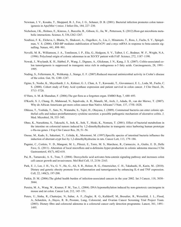
Newman, J. V., Kosaka, T., Sheppard, B. J., Fox, J. G., Schauer, D. B. (2001). Bacterial infection promotes colon tumor-igenesis in Apc(Min/+) mice. J Infect Dis, 184, 227–230.
Nicholson, J.K., Holmes, E., Kinross, J., Burcelin, R., Gibson, G., Jia, W., Pettersson, S. (2012).Host-gut microbiota meta-bolic interactions. Science, 8, 336,1262-1267.
Noubissi, F. K., Elcheva, I., Bhatia, N., Shakoori, A., Ougolkov, A., Liu, J., Minamoto, T., Ross, J., Fuchs, S. Y., Spiegel-man, V. S. (2006). CRD-BP mediates stabilization of betaTrCP1 and c-myc mRNA in response to beta-catenin sig-nalling. Nature, 441, 898–901.
Novelli, M. R., Williamson, J. A., Tomlinson, I. P., Elia, G., Hodgson, S. V., Talbot, I. C., Bodmer, W. F., Wright, N.A. (1996). Polyclonal origin of colonic adenomas in an XO/XY patient with FAP. Science, 272, 1187–1190.
Nowak, J., Weylandt, K. H., Habbel, P., Wang, J., Dignass, A., Glickman, J. N., Kang, J. X. (2007). Colitis-associated co-lon tumorigenesis is suppressed in transgenic mice rich in endogenous n-3 fatty acids. Carcinogenesis, 28, 1991–1995.
Nuding, S., Fellermann, K., Wehkamp, J., Stange, E. F. (2007).Reduced mucosal antimicrobial activity in Crohn’s disease of the colon. Gut, 56, 1240–1247.
Ogino, S., Nosho, K., Meyerhardt, J. A., Kirkner, G. J., Chan, A. T., Kawasaki, T., Giovannucci, E. L., Loda, M., Fuchs, C. S. (2008). Cohort study of Fatty Acid synthase expression and patient survival in colon cancer. J Clin Oncol, 26, 5713–5720.
O’Hara, A. M. & Shanahan, F. (2006).The gut flora as a forgotten organ. EMBO Rep, 7, 688–693.
O'Keefe, S. J., Chung, D., Mahmoud, N., Sepulveda, A. R., Manafe, M., Arch, J., Adada, H., van der Merwe, T. (2007). Why do African Americans get more colon cancer than Native Africans? J Nutr, 137, 175S–182S.
Ohkusa, T., Yoshida, T., Sato, N., Watanabe, S., Tajiri, H., Okayasu, I. (2009). Commensal bacteria can enter colonic epi-thelial cells and induce proinflammatory cytokine secretion: a possible pathogenic mechanism of ulcerative colitis. J. Med. Microbiol, 58, 535–545.
Ohno, K., Narushima, S., Takeuchi, S., Itoh, K., Itoh, T., Hioki, K., Nomura, T. (2001). Effect of bacterial metabolism in the intestine on colorectal tumors induced by 1,2-dimethylhydrazine in transgenic mice harboring human prototype c-Ha-ras genes. J Exp Clin Cancer Res, 20, 51–56.
Onoue, M., Kado, S., Sakaitani, Y., Uchida, K., Morotomi, M. (1997).Specific species of intestinal bacteria influence the induction of aberrant crypt foci by 1,2-dimethylhydrazine in rats. Cancer Lett, 113, 179–186.
Pagnini, C., Corleto, V. D., Mangoni, M. L., Pilozzi, E., Torre, M. S., Marchese, R., Carnuccio, A., Giulio, E. D., Delle Fave, G. (2011). Alteration of local microflora and α-defensins hyper-production in colonic adenoma mucosa.J Clin Gastroenterol, 45(7), 602-610.
Pai, R., Tarnawski, A. S., Tran, T. (2004). Deoxycholic acid activates beta-catenin signaling pathway and increases colon cell cancer growth and invasiveness. Mol Biol Cell, 15, 2156–2163.
Park, E. J., Lee, J. H., Yu, G. Y., He, G., Ali, S. R., Holzer, R. G., Osterreicher, C. H., Takahashi, H., Karin, M.. (2010). Dietary and genetic obesity promote liver inflammation and tumorigenesis by enhancing IL-6 and TNF expression. Cell, 22, 140(2), 197-208.
Parkin, D. M. (2006).The global health burden of infection-associated cancers in the year 2002. Int J Cancer, 118, 3030–3044.
Pereira, M. A., Wang, W., Kramer, P. M., Tao, L. (2004). DNA hypomethylation induced by non-genotoxic carcinogens in mouse and rat colon. Cancer Lett, 212, 145–151.
Peters, U., Sinha, R., Chatterjee, N., Subar, A. F., Ziegler, R. G., Kulldorff, M., Bresalier, R., Weissfeld, J. L., Flood, A., Schatzkin, A., Hayes, R. B., Prostate, Lung, Colorectal, and Ovarian Cancer Screening Trial Project Team. (2003). Dietary fibre and colorectal adenoma in a colorectal cancer early detection programme. Lancet, 361, 1491–1495.
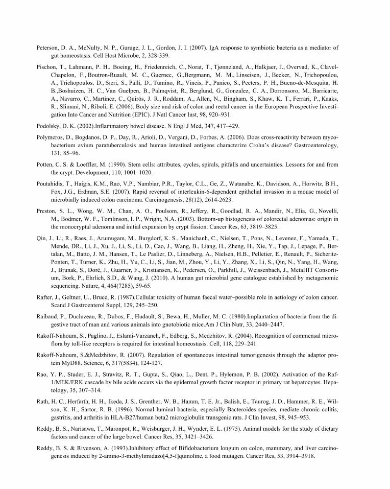
Peterson, D. A., McNulty, N. P., Guruge, J. L., Gordon, J. I. (2007). IgA response to symbiotic bacteria as a mediator of gut homeostasis. Cell Host Microbe, 2, 328-339.
Pischon, T., Lahmann, P. H., Boeing, H., Friedenreich, C., Norat, T., Tjønneland, A., Halkjaer, J., Overvad, K., Clavel-Chapelon, F., Boutron-Ruault, M. C., Guernec, G.,Bergmann, M. M., Linseisen, J., Becker, N., Trichopoulou, A., Trichopoulos, D., Sieri, S., Palli, D., Tumino, R., Vineis, P., Panico, S., Peeters, P. H., Bueno-de-Mesquita, H. B.,Boshuizen, H. C., Van Guelpen, B., Palmqvist, R., Berglund, G., Gonzalez, C. A., Dorronsoro, M., Barricarte, A., Navarro, C., Martinez, C., Quirós, J. R., Roddam, A., Allen, N., Bingham, S., Khaw, K. T., Ferrari, P., Kaaks, R., Slimani, N., Riboli, E. (2006). Body size and risk of colon and rectal cancer in the European Prospective Investi-gation Into Cancer and Nutrition (EPIC). J Natl Cancer Inst, 98, 920–931.
Podolsky, D. K. (2002).Inflammatory bowel disease. N Engl J Med, 347, 417–429.
Polymeros, D., Bogdanos, D. P., Day, R., Arioli, D., Vergani, D., Forbes, A. (2006). Does cross-reactivity between myco-bacterium avium paratuberculosis and human intestinal antigens characterize Crohn’s disease? Gastroenterology, 131, 85–96.
Potten, C. S. & Loeffler, M. (1990). Stem cells: attributes, cycles, spirals, pitfalls and uncertainties. Lessons for and from the crypt. Development, 110, 1001–1020.
Poutahidis, T., Haigis, K.M., Rao, V.P., Nambiar, P.R., Taylor, C.L., Ge, Z., Watanabe, K., Davidson, A., Horwitz, B.H., Fox, J.G., Erdman, S.E. (2007). Rapid reversal of interleukin-6-dependent epithelial invasion in a mouse model of microbially induced colon carcinoma. Carcinogenesis, 28(12), 2614-2623.
Preston, S. L., Wong, W. M., Chan, A. O., Poulsom, R., Jeffery, R., Goodlad, R. A., Mandir, N., Elia, G., Novelli, M., Bodmer, W. F., Tomlinson, I. P., Wright, N.A. (2003). Bottom-up histogenesis of colorectal adenomas: origin in the monocryptal adenoma and initial expansion by crypt fission. Cancer Res, 63, 3819–3825.
Qin, J., Li, R., Raes, J., Arumugam, M., Burgdorf, K. S., Manichanh, C., Nielsen, T., Pons, N., Levenez, F., Yamada, T., Mende, DR., Li, J., Xu, J., Li, S., Li, D., Cao, J., Wang, B., Liang, H., Zheng, H., Xie, Y., Tap, J., Lepage, P., Ber-talan, M., Batto, J. M., Hansen, T., Le Paslier, D., Linneberg, A., Nielsen, H.B., Pelletier, E., Renault, P., Sicheritz-Ponten, T., Turner, K., Zhu, H., Yu, C., Li, S., Jian, M., Zhou, Y., Li, Y., Zhang, X., Li, S., Qin, N., Yang, H., Wang, J., Brunak, S., Doré, J., Guarner, F., Kristiansen, K., Pedersen, O., Parkhill, J., Weissenbach, J., MetaHIT Consorti-um, Bork, P., Ehrlich, S.D., & Wang, J. (2010). A human gut microbial gene catalogue established by metagenomic sequencing. Nature, 4, 464(7285), 59-65.
Rafter, J., Geltner, U., Bruce, R. (1987).Cellular toxicity of human faecal water–possible role in aetiology of colon cancer. Scand J Gastroenterol Suppl, 129, 245–250.
Raibaud, P., Ducluzeau, R., Dubos, F., Hudault, S., Bewa, H., Muller, M. C. (1980).Implantation of bacteria from the di-gestive tract of man and various animals into gnotobiotic mice.Am J Clin Nutr, 33, 2440–2447.
Rakoff-Nahoum, S., Paglino, J., Eslami-Varzaneh, F., Edberg, S., Medzhitov, R. (2004). Recognition of commensal micro-flora by toll-like receptors is required for intestinal homeostasis. Cell, 118, 229–241.
Rakoff-Nahoum, S.&Medzhitov, R. (2007). Regulation of spontaneous intestinal tumorigenesis through the adaptor pro-tein MyD88. Science, 6, 317(5834), 124-127.
Rao, Y. P., Studer, E. J., Stravitz, R. T., Gupta, S., Qiao, L., Dent, P., Hylemon, P. B. (2002). Activation of the Raf-1/MEK/ERK cascade by bile acids occurs via the epidermal growth factor receptor in primary rat hepatocytes. Hepa-tology, 35, 307–314.
Rath, H. C., Herfarth, H. H., Ikeda, J. S., Grenther, W. B., Hamm, T. E. Jr., Balish, E., Taurog, J. D., Hammer, R. E., Wil-son, K. H., Sartor, R. B. (1996). Normal luminal bacteria, especially Bacteroides species, mediate chronic colitis, gastritis, and arthritis in HLA-B27/human beta2 microglobulin transgenic rats. J Clin Invest, 98, 945–953.
Reddy, B. S., Narisawa, T., Maronpot, R., Weisburger, J. H., Wynder, E. L. (1975). Animal models for the study of dietary factors and cancer of the large bowel. Cancer Res, 35, 3421–3426.
Reddy, B. S. & Rivenson, A. (1993).Inhibitory effect of Bifidobacterium longum on colon, mammary, and liver carcino-genesis induced by 2-amino-3-methylimidazo[4,5-f]quinoline, a food mutagen. Cancer Res, 53, 3914–3918.
Renwick, A. G. & Drasar, B. S. (1976).Environmental carcinogens and large bowel cancer.Nature, 263, 234–235.
Ricci-Vitiani, L., Lombardi, D. G., Pilozzi, E., Biffoni, M., Todaro, M., Peschle, C., De Maria, R. (2007). Identification and expansion of human colon-cancer-initiating cells. Nature, 445, 111–115.
Roediger, W. E. (1980). Role of anaerobic bacteria in the metabolic welfare of the colonic mucosa in man.Gut, 21, 793–798.
Roediger, W. E., Duncan, A., Kapaniris, O., Millard, S. (1993). Reducing sulfur compounds of the colon impair colono-cyte nutrition: implications for ulcerative colitis. Gastroenterology, 104, 802–809.
Rogler, G., Brand, K., Vogl, D., Page, S., Hofmeister, R., Andus, T., Knuechel, R., Baeuerle, P. A., Schölmerich, J., Gross, V. (1998). Nuclear factor kappaB is activated in macrophages and epithelial cells of inflamed intestinal mucosa. Ga-stroenterology, 115, 357–369.
Rolhion, N. & Darfeuille-Michaud, A. (2007). Adherent-invasive Escherichia coli in inflammatory bowel disease. In-flamm Bowel Dis, 13, 1277-1283.
Roncucci, L., Modica, S., Pedroni, M., Tamassia, M. G., Ghidoni, M., Losi, L., Fante, R., Di Gregorio, C., Manenti, A., Gafa, L., Ponz de Leon, M,.L. (1998). Aberrant crypt foci in patients with colorectal cancer. Br J Cancer, 77, 2343–2348.
Round, J. L. & Mazmanian, S. K. (2009). The gut microbiota shapes intestinal immune responses during health and dis-ease. Nat Rev Immunol, 9, 313–323.
Round, J. L. & Mazmanian, S. K. (2010).Inducible Foxp3+ regulatory T-cell development by a commensal bacterium of the intestinal microbiota. Proc. Natl Acad. Sci. USA, 107, 12204–12209.
Rutgeerts, P., Goboes, K., Peeters, M., Hiele, M., Penninckx, F., Aerts, R., Kerremans, R., Vantrappen, G. (1991).Effect of faecal stream diversion on recurrence of Crohn’s disease in the neoterminal ileum. Lancet, 338, 771–774.
Ryan, P., Kelly, R. G., Lee, G., Collins, J. K., O'Sullivan, G. C., O'Connell, J., Shanahan, F. (2004). Bacterial DNA within granulomas of patients with Crohn’s disease–detection by laser capture microdissection and PCR. Am J Gastroenter-ol, 99, 1539–1543.
Salyers, A. A., Vercellotti, J. R., West, S. E., Wilkins, T. D. (1977). Fermentation of mucin and plant polysaccharides by strains of Bacteroides from the human colon. Appl Environ Microbiol, 33, 319–322.
Salyers, A. A., Gherardini, F., O’Brien, M. (1981).Utilization of xylan by two species of human colonic Bacteroides. Appl Environ Microbiol, 41, 1065–1068.
Sanapareddy, N., Legge, R. M., Jovov, B., McCoy, A., Burcal, L., Araujo-Perez, F., Randall, T. A., Galanko, J., Benson, A., Sandler, R. S., Rawls, J. F., Abdo, Z., Fodor, A. A., Keku, T. O. (2012). Increased rectal microbial richness is as-sociated with the presence of colorectal adenomas in humans. ISME J, 6(10), 1858-1868.
Sartor, R. B. (2008). Microbial influences in inflammatory bowel dis- eases. Gastroenterology, 134, 577-594.
Sasaki, M., Sitaraman, S. V., Babbin, B. A., Gerner-Smidt, P., Ribot, E. M., Garrett, N., Alpern, J. A., Akyildiz, A., Theiss, A. L., Nusrat, A., Klapproth, J. M. (2007). Invasive Escherichia coli are a feature of Crohn's disease. Lab Invest, 87, 1042-1054.
Savage, D. C. (1977).Microbial ecology of the gastrointestinal tract. Annu Rev Microbiol, 31, 107–133.
Scanlan, P. D., Shanahan, F., Clune, Y., Collins, J. K., O'Sullivan, G. C., O'Riordan, M., Holmes, E., Wang, Y., Marchesi, J. R..(2008). Culture-independent analysis of the gut microbiota in colorectal cancer and polyposis.Environ Micro-biol, 10, 789–798.
Scanlan, P. D., Shanahan, F., Marchesi, J. R. (2009). Culture-independent analysis of desulfovibrios in the human distal colon of healthy, colorectal cancer and polypectomized individuals. FEMS Microbiol Ecol, 69(2), 213-221.
Schatzkin, A., Lanza, E., Corle, D., Lance, P., Iber, F., Caan, B., Shike, M., Weissfeld, J., Burt, R., Cooper, M. R., Kikendall, J. W., Cahill, J. (2000). Lack of effect of a low-fat, high-fiber diet on the recurrence of colorectal ade-nomas. Polyp Prevention Trial Study Group. N Engl J Med, 342, 1149–1155.
Schoeffner, D. J. & Thorgeirsson, U. P. (2000). Susceptibility of nonhuman primates to carcinogens of human relevance.In Vivo, 14, 149–156.
Schoppmann, S. F., Birner, P., Stöckl, J., Kalt, R., Ullrich, R., Caucig, C., Kriehuber, E., Nagy, K., Alitalo, K., Kerjaschki, D. Tumor-associated macrophages express lymphatic endothelial growth factors and are related to peritumoral lym-phangiogenesis. Am J Pathol, 161, 947–956.
Schreiter, K., Hausmann, M., Spoettl, T., Strauch, U. G., Bataille, F., Schoelmerich, J., Herfarth, H., Falk, W., Rogler, G. (2005). Glycoprotein (gp) 96 expression: induced during differentiation of intestinal macrophages but impaired in Crohn’s disease. Gut, 54, 935–943.
Schultsz, C., Berg, F. M. V. D., Kate, F. W. T., Tytgat, G. N., Dankert, J. (1999). The intestinal mucus layer from patients with inflammatory bowel disease harbors high numbers of bacteria compared with controls. Gastroenterology, 117, 1089–1097.
Schwiertz, A., Taras, D., Schäfer, K., Beijer, S., Bos, N. A., Donus, C., Hardt, P. D. (2010). Microbiota and SCFA in lean and overweight healthy subjects. Obesity (Silver Spring), 18(1), 190-195.
Sears, C. L. (2009). Enterotoxigenic Bacteroides fragilis: a rogue among symbiotes.Clin microbiol Rev, 22: 349-369.
Seibold, F., Brandwein, S., Simpson, S., Terhorst, C., Elson, C. O. (1998). pANCA represents a cross-reactivity to enteric bacterial antigens. J Clin Immunol, 18, 153–160.
Selgrad, M., Malfertheiner, P., Fini, L., Goel, A., Boland, C. R., Ricciardiello, L. (2008). The role of viral and bacterial pathogens in gastrointestinal cancer.J Cell Physiol, 216, 378–388.
Shen, X. J., Rawls, J. F., Randall, T., Burcal, L., Mpande, C. N., Jenkins, N., Jovov, B., Abdo, Z., Sandler, R. S., Keku, T. O. (2010). Molecular characterization of mucosal adherent bacteria and associations with colorectal adenomas.Gut Microbes, 1(3), 138-147.
Shih, I. M., Wang, T. L., Traverso, G., Romans, K., Hamilton, S. R., Ben-Sasson, S., Kinzler, K. W., Vogelstein, B. (2001). Top-down morphogenesis of colorectal tumors. Proc Natl Acad Sci U S A, 98, 2640–2645.
Silvi, S., Rumney, C. J., Cresci, A., Rowland, I. R. (1999). Resistant starch modifies gut microflora and microbial metabo-lism in human flora-associated rats inoculated with faeces from Italian and UK donors. J Appl Microbiol, 86, 521–530.
Sobhani, I., Tap, J., Roudot-Thoraval, F., Roperch, J. P., Letulle, S., Langella, P., Corthier, G., Tran Van Nhieu, J., Furet, J. P. (2011). Microbial dysbiosis in colorectal cancer (CRC) patients. PLoS One, 27, 6(1), e16393.
Sokol, H., Pigneur, B., Watterlot, L., Lakhdari, O., Bermúdez-Humarán, L. G., Gratadoux, J. J., Blugeon, S., Bridonneau, C., Furet, J. P., Corthier, G., Grangette, C., Vasquez, N., Pochart, P., Trugnan, G., Thomas, G., Blottière, H. M., Do-ré, J., Marteau, P., Seksik, P., Langella, P. (2008). Faecalibacterium prausnitzii is an anti-inflammatory commensal bacterium identified by gut microbiota analysis of Crohn disease patients. Proc Natl Acad Sci U S A, 105, 16731–16736.
Sonnenburg, J. L., Xu, J., Leip, D. D., Chen, C. H., Westover, B. P., Weatherford, J., Buhler, J. D., Gordon, J. I.(2005). Glycan foraging in vivo by an intestine-adapted bacterial symbiont. Science, 307, 1955–1959.
Strauss, J., Kaplan, G. G., Beck, P. L., Rioux, K., Panaccione, R., Devinney, R., Lynch, T., Allen-Vercoe, E. (2011). Inva-sive potential of gut mucosa-derived Fusobacterium nucleatum positively correlates with IBD status of the host. In-flamm Bowel Dis, 17, 1971-1978.
Strocchi, A., Furne, J., Ellis, C., Levitt, M. D. (1994). Methanogens outcompete sulphate reducing bacteria for H2 in the human colon. Gut, 35, 1098–1101.
Swidsinski, A., Weber, J., Loening-Baucke, V., Hale, L. P., Lochs, H. (2005).Spatial organization and composition of the mucosal flora in patients with inflammatory bowel disease. J Clin Microbiol, 43, 3380–3389.
Swidsinski, A., Loening-Baucke, V., Theissig, F., Engelhardt, H., Bengmark, S., Koch, S., Lochs, H., Dörffel, Y. (2007). Comparative study of the intestinal mucus barrier in normal and inflamed colon. Gut, 56, 343-350.
Szekely, J. & Gates, K. S. (2006).Noncovalent DNA binding and the mechanism of oxidative DNA damage by fecapen-taene-12. Chem Res Toxicol, 19, 117–121.
Takayama, T., Katsuki, S., Takahashi, Y., Ohi, M., Nojiri, S., Sakamaki, S., Kato, J., Kogawa, K., Miyake, H., Niitsu, Y. (1998). Aberrant crypt foci of the colon as precursors of adenoma and cancer. N Engl J Med, 339, 1277–1284.
The Cancer Genome Atlas Network.Comprehensive molecular characterization of human colon and rectal cancer. (2012). Nature, 487, 330-337.
Thompson-Chagoyán, O. C., Maldonado, J., Gil, A. (2007).Colonization and impact of disease and other factors on intesti-nal microbiota. Dig Dis Sci, 52, 2069–2077.
Thomas, G. A., Swift, G. L., Green, J. T., Newcombe, R. G., Braniff-Mathews, C., Rhodes, J., Wilkinson, S., Strohmeyer, G., Kreuzpainter, G. (1998). Controlled trial of antituberculous chemotherapy in Crohn’s disease: a five year follow up study. Gut, 42, 497–500.
Tjalsma, H., Boleij, A., Marchesi, J. R., Dutilh, B. E. (2012). A bacterial driver-passenger model for colorectal cancer: be-yond the usual suspects. Nat Rev Microbiol, 10, 575-582.
Tong, J.L., Ran, Z.H., Shen, J., Fan, G.Q., Xiao, S.D. (2008). Association between fecal bile acids and colorectal cancer: a meta-analysis of observational studies. Yonsei Med J, 31, 49(5), 792-803.
Turnbaugh, P. J., Ley, R. E., Mahowald, M. A., Magrini, V., Mardis, E. R., Gordon, J. I. (2006). An obesity-associated gut microbiome with increased capacity for energy harvest. Nature, 21, 444, 1027-1031.
Turnbaugh, P.J., Bäckhed, F., Fulton, L., Gordon, J.I. (2008). Diet-induced obesity is linked to marked but reversible alter-ations in the mouse distal gut microbiome. Cell Host Microbe, 17, 3, 213-223.
Turnbaugh, P.J., Ridaura, V.K., Faith, J.J., Rey, F.E., Knight, R., Gordon, J.I. (2009). The effect of diet on the human gut microbiome: a metagenomic analysis in humanized gnotobiotic mice. Sci Transl Med, 11, 1, 6ra14. (A).
Turnbaugh, P. J., Hamady, M., Yatsunenko, T., Cantarel, B. L., Duncan, A., Ley, R. E., Sogin, M. L, Jones, W. J., Roe, B. A., Affourtit, J. P., Egholm, M., Henrissat, B., Heath, A.C., Knight, R., Gordon, J.I. (2009). A core gut microbiome in obese and lean twins. Nature, 22, 457, 480-484. (B).
Umesaki, Y., Okada, Y., Matsumoto, S., Imaoka, A., Setoyama, H. (1995). Segmented filamentous bacteria are indigenous intestinal bacteria that activate intraepithelial lymphocytes and induce MHC class II molecules and fucosyl asialo GM1 glycolipids on the small intestinal epithelial cells in the ex-germ-free mouse. Microbiol Immunol, 39, 555–562.
Uronis, J.M., Mühlbauer, M., Herfarth, H.H., Rubinas, T.C., Jones, G.S., Jobin, C. (2009).Modulation of the intestinal mi-crobiota alters colitis-associated colorectal cancer susceptibility. PLoS One, 24,4(6), e6026.
Vaishnava, S., Behrendt, C. L., Ismail, A. S., Eckmann, L., Hooper, L. V. (2008). Paneth cells directly sense gut commen-sals and maintain homeostasis at the intestinal host-microbial interface. Proc Natl Acad Sci U S A, 105, 20858–20863.
Van der Waaij, L. A., Harmsen, H. J., Madjipour, M., Kroese, F. G., Zwiers, M., van Dullemen, H. M., de Boer, N. K., Welling, G. W., Jansen, P. L. (2005). Bacterial population analysis of human colon and terminal ileum biopsies with 16S rRNA-based fluorescent probes: commensal bacteria live in suspension and have no direct contact with epi-thelial cells. Inflamm Bowel Dis, 11, 865-871.
Velcich, A., Yang, W., Heyer, J., Fragale, A., Nicholas, C., Viani, S., Kucherlapati, R., Lipkin, M., Yang, K., Augenlicht, L. (2002). Colorectal cancer in mice genetically deficient in the mucin Muc2. Science, 295, 1726-1729.
Vijay-Kumar, M., Aitken, J. D., Carvalho, F. A., Cullender, T. C., Mwangi, S., Srinivasan, S., Sitaraman, S. V., Knight, R., Ley, R. E., Gewirtz, A. T. (2010). Metabolic syndrome and altered gut microbiota in mice lacking Toll-like re-ceptor 5. Science, 9, 328, 228-231.
Vulevic, J., McCartney, A. L., Gee, J. M., Johnson, I. T., Gibson, G. R. (2004). Microbial species involved in production of 1,2-sn-diacylglycerol and effects of phosphatidylcholine on human fecal microbiota. Appl Environ Microbiol, 70, 5659–5666.
Wallis, R. S., Broder, M. S., Wong, J. Y., Hanson, M. E., Beenhouwer, D. O. (2004). Granulomatous infectious diseases associated with tumor necrosis factor antagonists. Clin Infect Dis, 38,1261–1265.
Wang, T., Cai, G., Qiu, Y., Fei, N., Zhang, M., Pang, X., Jia, W., Cai, S., Zhao, L. (2012). Structural segregation of gut microbiota between colorectal cancer patients and healthy volunteers.ISME J, 6(2), 320-329.
World Cancer Research Fund/American Institute for Cancer Research. (2007). Food, Nutrition, Physical Activity, and the Prevention of Cancer: a Global Perspective. AICR, Washington DC.
Wostmann, B. S., Larkin, C., Moriarty, A., Bruckner-Kardoss, E. (1983).Dietary intake, energy metabolism, and excretory losses of adult male germfree Wistar rats. Lab Anim Sci, 33, 46–50.
Wu, S., Rhee, K. J., Albesiano, E., Rabizadeh, S., Wu, X., Yen, H. R., Huso, D. L., Brancati, F. L., Wick, E., McAllister, F., Housseau, F., Pardoll, D. M., Sears, C. L. (2009). A human colonic commensal promotes colon tumorigenesis via activation of T helper type 17 T cell responses. Nat Med, 15(9), 1016-1022.
Xavier, R. J. &Podolsky, D. K. (2007). Unravelling the pathogenesis of inflammatory bowel disease. Nature. 26, 448(7152), 427-434.
Xiao, H., Gulen, M. F., Qin, J., Yao, J., Bulek, K., Kish, D., Altuntas, C. Z., Wald, D., Ma, C., Zhou, H., Tuohy, V. K., Fairchild, R. L., de la Motte, C., Cua, D., Vallance, B. A., Li, X. (2007). The Toll-interleukin-1 receptor member SIGIRR regulates colonic epithelial homeostasis, inflammation, and tumorigenesis. Immunity, 26, 461-475.
Yamamoto, S., Mitsumori, K., Kodama, Y., Matsunuma, N., Manabe, S., Okamiya, H., Suzuki, H., Fukuda, T., Sakamaki, Y., Sunaga, M., Nomura, G., Hioki, K., Wakana, S., Nomura, T., Hayashi, Y. (1996). Rapid induction of more ma-lignant tumors by various genotoxic carcinogens in transgenic mice harboring a human prototype c-Ha-ras gene than in control non-transgenic mice.Carcinogenesis, 17, 2455–2461.
Yin, M. J., Yamamoto, Y., Gaynor, R. B. (1998). The anti-inflammatory agents aspirin and salicylate inhibit the activity of I(kappa)B kinase-beta. Nature, 396, 77–80.
Zarkovic, M., Qin, X., Nakatsuru, Y., Oda, H., Nakamura, T., Shamsuddin, A. M., Ishikawa, T. (1993). Tumor promotion by fecapentaene-12 in a rat colon carcinogenesis model. Carcinogenesis, 14, 1261–1264.
Zeissig, S., Bürgel, N., Günzel, D., Richter, J., Mankertz, J., Wahnschaffe, U., Kroesen, A. J., Zeitz, M., Fromm, M., Schulzke, J. D. (2007). Changes in expression and distribution of claudin 2, 5 and 8 lead to discontinuous tight junc-tions and barrier dysfunction in active Crohn’s disease. Gut, 56, 61–72.
Zgouras, D., Wächtershäuser, A., Frings, D., Stein, J. (2003). Butyrate impairs intestinal tumor cell-induced angiogenesis by inhibiting HIF-1alpha nuclear translocation. Biochem Biophys Res Commun, 300, 832–838.
Zoetendal, E. G., Akkermans, A. D., Vos, W. M. D. (1998). Temperature gradient gel electrophoresis analysis of 16S rRNA from human fecal samples reveals stable and host-specific communities of active bacteria. Appl Environ Mi-crobiol, 64, 3854–3859.
Zoetendal, E. G., Ben-Amor, K., Akkermans, A. D., Abee, T., de Vos, W. M. (2001). DNA isolation protocols affect the detection limit of PCR approaches of bacteria in samples from the human gastrointestinal tract. Syst Appl Microbiol, 24, 405–410.
Zoetendal, E. G., von Wright, A., Vilpponen-Salmela, T., Ben-Amor, K., Akkermans, A. D. L., de Vos, W. M. (2002). Mucosa-associated bacteria in the human gastrointestinal tract are uniformly distributed along the colon and differ from the community recovered from feces. Appl Environ Microbiol, 68, 3401–3407.
Zhu, Y., Hua, P., Rafiq, S., Waffner, E. J., Duffey, M. E., Lance, P. (2002). Ca2+- and PKC-dependent stimulation of PGE2 synthesis by deoxycholic acid in human colonic fibroblasts. Am J Physiol Gastrointest Liver Physiol, 283, G503–510.
Zychlinsky, A. & Sansonetti, P. (1997). Perspectives series: host/pathogen interactions. Apoptosis in bacterial pathogene-sis. J Clin Invest, 100, 493–495.