Embed Size (px)
Citation preview
INFECTION AND IMMUNITY,0019-9567/97/$04.0010
July 1997, p. 2528–2536 Vol. 65, No. 7
Copyright © 1997, American Society for Microbiology
Intimin-Dependent Binding of Enteropathogenic Escherichiacoli to Host Cells Triggers Novel Signaling Events, Including
Tyrosine Phosphorylation of Phospholipase C-g1BRENDAN KENNY AND B. BRETT FINLAY*
Biotechnology Laboratory, University of British Columbia, Vancouver, British Columbia, Canada V6T 1Z3
Received 3 December 1996/Returned for modification 12 February 1997/Accepted 9 April 1997
Enteropathogenic Escherichia coli (EPEC) interactions with HeLa epithelial cells induced the tyrosinephosphorylation of a host protein of approximately 150 kDa, Hp150. Phosphorylation of this protein band wasdependent on the interaction of the EPEC protein intimin with epithelial cell surfaces and was correlated withpedestal formation. Hp150 phosphorylation was specifically inhibited by the addition of cytochalasin D, aninhibitor of actin polymerization, although this appeared to be an indirect effect preventing interaction ofintimin with its receptor, tyrosine-phosphorylated Hp90, and thus triggering Hp150 phosphorylation. Thissuggests the involvement of an actin-based movement of membrane-bound tyrosine-phosphorylated Hp90 toallow its interaction with intimin. Analysis of the tyrosine-phosphorylated Hp150 protein demonstrated that itis heterogeneous in composition, with phospholipase C-g1 (PLC-g1) being a minor component. Activation ofPLC-g1 by tyrosine phosphorylation leads to inositol triphosphate and Ca21 fluxes, events detected followingEPEC infection. EPEC also induced tyrosine dephosphorylation of host proteins, including a 240-kDa hostprotein (Hp240), following EPEC infection. Protein dephosphorylation appears to be a signaling event whichoccurs independently of intimin. Inhibition of host tyrosine dephosphorylation events by the addition of thetyrosine phosphatase inhibitor sodium vanadate did not prevent actin accumulation beneath the adherentbacteria. We conclude that EPEC induces two sets of signaling events following infection. One set is dependenton EPEC proteins secreted by the type III secretion pathway (EspA and EspB) which induces Hp90 tyrosinephosphorylation and dephosphorylation of host phosphotyrosine proteins. The second set, which is alsodependent on the first signaling events, requires intimin interaction with its receptor, tyrosine-phosphorylatedHp90, to trigger Hp150 and PLC-g1 tyrosine phosphorylation as well as pedestal formation. Inhibition ofpedestal formation by tyrosine kinase inhibitors indicates an important role for tyrosine phosphorylationevents during EPEC subversion of host processes.
Enteropathogenic Escherichia coli (EPEC) is a gram-nega-tive bacterium responsible for both acute and persistent infan-tile diarrhea (24) and remains a major cause of morbidity andmortality in underdeveloped countries. Both in vivo and invitro studies indicate that EPEC initially attaches noninti-mately to host epithelial cells, which leads to localized efface-ment of host cell microvilli (15, 18, 22). This event is thenfollowed by intimate contact with the host cell and the accu-mulation of host cytoskeletal proteins beneath the bacteria,resulting in the formation of a pedestal-like structure (9, 19, 25,27). Several host and bacterial factors that are associated withthese events have been identified. These include the inductionof host cell inositol triphosphate (IP3) and Ca21 fluxes as wellas host protein phosphorylation (1, 8, 10, 21), including ty-rosine phosphorylation of a 90-kDa epithelial membrane pro-tein (Hp90) (25). These signaling events are dependent on thesecretion of at least two proteins, EspA and EspB, by a dedi-cated type III export pathway (11, 14, 16, 17). The EPECtransposon mutant Cfm-14 is defective for activating signalingin host cells (4) and is deficient in type III-mediated EspA andEspB protein secretion (16). In addition to these EPEC-se-creted proteins, a bacterial outer membrane protein, intimin(EaeA), is required for the organization of the host cytoskel-etal proteins beneath adherent bacteria (15, 25). Recent ex-
periments have shown that tyrosine-phosphorylated Hp90 isthe epithelial receptor for the intimin protein (27). OtherEPEC loci which play a more indirect role in host signaling,have been identified, including bfp, a plasmid-encoded locuswhich mediates bundle-forming pilus production required formicrocolony formation and nonintimate adherence (6). Alsolocated on the pMAR plasmid is the per regulon, which isinvolved in the positive regulation of intimin and Bfp expres-sion as well as the regulation of EPEC protein secretion (13,16).
In this study, we report previously unidentified tyrosinephosphorylation and dephosphorylation events that occur inHeLa cells following EPEC infection. We identified one of thehost proteins which becomes tyrosine phosphorylated but onlyafter intimin interacts with the host cell. We also investigatedthe nature and role of these tyrosine phosphorylation events inEPEC-triggered signaling and pedestal formation.
MATERIALS AND METHODS
Bacterial strains. In this study, we used the wild-type EPEC strain E2348/69and mutant strains UMD864 (eaeB espB), CVD206 (eaeA), Cfm-14 (14-2-1 [1]),and UMD872 (espA) described elsewhere (4, 5, 7, 17). Bacteria were grown onLuria broth agar plates or Luria broth with kanamycin (50 mg/ml), nalidixic acid(50 mg/ml), or tetracycline (10 mg/ml) added as appropriate.
Cell lines. HeLa (ATCC CCL2), Caco-2 (ATCC HTB37), and HEp-2 (ATCCCCL23) cells were grown at 37°C in 5% CO2 in Dulbecco’s modified Eagle’smedium) supplemented with 10% (vol/vol) fetal calf serum (GIBCO).
Cellular fractionation and protein extraction. HeLa monolayers were infectedwith EPEC strains (multiplicity of infection, 100) for 3 h. In experiments involv-ing presignaling of HeLa cells, monolayers were incubated with the intimin-negative mutant (CVD206) for 2.5 h prior to washing and adding strain Cfm-14
* Corresponding author. Mailing address: Biotechnology Labora-tory, Room 237 Wesbrook Building, 6174 University Blvd., Vancouver,British Columbia, Canada V6T 1Z3. Phone: (604) 822-2210. Fax: (604)822-9830. E-mail: [email protected] or [email protected].
2528
on August 8, 2020 by guest
http://iai.asm.org/
Dow
nloaded from
for a further 3 h. The monolayers were washed two or three times in coldphosphate-buffered saline (PBS), and the HeLa cells were permeabilized byadding 0.2% saponin (Calbiochem) (23) buffer (in 50 mM Tris [pH 7.5] contain-ing 0.4 mM NaVO4, 1 mM NaF, and 0.1 mM phenylmethylsulfonyl fluoride).After 5 min of incubation on ice, the samples were centrifuged (16,000 relativecentrifugal force, 5 min, 4°C), and the soluble cytoplasmic protein fraction wasremoved. The insoluble pellet was washed in PBS, and the membrane proteinswere separated from the insoluble components by the addition of 1% TritonX-100 lysis buffer (same as saponin buffer described above but with the additionof Triton X-100) as described before (25). Samples were resuspended in Laem-mli sample buffer (20). With experiments using the epidermal growth factor(EGF) to stimulate the tyrosine phosphorylation of phospholipase C-g1 (PLC-g1), EGF was added at 100 ng/ml (final concentration) for 10 min prior toprotein extraction as described above.
Inhibitors of signal transduction. Staurosporine (1 mM [final concentration] indimethyl sulfoxide; Sigma), genistein (250 mM [final concentration] in H2O;Bethesda Research Laboratories), and cytochalasin D (1 mg/ml [final concentra-tion in dimethyl sulfoxide; Sigma) were used in these studies. Monolayers wereinfected simultaneously with EPEC and the various inhibitors. Since there is a60- to 90-min lag prior to EPEC adherence and signal induction (10, 16, 25, 30),the drug has sufficient time to inhibit host processes. When HeLa monolayerswere preinduced with the intimin-negative mutant as described above, the drugswere added at the same time as the Cfm-14 bacteria. Trypan blue (GIBCO; 0.1%[wt/vol] in PBS) exclusion assays were used to confirm that the combination ofdrugs and EPEC did not induce HeLa cell death over the course of the exper-iment, except in the case of genistein, where 20 to 40% cell death was observed.
Immunoprecipitation studies. A total of 2.5 mg of anti-bovine PLC-g1 anti-bodies conjugated to protein A-agarose (mixed immunoglobulins G; UpstateBiotechnology, Inc.) or 15 mg of agarose-conjugated antiphosphotyrosine anti-bodies (immunoglobulin G2bk; Upstate Biotechnology, Inc.) was added to theHeLa cytoplasmic and/or membrane fractions derived from 3 3 106 HeLa cellsprepared as described above. After overnight incubation at 4°C with gentlerotation, the beads were pelleted (6,000 relative centrifugal force) for 1 min andthen washed four times in 1% Triton X-100 lysis buffer (see above) prior toresuspension in Laemmli sample gel buffer (20).
Western blot (immunoblot) analysis. Protein samples were resolved by sodiumdodecyl sulfate–6% polyacrylamide gel electrophoresis (SDS–6% PAGE) (20),and the proteins were transferred to nitrocellulose as described elsewhere (28).Blots were blocked in 5% bovine serum albumin–0.02% Tween 20 in PBS priorto incubation with antiphosphotyrosine (4G10; Upstate Biotechnology, Inc.;1:2,000 dilution), PLC-g1 (1:5,000), or PLC-g2 (Santa Cruz Biotech; 1:200)specific antibodies. Bands bound by these antibodies were detected by alkalinephosphatase-conjugated secondary antibodies as described previously (25).
Immunofluorescence microscopy. HeLa cells seeded on glass coverslips wereinfected with various EPEC strains in the presence or absence of drugs asdescribed above. After infection, the monolayers were washed and fixed in 2.5%paraformaldehyde. Cells were permeabilized with 0.1% Triton X-100 in PBS andstained for filamentous actin (with phallodin-Texas red; Molecular Probes) orwith antiphosphotyrosine antibodies (4G10; Upstate Biotechnology, Inc.) withan appropriate secondary fluorescein-conjugated antibody as described previ-ously (25).
Data imaging. Photographic negatives or raw data of the figures were scannedinto Adobe Photoshop with an AGFA Studio Scanner or a Kodak professionalRFS 2035 Plus film scanner, where they were labeled before printing with aMitsubishi S3600-30U color printer.
RESULTS
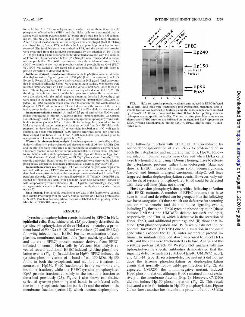
Tyrosine phosphorylation events induced by EPEC in HeLaepithelial cells. Rosenshine et al. (25) previously described thetyrosine phosphorylation of three HeLa cell proteins, a prom-inent band of 90 kDa (Hp90) and two others (72 and 39 kDa),following infection with EPEC. Further examination of cyto-plasmic, membrane, and insoluble (host nuclei, cytoskeleton,and adherent EPEC) protein extracts derived from EPEC-infected or control HeLa cells by Western blot analysis re-vealed several additional EPEC-induced tyrosine phosphory-lation events (Fig. 1). In addition to Hp90, EPEC induced thetyrosine phosphorylation of a band of ca. 150 kDa, Hp150,found in both the cytoplasmic and membrane fractions. Incontrast to Hp150, Hp90 fractionated in the membrane andinsoluble fractions, while the EPEC tyrosine-phosphorylatedEp85 protein fractionated solely in the insoluble fraction asdescribed previously (25). Figure 1 also shows two distinctseries of tyrosine-phosphorylated bands in uninfected cells,one in the cytoplasmic fraction (series I) and the other in themembrane fraction (series II), which become dephosphory-
lated following infection with EPEC. EPEC also induced ty-rosine dephosphorylation of a ca. 240-kDa protein found inboth the cytoplasmic and membrane fractions, Hp240, follow-ing infection. Similar results were observed when HeLa cellswere fractionated after using a Dounce homogenizer to releasethe cytoplasmic proteins rather than detergents (data notshown). EPEC infection of human colon adenocarcinoma,Caco-2, and human laryngeal carcinoma, HEp-2, cell linestriggered similar dephosphorylation events. However, only mi-nor levels of tyrosine-phosphorylated Hp90 could be detectedwith these cell lines (data not shown).
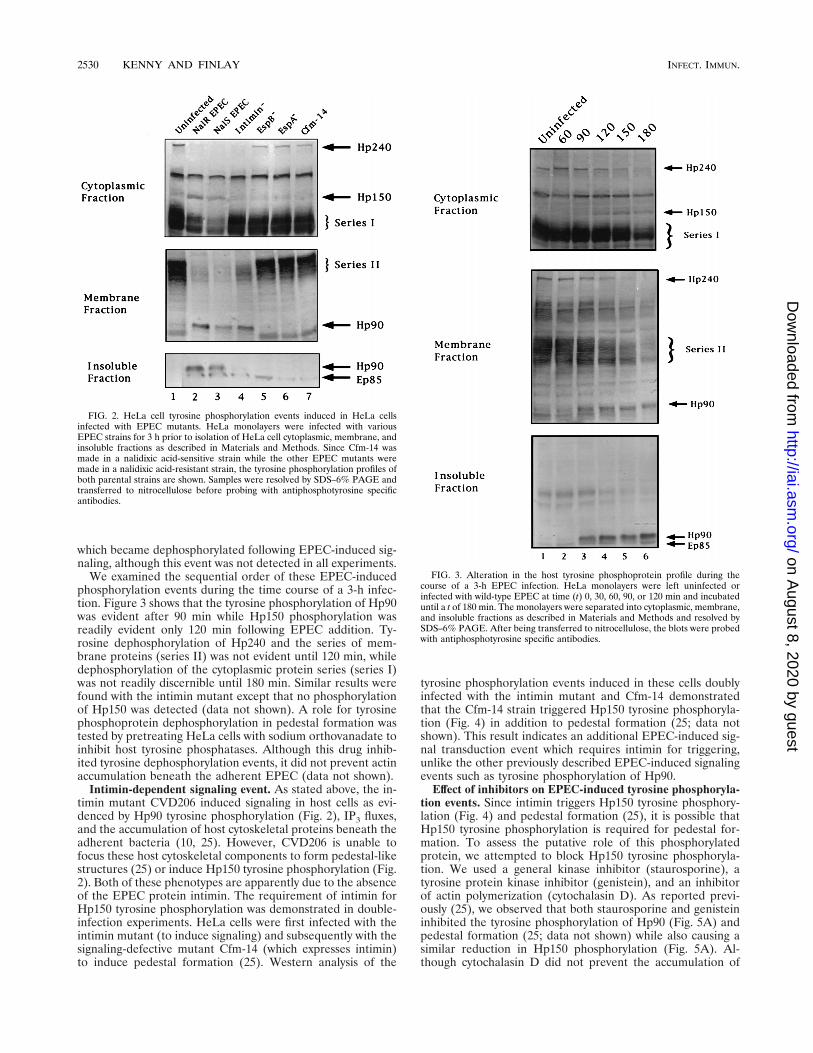
Host tyrosine phosphorylation profiles following infectionwith EPEC mutants. A number of EPEC mutants that havebeen isolated and described can be grouped into the followingtwo basic categories: (i) those which are defective for secretingone or more proteins and do not induce signaling events,including IP3 fluxes and Hp90 tyrosine phosphorylation (theseinclude UMD864 and UMD872, deleted for espB and espA,respectively, and Cfm-14, which is defective in the secretion ofEspA, EspB, and additional proteins); and (ii) those that in-duce Hp90 phosphorylation and IP3 fluxes but do not inducepedestal formation (CVD206) due to a mutation in the eaeAgene which encodes the EPEC outer membrane protein in-timin. The mutants described above were used to infect HeLacells, and the cells were fractionated as before. Analysis of theresulting protein extracts by Western blot analysis with an-tiphosphotyrosine specific antibodies demonstrated that thesignaling defective mutants (UMD864 [espB], UMD872 [espA],and Cfm-14 [type III secretion-defective mutant]) did not in-duce the tyrosine phosphorylation or dephosphorylationevents that normally follow wild-type infection (Fig. 2). Asexpected, CVD206, the intimin-negative mutant, inducedHp90 phosphorylation, although Hp90 remained almost exclu-sively in the membrane fraction (Fig. 2). However, CVD206did not induce Hp150 phosphorylation (Fig. 2). This resultindicated a role for intimin in Hp150 phosphorylation. Figure2 also shows another host membrane protein of about 85 kDa
FIG. 1. HeLa cell tyrosine phosphorylation events induced in EPEC-infectedHeLa cells. HeLa cells were fractionated into cytoplasmic, membrane, and in-soluble fractions as described in Materials and Methods. Samples were resolvedby SDS–6% PAGE and transferred to nitrocellulose before probing with an-tiphosphotyrosine specific antibodies. The host tyrosine phosphorylation eventsaltered after EPEC infection are indicated on the right, and Ep85 represents anEPEC tyrosine-phosphorylated protein (25). 1, EPEC-infected cells; 2, unin-fected cells.
VOL. 65, 1997 INTIMIN-DEPENDENT SIGNALING 2529
on August 8, 2020 by guest
http://iai.asm.org/
Dow
nloaded from
which became dephosphorylated following EPEC-induced sig-naling, although this event was not detected in all experiments.
We examined the sequential order of these EPEC-inducedphosphorylation events during the time course of a 3-h infec-tion. Figure 3 shows that the tyrosine phosphorylation of Hp90was evident after 90 min while Hp150 phosphorylation wasreadily evident only 120 min following EPEC addition. Ty-rosine dephosphorylation of Hp240 and the series of mem-brane proteins (series II) was not evident until 120 min, whiledephosphorylation of the cytoplasmic protein series (series I)was not readily discernible until 180 min. Similar results werefound with the intimin mutant except that no phosphorylationof Hp150 was detected (data not shown). A role for tyrosinephosphoprotein dephosphorylation in pedestal formation wastested by pretreating HeLa cells with sodium orthovanadate toinhibit host tyrosine phosphatases. Although this drug inhib-ited tyrosine dephosphorylation events, it did not prevent actinaccumulation beneath the adherent EPEC (data not shown).
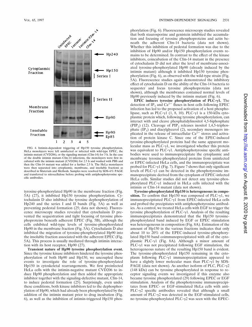
Intimin-dependent signaling event. As stated above, the in-timin mutant CVD206 induced signaling in host cells as evi-denced by Hp90 tyrosine phosphorylation (Fig. 2), IP3 fluxes,and the accumulation of host cytoskeletal proteins beneath theadherent bacteria (10, 25). However, CVD206 is unable tofocus these host cytoskeletal components to form pedestal-likestructures (25) or induce Hp150 tyrosine phosphorylation (Fig.2). Both of these phenotypes are apparently due to the absenceof the EPEC protein intimin. The requirement of intimin forHp150 tyrosine phosphorylation was demonstrated in double-infection experiments. HeLa cells were first infected with theintimin mutant (to induce signaling) and subsequently with thesignaling-defective mutant Cfm-14 (which expresses intimin)to induce pedestal formation (25). Western analysis of the
tyrosine phosphorylation events induced in these cells doublyinfected with the intimin mutant and Cfm-14 demonstratedthat the Cfm-14 strain triggered Hp150 tyrosine phosphoryla-tion (Fig. 4) in addition to pedestal formation (25; data notshown). This result indicates an additional EPEC-induced sig-nal transduction event which requires intimin for triggering,unlike the other previously described EPEC-induced signalingevents such as tyrosine phosphorylation of Hp90.
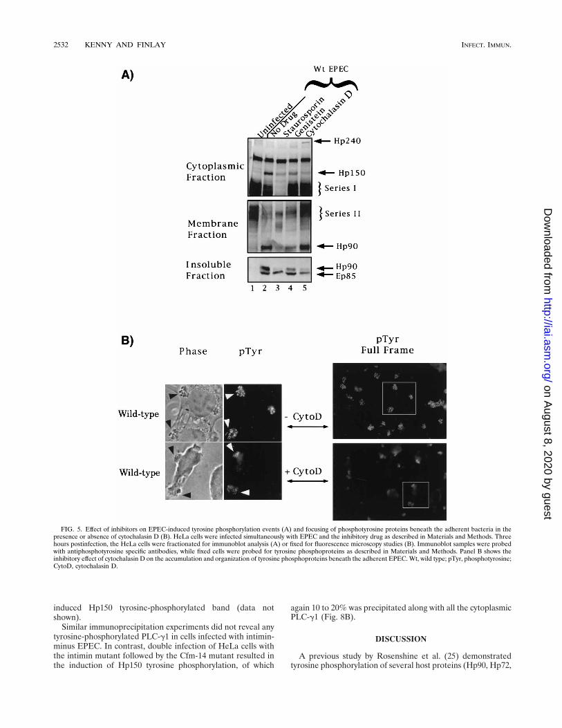
Effect of inhibitors on EPEC-induced tyrosine phosphoryla-tion events. Since intimin triggers Hp150 tyrosine phosphory-lation (Fig. 4) and pedestal formation (25), it is possible thatHp150 tyrosine phosphorylation is required for pedestal for-mation. To assess the putative role of this phosphorylatedprotein, we attempted to block Hp150 tyrosine phosphoryla-tion. We used a general kinase inhibitor (staurosporine), atyrosine protein kinase inhibitor (genistein), and an inhibitorof actin polymerization (cytochalasin D). As reported previ-ously (25), we observed that both staurosporine and genisteininhibited the tyrosine phosphorylation of Hp90 (Fig. 5A) andpedestal formation (25; data not shown) while also causing asimilar reduction in Hp150 phosphorylation (Fig. 5A). Al-though cytochalasin D did not prevent the accumulation of
FIG. 2. HeLa cell tyrosine phosphorylation events induced in HeLa cellsinfected with EPEC mutants. HeLa monolayers were infected with variousEPEC strains for 3 h prior to isolation of HeLa cell cytoplasmic, membrane, andinsoluble fractions as described in Materials and Methods. Since Cfm-14 wasmade in a nalidixic acid-sensitive strain while the other EPEC mutants weremade in a nalidixic acid-resistant strain, the tyrosine phosphorylation profiles ofboth parental strains are shown. Samples were resolved by SDS–6% PAGE andtransferred to nitrocellulose before probing with antiphosphotyrosine specificantibodies.
FIG. 3. Alteration in the host tyrosine phosphoprotein profile during thecourse of a 3-h EPEC infection. HeLa monolayers were left uninfected orinfected with wild-type EPEC at time (t) 0, 30, 60, 90, or 120 min and incubateduntil a t of 180 min. The monolayers were separated into cytoplasmic, membrane,and insoluble fractions as described in Materials and Methods and resolved bySDS–6% PAGE. After being transferred to nitrocellulose, the blots were probedwith antiphosphotyrosine specific antibodies.
2530 KENNY AND FINLAY INFECT. IMMUN.
on August 8, 2020 by guest
http://iai.asm.org/
Dow
nloaded from
tyrosine-phosphorylated Hp90 in the membrane fraction (Fig.5A) (27), it inhibited Hp150 tyrosine phosphorylation. Cy-tochalasin D also inhibited the tyrosine dephosphorylation ofHp240 and the series I and II bands (Fig. 5A) as well aspreventing pedestal formation (25; data not shown). Fluores-cence microscopy studies revealed that cytochalasin D pre-vented the sequestration and tight focusing of tyrosine phos-phoproteins beneath adherent EPEC (Fig. 5B), although thecells exhibited wild-type levels of tyrosine-phosphorylatedHp90 in the membrane fraction (Fig. 5A). Cytochalasin D alsoinhibited the migration of tyrosine-phosphorylated Hp90 intothe insoluble fraction associated with the adherent EPEC (Fig.5A). This process is usually mediated through intimin interac-tion with its host receptor, Hp90 (27).
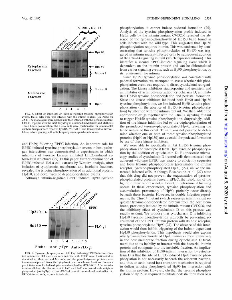
Transient nature of Hp90 tyrosine phosphorylation event.Since the tyrosine kinase inhibitors hindered the tyrosine phos-phorylation of both Hp90 and Hp150, we uncoupled theseevents to investigate the role of tyrosine-phosphorylatedHp150 in cytoskeletal rearrangements. We first preinfectedHeLa cells with the intimin-negative mutant CVD206 to in-duce Hp90 phosphorylation and then added the appropriateinhibitor together with the signaling-defective mutant, Cfm-14,to induce pedestal formation (25). Surprisingly, even underthese conditions, both kinase inhibitors led to the dephosphor-ylation of Hp90, which had already been phosphorylated by theaddition of the intimin mutant prior to drug incubation (Fig.6), as well as the inhibition of intimin-triggered Hp150 phos-
phorylation (Fig. 6). Fluorescence microscopy studies revealedthat both staurosporine and genistein inhibited the accumula-tion and focusing of tyrosine phosphoproteins and actin be-neath the adherent Cfm-14 bacteria (data not shown).Whether this inhibition of pedestal formation was due to theinhibition of Hp90 and/or Hp150 phosphorylation events re-mains to be determined. In contrast to the effect of the kinaseinhibitors, coincubation of the Cfm-14 mutant in the presenceof cytochalasin D did not alter the level of membrane-associ-ated tyrosine-phosphorylated Hp90 (already induced by theintimin mutant) although it inhibited Hp150 tyrosine phos-phorylation (Fig. 6), as observed with the wild-type strain (Fig.5A). Fluorescence studies again demonstrated the inhibitoryeffect of cytochalasin D on the ability of the Cfm-14 bacteria tosequester and focus tyrosine phosphoproteins (data notshown), although the membranes contained normal levels ofHp90 previously induced by the intimin mutant (Fig. 6).
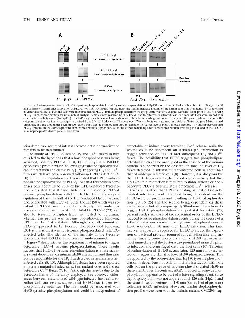
EPEC induces tyrosine phosphorylation of PLC-g1. Thedetection of IP3 and Ca21 fluxes in host cells following EPECinfection has led to the proposed activation of a host phospho-lipase, such as PLC-g1 (1, 8, 10). PLC-g1 is a 150-kDa cyto-plasmic protein which, following tyrosine phosphorylation, caninteract with and cleave phosphatidylinositol 4,5-biphosphate(PIP2) (12). Cleavage of PIP2 releases inositol 1,4,5-triphos-phate (IP3) and diacylglycerol (2), secondary messengers im-plicated in the release of intracellular Ca21 stores and activa-tion of protein kinase C. Since one of the EPEC-inducedtyrosine-phosphorylated proteins had the same apparent mo-lecular mass as PLC-g1, we investigated whether this proteinwas equivalent to PLC-g1. Antiphosphotyrosine specific anti-bodies were used to immunoprecipitate the cytoplasmic andmembrane tyrosine-phosphorylated proteins from uninfectedor EPEC-infected HeLa cells, and the immunoprecipitate wasprobed for PLC-g1 (Fig. 7). Figure 7 shows that only significantlevels of PLC-g1 can be detected in the phosphotyrosine im-munoprecipitate derived from the cytoplasm of EPEC-infectedHeLa cells. Similar studies did not detect any tyrosine-phos-phorylated PLC-g1 induced in HeLa cells infected with theintimin or Cfm-14 mutant (data not shown).
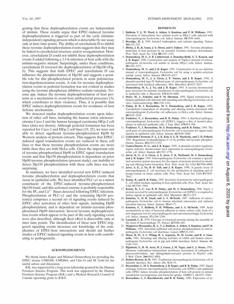
Tyrosine-phosphorylated Hp150 is heterogeneous in compo-sition. To confirm that Hp150 was composed of PLC-g1, weimmunoprecipitated PLC-g1 from EPEC-infected HeLa cellsand probed the precipitates with antiphosphotyrosine antibod-ies. As a control, we treated HeLa cells with EGF to trigger thetyrosine phosphorylation of PLC-g1. Analysis of the resultingimmunoprecipitates demonstrated that the Hp150 tyrosine-phosphorylated band induced by EGF or EPEC stimulationwas heterogeneous in composition (Fig. 8A). Estimation of theamount of Hp150 in the various fractions indicates that onlyabout 10 to 20% of the EPEC-induced tyrosine-phosphory-lated Hp150 band coimmunoprecipitated with all of the cyto-plasmic PLC-g1 (Fig. 8A). Although a minor amount ofPLC-g1 was not precipitated following EGF stimulation, theheterogeneous nature of the resulting Hp150 band is evident.The tyrosine-phosphorylated Hp150 remaining in the cyto-plasm following PLC-g1 immunoprecipitation appeared tohave a slightly lower molecular mass than PLC-g1 by SDS-PAGE (data not shown). As another isoform of PLC, PLC-g2(148 kDa) can be tyrosine phosphorylated in response to re-ceptor signaling events we investigated if this enzyme alsobecame tyrosine phosphorylated (29) following EPEC or EGFstimulation. Analysis of the phosphotyrosine immunoprecipi-tates from EPEC- or EGF-stimulated HeLa cells with anti-PLC-g2 specific antibodies revealed that although a smallamount of PLC-g2 was detected in the EGF-stimulated cells,no tyrosine-phosphorylated PLC-g2 was seen with the EPEC-
FIG. 4. Intimin-dependent triggering of Hp150 tyrosine phosphorylation.HeLa monolayers were left uninfected or infected with wild-type EPEC, theintimin mutant (CVD206), or the signaling mutant (Cfm-14) for 3 h. In the caseof the double intimin mutant–Cfm-14 infections, the monolayers were first in-cubated with the intimin mutant (CVD206) for 2.5 h and washed with PBS andthen the Cfm-14 mutant was added for a further 2.5 h. The HeLa monolayerswere then separated into cytoplasmic, membrane, and insoluble fractions asdescribed in Materials and Methods. Samples were resolved by SDS–6% PAGEand transferred to nitrocellulose before probing with antiphosphotyrosine spe-cific antibodies.
VOL. 65, 1997 INTIMIN-DEPENDENT SIGNALING 2531
on August 8, 2020 by guest
http://iai.asm.org/
Dow
nloaded from
induced Hp150 tyrosine-phosphorylated band (data notshown).
Similar immunoprecipitation experiments did not reveal anytyrosine-phosphorylated PLC-g1 in cells infected with intimin-minus EPEC. In contrast, double infection of HeLa cells withthe intimin mutant followed by the Cfm-14 mutant resulted inthe induction of Hp150 tyrosine phosphorylation, of which
again 10 to 20% was precipitated along with all the cytoplasmicPLC-g1 (Fig. 8B).
DISCUSSION
A previous study by Rosenshine et al. (25) demonstratedtyrosine phosphorylation of several host proteins (Hp90, Hp72,
FIG. 5. Effect of inhibitors on EPEC-induced tyrosine phosphorylation events (A) and focusing of phosphotyrosine proteins beneath the adherent bacteria in thepresence or absence of cytochalasin D (B). HeLa cells were infected simultaneously with EPEC and the inhibitory drug as described in Materials and Methods. Threehours postinfection, the HeLa cells were fractionated for immunoblot analysis (A) or fixed for fluorescence microscopy studies (B). Immunoblot samples were probedwith antiphosphotyrosine specific antibodies, while fixed cells were probed for tyrosine phosphoproteins as described in Materials and Methods. Panel B shows theinhibitory effect of cytochalasin D on the accumulation and organization of tyrosine phosphoproteins beneath the adherent EPEC. Wt, wild type; pTyr, phosphotyrosine;CytoD, cytochalasin D.
2532 KENNY AND FINLAY INFECT. IMMUN.
on August 8, 2020 by guest
http://iai.asm.org/
Dow
nloaded from
and Hp39) following EPEC infection. An important role forEPEC-induced tyrosine phosphorylation events in host-patho-gen interactions was demonstrated in experiments in whichinhibitors of tyrosine kinases inhibited EPEC-induced cy-toskeletal structures (25). In this paper, further examination ofEPEC-infected HeLa cell extracts by Western analysis, afterisolation of cytoplasmic, membrane, and insoluble fractions,revealed the tyrosine phosphorylation of an additional protein,Hp150, and novel tyrosine dephosphorylation events.
Although intimin-negative EPEC induces Hp90 tyrosine
phosphorylation, it cannot induce pedestal formation (25).Analysis of the tyrosine phosphorylation profile induced inHeLa cells by the intimin mutant CVD206 revealed the ab-sence of the tyrosine-phosphorylated Hp150 band found incells infected with the wild type. This suggested that Hp150phosphorylation requires intimin. This was confirmed by dem-onstrating that tyrosine phosphorylation of Hp150 was trig-gered in intimin mutant-infected cells by subsequent additionof the Cfm-14 signaling mutant (which expresses intimin). Thisidentifies a second EPEC-induced signaling event which isdependent on the intimin protein and can be differentiatedfrom earlier signaling events, such as Hp90 phosphorylation, byits requirement for intimin.
Since Hp150 tyrosine phosphorylation was correlated withpedestal formation, we attempted to assess whether this phos-phorylation event was required to direct cytoskeletal reorgani-zation. The kinase inhibitors staurosporine and genistein andan inhibitor of actin polymerization, cytochalasin D, all inhib-ited Hp150 tyrosine phosphorylation and pedestal formation.Since the kinase inhibitors inhibited both Hp90 and Hp150tyrosine phosphorylation, we first induced Hp90 tyrosine phos-phorylation (in the absence of Hp150 tyrosine phosphoryla-tion) by infection with the intimin mutant. We then added theappropriate drugs together with the Cfm-14 signaling mutantto trigger Hp150 tyrosine phosphorylation. Surprisingly, addi-tion of the kinase inhibitors led to the dephosphorylation ofthe preinduced tyrosine-phosphorylated Hp90, revealing thelabile nature of this event. Thus, it was not possible to deter-mine whether one or both of these tyrosine-phosphorylatedproteins (Hp90 or Hp150) are essential for pedestal formationby use of these kinase inhibitors.
We were able to specifically inhibit Hp150 tyrosine phos-phorylation and uncouple it from Hp90 tyrosine phosphoryla-tion by the addition of cytochalasin D. Fluorescence micros-copy studies of cytochalasin D-treated cells demonstrated thatadherent wild-type EPEC was unable to efficiently sequesterand focus tyrosine phosphoproteins (presumably the intiminreceptor, tyrosine-phosphorylated Hp90) (27), unlike the un-treated infected cells. Although Rosenshine et al. (27) statethat this drug did not prevent the sequestration of tyrosine-phosphorylated proteins beneath EPEC, the resolution of thefigure in their report is not sufficient to determine if focusingoccurs. In these experiments, tyrosine phosphorylation andaccumulation, presumably of Hp90, probably occur directlybeneath these bacteria. However, in double infection experi-ments, the Cfm-14 mutant (which expresses intimin) must se-quester tyrosine-phosphorylated proteins from the host mem-brane, previously induced by the intimin mutant CVD206, andthe inhibitory effect of cytochalasin D on this process wasreadily evident. We propose that cytochalasin D is inhibitingHp150 tyrosine phosphorylation indirectly by preventing re-cruitment of the EPEC intimin protein with its host receptor,tyrosine-phosphorylated Hp90 (27). The absence of this inter-action would then inhibit triggering of the intimin-dependentHp150 phosphorylation. This hypothesis would also explainwhy tyrosine-phosphorylated Hp90 remains almost exclusivelyin the host membrane fraction during cytochalasin D treat-ment due to its inability to interact with the bacterial intiminprotein and comigrate into the insoluble fraction. An implica-tion of this inhibition of Hp90-intimin interaction by cytocha-lasin D is that the site of EPEC-induced Hp90 tyrosine phos-phorylation is not necessarily beneath the adherent bacteria,and thus an actin-based host transport mechanism is requiredto deliver tyrosine-phosphorylated Hp90 for interaction withthe intimin protein. However, whether the tyrosine phosphor-ylation of Hp150 is required to initiate pedestal formation or is
FIG. 6. Effect of inhibitors on intimin-triggered tyrosine phosphorylationevents. HeLa cells were first infected with the intimin mutant (CVD206) for2.5 h. The monolayers were washed and then infected with the signaling mutant,Cfm-14, together with the inhibitory drug as described in Materials and Methods.Three hours postinfection, the HeLa cells were fractionated for immunoblotanalysis. Samples were resolved by SDS–6% PAGE and transferred to nitrocel-lulose before probing with antiphosphotyrosine specific antibodies.
FIG. 7. Tyrosine phosphorylation of PLC-g1 following EPEC infection. Con-trol uninfected HeLa cells or cells infected with EPEC were fractionated asdescribed in Materials and Methods, and the phosphotyrosine proteins wereimmunoprecipitated from the cytoplasmic and membrane fractions. Immuno-precipitates were loaded in duplicate and resolved by SDS-PAGE. After transferto nitrocellulose, the blot was cut in half; each half was probed with antiphos-photyrosine (Anti-pTyr) or anti-PLC-g1 specific monoclonal antibodies. 1,EPEC-infected cells; 2, uninfected cells.
VOL. 65, 1997 INTIMIN-DEPENDENT SIGNALING 2533
on August 8, 2020 by guest
http://iai.asm.org/
Dow
nloaded from
stimulated as a result of intimin-induced actin polymerizationremains to be determined.
The ability of EPEC to induce IP3 and Ca21 fluxes in hostcells led to the hypothesis that a host phospholipase was beingactivated, possibly PLC-g1 (1, 8, 10). PLC-g1 is a 150-kDacytoplasmic protein which, following tyrosine phosphorylation,can interact with and cleave PIP2 (12), triggering IP3 and Ca21
fluxes which have been observed following EPEC infection (8,10). Immunoprecipitation studies revealed that EPEC inducestyrosine phosphorylation of PLC-g1 but that this protein com-prises only about 10 to 20% of the EPEC-induced tyrosine-phosphorylated Hp150 band. Indeed, stimulation of PLC-g1tyrosine phosphorylation with EGF led to the coimmunopre-cipitation of less than half of the EGF-induced Hp150 tyrosinephosphorylated with PLC-g1. Since the Hp150 which was re-sistant to PLC-g1 precipitation had a slightly lower molecularmass and another isoform of PLC, 148-kDa PLC-g2 (29), canalso be tyrosine phosphorylated, we tested to determinewhether this protein was tyrosine phosphorylated followingEPEC or EGF stimulation. Although a small amount ofPLC-g2 appeared to be tyrosine phosphorylated followingEGF stimulation, it was not tyrosine phosphorylated in EPEC-infected cells. The identity of the majority of the tyrosine-phosphorylated 150-kDa band remains undetermined.
Figure 8 demonstrates the requirement of intimin to triggerdetectable PLC-g1 tyrosine phosphorylation. These resultssuggest that PLC-g1 tyrosine phosphorylation is a late signal-ing event dependent on intimin-Hp90 interaction and thus maynot be responsible for the IP3 flux detected in intimin mutant-infected cells (8, 10). Although IP3 fluxes have been reportedin intimin mutant-infected cells, this mutant does not inducedetectable Ca21 fluxes (8, 10). Although this may be due to thedetection limits of the assay employed, the observed differ-ences between mutant- and wild-type-infected host cells, to-gether with our results, suggest that EPEC may trigger twophospholipase activities. The first could be associated withHp90 tyrosine phosphorylation and could be independent of
detectable, or induce a very transient, Ca21 release, while thesecond could be dependent on intimin-Hp90 interaction totrigger activation of PLC-g1 and subsequent IP3 and Ca21
fluxes. The possibility that EPEC triggers two phospholipaseactivities which can be uncoupled in the absence of the intiminprotein is supported by the observation that the level of IP3fluxes detected in intimin mutant-infected cells is about halfthat of wild-type infected cells (8). However, it is also plausiblethat EPEC triggers a single phospholipase activity but thatHp90-intimin interaction is required to stably activate or phos-phorylate PLC-g1 to stimulate a detectable Ca21 release.
Our results show that EPEC signaling in host cells can bedivided into two events, the first being dependent on theEPEC-secreted proteins and resulting in Hp90 phosphoryla-tion (10, 16, 25) and the second being dependent on theseearlier events but also requiring Hp90-intimin interactions totrigger Hp150 phosphorylation and pedestal formation (25;present study). Analysis of the sequential order of the EPEC-induced tyrosine phosphorylation events during the course of a180-min infection showed that tyrosine phosphorylation ofHp90 was evident 90 min after EPEC infection. This timeinterval is apparently required for EPEC to induce the expres-sion of bacterial proteins required for cell adherence and sig-naling, since tyrosine phosphorylation of Hp90 can occur al-most immediately if the bacteria are preinduced in media priorto infection and centrifuged onto the host cells (26). Tyrosinephosphorylation of Hp150 occurs later, 120 min following in-fection, suggesting that it follows Hp90 phosphorylation. Thisis supported by the observation that Hp150 tyrosine phosphor-ylation is dependent not only on intimin interaction with hostcells but on the presence of tyrosine-phosphorylated Hp90 inthese membranes. In contrast, EPEC-induced tyrosine dephos-phorylation appears to be part of a later signaling event, sincedephosphorylation was not apparent until 120 min (Hp240 andthe series II set of proteins) or 180 min (series I set of proteins)following EPEC infection. However, similar dephosphoryla-tion profiles were seen with the intimin mutant CVD206, sug-
FIG. 8. Heterogeneous nature of Hp150 tyrosine-phosphorylated band. Tyrosine phosphorylation of Hp150 was induced in HeLa cells with EFG (100 ng/ml for 10min to induce tyrosine phosphorylation of PLC-g1) or wild-type EPEC (A) and EGF, the intimin-negative mutant, or the intimin and Cfm-14 mutants (B) as describedin Materials and Methods. HeLa cells were fractionated and PLC-g1 immunoprecipitated from the cytoplasmic fractions. Samples were also taken prior to and followingPLC-g1 immunoprecipitation for immunoblot analysis. Samples were resolved by SDS-PAGE and transferred to nitrocellulose, and separate blots were probed witheither antiphosphotyrosine (Anti-pTyr) or anti-PLC-g1 specific monoclonal antibodies. The relative loadings are indicated beneath the panels, where 1 denotes thecytoplasmic extract or immunoprecipitate derived from 3 3 105 HeLa cells. The developed Western blots were scanned into Adobe Photoshop (see Materials andMethods), and the area under each Hp150-related band was determined and used to estimate the percentage of Hp150 in each fraction. The phosphotyrosine andPLC-g1 profiles in the extracts prior to immunoprecipitation (upper panels), in the extract remaining after immunoprecipitation (middle panels), and in the PLC-g1immunoprecipitates (lower panels) are shown.
2534 KENNY AND FINLAY INFECT. IMMUN.
on August 8, 2020 by guest
http://iai.asm.org/
Dow
nloaded from
gesting that these dephosphorylation events are independentof intimin. These results argue that EPEC-induced tyrosinedephosphorylation is triggered as part of the early (intimin-independent) signaling process which is detectable by our assayonly at later time points. The ability of cytochalasin D to inhibitthese tyrosine dephosphorylation events suggests that they maybe linked to cytoskeletal structure and/or reorganization. How-ever, cytochalasin D could not inhibit these dephosphorylationevents if added following a 2.5-h infection of host cells with theintimin-negative mutant. Surprisingly, under these conditions,cytochalasin D reversed the dephosphorylation of Hp240 (Fig.6). This suggests that both EPEC and cytochalasin D caninfluence the phosphorylation of Hp240 and suggests a possi-ble role for this phosphorylated protein in actin polymeriza-tion-depolymerization events. A role for tyrosine dephosphor-ylation events in pedestal formation was not evident in studiesusing the tyrosine phosphatase inhibitor sodium vanadate. Yer-sinia spp. induce the tyrosine dephosphorylation of host pro-teins as a mechanism to avoid host antibacterial processes (3),which contributes to their virulence. Thus, it is possible thatEPEC induces dephosphorylation events for avoidance of hostdefense mechanisms.
We detected similar dephosphorylation events upon infec-tion of other cell lines, including the human colon adenocar-cinoma Caco-2 and the human laryngeal carcinoma HEp-2 celllines (data not shown). Although pedestal formation has beenreported for Caco-2 and HEp-2 cell lines (19, 25), we were notable to detect significant tyrosine-phosphorylated Hp90 byWestern analysis of protein extracts. This suggests that EPEC-induced signal transduction events are reduced in these celllines or that these tyrosine phosphorylation events are morelabile than they are with HeLa cells. Given the important roleof tyrosine-phosphorylated Hp90 in EPEC signal transductionevents and that Hp150 phosphorylation is dependent on priorHp90 tyrosine phosphorylation (present study), our inability todetect Hp150 phosphorylation with these cells lines was notsurprising.
In summary, we have identified several new EPEC-inducedtyrosine phosphorylation and dephosphorylation events thatoccur in epithelial cells. We have identified PLC-g1 as a minorconstituent of the EPEC-induced tyrosine-phosphorylatedHp150 band, and this activated enzyme is probably responsiblefor the IP3 and Ca21 fluxes detected following EPEC infection.Phosphorylation of PLC-g1 and the remaining Hp150 pro-tein(s) comprises a second set of signaling events induced byEPEC after activation of other host signals, including Hp90phosphorylation, and is dependent on intimin–tyrosine-phos-phorylated Hp90 interaction. Several tyrosine dephosphoryla-tion events which appear to be part of the early signaling eventwere also described, although their effect is discernible only atlater time points. The identification of these new EPEC-trig-gered signaling events increases our knowledge of the com-plexities of EPEC-host interactions and should aid furtherstudies of EPEC-induced signaling events in host cells contrib-uting to pathogenicity.
ACKNOWLEDGMENTS
We thank James Kaper and Michael Donnenberg for providing theEPEC strains UMD206, UMD864, and Cfm-14 and M. Gold for hisuseful advice and discussion.
B.K. was supported by a long-term fellowship granted by the HumanFrontiers Science Program. This work was supported by the HumanFrontiers Science Program (B.K.) and a Medical Research Council ofCanada operating grant to B.B.F.
REFERENCES
1. Baldwin, T. J., W. Ward, A. Aitken, S. Knutton, and P. H. Williams. 1991.Elevation of intracellular free calcium levels in HEp-2 cells infected withenteropathogenic Escherichia coli. Infect. Immun. 59:1599–1604.
2. Berridge, M. J. 1993. Inositol triphosphate and calcium signaling. Nature361:315–325.
3. Bliska, J. B., K. Guan, J. E. Dixon, and S. Falkow. 1991. Tyrosine phosphatehydrolysis of host proteins by an essential Yersinia virulence determinant.Proc. Natl. Acad. Sci. USA 88:1187–1191.
4. Donnenberg, M. S., S. B. Calderwood, A. Donohue-Rolfe, G. T. Keusch, andJ. B. Kaper. 1990. Construction and analysis of TnphoA mutants of entero-pathogenic Escherichia coli unable to invade HEp-2 cells. Infect. Immun.58:1565–1571.
5. Donnenberg, M. S., and J. B. Kaper. 1991. Construction of an eae deletionmutant of enteropathogenic Escherichia coli by using a positive-selectionsuicide vector. Infect. Immun. 59:4310–4317.
6. Donnenberg, M. S., J. A. Giron, J. P. Nataro, and J. B. Kaper. 1992. Aplasmid-encoded type IV fimbrial gene of enteropathogenic Escherichia coliassociated with localized adherence. Mol. Microbiol. 6:3427–3437.
7. Donnenberg, M. S., J. Yu, and J. B. Kaper. 1993. A second chromosomalgene necessary for intimate attachment of enteropathogenic Escherichia colito epithelial cells. J. Bacteriol. 175:4670–4680.
8. Dytoc, M., L. Fedorko, and P. M. Sherman. 1994. Signal transduction inhuman epithelial cells infected with attaching and effacing Escherichia coli invitro. Gastroenterology 106:1150–1161.
9. Finlay, B. B., I. Rosenshine, M. S. Donnenberg, and J. B. Kaper. 1992.Cytoskeletal composition of attaching and effacing lesions associated withenteropathogenic Escherichia coli adherence to HeLa cells. Infect. Immun.60:2541–2543.
10. Foubister, V., I. Rosenshine, and B. B. Finlay. 1994. A diarrheal pathogen,enteropathogenic Escherichia coli (EPEC), triggers a flux of inositol phos-phates in infected epithelial cells. J. Exp. Med. 179:993–998.
11. Foubister, V., I. Rosenshine, M. S. Donnenberg, and B. B. Finlay. 1994. TheeaeB gene of enteropathogenic Escherichia coli is necessary for signal trans-duction in epithelial cells. Infect. Immun. 62:3038–3040.
12. Goldschidt-Clermont, P. J., J. K. Kim, S. G. R. Machesky, and T. D. Pollard.1991. Regulation of phospholipase C-g1 by profilin and tyrosine phosphor-ylation. Science 251:1231–1233.
13. Gomez-Duarte, O. G., and J. B. Kaper. 1995. A plasmid-encoded regulatoryregion activates chromosomal eaeA expression in enteropathogenic Esche-richia coli. Infect. Immun. 63:1767–1776.
14. Jarvis, K. G., J. A. Giron, A. E. Jerse, T. K. McDaniel, M. S. Donnenberg,and J. B. Kaper. 1995. Enteropathogenic Escherichia coli contains a special-ized secretion system necessary for the export of proteins involved in attach-ing and effacing lesion formation. Proc. Natl. Acad. Sci. USA 92:7996–8000.
15. Jerse, A. E., J. Yu, B. D. Tall, and J. B. Kaper. 1990. A genetic locus ofenteropathogenic E. coli necessary for the production of attaching and ef-facing lesions on tissue culture cells. Proc. Natl. Acad. Sci. USA 87:7839–7843.
16. Kenny, B., and B. B. Finlay. 1995. Secretion of proteins by enteropathogenicE. coli which mediate signaling in host epithelial cells. Proc. Natl. Acad. Sci.USA 97:7991–7995.
17. Kenny, B., L.-C. Lai, B. B. Finlay, and M. S. Donnenberg. 1996. EspA, aprotein secreted by enteropathogenic Escherichia coli (EPEC), is required toinduce signals in epithelial cells. Mol. Microbiol. 20:313–323.
18. Knutton, S., D. R. Lloyd, and A. S. McNeish. 1987. Adhesion of entero-pathogenic Escherichia coli to human intestinal enterocytes and culturedintestinal mucosa. Infect. Immun. 55:69–77.
19. Knutton, S., T. Baldwin, P. H. Williams, and A. S. McNeish. 1989. Actinaccumulation at sites of bacterial adhesion to tissue culture cells: basis of anew diagnostic test for enteropathogenic and enterohemorrhagic Escherichiacoli. Infect. Immun. 57:1290–1298.
20. Laemmli, U. K. 1970. Cleavage of structural proteins during the assembly ofthe head of bacteriophage T4. Nature 227:680–685.
21. Manjarrez-Hernandez, H. A., T. J. Baldwin, A. Aitken, S. Knutton, and P. H.Williams. 1992. Intestinal epithelial cell protein phosphorylation in entero-pathogenic Escherichia coli diarrhoae. Lancet 339:521–523.
22. Moon, H. W., S. C. Whipp, R. A. Argenzio, M. M. Levine, and R. A. Gian-nella. 1983. Attaching and effacing activities of rabbit and human entero-pathogenic Escherichia coli in pig and rabbit intestines. Infect. Immun. 41:1340–1351.
23. Rijnboutt, S., H. M. Aerts, H. J. Geuze, J. M. Tager, and G. J. Strous. 1991.Mannose 6-phosphate-independent membrane association of cathepsin D,glucocerebrosidase, and sphingolipid-activated protein in HepG2 cells.J. Biol. Chem. 266:4862–4868.
24. Robins-Browne, R. M. 1987. Traditional enteropathogenic Escherichia coli ofinfantile diarrhea. Rev. Infect. Dis. 9:28–53.
25. Rosenshine, I., M. S. Donnenberg, J. B. Kaper, and B. B. Finlay. 1992. Signalexchange between enteropathogenic Escherichia coli (EPEC) and epithelialcells: EPEC induce tyrosine phosphorylation of host cell protein to initiatecytoskeletal rearrangement and bacterial uptake. EMBO J. 11:3551–3560.
26. Rosenshine, I., S. Ruschkowski, and B. B. Finlay. 1996. Expression of at-
VOL. 65, 1997 INTIMIN-DEPENDENT SIGNALING 2535
on August 8, 2020 by guest
http://iai.asm.org/
Dow
nloaded from
taching/effacing activity by enteropathogenic Escherichia coli depends ongrowth phase, temperature, and protein synthesis upon contact with epithe-lial cells. Infect. Immun. 64:966–973.
27. Rosenshine, I., S. Ruschkowski, M. Stein, D. J. Reinscheid, S. D. Mills, andB. B. Finlay. 1996. A pathogenic bacterium triggers epithelial signals to forma functional bacterial receptor that mediates actin psuedopod formation.EMBO J. 15:2613–2624.
28. Sambrook, J., E. F. Fritsch, and T. Maniatis. 1989. Molecular cloning: a
laboratory manual, 2nd ed. Cold Spring Harbor Laboratory Press, ColdSpring Harbor, N.Y.
29. Sultzman, L., C. Ellis, L.-L. Lin, T. Pawson, and J. Knopf. 1991. Platelet-derived growth factor increases the in vivo activity of phospholipase C-g1 andphospholipase C-g2. Mol. Cell. Biol. 11:2018–2025.
30. Vuopio-Varkila, J., and G. K. Schoolnik. 1991. Localized adherence by entero-pathogenic Escherichia coli is an inducible phenotype associated with the ex-pression of new outer membrane proteins. J. Exp. Med. 174:1167–1177.
Editor: A. O’Brien
2536 KENNY AND FINLAY INFECT. IMMUN.
on August 8, 2020 by guest
http://iai.asm.org/
Dow
nloaded from