Embed Size (px)
Citation preview
Ilme SchlichtingMPI Medical Research Heidelberg
Max Planck Advanced Study Group at CFEL, Hamburg
Into the future –a structure biologist’s dreams

Structural Biologist’s dreams
Cells are the basis of life
Wanted -High resolution temporal & spatial inventory of cells
Who is where, when, why
Comprehensive characterization of the players- 3D structures of macromolecules & assemblies- how do they function, structural changes-how do they acquire their structures, i.e. fold

Cells are the basis of life
F1/F0 ATPsynthase
ATP synthesis
Transcribed byRNA polymerase
mRNA mRNA
Translated and synthesizedin protein: ribosome
DNA: storage of genetic info
Viral infection

Some open issues in structural biologyStructures of big (weakly binding) complexes
membrane proteinstransient intermediates, foldingchromatin/ genome structurecellular organization at high resolution
Current limitations of crystallography, electron microscopyX-ray microscopy
Sample preparation (biochemistry) Crystals for crystallographic approachesData collection, radiation damageComputational approaches dealing with disorder
Current limitations of crystallography, electron microscopyX-ray microscopy
Sample preparation (biochemistry) Crystals for crystallographic approachesData collection, radiation damageComputational approaches dealing with disorder

particle injectionERLs (and FELs) have properties that
open new possibilities
• Which questions can be addressed ?What resolution is required/useful?
• What is needed to make this work?
High brightnessHigh coherenceSmall source sizeShort pulse length High repetition rate (except XFEL)Stability shot-to-shot (except seeded)

What component iswhere, when, why ?

• Depth resolution2D projection of 3D object- tomographic approaches
- multi-angular imaging using split beams
- curvature of Ewald sphere, small objects or sectioning(Bergh et al., Quart. Rev.Biophys.2008 )
3D-imaging of cells
Actin filamentsRibosomes, membranes
Cryoelectron tomography of Dictyostelium cells815 x 870 x 97 nmScience 298:1209 (2002)
• What does one see?cells are very crowded- identification of particles
(superposition, contrast, shape)
• Correlation with function- correlation with light microscopy, fluorescence labels, nonlin. nanocrystals (20-100nm, phasing?)(Pantazis et al PNAS 107:13535 (2010)

5 μm
3D imaging of cells
Square bacterium (Walsby, Nature 283:69(1980), 150 nm thick)
Gas vacuole
Hexagonal and tetragonalarrangement of cell wall subunits
J. Bact. 150:851 (1982)
Unicellular green alga Ostreococcus tauri
Smallest free-living eukaryote, a picoplancton, mean length 1±0.3 μ, width 0.7±0.2 μnaked, nonflagellated cell with a single mitochondrion and chloroplast
Courties et al Nature 370:255 (1994)

Nuclear pore: 100 nm wide, 50 MDa, 200 pores/yeast cell456 constituent proteins, 30 distinct ones
Nature 459, (2009)
Alber et al., Nature 450 (270)
Multidisciplinary approachBiophysics and proteomics modeling


Chromatin structure

Woodcock et al., J Cell Biol 99:42(1984)Helical ribbon, two-start helix
Organization of nucleosomes in DNA fibers
Schalch et al., Nature (2005)Dorigo et al., Science 2004
- Higher order chromatin structure (> 30 nm fiber)
- in vivo structure,changes associated with active/inactive states
- function: modification of histone tails, complexes with e.g. remodeling factors,
- Correlation sequence/ structure( first model of yeast genomeDuan et al., Nature (2010)

How many projections required?(Raines et al Nature 463:214 2010 applicable?)( Bergh et al Quat. Rev Biophys 2008)Add ”markers” for
phasing (large clusters, non-linear nanocrystals) site specific labeling for averaging of reproducible structuresin unique objects (?)
Cryogenic sample mount similar to EM. Holey carbon, graphene?Egg-carton like structure by surface modification
Structure determination of non-identical objects

3-dimensional genome organizationin viruses
Science 312: 1791 (2006) 1.7 nm resolution, 26422 particles
PNAS 2009
Electron microscopy:resolution of genomestructure limited by dynamic scattering,radiation damage
X-ray:greater penetration depthClassification(averaging with internal break in symmetry)

Complex unique vertex decorated by a spike in a giant algal virus
50 nm
Cherrier et al PNAS 116:1105 2009
Paramecium bursaria Chlorella virus-1
Unique vertex involved in infection
Virus brings ion channel to reduce pressure of host cell to inject its DNA
VP130
Genome 331-kbp codes for 11 tRNAs and 365 putative proteins, of which more than 100 are present in the mature virion.
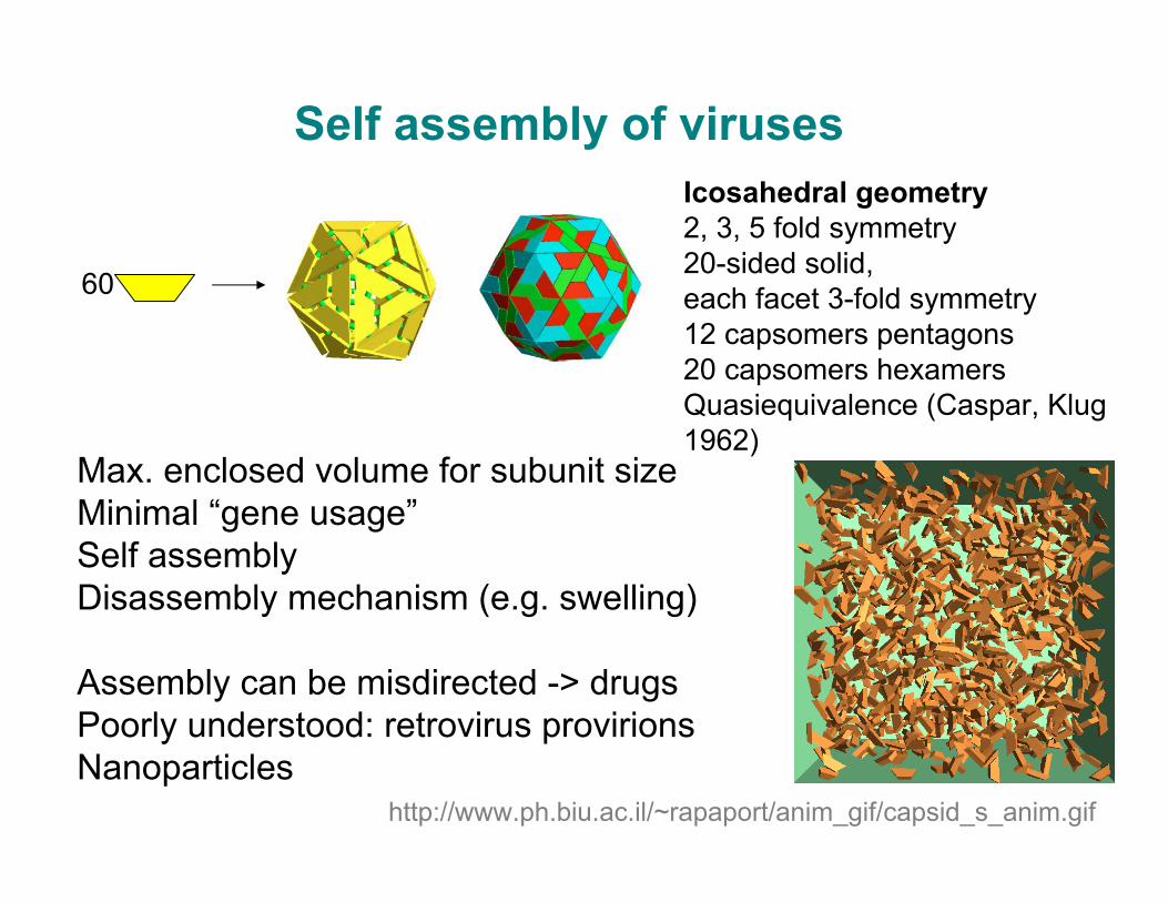
Self assembly of viruses
60
http://www.ph.biu.ac.il/~rapaport/anim_gif/capsid_s_anim.gif
Icosahedral geometry2, 3, 5 fold symmetry20-sided solid,each facet 3-fold symmetry12 capsomers pentagons20 capsomers hexamersQuasiequivalence (Caspar, Klug 1962)
Max. enclosed volume for subunit sizeMinimal “gene usage”Self assembly Disassembly mechanism (e.g. swelling)
Assembly can be misdirected -> drugsPoorly understood: retrovirus provirionsNanoparticles

• Initiate assembly by rapid mixing continuous flow (nanofluidics) , droplet mixing
• Collect time-series of SAXS/WAXS data • Analyze for angular cross correlations in intensity to analyze for local symmetries analogously to recent colloid study
(Wochner et al., PNAS 106:11511(2009) Altarelli et al., PHYS. REV. B 82, 104207 (2010)
Studying e.g. capsid self assembly

David Phillips, Royal Institution, London, 5th Nov 1965

Protein foldingFunnel-shaped energyLandscape:
Many high-energy statesrugged landscape
Few low-energy states
Stochastic process, initial hydrophobic collapsePhysical models vs bioinformatics approach of folding may allow to obtain deeper understanding of forces and dynamics that govern protein properties:
Predict conformational changes, e.g. induced fitRefine models beyond homology structuresImprovement for multi-domain or domain swapped or low homology models

Zipping and assembly mechanism
Dill et al Annu Rev Biophys 2008
Accessible via fast mixing and correlation approach to yield structural information? Complement with parallel IR/CD measurementsInformation beyond ensemble? Needs new software. Serves as input for computational models on folding
On fast time scales (ps-ns) peptide fragments search for local meta-stable structures (loops, beta-turns, helices)
Few are stable enough to survive for longer time scales, grow/zip into larger and more stable structures
On longer time scales, pairs or groups of substructures assemble into larger and more native like structures

Solution studies have great potential- Low and high resolution structural features in SAXS and
WAXS data, need better methods to extract those.Use coherence to exploit angular cross correlations to studyassembly reactions
- New mixing devices and high intensity, high repetition X-ray sources may allow routine microsecond studies, faster studiesby temperature-jump reaction initiation
- Structural changes occurring during reactions, in particular folding. characterization of the molten globule, distributionstudy of large proteins in dilute solutions to prevent aggregation?Simultaneously with IR, VUV (CD) spectroscopy, correlations?
Combination with labeling, alternative methods such asdouble electron electron resonance (DEER) spectroscopy

Concluding remarkNew sources provide new scientific optionsrequire development in both both hardware and software