Embed Size (px)
Citation preview

INTRACELLULAR RECEPTORS

The intracellular (nuclear) receptor superfamilySteroid hormones, thyroid hormones, retinoids and vitamin D

Intracellular receptor
HYDROPHOBIC:
- Non-polar molecules
- Gases
- Steroids

Regulation of transcription activity
• Regulatory mechanisms vary• Heterodimeric receptors - exclusively nuclear; without ligand, repress
transcription by binding to their cognate sites in DNA• Homodimeric receptors - mostly cytoplasmic (without ligands) & hormone
binding leads to nuclear translocation of receptors
• Without ligand - aggregation of receptor with inhibitor proteins (eg Hsp90)
• Steroid hormones are often required to dimerize with a partner to activate gene transcription
• Receptors for vitamin D, retinoic acid and thyroid hormone bind to responsive elements as heterodimers– Second component of the heterodimer is RXR monomer (i.e,
RXR-RAR; RXR-VDR)
Specificities of some receptors …

Intracellular signal molecules • small, lipid-soluble molecules such as steroid
hormones, retinoids, thyroid hormones, Vitamin D. (made from cholesterol)
• These molecules diffuse through plasma and nuclear membranes and interact directly with the transcription factors they control.

66
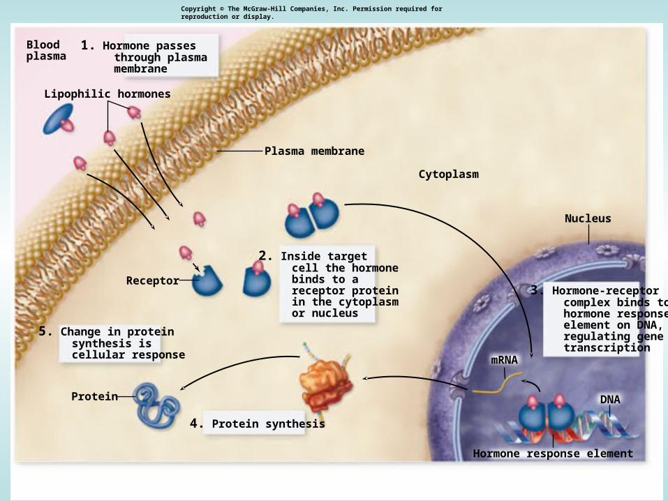
Lipophilic HormonesLipophilic Hormones
CCirculairculationtion in the blood in the blood bound to transport bound to transport proteinsproteins
Dissociation from carrier at target cellsDissociation from carrier at target cellsAction in the cellAction in the cell
-- Pass through the cell membrane and Pass through the cell membrane and bind bind to an intracellular receptor, either in to an intracellular receptor, either in the the cytoplasm or the nucleuscytoplasm or the nucleus-- Hormone-receptor complex binds to Hormone-receptor complex binds to hormone response elements hormone response elements in DNAin DNA -- Regulate gene expressionRegulate gene expression
77
Receptor
Bloodplasma
Protein
Lipophilic hormones
mRNA
DNA
Hormone response element
1. Hormone passes through plasma membrane
2. Inside target cell the hormone binds to a receptor protein in the cytoplasm or nucleus
3. Hormone-receptor complex binds to hormone response element on DNA, regulating gene transcription
4. Protein synthesis
5. Change in protein synthesis is cellular response
Cytoplasm
Plasma membrane
Nucleus
Copyright © The McGraw-Hill Companies, Inc. Permission required for reproduction or display.

Steroid Hormones
STEROID HORMONES:- sex steroids (estrogen, progesterone, testosterone)- corticosteroids (glucocorticoids and mineralcorticoids)
OTHER HORMONESThyroid hormone, vitamin D3, and retinoic acid have different structure and function but share the same mechanism of action with the other steroids.


BIOSYNTHESIS OF STEROIDS

Endocrine disruption
• Interference of xenobiotics with normal function of hormonal system
Possible consequences:
Disruption of homeostasis, reproduction, development, and/or behavior (and other hormone-controlled processes).
– Shift in sex ratio, defective sexual development– Low fecundity/fertility– Hypo-immunity, carcinogenesis– Malformations

Toxicants interact with hormonal system at different levels
Synthesis
Transport
Metabolization
Interaction with receptors
Stimulation
Suppression

biosynthesis and release of hormones
binding to plasmatic transport proteins
binding to nuclear hormonal receptor (HR)
activation of HR(dissociation of associated heat shock proteins, formation of homodimers)
binding of the activated receptor complex to specific DNA motifs - HREs
chromatin rearrangement and transcription of estrogen-inducible genes
effects at the cellular, tissue, organ, organism, and/or population level
e.g. modulation of CYP11A and/or CYP19 activities
e.g. down-regulation of receptor levels
e.g. modulation of other nuclear receptors(PPAR/RXR, RXR/TR)
STEROIDOGENESIS

Mechanisms of steroid hormones signalling
disruption
- Nonphysiological activation of hormone receptor (HR)
- Binding to HR without activation
- Decrease of HR cellular levels
- Disruption of the „master“ hormones (FSH/LH)
- Changes in hormone metabolism
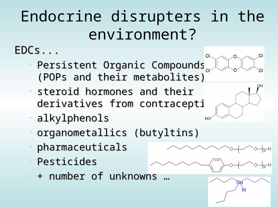
Endocrine disrupters in the environment?
EDCs...EDCs...• Persistent Organic Compounds Persistent Organic Compounds
(POPs and their metabolites)(POPs and their metabolites)• steroid hormones and their steroid hormones and their
derivatives from contraception pillsderivatives from contraception pills• alkylphenolsalkylphenols• organometallics (butyltins)organometallics (butyltins)• pharmaceuticalspharmaceuticals• PesticidesPesticides• + number of unknowns …+ number of unknowns …

ESTROGEN RECEPTOR – ER
the most studied target of EDCs

Estrogens:Estrogens:
• play a key role in female hormone regulation and signalling
• are responsible for metabolic, behavioural and morphologic changes occurring during stages of reproduction
• are involved in the growth, development and homeostasis of a number of tissues
• control the bone formation, regulation of homeostasis, cardiovascular system and behaviour
• regulate production, transport and concentration of testicular liquid and anabolic activity of androgens in males
•Synthesis in ovaries
• DISRUPTION -> investigated in aquatic biota & laboratory organisms (see notes on EDCs)
HO
OH
HO
OH
OH
17--estradiol estriol
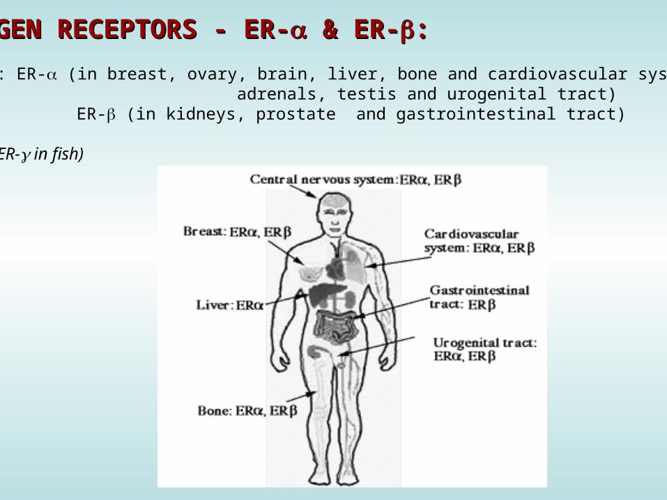
ESTROGEN RECEPTORS ESTROGEN RECEPTORS - ER-- ER- && ER- ER-::
subtype: ER- (in breast, ovary, brain, liver, bone and cardiovascular system, adrenals, testis and urogenital tract) ER- (in kidneys, prostate and gastrointestinal tract)
(ER- in fish)

Natural productsgenisteinnaringenincoumestrolzearalenone
Environmental pollutantDDTkeponePCBs/OH-PCBsPAHs and dioxins
Industrial chemicalsBisphenol ANonionic surfactantsPthalate estersendosulfan
PharmaceuticalsEthinyl estradiolDiethylstilbestrolgestodenenorgestrel
DEHP
Environmental estrogens (xenoestrogens, exoestrogens)Environmental estrogens (xenoestrogens, exoestrogens)
a diverse group of substances that do not necessarily share structural similarity to the prototypical estrogen (17-estradiol)May act as AGONISTS and/or ANTAGONISTS

Chemical group Substance REP
Estradiol 1
Estriol 6,3.10-3Endogenous hormones
Testosteron 9,6.10-6
Cuomestrol 6,8.10-3
PhytoestrogensGenistein 4,9.10-4
Pesticides o,p´-DDT 1,1.10-6
2,4,6-trichlorbiphenyl-4´-ol 1.10-2
2,5-dichlorobiphenyl-4´-ol 6,2.10-3PCBs
3,3´,5,5´tetrachlorobiphenyl-4,4´-diol 1,6.10-4
alkylphenoles 4-tert-oktylphenol 3,6.10-6
phthalates butylbenzylphthalate 4.10-6
REP (RElative Potencies) of selected compounds related to 17--estradiol derived from reporteryeast assay
Exoestrogens - Relative Potencies to bind to ERExoestrogens - Relative Potencies to bind to ER (REPs) (REPs)
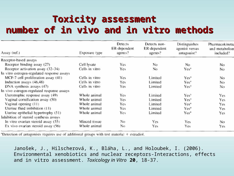
Toxicity assessment Toxicity assessment number of number of in vivo and in vitro methodsin vivo and in vitro methods
Janošek, J., Hilscherová, K., Bláha, L., and Holoubek, I. (2006). Environmental xenobiotics and nuclear receptors-Interactions, effects and in vitro assessment. Toxicology in Vitro 20, 18-37.

In vitro assaysIn vitro assays
•INTERACTION (BINDING) to the receptor• competitive ligand binding assay•Effect unknown (? Activation / suppression / no effect ?)
•Testing the effect at cellular level (interference with receptor biological activity)
• cell proliferation assay• endogenous protein expression (or enzyme activity) assay• reporter gene assay

mRNA
Estrogenor xenoestrogen
Light
DNA Binding
Luciferase
ER
ER-Responsive Genes
ERE-Luc
ER
Nuclear Factors
+
ER
P
Protein Phosphorylation of ER:Ligand-Independent Activation
ER ER
P
P
“Estrogenic Effects”
mRNA
Estrogenor xenoestrogen
Light
DNA Binding
Luciferase
ER
ER-Responsive Genes
ERE-Luc
ER
Nuclear Factors
+
ER
P
Protein Phosphorylation of ER:Ligand-Independent Activation
ER ER
PP
P
“Estrogenic Effects”
In vitro ER- mediated effects
luciferase reporter assay

ER- mediated effectsluciferase reporter assay
96 microwell platecultivation of transgenic cell lines
ER: breast carcinoma MVLN cells
SIMILAR DESIGN FOROTHER RECEPTORS(discussed below):AhR (H4IIE.luc cells) AR (MDA cells)RAR/RXR (P19 cells)
Cell lysis-> extraction of induced luciferase
Exposure (6 – 24 h)standards / samples
LuminoLuminescence determination(microplate luminescence reader)

In vivo assaysIn vivo assays
• uterotropic assay
• vaginal cornification assay
• standard test procedures for reproductive and developmental toxicity (e.g. FETAX)
• production of estrogen-inducible proteins (e.g. vittelogenin and zona radiata protein)

Kidd, K.A. et al. 2007. Collapse of a fish population following exposure to a synthetic estrogen. Proceedings of the National Academy of Sciences 104(21):8897-8901
Controls +Ethinylestradiol
5 ng/L (!)7 years
HO
OH

ANDROGEN RECEPTOR (AR)
effects known but less explored than ER

Androgens
- Role in males similar to the of estrogens in
females
- development of male sexual characteristics
- stimulating protein synthesis, growth of bones
- cell differenciation, spermatogenesis
- male type of behaviour

Androgens- Endogenous ligands – androgen hormones
-testosterone (T)
-dihydrotestosterone (DHT)
-androstanediol
-dehydroepiandrosterone
-androstenedione
T: synthesis in testis (Leydig cells)
in lesser extent in adrenals
DHT: Formed extratesticulary from T
-In several tissues (seminal vesicles, prostate, skin)
higher affinity to androgen receptor than T
-Daily production 5-10% of testosterone
OH
O
Testosterone
OH
ODihydrotestosterone

Mechanisms of androgen signalling disruption
1) Binding to AR
- Mostly competitive inhibition
- xenobiotics mostly DO NOT activate AR-dependent transcription
-Only few compounds are able to activate AR in the absence of androgen hormones, and these are also anti-androgenic in the presence of T/DHT (metabolites of fungicide
vinclozoline, some PAHs)
2) FSH/LH (gonadotropins) signalling disruption – less explored- FSH/LH expression - regulation via negative feedback by testosterone
- Suppression leads to alterations of spermatogenesis

Mechanisms of androgen signalling disruption
3) Alterations of testosterone synthesis
- Inhibition of P450scc needed for side chain cleavage of cholesterol (fungicide ketoconazol)
- Inhibition of 17--hydroxylase and other CYPs - – enzymes needed for testosterone synthesis (ketoconazol)
4) Testosterone metabolic clearance
- Induction of UDP-glucuronosyltransferase or monooxygenases CYP1A, 1B involved in androgen catabolism
- Pesticides endosulfan, mirex, o-p´-DDT

Effects of male exposure to antiandrogens
Exposure during prenatal development:- malformations of the reproductive tract
- reduced anogenital distance
- hypospadias (abnormal position of the urethral opening on the penis)
- vagina development
- undescendent ectopic testes
- atrophy of seminal vesicles and prostate gland
Exposure in prepubertal age:-delayed puberty
- reduced seminal vesicles
- reduced prostate
Exposure in adult age:- oligospermia -azoospermia -libido diminution

Antiandrogenic compound
tris-(4-chlorophenyl)-methanol- Ubiquitous contaminant of uncertain origin- Probable metabolite of DDT-mixture contaminant- Levels in human blood serum cca. 50nM- EC50 – cca. 200nM

AR-binding - potencies(Ref: DHT EC50 ~ 0.1 uM)
Compound IC50 (µM)Benz[a]anthracene 3.2
Benzo[a]pyrene 3.9
Dimethylbenz[a]anthracene 10.4
Chrysene 10.3
Dibenzo[a,h]anthracene activation in range 0.1-10µM
Bisphenol A 5
vinclozolin metabolites 9.7
hydroxyflutamide 5
Aroclor typical values 0.25-1.11
Individual PCBs typical values 64 - 87
tris-(4-chlorophenyl)-methanol 0.2

(Anti)androgenicity assessmentIn vivo Hershberger assay
- castrated rats treated with examined substance- Endpoint – after 4-7 days – seminal vesicles and ventral prostate weight
In vivo measurement of testosterone blood levels
In vitro cell proliferation assays – cell lines with androgen-dependent growth
- mammary carcinoma cell lines- prostatic carcinoma cell lines
¨Receptor-reporter assaysGene for luciferase (or GFP) under control of AR
AR-CALUX (human breast carcinoma T47D)PALM (human prostatic carcinoma PC-3)CHO515 (Chinese hamster ovary CHO)
Yeast transfected cellsbeta-galactosidase reporter
Treatment:tested chemical only
-> androgenicity Cotreatment with DHT
-> antiandrogenicity

Thyroid hormones

Thyroid hormones
Regulation of metabolism- increasing oxygen consumption
- modulating levels of other hormones (insulin, glucagon, somatotropin, adrenalin)
- important in cell differenciation
- crucial role in development of CNS, gonads and bones
Play crucial roles in stimulating metabolism, development and maturation
HYPOTHYROIDISM HYPERTHYROIDISM

Thyroid hormones
O OH
I
I
II
OH
O
NH2
O OH
I
I
II
OH
O
NH2
Thyroxine (T4)
O OH
I
I
IOH
O
NH2
3,5,3’-Triiodothyronine (T3)
O OH
I
I
IOH
O
NH2
O OH
I
I
IOH
O
NH2
3,5,3’-Triiodothyronine (T3)
- T4 – prohormone- 5´-deiodination leads to active form, T3
Thyroxine (T4)
Also called tetraiodothyronineContains 4 iodide ions
Triiodothyronine (T3)
Contains 3 iodide ions-Most T3 produced by deiodination in target tissues (deiodinases)

Enzymes involved in thyroid
metabolism
- Thyroid peroxidases - iodination of tyrosyl residues- coupling of iodinated tyrosyl residues
- Thyroid deiodinases- D1, D2 - activation of T4 into T3 via deiodination on „outer“ ring- D3 - deactivation into rT3 via deiodination on „inner“ ring
EDCs -> may affect metabolism of these key enzymes
O OH
I
I
II
OH
O
NH2
O OH
I
I
II
OH
O
NH2
„outer“
„inner“

Thyroid hormones are transported in the blood by thyroid binding proteins
- Regulating free T4 and T3 levels in blood- 3 types :
-Thyroid-binding prealbunin (transthyretin) (20-25%)-Albumin (5-10%)-Thyroid binding globulin (75%)
- NUMBER OF ENVIRONMENTAL TOXICANTS act at transport proteins-OH-PCBs, brominated and chlorinated flame retardants, DDT, dieldrin-OH-PCBs – equal affinity to TBP as T4 and T3 (!!!)
- More of free T4 in blood negative feedback to TSH release
=> increased depletion => increased weight, histological changes in thyroid gland
Observed after exposure to POPs in mammals, birds, fish

Competitive binding to TR
- Probably less important than binding to TBP
- Chemicals that affect thyroid signalling in vivo mostly don´t bind to TR (DDT, PCBs) or bind with much lesser affinity than T3 (OH-PCBs – 10000x)
Accelerated depletion of THUDP-glucuronosyltransferase – detoxication enzyme (II.biotransformation phase)
Induced by PCBs, dioxins Key enzyme in thyroid catabolism
Increased by disruption of TBP binding
Other possible effects of EDCs on Thyroid signalling

Effects of thyroid disruption
Disruption during prenatal development
- severe damage of CNS (cretenism, delayed eye opening, cognition)
- Megalotestis
- Histological changes in thyroid gland (goitre)
- nervous system fails to develop normally
- mental retardation
- skeletal development

Assessment of effects- In vivo approaches- TH serum levels – simple, nondestructive x variation within time of day, age,
sensitive to other than biochemical stresses- Thyroid gland weight and folicular cells number- Developmental toxicity assays - delayed eye opening, abnormalities in brain
development and cognition, increased testis weight and sperm counts- Perchlorate discharge test (TH synthesis)- Hepatic UDP-glucuronosyltransferase activity (marker of enhanced TH clearance
from serum)
- In vitro - Enzyme inhibition assays (thyroid peroxidase, deiodinases) – assessment of
thyroid metabolism- Competitive binding assays with TBP- TH- dependent proliferation assay (pituitary tumor GH3, thyroid tumors like
FRTL-5 cell line) or TSH-dependent proliferation assay (thyroid tumors)- Receptor-reporter gene assays with luciferase (monkey kidney CV-1, chinese
hamster ovary CHO or insect Sf9 cell lines)

Retinoids Vitamin A and its derivatives
Toxicants affect retinoid action but effects are much less explored

Suppressive effects in cancer development
Retinoids
Necessary for vision
Important for cell growth, apoptosis and differenciation
Development of embryonic, epithelial cells (gastrointestinal tract, skin, bones)
Antioxidative agent
Affect nervous and immune function
Regulation of development and homeostasis in tissues of vertebrates and invertebrates

Retinoids
Retinol (vitamin A)
OH
CH3CH3CH3
CH3
CH3
OH
CH3CH3CH3
CH3
CH3
O
CH3CH3CH3
CH3
CH3 CH3 CH3 CH3
CH3
CH3
Bond cleavage
Retinoic Acid
-karoten
Sources: from diet (dietary hormones)Retinyl esters – animal sourcesPlant carotenoids

RE: Retinol-Ester
R: Retinol
RBP: Retinol BindingProtein (LMW)
TTR: Transthyrethin (HMW)
TRANSPORT OF RETINOIDS

RAL - Retinal
CRBP – cellular retinol binding protein- binding of retinol, immediate decrease of retinol concentration
CRBAP – cellular retinoic acid binding protein- Controlling ratio free retinol/free retinoic acid
Retinoid binding proteins

Mode of action - Isoforms of RAR a RXR
- Both have isoforms andeach of them several subtypes
- Formation of homo- and heterodimers
- 48 possible RAR-RXR heterodimers =>sensitive regulation of gene expression
- RXR – heterodimers even with other receptors like VDR, TR, PPAR
- 3 basic subtypes
- all-trans-, 9-cis- and 13-cis-retinoic acid
- All-trans RA binds selectively to RAR
- Cis RA bind to both receptor types
Retinoic acid
R A R
R X R
R A R R X R
RARE RXRE
R etino idy
Gene expression

Disruption of retinoid signalling by xenobiotics
- Relatively little known
- Possible modes of action:- Metabolization of retinoids by detoxication enzymes- Disruption of binding retinoids to retinoid binding proteins- Retinoids as antioxidants may be consumed cause of oxidative stress caused by xenobiotics- Interference of chemicals (binding to RAR/RXR)
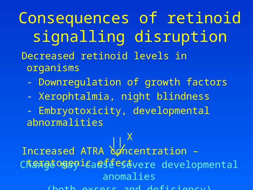
Consequences of retinoid signalling disruption
Decreased retinoid levels in organisms
- Downregulation of growth factors
- Xerophtalmia, night blindness
- Embryotoxicity, developmental abnormalities
X
Increased ATRA concentration – teratogenic effect
Change may cause severe developmental anomalies(both excess and deficiency)

Disruption of retinoid signalling by xenobiotics
Polluted areas – mostly decrease of retinoid levels in aquatic birds, mammals and fish
Disruption of retinoid transport: PCBs
Effects on retinoid receptors:-RAR, RXR binding and/or transactivation – pesticides (chlordane,
dieldrin, methoprene, tributyltin…)
-Effect on ATRA mediated response – TCDD, PAHs
Disruption of retinoid metabolism:
– PCDD/Fs, PAHs, PCBs, pesticides
- changes of serum concentrations of retinol and RA
- mobilization of hepatic storage forms
- in kidney, concentration of all forms elevated
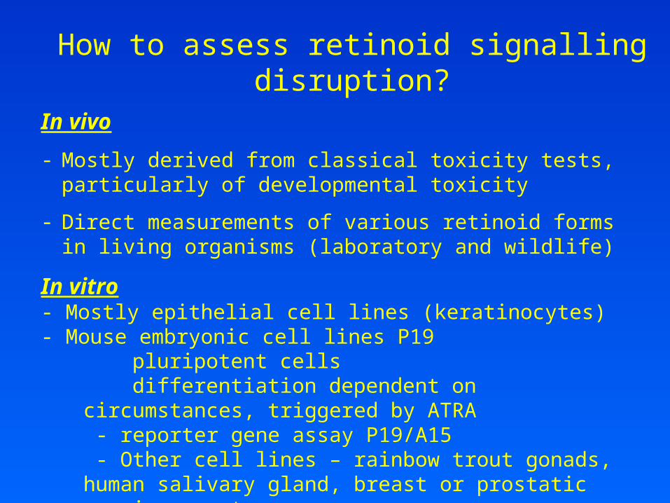
How to assess retinoid signalling disruption?
In vivo
- Mostly derived from classical toxicity tests, particularly of developmental toxicity
- Direct measurements of various retinoid forms in living organisms (laboratory and wildlife)
In vitro- Mostly epithelial cell lines (keratinocytes)- Mouse embryonic cell lines P19
pluripotent cells differentiation dependent on circumstances, triggered by ATRA - reporter gene assay P19/A15 - Other cell lines – rainbow trout gonads, human salivary gland, breast or prostatic carcinomas etc.
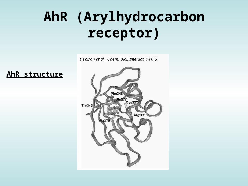
AhR (Arylhydrocarbon receptor)
Denison et al., Chem. Biol. Interact. 141: 3
AhR structure

AhR
• ligand-activated transcription factor
• activation of different responsive elements (genes)
• important mediator of toxicity of POPs – primary target of
coplanar aromatic substances
• regulator of xenobiotic metabolism and activation of
promutagens
• crossactivation/crosstalk with other receptors
• strongest known ligand TCDD

AhR activation:AhR activation:

AhR regulated genes:AhR regulated genes:
contain xenobiotic response elements (XRE) or dioxin responsive
elements (DRE) in their promoter region:
• phase I enzymes - CYP 1A1, CYP 1A2, CYP 1B1;
• phase II enzymes - UDP-glucuronosyltransferase, GST-Ya,
NADP(H):oxidoreductase;
• other genes - Bax, p27Kip1, Jun B, TGF- - regulation of cell cycle
and apoptosis;

6-formylindolo[3,2-b]carbazole (FICZ)potent endogenous physiological (natural) ligand of AhR

Denison & Nagy, Annu. Rev. Pharmacol. Toxicol. 43:309

„„Non-classical“ AhR ligandsNon-classical“ AhR ligands

Physiological role for AhR Physiological role for AhR not known not known (?)(?)
→ Effects in AhR-deficient mice:Effects in AhR-deficient mice:
• significant growth retardation;
• defective development of liver and immune system;
• retinoid accumulation in liver;
• abnormal kidney and hepatic vascular structures.
• resistant to BaP-induced carcinogenesis and TCDD-induced
teratogenesis;
• no inducible expression of CYP 1A1 and 2.

of
S c h m i d t & B r a d f i e l d , A n n u . R e v . C e l l D e v . B i o l . 1 2 : 5 5
of
S c h m i d t & B r a d f i e l d , A n n u . R e v . C e l l D e v . B i o l . 1 2 : 5 5
Schmidt & Bradfield, Annu. Rev. Cell Dev. Biol. 12:55
Biological responses & effects of TCDD (mostly related to AhR activation)

Compounds having similar toxicological properties as TCDD (strongest AhR ligand) may be evaluated by TEF/TEQ concept
TEF = Toxic Equivalency Factor (characteristic of the Chemical)TEQ = Toxic Equivalent (sum of TEFs x concentrations)
TEFs are consensus values based on REPs (relative potencies) across multiple species and/or endpoints. TEFs are based upon a number of endpoints, from chronic in vivo toxicity to in vitro toxicity with the former having the greatest importance in determining overall TEF.
TEQs provide a simple, single number that is indicative of overall toxicity of a sample containing a mixture of dioxins and dioxin-like compounds.
The total potency of a mixture can be expressed in TCDD TEQ concentration:
Toxic equivalency factors (TEF)/TEQ concept:Toxic equivalency factors (TEF)/TEQ concept:

Toxic equivalency factors for PCDDs, PCDFs Toxic equivalency factors for PCDDs, PCDFs and PCBs:and PCBs:
Eljarrat & Barceló, Trends Anal. Chem.22: 655
Final concentration is expressed as „Equivalents of TCDD“ (e.g. ng TEQ / kg = ng TCDD / kg)

Biomarkers/bioanalytical methods for AhR toxicityBiomarkers/bioanalytical methods for AhR toxicity
• in vivo: liver enlargement, reduction of thymus weight, wasting
syndrome, reproductive and developmental disorders
• in vivo biomarkers: EROD activity, CYP 1A1 and 1B1 expression
• in vitro:
EROD (ethoxyresorufin-O-deethylase activity)
in H4IIE rat hepatoma cells;
CALUX/CAFLUX assays;
GRAB assay (AhR-DNA binding)
yeast bioassay;
immunoassays;
detection of CYP1A mRNA or protein

AhR-mediated effectsluciferase reporter assay - H4IIE.luc cells
LightLight Luciferase
Nuclear Factors
1
P
+ AhRHSP90
HSP90
Src
HSP90
HSP90
Adapted from Blankenship (1994)
DRE -Luc
AhRARNT
Modulation of Gene Expression
ARNT
Src
“Activated”
2
P
PCytosolicProteins
MembraneProteins
Increased Protein Phosphorylation
Ligand (TCDD)
LightLight Luciferase
Nuclear Factors
1
P
+ AhRHSP90
HSP90
SrcSrc
HSP90
HSP90
Adapted from Blankenship (1994)
DRE -Luc
AhRARNT
Modulation of Gene Expression
ARNT
Src
“Activated”
2
P
PCytosolicProteins
MembraneProteins
Increased Protein Phosphorylation
Estrogens[AhR <->
ER]
In vitro CALUX/CAFLUX assaysIn vitro CALUX/CAFLUX assays

Detection of EROD activity:Detection of EROD activity:
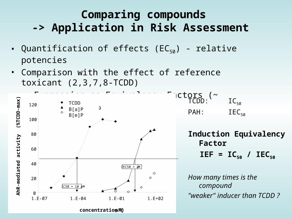
Comparing compounds-> Application in Risk Assessment
• Quantification of effects (EC50) - relative potencies
• Comparison with the effect of reference toxicant (2,3,7,8-TCDD)
• Expression as Equivalency Factors (~ TEFs)
0
20
40
60
80
100
120
1.E-07 1.E-04 1.E-01 1.E+02
TCDD4´-OH-PCB 794´-OH-PCB 3
Ah
R-m
ed
iate
d a
cti
vit
y
(%T
CD
D-m
ax
)
concentration (M)
IC50 = 10 pM
IEC50 = 2 M
B[a]PB[e]P
TCDD: IC50
PAH: IEC50
Induction Equivalency Factor
IEF = IC50 / IEC50
How many times is the compound
"weaker" inducer than TCDD ?


Crosstalk in signalling of nuclear receptors

Nuclear Receptors & Signalling Crosstalkpoorly characterized (toxicity) mechanisms
Nuclear receptors (AhR, ER, RAR/RXR ...) = Transcription factors with numerous cofactors and interactions (crosstalk)
In vitro assays for nongenotoxic effects
AhR
ER
hsp90hsp90
P
RAR
AhRhsp90hsp90
P
ERP
RARH3C CH3
CH3 CH3
COOH
CH3
P
HO
OH
H3C CH3CH3 CH3
COOH
CH3
?
AhRER
ERRAR
AhRRAR

Cross-talk between estrogen signalling pathways Cross-talk between estrogen signalling pathways and other receptorsand other receptors
• estrogen signalling pathways and other members of nuclear receptor superfamily
• estrogen signalling pathways and AhR
• estrogen signalling pathways and receptors for EGF and insuline
=> Many effects observed in vivo (higher cancer incidence, allergies …)
without known mechanisms … ? complex toxicity / crosstalk ?