Embed Size (px)
Citation preview
INTRACEREBRAL HEMORRHAGE (ICH) Vas20 (1)
Intracerebral Hemorrhage (ICH) Last updated: September 5, 2017
ETIOLOGY ................................................................................................................................................ 1 Etiology according to patient’s age .................................................................................................. 3
PRECIPITATING CONDITIONS .................................................................................................................. 3
RISK FACTORS ........................................................................................................................................ 3
PATHOLOGY, PATHOPHYSIOLOGY .......................................................................................................... 3
EPIDEMIOLOGY ........................................................................................................................................ 4 CLINICAL FEATURES ............................................................................................................................... 4
DIAGNOSIS................................................................................................................................................ 5 Blood ................................................................................................................................................ 5
EEG .................................................................................................................................................. 5
IMAGING ................................................................................................................................................ 5 Noncontrast CT ................................................................................................................................ 5
MRI .................................................................................................................................................. 6 VASCULAR IMAGING (ANGIOGRAPHY / CTA / MRA)............................................................................. 6
SPECIFIC ANATOMIC LOCATIONS ............................................................................................................ 7 Putaminal Hemorrhage ..................................................................................................................... 7 Thalamic Hemorrhage ...................................................................................................................... 7
Lobar Hemorrhage ........................................................................................................................... 7 Pontine Hemorrhage ......................................................................................................................... 8
Cerebellar Hemorrhage .................................................................................................................... 8
Caudate Hemorrhage ........................................................................................................................ 8 INTRAVENTRICULAR HEMORRHAGE....................................................................................................... 8
TREATMENT ............................................................................................................................................. 8 CONSERVATIVE MEASURES ................................................................................................................... 8
General measures ............................................................................................................................. 8
BP control ......................................................................................................................................... 8 ICP control ....................................................................................................................................... 9
Reversal of bleeding diathesis .......................................................................................................... 9 AED .................................................................................................................................................. 9
Neuroprotective strategies ................................................................................................................ 9
SURGICAL TREATMENT ........................................................................................................................ 10
PROGNOSIS ............................................................................................................................................. 11 ICH ...................................................................................................................................................... 11 IVH ..................................................................................................................................................... 11
SPECIAL SITUATIONS ............................................................................................................................. 11 LVAD (LEFT VENTRICULAR ASSIST DEVICE) ........................................................................................ 11
TRAUMATIC ICH → see p. TrH1 >>
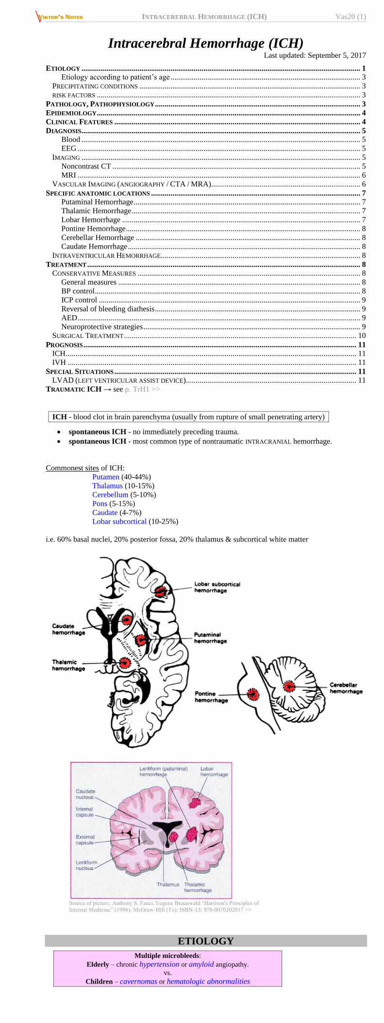
ICH - blood clot in brain parenchyma (usually from rupture of small penetrating artery)
spontaneous ICH - no immediately preceding trauma.
spontaneous ICH - most common type of nontraumatic INTRACRANIAL hemorrhage.
Commonest sites of ICH:
Putamen (40-44%)
Thalamus (10-15%)
Cerebellum (5-10%)
Pons (5-15%)
Caudate (4-7%)
Lobar subcortical (10-25%)
i.e. 60% basal nuclei, 20% posterior fossa, 20% thalamus & subcortical white matter
Source of picture: Anthony S. Fauci, Eugene Braunwald “Harrison's Principles of
Internal Medicine” (1998); McGraw-Hill (Tx); ISBN-13: 978-0070202917 >>
ETIOLOGY
Multiple microbleeds:
Elderly – chronic hypertension or amyloid angiopathy.
vs.
Children – cavernomas or hematologic abnormalities
INTRACEREBRAL HEMORRHAGE (ICH) Vas20 (2)
Any age - acute disseminated encephalomyelitis (s. acute hemorrhagic
leukoencephalopathy, Weston-Hurst disease).
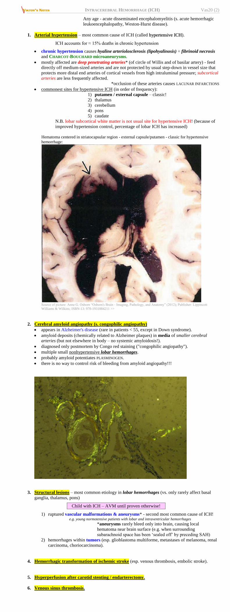
1. Arterial hypertension – most common cause of ICH (called hypertensive ICH).
ICH accounts for ≈ 15% deaths in chronic hypertension
chronic hypertension causes hyaline arteriolosclerosis (lipohyalinosis) ÷ fibrinoid necrosis
and CHARCOT-BOUCHARD microaneurysms.
mostly affected are deep penetrating arteries* (of circle of Willis and of basilar artery) - feed
directly off medium-sized arteries and are not protected by usual step-down in vessel size that
protects more distal end arteries of cortical vessels from high intraluminal pressure; subcortical
arteries are less frequently affected.
*occlusion of these arteries causes LACUNAR INFARCTIONS
commonest sites for hypertensive ICH (in order of frequency):
1) putamen / external capsule – classic!
2) thalamus
3) cerebellum
4) pons
5) caudate
N.B. lobar subcortical white matter is not usual site for hypertensive ICH! (because of
improved hypertension control, percentage of lobar ICH has increased)
Hematoma centered in striatocapsular region - external capsule/putamen - classic for hypertensive
hemorrhage:
Source of picture: Anne G. Osborn “Osborn's Brain ‐ Imaging, Pathology, and Anatomy” (2012); Publisher: Lippincott
Williams & Wilkins; ISBN-13: 978-1931884211 >>
2. Cerebral amyloid angiopathy (s. congophilic angiopathy)
appears in Alzheimer's disease (rare in patients < 55, except in Down syndrome).
amyloid deposits (chemically related to Alzheimer plaques) in media of smaller cerebral
arteries (but not elsewhere in body – no systemic amyloidosis!).
diagnosed only postmortem by Congo red staining ("congophilic angiopathy").
multiple small nonhypertensive lobar hemorrhages.
probably amyloid potentiates PLASMINOGEN.
there is no way to control risk of bleeding from amyloid angiopathy!!!
3. Structural lesions – most common etiology in lobar hemorrhages (vs. only rarely affect basal
ganglia, thalamus, pons)
Child with ICH – AVM until proven otherwise!
1) ruptured vascular malformations & aneurysms* - second most common cause of ICH! e.g. young normotensive patients with lobar and intraventricular hemorrhages
*aneurysms rarely bleed only into brain, causing local
hematoma near brain surface (e.g. when surrounding
subarachnoid space has been ‘sealed off’ by preceding SAH)
2) hemorrhages within tumors (esp. glioblastoma multiforme, metastases of melanoma, renal
carcinoma, choriocarcinoma).
4. Hemorrhagic transformation of ischemic stroke (esp. venous thrombosis, embolic stroke).
5. Hyperperfusion after carotid stenting / endarterectomy.
6. Venous sinus thrombosis.
INTRACEREBRAL HEMORRHAGE (ICH) Vas20 (3)
ETIOLOGY ACCORDING TO PATIENT’S AGE
YOUNG PERSONS – vascular disorders (AVM, aneurysm, vasculitis), drug abuse (amphetamines,
cocaine), hematologic abnormalities
ELDERLY PERSONS – hypertension, amyloid angiopathy, tumors, coagulopathies (incl. anticoagulants).
PRECIPITATING conditions
1. Pregnancy (esp. with eclampsia)
eclampsia causes > 40% ICHs in pregnancy.
ICH is common cause of death from eclampsia.
2. Acute BP rises (can cause ICH even in absence of preexisting severe hypertension!), e.g.
sympatheticomimetic drugs (esp. cocaine, amphetamines).
3. Bleeding diatheses (esp. iatrogenic anticoagulation and thrombolysis, liver dysfunction) -
hemorrhages can occur at any site, tend to evolve slowly and be multiple.
4. Trauma (4-23% head injury cases) - multifocal inhomogeneous hemorrhages (most common in
frontal and temporal lobes). see p. TrH1 >>
5. Heavy alcohol consumption (acute or chronic).
6. Drug abuse (amphetamines, cocaine)
RISK FACTORS
1. Age > 70 (increases ICH risk 7x)
2. Male sex
3. Non-Caucasian race
4. Previous CVA (23x)
5. NSAID use – only DDIICCLLOOFFEENNAACC and MMEELLOOXXIICCAAMM (RR 1.27; 95% CI, 1.02– 1.59 and RR 1.27; 95%
CI, 1.08–1.50, respectively).
PATHOLOGY, PATHOPHYSIOLOGY
hematomas are at first soft and dissect along white matter fiber tracts (rather than destroying brain
tissue locally).
hematoma may spread (lobar and cerebellar hemorrhages tend to remain confined within
parenchyma):
a) intraparenchymal extensions
b) intraventricular extension (primary intraventricular hemorrhage is rare!) → acute
hydrocephalus
c) SAH
bleeding is spontaneously limited by resistance of surrounding tissue pressure (usually within 30
minutes);
– once bleeding stops, it generally does not start again.
– in severe cases, bleeding continues until death.
large hematoma causes mass effect → distorts structures (with ischemic pressure damage),
increases ICP → herniation.
if patient survives initial ICP changes, blood is absorbed over weeks ÷ months → cavity or cleft
(lined by glial scar and hemosiderin-containing macrophages) that may disconnect brain pathways.
– less frequently, blood clot is treated as FOREIGN BODY - calcifies and is surrounded by thick
glial membrane.
Putaminal hemorrhage (mass effect with midline shift):
Source of picture: “WebPath - The Internet Pathology Laboratory for Medical Education” (by Edward C. Klatt, MD) >>
Hypertensive basal ganglia hemorrhage:
Source of picture: “WebPath - The Internet Pathology Laboratory for Medical Education” (by Edward C. Klatt, MD) >>
INTRACEREBRAL HEMORRHAGE (ICH) Vas20 (4)
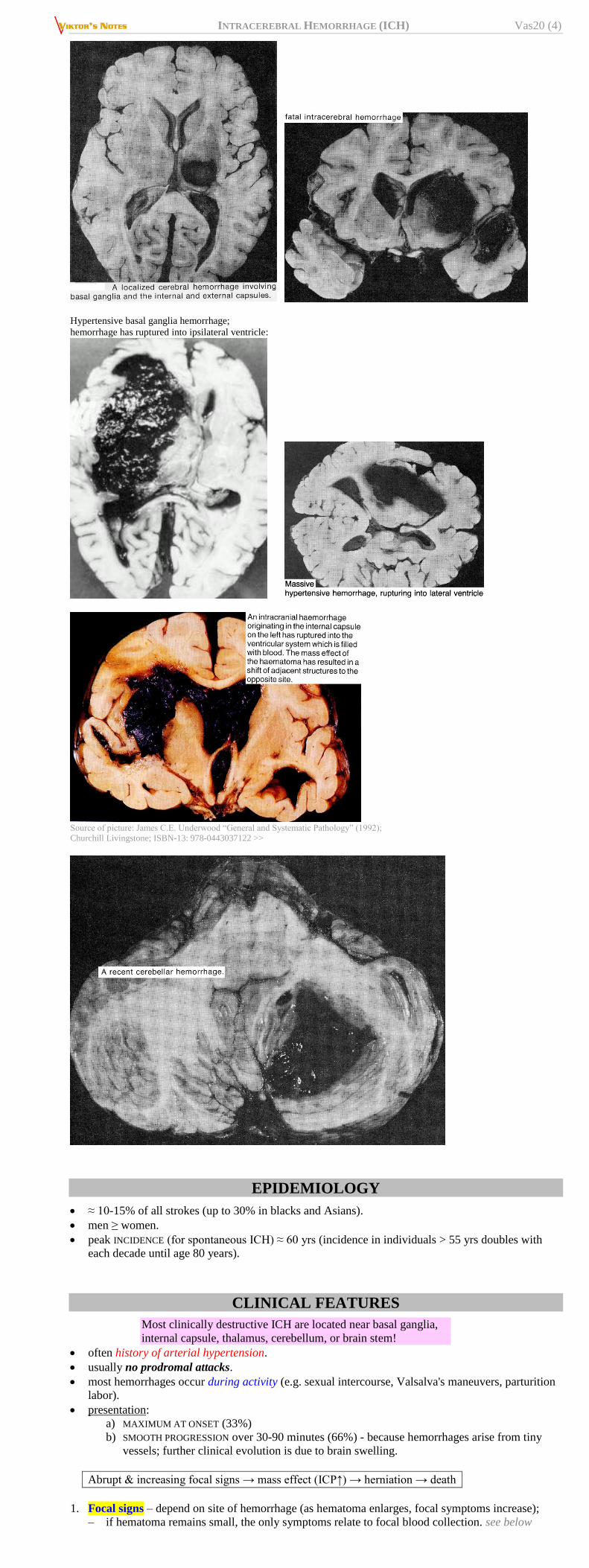
Hypertensive basal ganglia hemorrhage;
hemorrhage has ruptured into ipsilateral ventricle:
Source of picture: James C.E. Underwood “General and Systematic Pathology” (1992);
Churchill Livingstone; ISBN-13: 978-0443037122 >>
EPIDEMIOLOGY
≈ 10-15% of all strokes (up to 30% in blacks and Asians).
men ≥ women.
peak INCIDENCE (for spontaneous ICH) ≈ 60 yrs (incidence in individuals > 55 yrs doubles with
each decade until age 80 years).
CLINICAL FEATURES
Most clinically destructive ICH are located near basal ganglia,
internal capsule, thalamus, cerebellum, or brain stem!
often history of arterial hypertension.
usually no prodromal attacks.
most hemorrhages occur during activity (e.g. sexual intercourse, Valsalva's maneuvers, parturition
labor).
presentation:
a) MAXIMUM AT ONSET (33%)
b) SMOOTH PROGRESSION over 30-90 minutes (66%) - because hemorrhages arise from tiny
vessels; further clinical evolution is due to brain swelling.
Abrupt & increasing focal signs → mass effect (ICP↑) → herniation → death
1. Focal signs – depend on site of hemorrhage (as hematoma enlarges, focal symptoms increase);
– if hematoma remains small, the only symptoms relate to focal blood collection. see below
INTRACEREBRAL HEMORRHAGE (ICH) Vas20 (5)
2. Signs of mass effect (develop after hematoma becomes large enough to raise ICP):
1) headache (40-50%).
2) nausea & vomiting (40-50%).
3) normal ÷ decreased level of consciousness (50%); may progress to coma in 24-48 hrs
(consciousness is sometimes impaired at start – esp. pontine or thalamic hemorrhage).
3. Seizures (6-10%*)
*much more common with lobar hemorrhage (≈ 25% patients) - cortical irritation by blood.
4. Meningeal irritation – if bleeding extends to subarachnoid space.
CLINICAL SITE OF HEMORRHAGE
Putaminal Thalamic Pontine Cerebellar
Unconsciousness Later Later Early Late
Hemiparesis Yes Yes Quadriparesis Late
Sensory change Yes Yes Yes Late
Hemianopia Yes Yes – –
Pupils (Size /
Reaction)
Normal / + Small / ± Very small / + Normal / +
Gaze paresis Contralateral (eyes look to ICH)
Upward (eyes
look to nose tip)
Bilateral
(centrally
positioned eyes)
Ipsilateral (eyes
look away from
ICH)
Response to calorics Yes Yes – ±
Ocular bobbing – – Sometimes Sometimes
Gait lost – – Yes Yes
Vomiting Occasional Occasional Often Severe
Ocular signs are rapid method of localizing hemorrhages!
DIAGNOSIS
Lumbar puncture is contraindicated! – may cause herniation;
CSF does not provide definitive diagnostic information
– CSF is usually bloody several hours after hemorrhage, but sometimes it
is normal initially.
Either CT or MRI may be used for initial neuroimaging (but MRI may be more difficult to perform
because of impaired consciousness, vomiting, or agitation)
BLOOD
CBC, chemistries, coagulation studies (prothrombin time, PTT, bleeding time, platelet count), arterial
blood gas analysis (in patients with reduced alertness), toxicology screen.
EEG
- polymorphic slow waves over region.
IMAGING
NONCONTRAST CT
- very reliable! - accurately documents hematoma, mass effect, intraventricular hemorrhage,
hydrocephalus.
performed immediately in suspected acute ICH!
follow-up CT is frequently requested (changes in lesion size, ventricular system).
1/3 of patients have ICH size growth on repeat imaging!
fresh hematoma - homogeneous rounded area of increased density (≈ 100 HU) + mass effect (vs.
hemorrhagic infarctions - areas of increased density [blood] interspersed with areas of decreased
density [infarction]).
– acute hematoma volume ≥ 80 cm3 is usually fatal.
– no edema around fresh clot (!!!); but clot retraction → fine rim of low density.
– in severely anaemic patients (Hct ≤ 20%), hematomas can be isointense to surrounding brain.
– multifocal hemorrhages at poles (frontal, temporal, or occipital) suggest TRAUMATIC etiology.
– TUMORS can acquire similar density in contrast CT!
CT is always performed without contrast medium if hemorrhage is possible!
layering in clot (as if fluid-blood layer) or mixed iso-hyperdense picture:
a) hyperacute / ongoing bleeding
b) coagulopathic patient
blood may leak into ventricles:
a) adheres to ependyma or choroid plexus;
b) sinks to most dependent part of ventricular system (usually occipital horns) → fluid
level within ventricular fluid.
after several days, hematoma becomes less radiodense, from periphery towards centre (therefore
appears smaller); vasogenic edema develops in surrounding white matter (IV contrast → ring
enhancement*).
*vs. gyral enhancement typical of infarction
after 2 weeks, CT density becomes similar to that of brain or CSF (i.e. isointense); surrounding rim
of contrast enhancement may persist for months.
in chronic stage, lesion becomes hypodense slit-like cavity (many disappear into isodense tissue) -
resembles infarct; H: MRI.
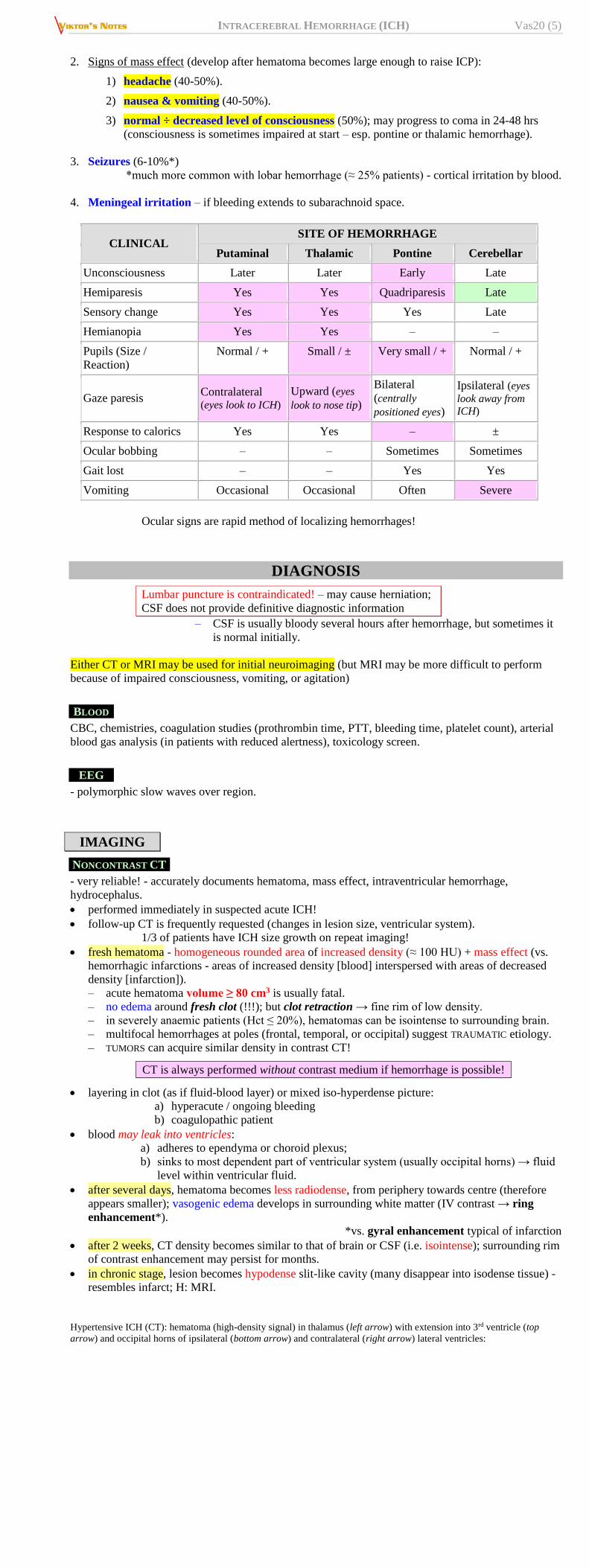
Hypertensive ICH (CT): hematoma (high-density signal) in thalamus (left arrow) with extension into 3rd ventricle (top
arrow) and occipital horns of ipsilateral (bottom arrow) and contralateral (right arrow) lateral ventricles:
INTRACEREBRAL HEMORRHAGE (ICH) Vas20 (6)
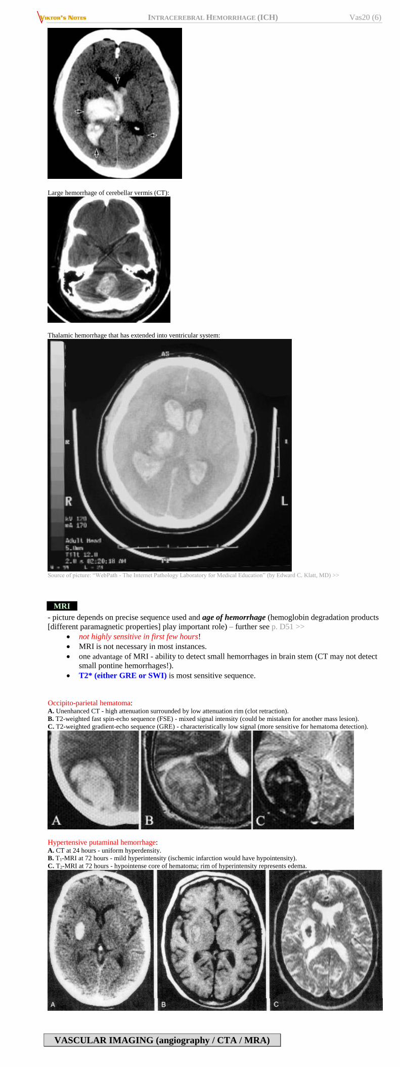
Large hemorrhage of cerebellar vermis (CT):
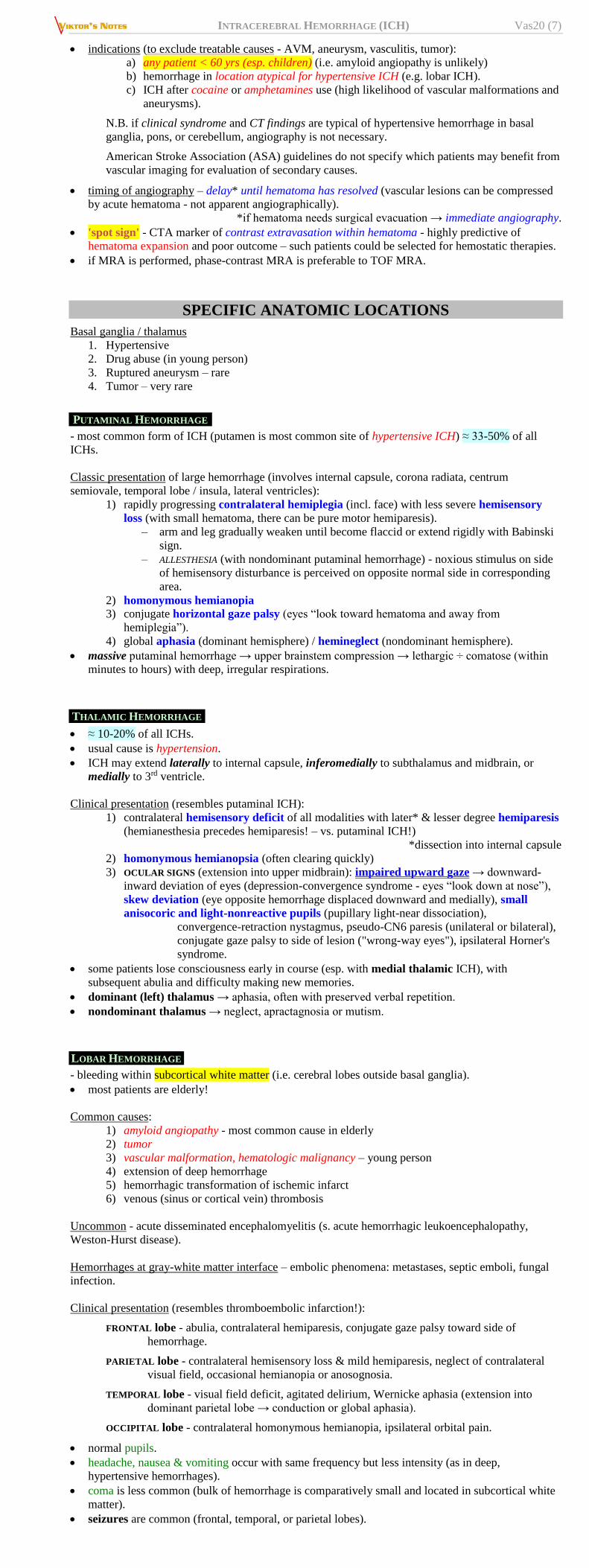
Thalamic hemorrhage that has extended into ventricular system:
Source of picture: “WebPath - The Internet Pathology Laboratory for Medical Education” (by Edward C. Klatt, MD) >>
MRI
- picture depends on precise sequence used and age of hemorrhage (hemoglobin degradation products
[different paramagnetic properties] play important role) – further see p. D51 >>
not highly sensitive in first few hours!
MRI is not necessary in most instances.
one advantage of MRI - ability to detect small hemorrhages in brain stem (CT may not detect
small pontine hemorrhages!).
T2* (either GRE or SWI) is most sensitive sequence.
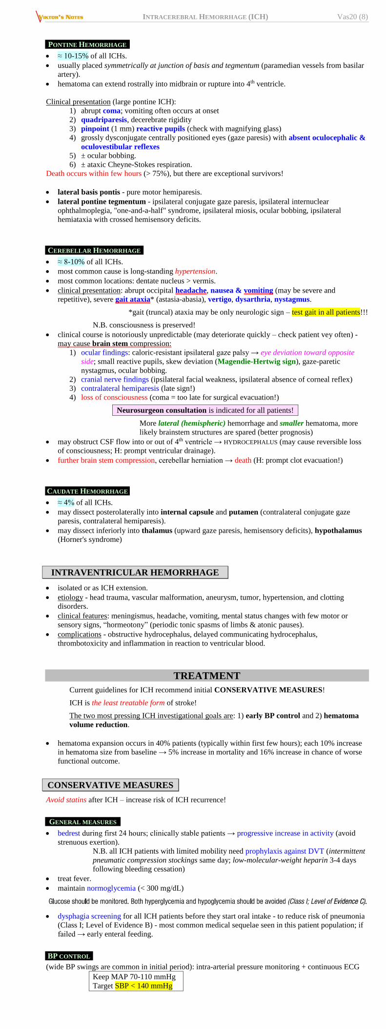
Occipito-parietal hematoma: A. Unenhanced CT - high attenuation surrounded by low attenuation rim (clot retraction).
B. T2-weighted fast spin-echo sequence (FSE) - mixed signal intensity (could be mistaken for another mass lesion).
C. T2-weighted gradient-echo sequence (GRE) - characteristically low signal (more sensitive for hematoma detection).
Hypertensive putaminal hemorrhage: A. CT at 24 hours - uniform hyperdensity.
B. T1-MRI at 72 hours - mild hyperintensity (ischemic infarction would have hypointensity).
C. T2-MRI at 72 hours - hypointense core of hematoma; rim of hyperintensity represents edema.
VASCULAR IMAGING (angiography / CTA / MRA)
INTRACEREBRAL HEMORRHAGE (ICH) Vas20 (7)
indications (to exclude treatable causes - AVM, aneurysm, vasculitis, tumor):
a) any patient < 60 yrs (esp. children) (i.e. amyloid angiopathy is unlikely)
b) hemorrhage in location atypical for hypertensive ICH (e.g. lobar ICH).
c) ICH after cocaine or amphetamines use (high likelihood of vascular malformations and
aneurysms).
N.B. if clinical syndrome and CT findings are typical of hypertensive hemorrhage in basal
ganglia, pons, or cerebellum, angiography is not necessary.
American Stroke Association (ASA) guidelines do not specify which patients may benefit from
vascular imaging for evaluation of secondary causes.
timing of angiography – delay* until hematoma has resolved (vascular lesions can be compressed
by acute hematoma - not apparent angiographically).
*if hematoma needs surgical evacuation → immediate angiography.
'spot sign' - CTA marker of contrast extravasation within hematoma - highly predictive of
hematoma expansion and poor outcome – such patients could be selected for hemostatic therapies.
if MRA is performed, phase-contrast MRA is preferable to TOF MRA.
SPECIFIC ANATOMIC LOCATIONS
Basal ganglia / thalamus
1. Hypertensive
2. Drug abuse (in young person)
3. Ruptured aneurysm – rare
4. Tumor – very rare
PUTAMINAL HEMORRHAGE
- most common form of ICH (putamen is most common site of hypertensive ICH) ≈ 33-50% of all
ICHs.
Classic presentation of large hemorrhage (involves internal capsule, corona radiata, centrum
semiovale, temporal lobe / insula, lateral ventricles):
1) rapidly progressing contralateral hemiplegia (incl. face) with less severe hemisensory
loss (with small hematoma, there can be pure motor hemiparesis).
– arm and leg gradually weaken until become flaccid or extend rigidly with Babinski
sign.
– ALLESTHESIA (with nondominant putaminal hemorrhage) - noxious stimulus on side
of hemisensory disturbance is perceived on opposite normal side in corresponding
area.
2) homonymous hemianopia 3) conjugate horizontal gaze palsy (eyes “look toward hematoma and away from
hemiplegia”).
4) global aphasia (dominant hemisphere) / hemineglect (nondominant hemisphere).
massive putaminal hemorrhage → upper brainstem compression → lethargic ÷ comatose (within
minutes to hours) with deep, irregular respirations.
THALAMIC HEMORRHAGE
≈ 10-20% of all ICHs.
usual cause is hypertension.
ICH may extend laterally to internal capsule, inferomedially to subthalamus and midbrain, or
medially to 3rd ventricle.
Clinical presentation (resembles putaminal ICH):
1) contralateral hemisensory deficit of all modalities with later* & lesser degree hemiparesis
(hemianesthesia precedes hemiparesis! – vs. putaminal ICH!)
*dissection into internal capsule
2) homonymous hemianopsia (often clearing quickly)
3) OCULAR SIGNS (extension into upper midbrain): impaired upward gaze → downward-
inward deviation of eyes (depression-convergence syndrome - eyes “look down at nose”),
skew deviation (eye opposite hemorrhage displaced downward and medially), small
anisocoric and light-nonreactive pupils (pupillary light-near dissociation),
convergence-retraction nystagmus, pseudo-CN6 paresis (unilateral or bilateral),
conjugate gaze palsy to side of lesion ("wrong-way eyes"), ipsilateral Horner's
syndrome.
some patients lose consciousness early in course (esp. with medial thalamic ICH), with
subsequent abulia and difficulty making new memories.
dominant (left) thalamus → aphasia, often with preserved verbal repetition.
nondominant thalamus → neglect, apractagnosia or mutism.
LOBAR HEMORRHAGE
- bleeding within subcortical white matter (i.e. cerebral lobes outside basal ganglia).
most patients are elderly!
Common causes:
1) amyloid angiopathy - most common cause in elderly
2) tumor
3) vascular malformation, hematologic malignancy – young person
4) extension of deep hemorrhage
5) hemorrhagic transformation of ischemic infarct
6) venous (sinus or cortical vein) thrombosis
Uncommon - acute disseminated encephalomyelitis (s. acute hemorrhagic leukoencephalopathy,
Weston-Hurst disease).
Hemorrhages at gray-white matter interface – embolic phenomena: metastases, septic emboli, fungal
infection.
Clinical presentation (resembles thromboembolic infarction!):
FRONTAL lobe - abulia, contralateral hemiparesis, conjugate gaze palsy toward side of
hemorrhage.
PARIETAL lobe - contralateral hemisensory loss & mild hemiparesis, neglect of contralateral
visual field, occasional hemianopia or anosognosia.
TEMPORAL lobe - visual field deficit, agitated delirium, Wernicke aphasia (extension into
dominant parietal lobe → conduction or global aphasia).
OCCIPITAL lobe - contralateral homonymous hemianopia, ipsilateral orbital pain.
normal pupils.
headache, nausea & vomiting occur with same frequency but less intensity (as in deep,
hypertensive hemorrhages).
coma is less common (bulk of hemorrhage is comparatively small and located in subcortical white
matter).
seizures are common (frontal, temporal, or parietal lobes).
INTRACEREBRAL HEMORRHAGE (ICH) Vas20 (8)
PONTINE HEMORRHAGE
≈ 10-15% of all ICHs.
usually placed symmetrically at junction of basis and tegmentum (paramedian vessels from basilar
artery).
hematoma can extend rostrally into midbrain or rupture into 4th ventricle.
Clinical presentation (large pontine ICH):
1) abrupt coma; vomiting often occurs at onset
2) quadriparesis, decerebrate rigidity
3) pinpoint (1 mm) reactive pupils (check with magnifying glass)
4) grossly dysconjugate centrally positioned eyes (gaze paresis) with absent oculocephalic &
oculovestibular reflexes 5) ± ocular bobbing.
6) ± ataxic Cheyne-Stokes respiration.
Death occurs within few hours (> 75%), but there are exceptional survivors!
lateral basis pontis - pure motor hemiparesis.
lateral pontine tegmentum - ipsilateral conjugate gaze paresis, ipsilateral internuclear
ophthalmoplegia, "one-and-a-half" syndrome, ipsilateral miosis, ocular bobbing, ipsilateral
hemiataxia with crossed hemisensory deficits.
CEREBELLAR HEMORRHAGE
≈ 8-10% of all ICHs.
most common cause is long-standing hypertension.
most common locations: dentate nucleus > vermis.
clinical presentation: abrupt occipital headache, nausea & vomiting (may be severe and
repetitive), severe gait ataxia* (astasia-abasia), vertigo, dysarthria, nystagmus.
*gait (truncal) ataxia may be only neurologic sign – test gait in all patients!!!
N.B. consciousness is preserved!
clinical course is notoriously unpredictable (may deteriorate quickly – check patient vey often) -
may cause brain stem compression:
1) ocular findings: caloric-resistant ipsilateral gaze palsy → eye deviation toward opposite
side; small reactive pupils, skew deviation (Magendie-Hertwig sign), gaze-paretic
nystagmus, ocular bobbing.
2) cranial nerve findings (ipsilateral facial weakness, ipsilateral absence of corneal reflex)
3) contralateral hemiparesis (late sign!)
4) loss of consciousness (coma = too late for surgical evacuation!)
Neurosurgeon consultation is indicated for all patients!
More lateral (hemispheric) hemorrhage and smaller hematoma, more
likely brainstem structures are spared (better prognosis)
may obstruct CSF flow into or out of 4th ventricle → HYDROCEPHALUS (may cause reversible loss
of consciousness; H: prompt ventricular drainage).
further brain stem compression, cerebellar herniation → death (H: prompt clot evacuation!)
CAUDATE HEMORRHAGE
≈ 4% of all ICHs.
may dissect posterolaterally into internal capsule and putamen (contralateral conjugate gaze
paresis, contralateral hemiparesis).
may dissect inferiorly into thalamus (upward gaze paresis, hemisensory deficits), hypothalamus
(Horner's syndrome)
INTRAVENTRICULAR HEMORRHAGE
isolated or as ICH extension.
etiology - head trauma, vascular malformation, aneurysm, tumor, hypertension, and clotting
disorders.
clinical features: meningismus, headache, vomiting, mental status changes with few motor or
sensory signs, “hormeotony” (periodic tonic spasms of limbs & atonic pauses).
complications - obstructive hydrocephalus, delayed communicating hydrocephalus,
thrombotoxicity and inflammation in reaction to ventricular blood.
TREATMENT
Current guidelines for ICH recommend initial CONSERVATIVE MEASURES!
ICH is the least treatable form of stroke!
The two most pressing ICH investigational goals are: 1) early BP control and 2) hematoma
volume reduction.
hematoma expansion occurs in 40% patients (typically within first few hours); each 10% increase
in hematoma size from baseline → 5% increase in mortality and 16% increase in chance of worse
functional outcome.
CONSERVATIVE MEASURES
Avoid statins after ICH – increase risk of ICH recurrence!
GENERAL MEASURES
bedrest during first 24 hours; clinically stable patients → progressive increase in activity (avoid
strenuous exertion).
N.B. all ICH patients with limited mobility need prophylaxis against DVT (intermittent
pneumatic compression stockings same day; low-molecular-weight heparin 3-4 days
following bleeding cessation)
treat fever.
maintain normoglycemia (< 300 mg/dL)
dysphagia screening for all ICH patients before they start oral intake - to reduce risk of pneumonia
(Class I; Level of Evidence B) - most common medical sequelae seen in this patient population; if
failed → early enteral feeding.
BP CONTROL
(wide BP swings are common in initial period): intra-arterial pressure monitoring + continuous ECG
Keep MAP 70-110 mmHg
Target SBP < 140 mmHg
INTRACEREBRAL HEMORRHAGE (ICH) Vas20 (9)
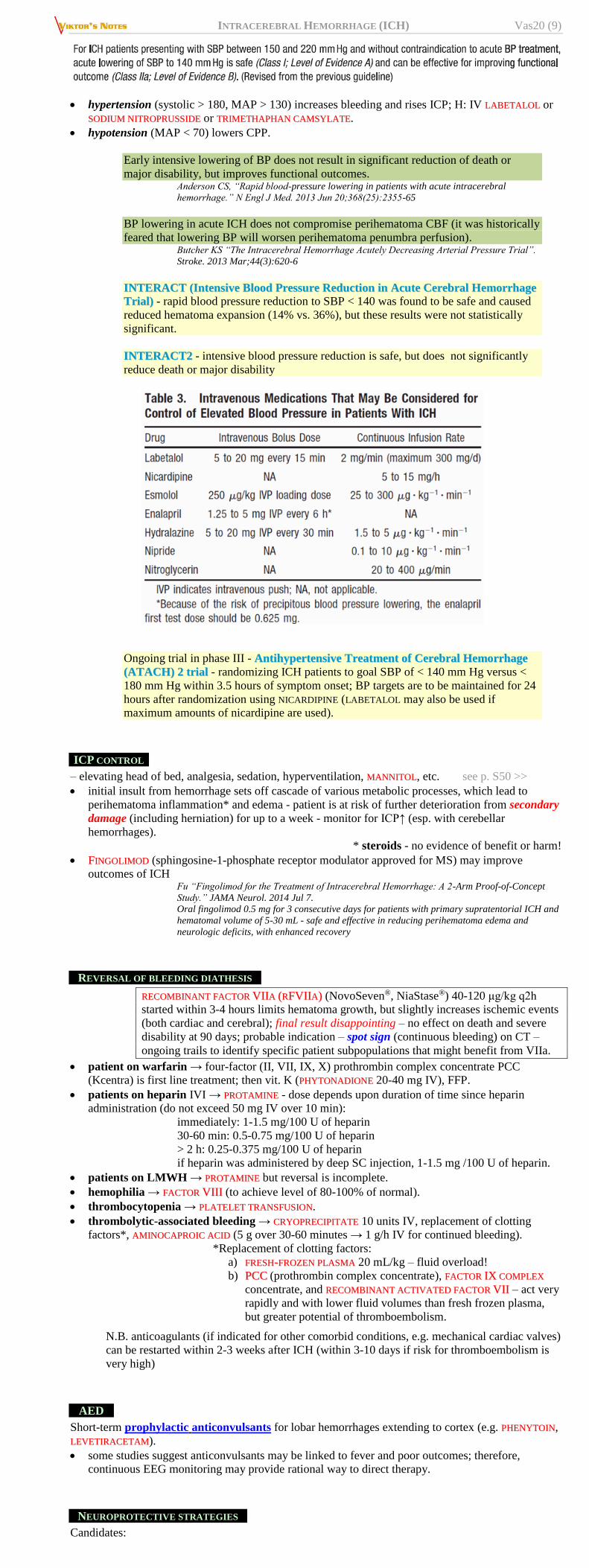
hypertension (systolic > 180, MAP > 130) increases bleeding and rises ICP; H: IV LLAABBEETTAALLOOLL or
SSOODDIIUUMM NNIITTRROOPPRRUUSSSSIIDDEE or TTRRIIMMEETTHHAAPPHHAANN CCAAMMSSYYLLAATTEE.
hypotension (MAP < 70) lowers CPP.
Early intensive lowering of BP does not result in significant reduction of death or
major disability, but improves functional outcomes. Anderson CS, “Rapid blood-pressure lowering in patients with acute intracerebral
hemorrhage.” N Engl J Med. 2013 Jun 20;368(25):2355-65
BP lowering in acute ICH does not compromise perihematoma CBF (it was historically
feared that lowering BP will worsen perihematoma penumbra perfusion). Butcher KS “The Intracerebral Hemorrhage Acutely Decreasing Arterial Pressure Trial”.
Stroke. 2013 Mar;44(3):620-6
IINNTTEERRAACCTT ((IInntteennssiivvee BBlloooodd PPrreessssuurree RReedduuccttiioonn iinn AAccuuttee CCeerreebbrraall HHeemmoorrrrhhaaggee
TTrriiaall)) - rapid blood pressure reduction to SBP < 140 was found to be safe and caused
reduced hematoma expansion (14% vs. 36%), but these results were not statistically
significant.
IINNTTEERRAACCTT22 - intensive blood pressure reduction is safe, but does not significantly
reduce death or major disability
Ongoing trial in phase III - AAnnttiihhyyppeerrtteennssiivvee TTrreeaattmmeenntt ooff CCeerreebbrraall HHeemmoorrrrhhaaggee
((AATTAACCHH)) 22 ttrriiaall - randomizing ICH patients to goal SBP of < 140 mm Hg versus <
180 mm Hg within 3.5 hours of symptom onset; BP targets are to be maintained for 24
hours after randomization using NICARDIPINE (LABETALOL may also be used if
maximum amounts of nicardipine are used).
ICP CONTROL
– elevating head of bed, analgesia, sedation, hyperventilation, MMAANNNNIITTOOLL, etc. see p. S50 >>
initial insult from hemorrhage sets off cascade of various metabolic processes, which lead to
perihematoma inflammation* and edema - patient is at risk of further deterioration from secondary
damage (including herniation) for up to a week - monitor for ICP↑ (esp. with cerebellar
hemorrhages).
* steroids - no evidence of benefit or harm!
FFIINNGGOOLLIIMMOODD (sphingosine-1-phosphate receptor modulator approved for MS) may improve
outcomes of ICH Fu “Fingolimod for the Treatment of Intracerebral Hemorrhage: A 2-Arm Proof-of-Concept
Study.” JAMA Neurol. 2014 Jul 7.
Oral fingolimod 0.5 mg for 3 consecutive days for patients with primary supratentorial ICH and
hematomal volume of 5-30 mL - safe and effective in reducing perihematoma edema and
neurologic deficits, with enhanced recovery
REVERSAL OF BLEEDING DIATHESIS
RREECCOOMMBBIINNAANNTT FFAACCTTOORR VVIIIIAA ((RRFFVVIIIIAA)) (NovoSeven®, NiaStase®) 40-120 μg/kg q2h
started within 3-4 hours limits hematoma growth, but slightly increases ischemic events
(both cardiac and cerebral); final result disappointing – no effect on death and severe
disability at 90 days; probable indication – spot sign (continuous bleeding) on CT –
ongoing trails to identify specific patient subpopulations that might benefit from VIIa.
patient on warfarin → four-factor (II, VII, IX, X) prothrombin complex concentrate PCC
(Kcentra) is first line treatment; then vit. K (PPHHYYTTOONNAADDIIOONNEE 20-40 mg IV), FFP.
patients on heparin IVI → PPRROOTTAAMMIINNEE - dose depends upon duration of time since heparin
administration (do not exceed 50 mg IV over 10 min):
immediately: 1-1.5 mg/100 U of heparin
30-60 min: 0.5-0.75 mg/100 U of heparin
> 2 h: 0.25-0.375 mg/100 U of heparin
if heparin was administered by deep SC injection, 1-1.5 mg /100 U of heparin.
patients on LMWH → PPRROOTTAAMMIINNEE but reversal is incomplete.
hemophilia → FFAACCTTOORR VVIIIIII (to achieve level of 80-100% of normal).
thrombocytopenia → PPLLAATTEELLEETT TTRRAANNSSFFUUSSIIOONN.
thrombolytic-associated bleeding → CCRRYYOOPPRREECCIIPPIITTAATTEE 10 units IV, replacement of clotting
factors*, AAMMIINNOOCCAAPPRROOIICC AACCIIDD (5 g over 30-60 minutes → 1 g/h IV for continued bleeding).
*Replacement of clotting factors:
a) FFRREESSHH--FFRROOZZEENN PPLLAASSMMAA 20 mL/kg – fluid overload!
b) PPCCCC (prothrombin complex concentrate), FFAACCTTOORR IIXX CCOOMMPPLLEEXX
concentrate, and RREECCOOMMBBIINNAANNTT AACCTTIIVVAATTEEDD FFAACCTTOORR VVIIII – act very
rapidly and with lower fluid volumes than fresh frozen plasma,
but greater potential of thromboembolism.
N.B. anticoagulants (if indicated for other comorbid conditions, e.g. mechanical cardiac valves)
can be restarted within 2-3 weeks after ICH (within 3-10 days if risk for thromboembolism is
very high)
AED
Short-term prophylactic anticonvulsants for lobar hemorrhages extending to cortex (e.g. PPHHEENNYYTTOOIINN,
LLEEVVEETTIIRRAACCEETTAAMM).
some studies suggest anticonvulsants may be linked to fever and poor outcomes; therefore,
continuous EEG monitoring may provide rational way to direct therapy.
NEUROPROTECTIVE STRATEGIES
Candidates:
INTRACEREBRAL HEMORRHAGE (ICH) Vas20 (10)
1) STATINS – improve outcomes!!! (but avoid in amyloid angiopathy bleeds!!!)
2) MINOCYCLINE
3) DEFEROXAMINE
4) Hypothermia; more effective in combination with magnesium.
SURGICAL TREATMENT
A. Open surgical evacuation via craniotomy (ultrasonography can confirm clot localization) – esp. for
lobar clots within 1 cm of surface.
surgery between 24-48 h is the best time - vessel has stopped leaking (either spontaneously, or
after hemostatic therapy); if earlier - increased risk of rebleeding.
aspirate, irrigate; most authors recommend leaving small bits of clot on vessels in order to avoid
new hemorrhage.
hemostasis: bipolar coagulation, cotton balls with peroxide, SurgiFoam / FloSeal; may finish by
Surgicel on hematoma walls.
B. Stereotactic aspiration via burr hole; clot mobilization methods:
a) fibrinolytic agent instillation (e.g. urokinase infusion into clot cavity within 72
hrs reduces clot burden and risk for death; but rebleeding↑ → functional outcome
is not improved).
b) mechanical rotors.
INDICATIONS
1. Cerebellar hemorrhages compressing vital structures in medulla (suggested by declining level
of consciousness, posturing, altered respiration, shifted or obliterated 4th ventricle,
hydrocephalus) - surgical removal of hemorrhage as soon as possible (Class I; Level of
Evidence B).
most cerebellar hematomas > 3 cm require surgical evacuation within hours.
surgery is not indicated - GCS score ≥ 14 (some investigators say ≥ 9) with
small hemorrhage (< 3-4 cm) without hydrocephalus.
contraindication (poor surgery results) - large midline hemorrhage with lost all
brain stem functions and flaccid coma.
small time margin between alert state (surgery still not indicated)
and irreversible coma (surgery is too late)
consider preoperative MANNITOL 1 g/kg.
EVD has risk of upward herniation of cerebellum and does not relieve
brainstem compression.
Initial treatment with ventricular drainage rather than surgical evacuation is not
recommended (Class III; Level of Evidence C).
2. Supratentorial hemorrhages with signs of herniation, declining sensorium (esp. if clot is on
nondominant side and ≤ 1 cm from cortical surface) – surgical evacuation and/or decompressive
craniectomy might be considered life-saving (Class IIb; Level of Evidence C).
Routine evacuation of supratentorial ICH by standard craniotomy within 96 hours of
ictus is not recommended!
N.B. patients with deep ICH esp. with IVH do worse with surgery; but patients with
poor prognosis (GCS 9-12) are better off with early surgery! – SSTTIICCHH IIII ttrriiaall
SSTTIICCHH II ttrriiaall - craniotomy is as safe as medical treatment and small trend (2%-4%)
mRS benefit favored surgery.
SSTTIICCHH IIII ttrriiaall - no clear benefit from early surgery!
MMIISSTTIIEE ((MMiinniimmaallllyy IInnvvaassiivvee SSuurrggeerryy PPlluuss RRtt--PPAA ffoorr IICCHH EEvvaaccuuaattiioonn)) IIIIII ttrriiaall -
minimally invasive surgery aspiration plus 1 mg rt-PA through intraclot catheter
q8hrs (up to 9 doses total) vs. medical therapy alone; inclusion: spontaneous, non-
traumatic supratentorial ICH ≥ 30 mL with or without intraventricular hemorrhage
(IVH) not requiring EVD, with GCS ≤ 14 or NIHSS ≥ 6, in 18-80 yo patient with
symptom onset within 24 hours of diagnostic CT, initiation of treatment from 12 to 72
hours of diagnostic CT, with first dose given within 76 hours of the diagnostic CT.
N.B. surgery is not beneficial for hemorrhages in putamen, thalamus, and pons.
In general, surgical evacuation is seldom justified
- does not substantially improve mortality + considerably increases risk of severe
residual neurologic disability if patient survives.
best candidates are patients with increasing moderate ÷ large hematomas who are still awake (GCS
≥ 9).
N.B. patients with massive hemorrhage who are in coma are not likely to benefit!
OTHER SURGICAL MEASURES:
1. Hemicraniectomy - option for younger patients with rapidly declining conscious state and
imminent herniation.
2. Ventricular drainage for acute obstructive hydrocephalus (esp. in cerebellar hematomas,
intraventricular hemorrhage); endoscopic neurosurgical techniques for IVH evacuation may be
advantageous compared with EVD.
N.B. INTRAVENTRICULAR HEMORRHAGE must be treated with EVD and can be treated with low-
dose intraventricular fibrinolytics (catheter-based clot lysis) to dissolve clot quicker (e.g.
1.0 mg ttPPAA q 8-12 hrs) - dramatically reduced morbidity & mortality!!!
– EVD must go into clot
– clamp ventriculostomy 30-60 minutes and monitor for increased ICP
– monitor daily with CT.
– clots dissolve on average within 3-4 days.
– clotted intraventricular catheter: alteplase 0.5 mg IT once, reassess
– complications (tPA, frequent EVD access).
CLEAR (Clot Lysis Evaluating Accelerated Resolution of intraventricular hemorrhage)
III trial – intraventricular tPA in patients with small ICH but with IVH (to test treatment for
IVH and not to be obscured by large ICH):
– does not improve good functional outcome (mRS 0-3: 48% in alteplase group,
45% in saline group), but gives 10% reduction in mortality without increasing
the number of patients left in a vegetative state or requiring nursing home care
(best results in patients with > 20 mL or > 90% of blood removed; no benefit of
IVH blood is < 20 mL to start).
Complication CLEAR III trial patients Literature meta-analysis
Hemorrhage 16.8% (2.4% symptomatic
hemorrhages) – both saline
and alteplase groups
8.4% (0.7% symptomatic
hemorrhage)
Infection 4.4% 7.9%
Alteplase is associated with reduction in bacterial
ventriculitis (P = .05).
CLEAR IV trial – patients with larger clots - awaiting a funding application
INTRACEREBRAL HEMORRHAGE (ICH) Vas20 (11)
3. Aneurysm repair, removal of bleeding AVM or tumor, i.e. bleeding structural / vascular lesion is
also indication for surgery.
4. Ventriculoperitoneal shunt for chronic hydrocephalus.
– predictors of development of shunt-dependent hydrocephalus after ICH:
thalamic ICH, persistently elevated ICP.
FUTURE APPROACHES
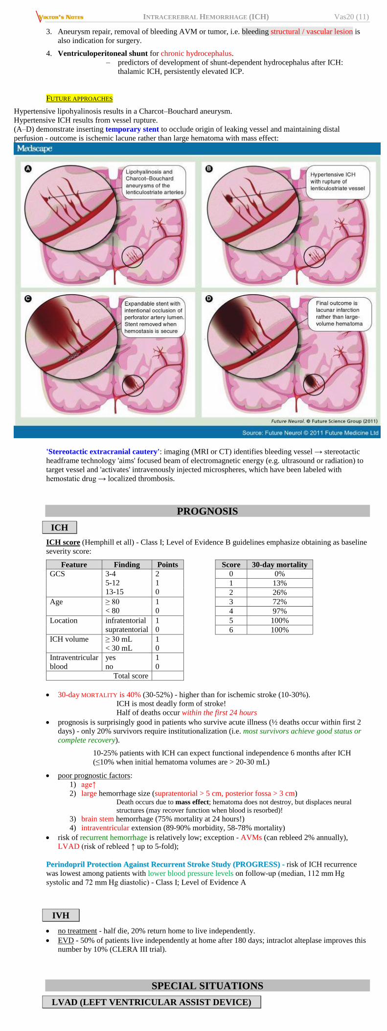
Hypertensive lipohyalinosis results in a Charcot–Bouchard aneurysm.
Hypertensive ICH results from vessel rupture.
(A–D) demonstrate inserting temporary stent to occlude origin of leaking vessel and maintaining distal
perfusion - outcome is ischemic lacune rather than large hematoma with mass effect:
'Stereotactic extracranial cautery': imaging (MRI or CT) identifies bleeding vessel → stereotactic
headframe technology 'aims' focused beam of electromagnetic energy (e.g. ultrasound or radiation) to
target vessel and 'activates' intravenously injected microspheres, which have been labeled with
hemostatic drug → localized thrombosis.
PROGNOSIS
ICH
ICH score (Hemphill et all) - Class I; Level of Evidence B guidelines emphasize obtaining as baseline
severity score:
Feature Finding Points Score 30-day mortality
GCS 3-4
5-12
13-15
2
1
0
0 0%
1 13%
2 26%
Age ≥ 80
< 80
1
0
3 72%
4 97%
Location infratentorial
supratentorial
1
0
5 100%
6 100%
ICH volume ≥ 30 mL
< 30 mL
1
0
Intraventricular
blood
yes
no
1
0
Total score
30-day MORTALITY is 40% (30-52%) - higher than for ischemic stroke (10-30%).
ICH is most deadly form of stroke!
Half of deaths occur within the first 24 hours
prognosis is surprisingly good in patients who survive acute illness (½ deaths occur within first 2
days) - only 20% survivors require institutionalization (i.e. most survivors achieve good status or
complete recovery).
10-25% patients with ICH can expect functional independence 6 months after ICH
(≤10% when initial hematoma volumes are > 20-30 mL)
poor prognostic factors:
1) age↑
2) large hemorrhage size (supratentorial > 5 cm, posterior fossa > 3 cm) Death occurs due to mass effect; hematoma does not destroy, but displaces neural
structures (may recover function when blood is resorbed)!
3) brain stem hemorrhage (75% mortality at 24 hours!)
4) intraventricular extension (89-90% morbidity, 58-78% mortality)
risk of recurrent hemorrhage is relatively low; exception - AVMs (can rebleed 2% annually),
LVAD (risk of rebleed ↑ up to 5-fold);
PPeerriinnddoopprriill PPrrootteeccttiioonn AAggaaiinnsstt RReeccuurrrreenntt SSttrrookkee SSttuuddyy ((PPRROOGGRREESSSS)) -- risk of ICH recurrence
was lowest among patients with lower blood pressure levels on follow-up (median, 112 mm Hg
systolic and 72 mm Hg diastolic) - Class I; Level of Evidence A
IVH
no treatment - half die, 20% return home to live independently.
EVD - 50% of patients live independently at home after 180 days; intraclot alteplase improves this
number by 10% (CLERA III trial).
SPECIAL SITUATIONS
LVAD (LEFT VENTRICULAR ASSIST DEVICE)

INTRACEREBRAL HEMORRHAGE (ICH) Vas20 (12)
Two types of LVADs:
1) pulsatile flow
2) nonpulsatile flow (more and more popular) – cannot use BP cuff; use A-line – see MAP
N.B. MAP > 90 mmHg is abnormal (risk of ICH↑)
most important prognostic factor – GCS at presentation (no patients with GCS ≤ 11 did survive 30
days).
patient is usually on Aspirin and warfarin; when to restart:
[classic AFib with worst CHAD – annual stroke risk is only 18-20%]
– experts usually restart Aspirin in 7-14 days and warfarin in 14-21 days; no
thrombotic complications reported from withholding so long.
– once restarted, risk of rebleed ↑ 5-fold in one Italian study but no increased risk in
one Canadian study.
BIBLIOGRAPHY for ch. “Neurovascular Disorders” → follow this LINK >>
AHA/American Stroke Assoc 2015 “Guidelines for the Management of Spontaneous Intracerebral
Hemorrhage”
Viktor’s Notes℠ for the Neurosurgery Resident
Please visit website at www.NeurosurgeryResident.net